This is a modern-English version of Surgical Anatomy, originally written by Maclise, Joseph.
It has been thoroughly updated, including changes to sentence structure, words, spelling,
and grammar—to ensure clarity for contemporary readers, while preserving the original spirit and nuance. If
you click on a paragraph, you will see the original text that we modified, and you can toggle between the two versions.
Scroll to the bottom of this page and you will find a free ePUB download link for this book.
[Transcriber’s Notes]
[Transcriber’s Notes]
Thanks to Carol Presher of Timeless Antiques, Valley, Alabama, for lending the original book for this production. The 140 year old binding had disintegrated, but the paper and printing was in amazingly good condition, particularly the multicolor images.
Thanks to Carol Presher of Timeless Antiques, Valley, Alabama, for lending the original book for this production. The 140-year-old binding had fallen apart, but the paper and printing were in surprisingly good shape, especially the multicolor images.
Thanks also to the Mayo Clinic. This book has increased my appreciation of their skilled care of my case by showing the many ways that things could go wrong.
Thanks also to the Mayo Clinic. This book has deepened my appreciation for their expert care of my case by highlighting the many ways things could go wrong.
Footnotes are indicated by “[Footnote]” where they appear in the text. The body of the footnote appears immediately following the complete paragraph. If more than one footnote appears in the same paragraph, they are numbered.
Footnotes are marked by “[Footnote]” where they show up in the text. The full text of the footnote follows right after the complete paragraph. If there are multiple footnotes in the same paragraph, they are numbered.
A few obvious misspellings have been corrected. Several cases of alternate spelling of the same(?) word have not been modified.
A few obvious misspellings have been fixed. Several instances of different spellings of the same word have not been changed.
Pages have been reorganized to avoid splitting sentences and paragraphs. Each image is inserted immediately following its description.
Pages have been rearranged to prevent cutting off sentences and paragraphs. Each image is placed right after its description.
Some of the plates did not fit on the scanner and were captured as two separate images. The merged images show some artifacts of the merge process due to slightly different lighting of the page. The contrast and gamma values have been adjusted to restore the images.
Some of the plates didn't fit on the scanner and were captured as two separate images. The combined images show some artifacts from the merging process because of slightly different lighting on the page. The contrast and gamma values have been adjusted to improve the images.
In this HTML format the images have been resized to fit on smaller displays. Each image is also a link to the original size version. Click on the image to see the original.
In this HTML format, the images have been resized to fit smaller screens. Each image is also a link to the full-size version. Click on the image to view the original.
[End Transcriber’s Notes]
[End Transcriber’s Notes]
![[Illustration]](images/cover.jpg)
SURGICAL ANATOMY
BY
JOSEPH MACLISE
FELLOW OF THE ROYAL COLLEGE OF SURGEONS.
WITH SIXTY-EIGHT COLOURED PLATES.
![[Illustration]](images/0Title1Pic.jpg)
PHILADELPHIA:
BLANCHARD AND LEA.
1859.
I INSCRIBE THIS WORK TO
THE GENTLEMEN WITH WHOM AS A FELLOW-STUDENT I WAS ASSOCIATED
AT THE
London University College:
I DEDICATE THIS WORK TO
THE GENTLEMEN WITH WHOM I STUDIED AS A CLASSMATE
AT THE
London University College:
AND IN AN ESPECIAL MANNER, IN THEIR NAME AS WELL AS MY OWN, I AVAIL MYSELF OF
THE OPPORTUNITY TO RECORD,
ON THIS PAGE,
ALBEIT IN CHARACTERS LESS IMPRESSIVE THAN THOSE WHICH ARE WRITTEN
ON THE LIVING TABLET OF MEMORY,
THE DEBT OF GRATITUDE WHICH WE OWE
TO THE LATE
AND IN A SPECIAL WAY, ON THEIR BEHALF AS WELL AS MY OWN, I TAKE THIS OPPORTUNITY TO RECORD,
ON THIS PAGE,
ALBEIT IN CHARACTERS LESS IMPRESSIVE THAN THOSE WHICH ARE WRITTEN
ON THE LIVING TABLET OF MEMORY,
THE DEBT OF GRATITUDE WE OWE
TO THE LATE
SAMUEL COOPER, F.R.S., AND ROBERT LISTON, F.R.S.,
SAMUEL COOPER, F.R.S., AND ROBERT LISTON, F.R.S.,
TWO AMONG THE MANY DISTINGUISHED PROFESSORS OF THAT INSTITUTION,
WHOSE PUPILS WE HAVE BEEN,
AND FROM WHOM WE INHERIT THAT BETTER POSSESSION THAN LIFE ITSELF,
AN ASPIRATION FOR THE LIGHT OF SCIENCE.
TWO AMONG THE MANY DISTINGUISHED PROFESSORS OF THAT INSTITUTION,
WHOSE STUDENTS WE HAVE BEEN,
AND FROM WHOM WE INHERIT THAT GREATER GIFT THAN LIFE ITSELF,
A DESIRE FOR THE LIGHT OF SCIENCE.
JOSEPH MACLISE.
JOSEPH MACLISE.
PREFACE.
The object of this work is to present to the student of medicine and the practitioner removed from the schools, a series of dissections demonstrative of the relative anatomy of the principal regions of the human body. Whatever title may most fittingly apply to a work with this intent, whether it had better be styled surgical or medical, regional, relative, descriptive, or topographical anatomy, will matter little, provided its more salient or prominent character be manifested in its own form and feature. The work, as I have designed it, will itself show that my intent has been to base the practical upon the anatomical, and to unite these wherever a mutual dependence was apparent.
The goal of this work is to provide medical students and practicing professionals outside of academic settings with a series of dissections that illustrate the relative anatomy of key areas of the human body. The specific title that best describes this work—whether it should be considered surgical, medical, regional, relative, descriptive, or topographical anatomy—matters little, as long as its main purpose is clear in its format and content. As I've planned, this work will demonstrate my intention to link practical knowledge with anatomical understanding, connecting them wherever their interdependence is evident.
That department of anatomical research to which the name topographical strictly applies, as confining itself to the mere account of the form and relative location of the several organs comprising the animal body, is almost wholly isolated from the main questions of physiological and transcendental interest, and cannot, therefore, be supposed to speak in those comprehensive views which anatomy, taken in its widest signification as a science, necessarily includes. While the anatomist contents himself with describing the form and position of organs as they appear exposed, layer after layer, by his dissecting instruments, he does not pretend to soar any higher in the region of science than the humble level of other mechanical arts, which merely appreciate the fitting arrangement of things relative to one another, and combinative to the whole design of the form or machine of whatever species this may be, whether organic or inorganic. The descriptive anatomist of the human body aims at no higher walk in science than this, and hence his nomenclature is, as it is, a barbarous jargon of words, barren of all truthful signification, inconsonant with nature, and blindly irrespective of the cognitio certa ex principiis certis exorta.
That area of anatomical research referred to as topographical focuses solely on describing the shape and relative location of the various organs in the animal body. It is largely disconnected from the more significant physiological and philosophical questions and, therefore, doesn't engage in the broader perspectives that anatomy, in its most expansive sense as a science, inherently encompasses. While the anatomist is satisfied with outlining the shape and position of organs as they are revealed, layer by layer, through dissection, he doesn't aim to reach any higher scientific understanding than the basic level of other mechanical arts, which only evaluate how things are arranged in relation to one another and contribute to the overall design of an object, whether it’s organic or inorganic. The descriptive anatomist studying the human body doesn’t aspire to a higher level of science than this, which is why his terminology often resembles a confusing mix of words, lacking genuine significance, misaligned with nature, and blindly ignoring the cognitio certa ex principiis certis exorta.
Still, however, this anatomy of form, although so much requiring purification of its nomenclature, in order to clothe it in the high reaching dignity of a science, does not disturb the medical or surgical practitioner, so far as their wants are concerned. Although it may, and actually does, trammel the votary who aspires to the higher generalizations and the development of a law of formation, yet, as this is not the object of the surgical anatomist, the nomenclature, such as it is, will answer conveniently enough the present purpose.
Still, this analysis of form, even though it needs a better naming system to give it the respect of a science, doesn't trouble medical or surgical practitioners when it comes to their needs. While it might limit those who aim for broader theories and the development of a formation law, this isn't the goal of the surgical anatomist, so the existing naming works just fine for the current purpose.
The anatomy of the human form, contemplated in reference to that of all other species of animals to which it bears comparison, constitutes the study of the comparative anatomist, and, as such, establishes the science in its full intent. But the anatomy of the human figure, considered as a species, per se, is confessedly the humblest walk of the understanding in a subject which, as anatomy, is relationary, and branches far and wide through all the domain of an animal kingdom. While restricted to the study of the isolated human species, the cramped judgment wastes in such narrow confine; whereas, in the expansive gaze over all allying and allied species, the intellect bodies forth to its vision the full appointed form of natural majesty; and after having experienced the manifold analogies and differentials of the many, is thereby enabled, when it returns to the study of the one, to view this one of human type under manifold points of interest, to the appreciation of which the understanding never wakens otherwise. If it did not happen that the study of the human form (confined to itself) had some practical bearing, such study could not deserve the name of anatomical, while anatomical means comparative, and whilst comparison implies inductive reasoning.
The study of human anatomy, when compared to that of other animal species, forms the basis of comparative anatomy and fully establishes the science behind it. However, examining human anatomy as a separate category is admittedly the simplest approach within a field that, as anatomy, is related and branches out broadly across the entire animal kingdom. Focusing solely on the study of humanity limits understanding to a narrow perspective; but when looking at various related and allied species, the intellect can envision the complete natural majesty of form. After exploring the many similarities and differences among these species, one can return to the study of humans and appreciate this unique type from multiple angles that would otherwise go unnoticed. If the study of the human form were not connected to practical applications, it wouldn't truly be considered anatomical since anatomy inherently involves comparison, and comparison requires inductive reasoning.
However, practical anatomy, such as it is, is concerned with an exact knowledge of the relationship of organs as they stand in reference to each other, and to the whole design of which these organs are the integral parts. The figure, the capacity, and the contents of the thoracic and abdominal cavities, become a study of not more urgent concernment to the physician, than are the regions named cervical, axillary, inguinal, &c., to the surgeon. He who would combine both modes of a relationary practice, such as that of medicine and surgery, should be well acquainted with the form and structures characteristic of all regions of the human body; and it may be doubted whether he who pursues either mode of practice, wholly exclusive of the other, can do so with honest purpose and large range of understanding, if he be not equally well acquainted with the subject matter of both. It is, in fact, more triflingly fashionable than soundly reasonable, to seek to define the line of demarcation between the special callings of medicine and surgery, for it will ever be as vain an endeavour to separate the one from the other without extinguishing the vitality of both, as it would be to sunder the trunk from the head, and give to each a separate living existence. The necessary division of labour is the only reason that can be advanced in excuse of specialisms; but it will be readily agreed to, that that practitioner who has first laid within himself the foundation of a general knowledge of matters relationary to his subject, will always be found to pursue the speciality according to the light of reason and science.
However, practical anatomy, as it stands, focuses on a clear understanding of how organs relate to each other and to the overall design of which they are essential parts. The shape, size, and contents of the thoracic and abdominal cavities are just as important for a physician to study as the cervical, axillary, inguinal regions, etc., are for a surgeon. Anyone who wants to blend both fields of practice—medicine and surgery—needs to have a good understanding of the form and structures that characterize all areas of the human body. It can be argued that someone who only focuses on one of these practices without acknowledging the other cannot truly do so with integrity and a broad understanding, unless they are also knowledgeable about both areas. In reality, it’s often more trendy than sensible to try to draw a clear line between medicine and surgery, as trying to separate the two would be as futile as trying to detach the trunk from the head and giving each a separate life. The necessary division of labor is the only valid reason for specializations, but it’s widely accepted that a practitioner who has built a solid foundation of general knowledge related to their field will always approach their specialty in alignment with reason and science.
Anatomy—the  the knowledge based on principle—is the
foundation of the curative art, cultivated as a science in all its branchings;
and comparison is the nurse of reason, which we are fain to make our guide in
bringing the practical to bear productively. The human body, in a state of
health, is the standard whereunto we compare the same body in a state of
disease. The knowledge of the latter can only exist by the knowledge of the
former, and by the comparison of both.
the knowledge based on principle—is the
foundation of the curative art, cultivated as a science in all its branchings;
and comparison is the nurse of reason, which we are fain to make our guide in
bringing the practical to bear productively. The human body, in a state of
health, is the standard whereunto we compare the same body in a state of
disease. The knowledge of the latter can only exist by the knowledge of the
former, and by the comparison of both.
Anatomy—the the knowledge based on principle—is the foundation of healing, developed as a science across all its branches; and comparison is essential for reasoning, which we seek to use as our guide in applying practical knowledge effectively. The human body, when healthy, is the benchmark against which we measure the same body when it is diseased. Understanding disease can only come from understanding health, and from comparing both states.
Comparison may be fairly termed the pioneer to all certain knowledge. It is a potent instrument—the only one, in the hands of the pathologist, as well as in those of the philosophic generalizer of anatomical facts, gathered through the extended survey of an animal kingdom. We best recognise the condition of a dislocated joint after we have become well acquainted with the contour of its normal state; all abnormal conditions are best understood by a knowledge of what we know to be normal character. Every anatomist is a comparer, in a greater or lesser degree; and he is the greatest anatomist who compares the most generally.
Comparison can be considered the foundation of all certain knowledge. It’s a powerful tool—the only one available to pathologists and to those who philosophically generalize anatomical facts gathered from extensive studies of the animal kingdom. We best understand the condition of a dislocated joint after we’ve become familiar with the shape of its normal state; all abnormal conditions are best understood by knowing what we recognize as normal. Every anatomist is a comparer to some degree, and the best anatomist is the one who makes the most comparisons.
Impressed with this belief, I have laid particular emphasis on imitating the character of the normal form of the human figure, taken as a whole; that of its several regions as parts of this whole, and that of the various organs (contained within those regions) as its integrals or elements. And in order to present this subject of relative anatomy in more vivid reality to the understanding of the student, I have chosen the medium of illustrating by figure rather than by that of written language, which latter, taken alone, is almost impotent in a study of this nature.
Impressed by this belief, I have focused on mimicking the normal shape of the human body as a whole; including its various regions as parts of this whole, and the different organs (found within those regions) as its components or elements. To make this topic of relative anatomy easier to understand for students, I chose to use illustrations rather than just written descriptions, which alone are quite ineffective for studying this subject.
It is wholly impossible for anyone to describe form in words without the aid of figures. Even the mathematical strength of Euclid would avail nothing, if shorn of his diagrams. The professorial robe is impotent without its diagrams. Anatomy being a science existing by demonstration, (for as much as form in its actuality is the language of nature,) must be discoursed of by the instrumentality of figure.
It’s completely impossible for anyone to describe shape in words without using drawings. Even the mathematical skills of Euclid wouldn’t mean much without his diagrams. A professor's authority is powerless without their illustrations. Since anatomy is a science based on demonstration (because the shape of something in reality is nature's language), it has to be discussed with the help of figures.
An anatomical illustration enters the understanding straight-forward in a direct passage, and is almost independent of the aid of written language. A picture of form is a proposition which solves itself. It is an axiom encompassed in a frame-work of self-evident truth. The best substitute for Nature herself, upon which to teach the knowledge of her, is an exact representation of her form.
An anatomical illustration conveys understanding directly and almost doesn't require written language. An image of a shape presents a concept that explains itself. It's a fundamental truth wrapped in a framework of self-evidence. The best alternative to Nature herself, for teaching her knowledge, is an accurate depiction of her form.
Every surgical anatomist will (if he examine himself) perceive that, previously to undertaking the performance of an operation upon the living body, he stands reassured and self-reliant in that degree in which he is capable of conjuring up before his mental vision a distinct picture of his subject. Mr. Liston could draw the same anatomical picture mentally which Sir Charles Bell’s handicraft could draw in reality of form and figure. Scarpa was his own draughtsman.
Every surgical anatomist will (if he examines himself) realize that, before performing surgery on a living person, he feels confident and self-assured to the extent that he can vividly imagine a clear image of his subject. Mr. Liston could mentally recreate the same anatomical picture that Sir Charles Bell could produce in the physical form. Scarpa was his own illustrator.
If there may be any novelty now-a-days possible to be recognised upon the out-trodden track of human relative anatomy, it can only be in truthful and well-planned illustration. Under this view alone may the anatomist plead an excuse for reiterating a theme which the beautiful works of Cowper, Haller, Hunter, Scarpa, Soemmering, and others, have dealt out so respectably. Except the human anatomist turns now to what he terms the practical ends of his study, and marshals his little knowledge to bear upon those ends, one may proclaim anthropotomy to have worn itself out. Dissection can do no more, except to repeat Cruveilhier. And that which Cruveilhier has done for human anatomy, Muller has completed for the physiological interpretation of human anatomy; Burdach has philosophised, and Magendie has experimented to the full upon this theme, so far as it would permit. All have pushed the subject to its furthest limits, in one aspect of view. The narrow circle is footworn. All the needful facts are long since gathered, sown, and known. We have been seekers after those facts from the days of Aristotle. Are we to put off the day of attempting interpretation for three thousand years more, to allow the human physiologist time to slice the brain into more delicate atoms than he has done hitherto, in order to coin more names, and swell the dictionary? No! The work must now be retrospective, if we would render true knowledge progressive. It is not a list of new and disjointed facts that Science at present thirsts for; but she is impressed with the conviction that her wants can alone be supplied by the creation of a new and truthful theory,—a generalization which the facts already known are sufficient to supply, if they were well ordered according to their natural relationship and mutual dependence. “Le temps viendra peut-etre,” says Fontenelle, “que l’on joindra en un corps regulier ces membres epars; et, s’ils sont tels qu’on le souhaite, ils s’assembleront en quelque sorte d’eux-memes. Plusieurs verites separees, des qu’elles sont en assez grand nombre, offrent si vivement a l’esprit leurs rapports et leur mutuelle dependance, qu’il semble qu’apres les avoir detachees par une espece de violence les unes des autres, elles cherchent naturellement a se reunir.”—(Preface sur l’utilite des Sciences, &c.)
If there's any novelty nowadays to be recognized in the well-trodden path of human anatomy, it can only come from clear and well-planned illustrations. Only under this view can the anatomist justify revisiting a subject that has been so respectfully covered by the remarkable works of Cowper, Haller, Hunter, Scarpa, Soemmering, and others. Unless the human anatomist shifts focus to what he calls the practical applications of his study and directs his limited knowledge toward those ends, one might declare that human anatomy has reached its limit. Dissection can do no more than repeat what Cruveilhier has done. Just as Cruveilhier has contributed to human anatomy, Muller has advanced our physiological understanding; Burdach has provided philosophical insights, and Magendie has thoroughly experimented within this topic as far as possible. All have pushed the subject to its outer limits from one perspective. The narrow field has been thoroughly explored. All the necessary facts have long been collected, shared, and understood. We have been searching for these facts since the days of Aristotle. Should we delay interpretation for another three thousand years, allowing the human physiologist time to dissect the brain into even finer particles than before, just to create more terminology and expand the dictionary? No! The work must now be backward-looking if we aim to make true knowledge progressive. Science does not currently thirst for a list of new and disjointed facts; she is convinced that her needs can only be met by developing a new and accurate theory—a generalization that the facts already known can adequately provide if they are properly organized according to their natural relationships and interdependencies. “Le temps viendra peut-etre,” says Fontenelle, “que l’on joindra en un corps regulier ces membres epars; et, s’ils sont tels qu’on le souhaite, ils s’assembleront en quelque sorte d’eux-memes. Plusieurs verites separees, des qu’elles sont en assez grand nombre, offrent si vivement a l’esprit leurs rapports et leur mutuelle dependance, qu’il semble qu’apres les avoir detachees par une espece de violence les unes des autres, elles cherchent naturellement a se reunir.”—(Preface sur l’utilite des Sciences, &c.)
The comparison of facts already known must henceforward be the scalpel which we are to take in hand. We must return by the same road on which we set out, and reexamine the things and phenomena which, as novices, we passed by too lightly. The travelled experience may now sit down and contemplate.
The comparison of known facts will now be the tool we use. We need to retrace the path we took and take a closer look at the things and events we overlooked as beginners. Our gained experience can now take a moment to reflect.
That which I have said and proved elsewhere in respect to the skeleton system may, with equal truth, be remarked of the nervous system—namely, that the question is not in how far does the limit of diversity extend through the condition of an evidently common analogy, but by what rule or law the uniform ens is rendered the diverse entity? The womb of anatomical science is pregnant of the true interpretation of the law of unity in variety; but the question is of longer duration than was the life of the progenitor. Though Aristotle and Linnaeus, and Buffon and Cuvier, and Geoffroy St. Hilaire and Leibnitz, and Gothe, have lived and spoken, yet the present state of knowledge proclaims the Newton of physiology to be as yet unborn. The iron scalpel has already made acquaintance with not only the greater parts, but even with the infinitesimals of the human body; and reason, confined to this narrow range of a subject, perceives herself to be imprisoned, and quenches her guiding light in despair. Originality has outlived itself; and discovery is a long-forgotten enterprise, except as pursued in the microcosm on the field of the microscope, which, it must be confessed, has drawn forth demonstrations only commensurate in importance with the magnitude of the littleness there seen.
What I’ve mentioned and proven before about the skeletal system can also be said about the nervous system—specifically, the issue isn’t how far diversity extends through a clear common analogy, but what rule or law transforms a uniform entity into a diverse one. The foundation of anatomical science holds the key to truly understanding the law of unity in variety; however, this question has persisted longer than the life of its originator. Though Aristotle, Linnaeus, Buffon, Cuvier, Geoffroy St. Hilaire, Leibnitz, and Goethe have all come and gone, the current state of knowledge indicates that the Newton of physiology has yet to be born. The iron scalpel has investigated not only the major parts but even the tiniest details of the human body; yet, reason, confined to this limited scope, feels trapped and extinguishes its guiding light in despair. Originality has outlasted itself, and discovery has become a long-forgotten pursuit, except in the microcosm of the microscope, which, it must be admitted, has only produced findings that are significant relative to the scale of the tiny things it reveals.
The subject of our study, whichever it happen to be, may appear exhausted of all interest, and the promise of valuable novelty, owing to two reasons:—It may be, like descriptive human anatomy, so cold, poor and sterile in its own nature, and so barren of product, that it will be impossible for even the genius of Promethean fire to warm it; or else, like existing physiology, the very point of view from which the mental eye surveys the theme, will blight the fair prospect of truth, distort induction, and clog up the paces of ratiocination. The physiologist of the present day is too little of a comparative anatomist, and far too closely enveloped in the absurd jargon of the anthropotomist, ever to hope to reveal any great truth for science, and dispel the mists which still hang over the phenomena of the nervous system. He is steeped too deeply in the base nomenclature of the antique school, and too indolent to question the import of Pons, Commissure, Island, Taenia, Nates, Testes, Cornu, Hippocamp, Thalamus, Vermes, Arbor Vitro, Respiratory Tract, Ganglia of Increase, and all such phrase of unmeaning sound, ever to be productive of lucid interpretation of the cerebro-spinal ens. Custom alone sanctions his use of such names; but
The topic of our study, no matter what it is, might seem completely drained of interest and the potential for valuable new insights for two reasons: It could be, like descriptive human anatomy, so dull, poor, and lifeless that not even the most brilliant minds could bring it to life; or, like current physiology, the perspective from which we examine the subject might obscure the truth, distort our reasoning, and block clear thinking. Today's physiologist lacks the comparative anatomy background and is too wrapped up in the absurd terminology of anthropotomy to hope to uncover any significant truths for science or clear away the confusion surrounding the nervous system phenomena. He is too immersed in the outdated language of the old school and too lazy to question the meaning of terms like Pons, Commissure, Island, Taenia, Nates, Testes, Cornu, Hippocamp, Thalamus, Vermes, Arbor Vitro, Respiratory Tract, Ganglia of Increase, and all those similar phrases that are just empty sounds, preventing any clear understanding of the cerebro-spinal system. Tradition alone supports his use of such names, but
“Custom calls him to it!
What custom wills; should custom always do it,
The dust on antique time would lie unswept,
And mountainous error be too highly heaped,
For truth to overpeer.”
“Tradition demands it!
What tradition wants; if tradition always did it,
The dust of ancient times would remain uncleaned,
And massive mistakes would pile up too high,
For truth to shine through.”
Of the illustrations of this work I may state, in guarantee of their anatomical accuracy, that they have been made by myself from my own dissections, first planned at the London University College, and afterwards realised at the Ecole Pratique, and School of Anatomy adjoining the Hospital La Pitie, Paris, a few years since. As far as the subject of relative anatomy could admit of novel treatment, rigidly confined to facts unalterable, I have endeavoured to give it.
Of the illustrations in this work, I can assure you of their anatomical accuracy since I created them myself from my own dissections. I initially planned these at London University College and later completed them at the Ecole Pratique and the School of Anatomy next to the Hospital La Pitie in Paris a few years ago. As much as the topic of relative anatomy allows for new approaches, strictly adhering to the unchangeable facts, I have tried to present it.
The unbroken surface of the human figure is as a map to the surgeon, explanatory of the anatomy arranged beneath; and I have therefore left appended to the dissected regions as much of the undissected as was necessary. My object was to indicate the interior through the superficies, and thereby illustrate the whole living body which concerns surgery, through its dissected dead counterfeit. We dissect the dead animal body in order to furnish the memory with as clear an account of the structure contained in its living representative, which we are not allowed to analyse, as if this latter were perfectly translucent, and directly demonstrative of its component parts.
The smooth surface of the human body serves as a map for the surgeon, revealing the anatomy underneath; that's why I've included as much of the skin as needed alongside the dissected areas. My goal was to show the inner workings through the outer layer, illustrating the entire living body relevant to surgery by using its dissected, lifeless replica. We dissect the body of a dead animal to provide a clear understanding of the structure found in its living version, which we can’t analyze as if it were completely transparent and directly showing its parts.
J. M
J.M.
TABLE OF CONTENTS.
PREFACE
INTRODUCTORY TO THE STUDY OF ANATOMY AS A SCIENCE.
PREFACE
INTRODUCTION TO THE STUDY OF ANATOMY AS A SCIENCE.
THE FORM OF THE THORAX, AND THE RELATIVE POSITION OF ITS CONTAINED PARTS—THE LUNGS, HEART, AND LARGER BLOOD VESSELS.
THE SHAPE OF THE THORAX, AND THE RELATIVE POSITION OF ITS CONTAINED PARTS—THE LUNGS, HEART, AND LARGER BLOOD VESSELS.
The structure, mechanism, and respiratory motions of the thoracic apparatus.
Its varieties in form, according to age and sex. Its deformities. Applications
to the study of physical diagnosis.
The structure, function, and breathing movements of the chest area. Its different shapes based on age and gender. Its abnormalities. Uses in the field of physical diagnosis.
THE SURGICAL FORM OF THE SUPERFICIAL, CERVICAL, AND FACIAL REGIONS, AND THE RELATIVE POSITION OF THE PRINCIPAL BLOOD VESSELS, NERVES, ETC.
THE SURGICAL ASPECTS OF THE SUPERFICIAL CERVICAL AND FACIAL AREAS, AND THE POSITION OF THE MAIN BLOOD VESSELS, NERVES, ETC.
The cervical surgical triangles considered in reference to the position of the
subclavian and carotid vessels, &c. Venesection in respect to the external
jugular vein. Anatomical reasons for avoiding transverse incisions in the neck.
The parts endangered in surgical operations on the parotid and submaxillary
glands, &c.
The cervical surgical triangles related to the positioning of the subclavian and carotid vessels, etc. Bloodletting concerning the external jugular vein. Anatomical reasons for steering clear of horizontal cuts in the neck. The structures at risk during surgical procedures on the parotid and submaxillary glands, etc.
THE SURGICAL FORM OF THE DEEP CERVICAL AND FACIAL REGIONS, AND THE RELATIVE POSITION OF THE PRINCIPAL BLOOD VESSELS, NERVES, ETC.
THE SURGICAL STRUCTURE OF THE DEEP CERVICAL AND FACIAL AREAS, AND THE RELATIVE POSITION OF THE MAIN BLOOD VESSELS, NERVES, ETC.
The course of the carotid and subclavian vessels in reference to each other, to
the surface, and to their respective surgical triangles. Differences in the
form of the neck in individuals of different age and sex. Special relations of
the vessels. Physiological remarks on the carotid artery. Peculiarities in the
relative position of the subclavian artery.
The path of the carotid and subclavian vessels in relation to each other, to the surface, and to their specific surgical triangles. Variations in the shape of the neck among individuals of different ages and genders. Unique connections of the vessels. Physiological notes on the carotid artery. Distinctive features in the positioning of the subclavian artery.
THE SURGICAL DISSECTION OF THE SUBCLAVIAN AND CAROTID REGIONS, AND THE RELATIVE ANATOMY OF THEIR CONTENTS.
THE SURGICAL DISSECTION OF THE SUBCLAVIAN AND CAROTID AREAS, AND THE RELATIVE ANATOMY OF WHAT'S INSIDE THEM.
General observations. Abnormal complications of the carotid and subclavian
arteries. Relative position of the vessels liable to change by the motions of
the head and shoulder. Necessity for a fixed surgical position in operations
affecting these vessels. The operations for tying the carotid or the subclavian
at different situations in cases of aneurism, &c. The operation for tying
the innominate artery. Reasons of the unfavourable results of this
proceeding.
General observations. Unusual complications of the carotid and subclavian arteries. The relative position of the vessels can change with movements of the head and shoulder. It's crucial to have a steady surgical position during procedures involving these vessels. The procedures for clamping the carotid or the subclavian arteries in various situations, such as aneurysms, etc. The procedure for clamping the innominate artery. Reasons for the unfavorable outcomes of this procedure.
THE SURGICAL DISSECTION OF THE EPISTERNAL OR TRACHEAL REGION, AND THE RELATIVE POSITION OF ITS MAIN BLOOD VESSELS, NERVES, ETC.
THE SURGICAL DISSECTION OF THE EPISTERNAL OR TRACHEAL REGION, AND THE RELATIVE POSITION OF ITS MAIN BLOOD VESSELS, NERVES, ETC.
Varieties of the primary aortic branches explained by the law of metamorphosis.
The structures at the median line of the neck. The operations of tracheotomy
and laryngotomy in the child and adult, The right and left brachio-cephalic
arteries and their varieties considered surgically.
Varieties of the main aortic branches explained by the law of transformation. The structures at the center of the neck. The procedures of tracheotomy and laryngotomy in children and adults. The right and left brachiocephalic arteries and their surgical variations examined.
THE SURGICAL DISSECTION OF THE AXILLARY AND BRACHIAL REGIONS, DISPLAYING THE RELATIVE POSITION OF THEIR CONTAINED PARTS.
THE SURGICAL DISSECTION OF THE AXILLARY AND BRACHIAL REGIONS, SHOWING THE RELATIVE POSITION OF THEIR CONTAINED PARTS.
The operation for tying the axillary artery. Remarks on fractures of the
clavicle and dislocation of the humerus in reference to the axillary vessels.
The operation for tying the brachial artery near the axilla. Mode of
compressing this vessel against the humerus.
The procedure for tying off the axillary artery. Comments on clavicle fractures and humerus dislocations in relation to the axillary blood vessels. The procedure for tying off the brachial artery near the armpit. Method for compressing this vessel against the humerus.
THE SURGICAL FORMS OF THE MALE AND FEMALE AXILLAE COMPARED.
THE SURGICAL FORMS OF THE MALE AND FEMALE AXILLAE COMPARED.
The mammary and axillary glands in health and disease. Excision of these
glands. Axillary abscess. General surgical observations on the axilla.
The mammary and axillary glands in health and illness. Removal of these glands. Axillary abscess. General surgical notes on the axilla.
THE SURGICAL DISSECTION OF THE BEND OF THE ELBOW AND THE FOREARM, SHOWING THE RELATIVE POSITION OF THE VESSELS AND NERVES.
THE SURGICAL DISSECTION OF THE ELBOW BEND AND FOREARM, SHOWING THE RELATIVE POSITION OF THE VESSELS AND NERVES.
General remarks. Operation for tying the brachial artery at its middle and
lower thirds. Varieties of the brachial artery. Venesection at the bend of the
elbow. The radial and ulnar pulse. Operations for tying the radial and ulnar
arteries in several parts.
General remarks. Surgery to tie off the brachial artery in its middle and lower thirds. Different types of the brachial artery. Bloodletting at the crease of the elbow. The radial and ulnar pulse. Procedures for tying off the radial and ulnar arteries in various locations.
THE SURGICAL DISSECTION OF THE WRIST AND HAND.
THE SURGICAL DISSECTION OF THE WRIST AND HAND.
General observations. Superficial and deep palmar arches. Wounds of these
vessels requiring a ligature to be applied to both ends. General surgical
remarks on the arteries of the upper limb. Palmar abscess, &c.
General observations. Superficial and deep palmar arches. Injuries to these vessels that need a ligature to be applied to both ends. General surgical comments on the arteries of the upper limb. Palmar abscess, etc.
THE RELATIVE POSITION OF THE CRANIAL, NASAL, ORAL, AND PHARYNGEAL CAVITIES, ETC.
THE RELATIVE POSITION OF THE CRANIAL, NASAL, ORAL, AND PHARYNGEAL CAVITIES, ETC.
Fractures of the cranium, and the operation of trephining anatomically
considered. Instrumental measures in reference to the fauces, tonsils,
oesophagus, and lungs.
Fractures of the skull, and the procedure of trephining examined anatomically. Surgical procedures related to the throat, tonsils, esophagus, and lungs.
THE RELATIVE POSITION OF THE SUPERFICIAL ORGANS OF THE THORAX AND ABDOMEN.
THE RELATIVE POSITION OF THE SURFACE ORGANS OF THE CHEST AND ABDOMEN.
Application to correct physical diagnosis. Changes in the relative position of
the organs during the respiratory motions. Changes effected by disease.
Physiological remarks on wounds of the thorax and on pleuritic effusion.
Symmetry of the organs, &c.
Application to correct physical diagnosis. Changes in the position of the organs during breathing. Changes caused by disease. Observations on injuries to the chest and on pleural effusion. Symmetry of the organs, etc.
THE RELATIVE POSITION OF THE DEEPER ORGANS OF THE THORAX AND THOSE OF THE ABDOMEN.
THE RELATIVE POSITION OF THE DEEPER ORGANS OF THE CHEST AND THOSE OF THE ABDOMEN.
Of the heart in reference to auscultation and percussion. Of the lungs, ditto.
Relative capacity of the thorax and abdomen as influenced by the motions of the
diaphragm. Abdominal respiration. Physical causes of abdominal herniae.
Enlarged liver as affecting the capacity of the thorax and abdomen.
Physiological remarks on wounds of the lungs. Pneumothorax, emphysema,
&c.
Of the heart regarding listening and tapping. Of the lungs, likewise.
The relative capacity of the chest and abdomen as affected by the movements of the diaphragm. Abdominal breathing. Physical causes of abdominal hernias.
An enlarged liver and its impact on the capacity of the chest and abdomen. Physiological notes on lung injuries. Pneumothorax, emphysema, etc.
THE RELATIONS OF THE PRINCIPAL BLOODVESSELS TO THE VISCERA OF THE THORACICO-ABDOMINAL CAVITY.
THE RELATIONSHIP OF THE MAIN BLOOD VESSELS TO THE ORGANS OF THE THORACIC-ABDOMINAL CAVITY.
Symmetrical arrangement of the vessels arising from the median
thoracico-abdominal aorta, &c. Special relations of the aorta. Aortic
sounds. Aortic aneurism and its effects on neighbouring organs. Paracentesis
thoracis. Physical causes of dropsy. Hepatic abscess. Chronic enlargements of
the liver and spleen as affecting the relative position of other parts. Biliary
concretions. Wounds of the intestines. Artificial anus.
Symmetrical arrangement of the vessels coming from the median thoracico-abdominal aorta, etc. Special relationships of the aorta. Aortic sounds. Aortic aneurysm and its effects on nearby organs. Thoracentesis. Physical causes of edema. Liver abscess. Chronic enlargements of the liver and spleen affecting the relative position of other parts. Biliary stones. Intestinal injuries. Artificial anus.
THE RELATION OF THE PRINCIPAL BLOODVESSELS OF THE THORAX AND ABDOMEN TO THE OSSEOUS SKELETON.
THE RELATION OF THE MAIN BLOOD VESSELS OF THE CHEST AND ABDOMEN TO THE BONE STRUCTURE.
The vessels conforming to the shape of the skeleton. Analogy between the
branches arising from both ends of the aorta. Their normal and abnormal
conditions. Varieties as to the length of these arteries considered surgically.
Measurements of the abdomen and thorax compared. Anastomosing branches of the
thoracic and abdominal parts of the aorta.
The blood vessels take the shape of the skeleton. There is a comparison between the branches that come from both ends of the aorta. Their typical and atypical conditions. Different lengths of these arteries are looked at surgically. Measurements of the abdomen and chest are compared. Connecting branches of the thoracic and abdominal sections of the aorta.
THE RELATION OF THE INTERNAL PARTS TO THE EXTERNAL SURFACE.
THE RELATIONSHIP BETWEEN THE INTERNAL PARTS AND THE OUTER SURFACE.
In health and disease. Displacement of the lungs from pleuritic effusion.
Paracentesis thoracis. Hydrops pericardii. Puncturation. Abdominal and ovarian
dropsy as influencing the position of the viscera. Diagnosis of both dropsies.
Paracentesis abdominis. Vascular obstructions and their effects.
In health and disease. Shift of the lungs due to pleural effusion. Thoracentesis. Pericardial effusion. Puncture. Abdominal and ovarian swelling affecting the positioning of the organs. Diagnosis of both types of swelling. Abdominal paracentesis. Blood vessel blockages and their impacts.
THE SURGICAL DISSECTION OF THE SUPERFICIAL PARTS AND BLOODVESSELS OF THE INGUINO-FEMORAL REGION.
THE SURGICAL DISSECTION OF THE SURFACE LAYERS AND BLOOD VESSELS OF THE INGUINO-FEMORAL REGION.
Physical causes of the greater frequency of inguinal and femoral herniae. The
surface considered in reference to the subjacent parts.
Physical causes of the higher occurrence of inguinal and femoral hernias. The surface analyzed in relation to the underlying parts.
THE SURGICAL DISSECTION OF THE FIRST, SECOND, THIRD, AND FOURTH LAYERS OF THE INGUINAL REGION, IN CONNEXION WITH THOSE OF THE THIGH.
THE SURGICAL DISSECTION OF THE FIRST, SECOND, THIRD, AND FOURTH LAYERS OF THE INGUINAL REGION, IN CONNECTION WITH THOSE OF THE THIGH.
The external abdominal ring and spermatic cord. Cremaster muscle—how formed.
The parts considered in reference to inguinal hernia. The saphenous opening,
spermatic cord, and femoral vessels in relation to femoral hernia.
The outer abdominal ring and spermatic cord. Cremaster muscle—how it’s formed. The components examined in relation to inguinal hernia. The saphenous opening, spermatic cord, and femoral vessels regarding femoral hernia.
THE SURGICAL DISSECTION OF THE FIFTH, SIXTH, SEVENTH, AND EIGHTH LAYERS OF THE INGUINAL REGION, AND THEIR CONNEXION WITH THOSE OF THE THIGH.
THE SURGICAL DISSECTION OF THE FIFTH, SIXTH, SEVENTH, AND EIGHTH LAYERS OF THE INGUNAL REGION, AND THEIR CONNECTION WITH THOSE OF THE THIGH.
The conjoined tendon, internal inguinal ring, and cremaster muscle, considered
in reference to the descent of the testicle and of the hernia. The structure
and direction of the inguinal canal.
The conjoined tendon, internal inguinal ring, and cremaster muscle are looked at in relation to the descent of the testicle and the hernia. This involves the structure and direction of the inguinal canal.
THE DISSECTION OF THE OBLIQUE OR EXTERNAL, AND OF THE DIRECT OR INTERNAL INGUINAL HERNIA.
THE DISSECTION OF THE OBLIQUE OR EXTERNAL, AND OF THE DIRECT OR INTERNAL INGUINAL HERNIA.
Their points of origin and their relations to the inguinal rings. The triangle
of Hesselbach. Investments and varieties of the external inguinal hernia, its
relations to the epigastric artery, and its position in the canal. Bubonocele,
complete and scrotal varieties in the male. Internal inguinal hernia considered
in reference to the same points. Corresponding varieties of both herniae in the
female.
Their points of origin and their connections to the inguinal rings. The triangle of Hesselbach. Types and variations of the external inguinal hernia, its relationship to the epigastric artery, and its location in the canal. Bubonocele, complete and scrotal types in males. Internal inguinal hernia discussed in relation to the same points. Corresponding types of both hernias in females.
THE DISTINCTIVE DIAGNOSIS BETWEEN EXTERNAL AND INTERNAL INGUINAL HERNIAE, THE TAXIS, SEAT OF STRICTURE, AND THE OPERATION.
THE DISTINCTIVE DIAGNOSIS BETWEEN EXTERNAL AND INTERNAL INGUINAL HERNIAE, THE TAXIS, SEAT OF STRICTURE, AND THE OPERATION.
Both herniae compared as to position and structural characters. The
co-existence of both rendering diagnosis difficult. The oblique changing to the
direct hernia as to position, but not in relation to the epigastric artery. The
taxis performed in reference to the position of both as regards the canal and
abdominal rings. The seat of stricture varying. The sac. The lines of incision
required to avoid the epigastric artery. Necessity for opening the sac.
Both hernias were compared in terms of their position and structural features. The presence of both makes diagnosis challenging. The oblique hernia changes to a direct hernia in terms of position, but not concerning the epigastric artery. The reduction technique was applied with respect to the position of both in relation to the canal and abdominal rings. The location of the stricture varies. The sac. The incision lines needed to avoid damaging the epigastric artery. It's essential to open the sac.
DEMONSTRATIONS OF THE NATURE OF CONGENITAL AND INFANTILE INGUINAL HERNIAE, AND OF HYDROCELE.
DEMONSTRATIONS OF THE NATURE OF CONGENITAL AND INFANTILE INGUINAL HERNIAS, AND OF HYDROCELE.
Descent of the testicle. The testicle in the scrotum. Isolation of its tunica
vaginalis. The tunica vaginalis communicating with the abdomen. Sacculated
serous spermatic canal. Hydrocele of the isolated tunica vaginalis. Congenital
hernia and hydrocele. Infantile hernia. Oblique inguinal hernia. How formed and
characterized.
Descent of the testicle. The testicle in the scrotum. Separation of its tunica vaginalis. The tunica vaginalis connected to the abdomen. Sacculated serous spermatic canal. Hydrocele of the separated tunica vaginalis. Congenital hernia and hydrocele. Infant hernia. Oblique inguinal hernia. How it forms and what it’s characterized by.
DEMONSTRATIONS OF THE ORIGIN AND PROGRESS OF INGUINAL HERNIAE IN GENERAL.
DEMONSTRATIONS OF THE ORIGIN AND PROGRESS OF INGUINAL HERNIAE IN GENERAL.
Formation of the serous sac. Formation of congenital hernia. Hernia in the
canal of Nuck. Formation of infantile hernia. Dilatation of the serous sac.
Funnel-shaped investments of the hernia. Descent of the hernia like that of the
testicle. Varieties of infantile hernia. Sacculated cord. Oblique internal
inguinal hernia—cannot be congenital. Varieties of internal hernia. Direct
external hernia. Varieties of the inguinal canal.
Formation of the serous sac. Creation of a congenital hernia. Hernia in the canal of Nuck. Development of infantile hernia. Expansion of the serous sac. Funnel-shaped coverings of the hernia. Descent of the hernia similar to that of the testicle. Types of infantile hernia. Sacculated cord. Oblique internal inguinal hernia—cannot be congenital. Types of internal hernia. Direct external hernia. Types of the inguinal canal.
THE DISSECTION OF FEMORAL HERNIA AND THE SEAT OF STRICTURE.
THE DISSECTION OF FEMORAL HERNIA AND THE LOCATION OF STRICTURE.
Compared with the inguinal variety. Position and relations. Sheath of the
femoral vessels and of the hernia. Crural ring and canal. Formation of the sac.
Saphenous opening. Relations of the hernia. Varieties of the obturator and
epigastric arteries. Course of the hernia. Investments. Causes and situations
of the stricture.
Compared to the inguinal type. Position and connections. Sheath of the femoral vessels and of the hernia. Crural ring and canal. Creation of the sac. Saphenous opening. Connections of the hernia. Types of the obturator and epigastric arteries. Path of the hernia. Coverings. Causes and locations of the stricture.
DEMONSTRATIONS OF THE ORIGIN AND PROGRESS OF FEMORAL HERNIA; ITS DIAGNOSIS, THE TAXIS, AND THE OPERATION.
DEMONSTRATIONS OF THE ORIGIN AND PROGRESS OF FEMORAL HERNIA; ITS DIAGNOSIS, THE TAxIS, AND THE OPERATION.
Its course compared with that of the inguinal hernia. Its investments and
relations. Its diagnosis from inguinal hernia, &c. Its varieties. Mode of
performing the taxis according to the course of the hernia. The operation for
the strangulated condition. Proper lines in which incisions should be made.
Necessity for and mode of opening the sac.
Its path compared to that of the inguinal hernia. Its coverings and connections. How to distinguish it from inguinal hernia, etc. Its types. The method of performing the reduction based on the path of the hernia. The procedure for dealing with the strangulated situation. Correct lines for making incisions. The need for and method of opening the sac.
THE SURGICAL DISSECTION OF THE PRINCIPAL BLOODVESSELS AND NERVES OF THE ILIAC AND FEMORAL REGIONS.
THE SURGICAL DISSECTION OF THE MAIN BLOOD VESSELS AND NERVES OF THE ILIAC AND FEMORAL REGIONS.
The femoral triangle. Eligible place for tying the femoral artery. The
operations of Scarpa and Hunter. Remarks on the common femoral artery. Ligature
of the external iliac artery according to the seat of aneurism.
The femoral triangle. Suitable area for tying off the femoral artery. The operations of Scarpa and Hunter. Notes on the common femoral artery. Ligation of the external iliac artery based on the location of the aneurysm.
THE RELATIVE ANATOMY OF THE MALE PELVIC ORGANS.
THE RELATIVE ANATOMY OF THE MALE PELVIC ORGANS.
Physiological remarks on the functions of the abdominal muscles. Effects of
spinal injuries on the processes of defecation and micturition. Function of the
bladder. Its change of form and position in various states. Relation to the
peritonaeum. Neck of the bladder. The prostate. Puncturation of the bladder by
the rectum. The pudic artery.
Physiological observations on the functions of the abdominal muscles. Impacts of spinal injuries on the processes of bowel movements and urination. Role of the bladder. Its changes in shape and position in different conditions. Relationship with the peritoneum. Neck of the bladder. The prostate. Puncture of the bladder via the rectum. The pudendal artery.
THE SURGICAL DISSECTION OF THE SUPERFICIAL STRUCTURES OF THE MALE PERINAEUM.
THE SURGICAL DISSECTION OF THE SUPERFICIAL STRUCTURES OF THE MALE PERINEUM.
Remarks on the median line. Congenital malformations. Extravasation of urine
into the sac of the superficial fascia. Symmetry of the parts. Surgical
boundaries of the perinaeum. Median and lateral important parts to be avoided
in lithotomy, and the operation for fistula in ano.
Remarks on the median line. Congenital malformations. Leakage of urine into the sac of the superficial fascia. Symmetry of the parts. Surgical boundaries of the perineum. Important median and lateral areas to avoid during lithotomy and the procedure for anal fistula.
THE SURGICAL DISSECTION OF THE DEEP STRUCTURES OF THE MALE PERINAEUM; THE LATERAL OPERATION OF LITHOTOMY.
THE SURGICAL DISSECTION OF THE DEEP STRUCTURES OF THE MALE PERINEUM; THE LATERAL OPERATION OF LITHOTOMY.
Relative position of the parts at the base of the bladder. Puncture of the
bladder through the rectum and of the urethra in the perinaeum. General rules
for lithotomy.
Relative position of the parts at the base of the bladder. Puncture of the bladder through the rectum and of the urethra in the perineum. General rules for lithotomy.
THE SURGICAL DISSECTION OF THE MALE BLADDER AND URETHRA; LATERAL AND BILATERAL LITHOTOMY COMPARED.
THE SURGICAL DISSECTION OF THE MALE BLADDER AND URETHRA; LATERAL AND BILATERAL LITHOTOMY COMPARED.
Lines of incision in both operations. Urethral muscles—their analogies and
significations. Direction, form, length, structure, &c., of the urethra at
different ages. Third lobe of the prostate. Physiological remarks. Trigone
vesical. Bas fond of the bladder. Natural form of the prostate at different
ages.
Lines of incision in both procedures. Urethral muscles—their similarities and meanings. The direction, shape, length, structure, etc., of the urethra at different ages. Third lobe of the prostate. Physiological observations. Trigone of the bladder. Base of the bladder. Natural shape of the prostate at various ages.
CONGENITAL AND PATHOLOGICAL DEFORMITIES OF THE PREPUCE AND URETHRA; STRICTURES AND MECHANICAL OBSTRUCTIONS OF THE URETHRA.
CONGENITAL AND PATHOLOGICAL DEFORMITIES OF THE PREPUCE AND URETHRA; STRICTURES AND MECHANICAL OBSTRUCTIONS OF THE URETHRA.
General remarks. Congenital phymosis. Gonorrhoeal paraphymosis and phymosis.
Effect of circumcision. Protrusion of the glans through an ulcerated opening in
the prepuce. Congenital hypospadias. Ulcerated perforations of the urethra.
Congenital epispadias. Urethral fistula, stricture, and catheterism. Sacculated
urethra. Stricture opposite the bulb and the membranous portion of the urethra.
Observations respecting the frequency of stricture in these parts. Calculus at
the bulb. Polypus of the urethra. Calculus in its membranous portion. Stricture
midway between the meatus and bulb. Old callous stricture, its form, &c.
Spasmodic stricture of the urethra by the urethral muscles. Organic stricture.
Surgical observations.
General remarks. Congenital phimosis. Gonorrheal paraphimosis and phimosis. Effect of circumcision. The glans protruding through an ulcerated opening in the foreskin. Congenital hypospadias. Ulcerated perforations of the urethra. Congenital epispadias. Urethral fistula, stricture, and catheterization. Sacculated urethra. Stricture at the bulb and the membranous portion of the urethra. Notes on how often stricture occurs in these areas. Calculus at the bulb. Urethral polyp. Calculus in the membranous portion. Stricture located between the meatus and bulb. Old callous stricture, its shape, etc. Spasmodic stricture of the urethra due to the urethral muscles. Organic stricture. Surgical observations.
THE VARIOUS FORMS AND POSITIONS OF STRICTURES AND OTHER OBSTRUCTIONS OF THE URETHRA; FALSE PASSAGES; ENLARGEMENTS AND DEFORMITIES OF THE PROSTATE.
THE DIFFERENT TYPES AND LOCATIONS OF STRICTURES AND OTHER BLOCKAGES OF THE URETHRA; FALSE PASSAGES; ENLARGEMENTS AND ABNORMALITIES OF THE PROSTATE.
General remarks. Different forms of the organic stricture. Coexistence of
several. Prostatic abscess distorting and constricting the urethra. Perforation
of the prostate by catheters. Series of gradual enlargements of the third lobe
of the prostate. Distortion of the canal by the enlarged third lobe—by the
irregular enlargement of the three lobes—by a nipple-shaped excrescence at the
vesical orifice.
General remarks. Various types of organic narrowing. The presence of multiple forms. Prostatic abscess that distorts and constricts the urethra. Perforation of the prostate by catheters. A series of gradual enlargements of the third lobe of the prostate. Distortion of the canal caused by the enlarged third lobe—by the uneven enlargement of all three lobes—by a nipple-shaped growth at the bladder opening.
DEFORMITIES OF THE PROSTATE; DISTORTIONS AND OBSTRUCTIONS OF THE PROSTATIC URETHRA.
DEFORMITIES OF THE PROSTATE; DISTORTIONS AND BLOCKAGES OF THE PROSTATIC URETHRA.
Observations on the nature of the prostate—its signification. Cases of prostate
and bulb pouched by catheters. Obstructions of the vesical orifice. Sinuous
prostatic canal. Distortions of the vesical orifice. Large prostatic calculus.
Sacculated prostate. Triple prostatic urethra. Encrusted prostate. Fasciculated
bladder. Prostatic sac distinct from the bladder. Practical remarks. Impaction
of a large calculus in the prostate. Practical remarks.
Observations on the nature of the prostate—its significance. Cases of prostate and bulb treated with catheters. Blockages of the bladder opening. Curved prostatic canal. Deformations of the bladder opening. Large prostatic stone. Sacculated prostate. Triple prostatic urethra. Encrusted prostate. Fasciculated bladder. Prostatic sac separate from the bladder. Practical notes. Impact of a large stone in the prostate. Practical notes.
DEFORMITIES OF THE URINARY BLADDER; THE OPERATIONS OF SOUNDING FOR STONE; OF CATHETERISM AND OF PUNCTURING THE BLADDER ABOVE THE PUBES.
DEFORMITIES OF THE URINARY BLADDER; THE PROCEDURES FOR SOUNDING FOR STONE; OF CATHETERIZATION AND OF PUNCTURING THE BLADDER ABOVE THE PUBES.
General remarks on the causes of the various deformities, and of the formation
of stone. Lithic diathesis—its signification. The sacculated bladder considered
in reference to sounding, to catheterism, to puncturation, and to lithotomy.
Polypi in the bladder. Dilated ureters. The operation of catheterism. General
rules to be followed. Remarks on the operation of puncturing the bladder above
the pubes.
General comments on the causes of different deformities and the formation of stones. Lithic diathesis—what it means. The sacculated bladder discussed in relation to sounding, catheterization, puncturing, and lithotomy. Polyps in the bladder. Enlarged ureters. The procedure for catheterization. General guidelines to follow. Comments on the technique of puncturing the bladder above the pubic bone.
THE SURGICAL DISSECTION OF THE POPLITEAL SPACE, AND THE POSTERIOR CRURAL REGION.
THE SURGICAL DISSECTION OF THE POPLITEAL SPACE AND THE BACK OF THE LOWER LEG.
Varieties of the popliteal and posterior crural vessels. Remarks on popliteal
aneurism, and the operation for tying the popliteal artery, in wounds of this
vessel. Wounds of the posterior crural arteries requiring double ligatures. The
operations necessary for reaching these vessels.
Varieties of the popliteal and posterior leg vessels. Comments on popliteal aneurysm and the procedure for tying the popliteal artery in case of injuries to this vessel. Injuries to the posterior leg arteries that need double ligatures. The procedures required to access these vessels.
THE SURGICAL DISSECTION OF THE ANTERIOR CRURAL REGION; THE ANKLES AND THE FOOT.
THE SURGICAL DISSECTION OF THE FRONT LEG REGION; THE ANKLES AND THE FOOT.
Varieties of the anterior and posterior tibial and the peronaeal arteries. The
operations for tying these vessels in several situations. Practical
observations on wounds of the arteries of the leg and foot.
Varieties of the front and back tibial arteries and the fibular arteries. The procedures for tying off these vessels in different situations. Practical observations on wounds of the arteries in the leg and foot.
ON THE FORM AND DISTRIBUTION OF THE VASCULAR SYSTEM AS A WHOLE; ANOMALIES; RAMIFICATION; ANASTOMOSIS.
ON THE STRUCTURE AND DISTRIBUTION OF THE VASCULAR SYSTEM AS A WHOLE; ANOMALIES; BRANCHING; CONNECTIONS.
The double heart. Universal systemic capillary anastomosis. Its division, by the median line, into two great lateral fields—those subdivided into two systems or provinces—viz., pulmonary and systemic. Relation of pulmonary and systemic circulating vessels. Motions of the heart. Circulation of the blood through the lungs and system. Symmetry of the hearts and their vessels. Development of the heart and primary vessels. Their stages of metamorphosis simulating the permanent conditions of the parts in lower animals. The primitive branchial arches undergoing metamorphosis. Completion of these changes. Interpretation of the varieties of form in the heart and primary vessels. Signification of their normal condition. The portal system no exception to the law of vascular symmetry. Signification of the portal system. The liver and spleen as homologous organs,—as parts of the same whole quantity. Cardiac anastomosing vessels. Vasa vasorum. Anastomosing branches of the systemic aorta considered in reference to the operations of arresting by ligature the direct circulation through the arteries of the head, neck, upper limbs, pelvis, and lower limbs. The collateral circulation. Practical observations on the most eligible situations for tying each of the principal vessels, as determined by the greatest number of their anastomosing branches on either side of the ligature, and the largest amount of the collateral circulation that may be thereby carried on for the support of distal parts.
The double heart. Universal systemic capillary anastomosis. Its division, along the median line, into two large lateral regions—those further divided into two systems or provinces—namely, pulmonary and systemic. The relationship between pulmonary and systemic blood vessels. The movements of the heart. Blood circulation through the lungs and throughout the body. The symmetry of the hearts and their vessels. Development of the heart and primary blood vessels. Their stages of transformation mimicking the permanent conditions found in lower animals. The primitive branchial arches undergoing transformation. The completion of these changes. Understanding the variations in shape of the heart and primary blood vessels. The meaning of their normal condition. The portal system is no exception to the rule of vascular symmetry. The significance of the portal system. The liver and spleen as homologous organs—parts of the same whole. Cardiac anastomosing vessels. Vasa vasorum. The anastomosing branches of the systemic aorta considered in relation to the procedures for stopping the direct blood flow through the arteries of the head, neck, upper limbs, pelvis, and lower limbs. The collateral circulation. Practical observations on the best places to tie each of the main vessels, as identified by the greatest number of their anastomosing branches on either side of the tie, and the largest amount of collateral circulation that can be maintained to support distal parts.
COMMENTARY ON PLATES 1 & 2.
THE FORM OF THE THORACIC CAVITY, AND THE POSITION OF THE LUNGS, HEART, AND LARGER BLOODVESSELS.
In the human body there does not exist any such space as cavity, properly so called. Every space is occupied by its contents. The thoracic space is completely filled by its viscera, which, in mass, take a perfect cast or model of its interior. The thoracic viscera lie so closely to one another, that they respectively influence the form and dimensions of each other. That space which the lungs do not occupy is filled by the heart, &c., and vice versa. The thoracic apparatus causes no vacuum by the acts of either contraction or dilatation. Neither do the lungs or the heart. When any organ, by its process of growth, or by its own functional act, forces a space for itself, it immediately inhabits that space entirely at the expense of neighbouring organs. When the heart dilates, the pulmonary space contracts; and when the thoracic space increases, general space diminishes in the same ratio.
In the human body, there isn't any space that can be called a cavity. Every area is filled with its contents. The thoracic space is completely occupied by its organs, which perfectly fit the shape of the inside. The thoracic organs are so closely packed that they affect each other's shape and size. The space not taken up by the lungs is filled by the heart, and vice versa. The thoracic system doesn’t create a vacuum when it contracts or expands, nor do the lungs or heart. When an organ grows or performs its function and creates space for itself, it immediately fills that space at the expense of nearby organs. When the heart expands, the space for the lungs decreases, and when the thoracic space increases, the overall space decreases by the same amount.
The mechanism of the functions of respiration and circulation consists, during the life of the animal, in a constant oscillatory nisus to produce a vacuum which it never establishes. These vital forces of the respiratory and circulatory organs, so characteristic of the higher classes of animals, are opposed to the general forces of surrounding nature. The former vainly strive to make exception to the irrevocable law, that “nature abhors a vacuum.” This act of opposition between both forces constitutes the respiratory act, and thus the respiratory thoracic being (like a vibrating pendulum) manifests respiratory motion, not as an effort of volition originating solely with itself, but according to the measure of the force of either law; as entity is relationary, so is functionality likewise. The being is functional by relationship; and just as a pendulum is functional, by reason of the counteraction of two opposing forces,—viz., the force of motion and the force of gravity,—so is a thoracic cavity (considering it as a mechanical apparatus) functional by two opposing forces—the vital force and the surrounding physical force. The inspiration of thoracic space is the expiration of general space, and reciprocally.
The way respiration and circulation work in living animals involves a constant effort to create a vacuum that is never fully achieved. These vital forces of the respiratory and circulatory systems, which are distinctive to more advanced animals, act against the natural forces around them. The former futilely attempts to defy the unchangeable rule that "nature hates a vacuum." This struggle between the two forces forms the act of breathing, so the respiratory system—similar to a swinging pendulum—shows respiratory movement, not just as a product of its own will, but in relation to the strength of each law. Just as existence is about relationships, so is functionality. A being is functional through its relationships; and just as a pendulum operates due to the balance of two opposing forces—motion and gravity—so does the chest cavity (viewed as a mechanical system) function through two opposing forces: the vital force and the external physical force. Breathing in the chest space equates to breathing out into the general space, and vice versa.
The thoracic space is a symmetrical enclosure originally, which aftercoming necessities modify and distort in some degree. The spaces occupied by the opposite lungs in the adult body do not exactly correspond as to capacity, O O, Plate 1. Neither is the cardiac space, A E G D, Plate 1, which is traversed by the common median line, symmetrical. The asymmetry of the lungs is mainly owing to the form and position of the heart; for this organ inclines towards the left thoracic side. The left lung is less in capacity than the right, by so much space as the heart occupies in the left pulmonary side. The general form of the thorax is that of a cone, I I N N, Plate 1, bicleft through its perpendicular axis, H M. The line of bicleavage is exactly median, and passes through the centre of the sternum in front, and the centres of the dorsal vertebral behind. Between the dorsal vertebral and the sternum, the line of median cleavage is maintained and sketched out in membrane. This membranous middle is formed by the adjacent sides of the opposite pleural or enveloping bags in which the lungs are enclosed. The heart, A, Plate 1, is developed between these two pleural sacs, F F, and separates them from each other to a distance corresponding to its own size. The adjacent sides of the two pleural sacs are central to the thorax, and form that space which is called mediastinum; the heart is located in this mediastinum, U E, Plate 1. The extent of the thoracic region ranges perpendicularly from the root of the neck, Q, Plate 1, to the roof of the abdomen—viz., the diaphragm, P, transversely from the ribs of one side, I N, Plate 1, to those of the other, and antero-posteriorly from the sternum, H M, to the vertebral column. All this space is pulmonary, except the cardiac or median space, which, in addition to the heart, A, Plate 1, and great bloodvessels, G C B, contains the oesophagus, bronchi, &c. The ribs are the true enclosures of thoracic space, and, generally, in mammalian forms, they fail or degenerate at that region of the trunk which is not pulmonary or respiratory. In human anatomy, a teleological reason is given for this—namely, that of the ribs being mechanically subservient to the function of respiration alone. But the transcendental anatomists interpret this fact otherwise, and refer it to the operation of a higher law of formation.
The thoracic space is a symmetrical enclosure initially, but it becomes somewhat altered due to the necessities that arise later. The spaces occupied by the lungs on opposite sides of the adult body don't perfectly match in size, O O, Plate 1. The space for the heart, A E G D, Plate 1, which runs along the midline, is also not symmetrical. The asymmetry of the lungs primarily results from the shape and position of the heart, which leans toward the left side of the thorax. The left lung has a smaller capacity than the right by the amount of space the heart takes up on the left side. The overall shape of the thorax resembles a cone, I I N N, Plate 1, split down its vertical axis, H M. The line of this split is exactly in the middle and runs through the center of the sternum in front and the centers of the dorsal vertebrae in the back. Between the dorsal vertebrae and the sternum, the median line of separation is maintained and outlined in membrane. This membranous middle is created by the adjacent sides of the pleural sacs that surround the lungs. The heart, A, Plate 1, develops between these two pleural sacs, F F, and pushes them apart to a distance that matches its own size. The adjacent sides of the two pleural sacs are central to the thorax and form the space known as the mediastinum; the heart is located in this mediastinum, U E, Plate 1. The thoracic region extends vertically from the base of the neck, Q, Plate 1, to the top of the abdomen—specifically, the diaphragm, P, horizontally from the ribs on one side, I N, Plate 1, to those on the other, and front to back from the sternum, H M, to the spinal column. This entire space is for the lungs, except for the heart’s or median space, which, in addition to the heart, A, Plate 1, and large blood vessels, G C B, also includes the esophagus, bronchi, etc. The ribs form the actual boundaries of the thoracic space, and generally, in mammalian forms, they reduce or disappear in the section of the trunk that isn't involved in breathing. In human anatomy, one explanation for this is that the ribs are mainly designed to support respiratory function. However, transcendental anatomists view this fact differently, attributing it to a higher principle of formation.
The capacity of the thorax is influenced by the capacity of the abdomen and its contents. In order to admit of full inspiration and pulmonary expansion, the abdominal viscera recede in the same ratio as the lungs dilate. The diaphragm, P P, Plate 1, or transverse musculo-membranous partition which divides the pulmonary and alimentary cavities, is, by virtue of its situation, as mechanically subservient to the abdomen as to the thorax. And under general notice, it will appear that even the abdominal muscles are as directly related to the respiratory act as those of the thorax. The connexion between functions is as intimate and indissoluble as the connexion between organs in the same body. There can be no more striking proof of the divinity of design than by such revelations as anatomical science everywhere manifests in facts such as this—viz., that each organ serves in most cases a double, and in many a triple purpose, in the animal economy.
The size of the chest is affected by the size of the abdomen and what’s inside it. To allow for deep breathing and lung expansion, the abdominal organs move down as the lungs expand. The diaphragm, P P, Plate 1, or the fibrous-muscle partition that separates the lungs from the digestive system, is, because of its position, functionally important to both the abdomen and the chest. Overall, it seems that even the abdominal muscles are just as involved in breathing as those in the chest. The connection between different functions is as strong and inseparable as the connection between organs in the same body. There is no clearer evidence of the brilliance of design than the insights provided by anatomical science, which shows that most organs often serve multiple functions, and many serve two or even three purposes in the body.
The apex of the lung projects into the root of the neck, even to a higher level, Q, Plate 1, than that occupied by the sternal end of the clavicle, K. If the point of a sword were pushed through the neck above the clavicle, at K, Plate 1, it would penetrate the apex of the right lung, where the subclavian artery, Q, Plate 1, arches over it. In connexion with this fact, I may mention it as very probable that the bruit, or continuous murmur which we hear through the stethoscope, in chlorotic females, is caused by the pulsation of the subclavian artery against the top of the lung. The stays or girdle which braces the loins of most women prevents the expansion of the thoracic apparatus, naturally attained by the descent of the diaphragm; and hence, no doubt, the lung will distend inordinately above towards the neck. It is an interesting fact for those anatomists who study the higher generalizations of their science, that at those very localities—viz., the neck and loins, where the lungs by their own natural effort are prone to extend themselves in forced inspiration—happen the “anomalous” creations of cervical and lumbar ribs. The subclavian artery is occasionally complicated by the presence of these costal appendages.
The top of the lung extends into the root of the neck, even higher than the sternal end of the clavicle. If a sword were to be pushed through the neck above the clavicle, it would reach the top of the right lung, where the subclavian artery arches over it. It's likely that the continuous murmur we hear through the stethoscope in chlorotic females is due to the pulsation of the subclavian artery against the top of the lung. The corset or girdle that supports the waists of many women prevents the natural expansion of the chest that occurs when the diaphragm lowers, causing the lung to inflate excessively upward toward the neck. It’s interesting for anatomists who examine broader patterns in their field that at those locations—the neck and lower back—where the lungs tend to stretch during deep breaths, we see the unusual formation of cervical and lumbar ribs. The subclavian artery can sometimes be affected by the presence of these rib-like structures.
If the body be transfixed through any one of the intercostal spaces, the instrument will surely wound some part of the lung. If the thorax be pierced from any point whatever, provided the instrument be directed towards a common centre, A, Plate 1, the lung will suffer lesion; for the heart is, almost completely, in the healthy living body, enveloped in the lungs. So true is it that all the costal region (the asternal as well as the sternal) is a pulmonary enclosure, that any instrument which pierces intercostal space must wound the lung.
If the body is pierced through any of the spaces between the ribs, the instrument will definitely harm some part of the lung. If the chest is punctured from any direction, as long as the instrument is aimed at a central point, A, Plate 1, the lung will be damaged; because the heart is almost entirely surrounded by the lungs in a healthy body. It's so accurate that the entire rib area (both the sternum and the area without a sternum) is a space occupied by the lungs, that any instrument that goes through the spaces between the ribs will injure the lung.
As the sternal ribs degenerate into the “false” asternal or incomplete ribs from before, obliquely backward down to the last dorsal vertebra, so the thoracic space takes form. The lungs range through a much larger space, therefore, posteriorly than they do anteriorly.
As the sternum ribs break down into the "false" asternal or incomplete ribs mentioned earlier, they angle backward towards the last dorsal vertebra, shaping the thoracic cavity. Because of this, the lungs occupy a much larger space in the back than they do in the front.
The form of the thorax, in relation to that of the abdomen, may be learned from the fact that a gunshot, which shall enter a little below N, Plate 1, and, after traversing the body transversely, shall pass out at a corresponding point at the opposite side, would open the thorax and the abdomen into a common cavity; for it would pierce the thorax at N, the arching diaphragm at the level of M, and thereat enter the belly; then it would enter the thorax again at P, and make exit below N, opposite. If a cutting instrument were passed horizontally from before backward, a little below M, it would first open the abdomen, then pierce the arching diaphragm, and pass into the thorax, opposite the ninth or eighth dorsal vertebra.
The shape of the chest, in relation to the abdomen, can be understood from the observation that a gunshot, entering just below point N in Plate 1, and traveling across the body, would exit at a corresponding spot on the other side. This would create a connection between the chest and the abdomen, as it would penetrate the chest at N, the curved diaphragm at the level of M, and then enter the abdomen. Afterward, it would re-enter the chest at P and exit below N on the opposite side. If a cutting tool were inserted horizontally from front to back, just below M, it would first open the abdomen, then pierce the curved diaphragm, and enter the chest at the level of the ninth or eighth dorsal vertebra.
The outward form or superficies masks in some degree the form of the interior. The width of the thorax above does not exceed the diameter between the points I I, of Plate 1, or the points W W, of Plate 2. If we make percussion directly from before backwards at any place external to I, Plate 1, we do not render the lung vibrative. The diameters between I I and N N, Plate 1, are not equal; and these measures will indicate the form of the thorax in the living body, between the shoulders above and the loins below.
The outer shape somewhat hides the internal structure. The width of the chest at the top doesn't go beyond the distance between the points I I on Plate 1 or the points W W on Plate 2. If we strike directly from front to back at any point outside of I on Plate 1, we won't make the lung vibrate. The distances between I I and N N on Plate 1 aren't the same, and these measurements will reflect the shape of the chest in a living body, from the shoulders above to the lower back below.
The position of the heart in the thorax varies somewhat with several bodies. The size of the heart, even in a state of perfect health, varies also in subjects of corresponding ages, a condition which is often mistaken for pathological. For the most part, its form occupies a space ranging from two or three lines right of the right side of the sternum to the middle of the shafts of the fifth and sixth ribs of the left side. In general, the length of the osseous sternum gives the exact perpendicular range of the heart, together with its great vessels.
The location of the heart in the chest varies a bit among different people. The size of the heart, even when someone is perfectly healthy, also differs among individuals of similar ages, which is often misinterpreted as a medical issue. Typically, its shape takes up space extending from two or three lines to the right of the right side of the sternum to the middle of the shafts of the fifth and sixth ribs on the left side. Generally, the length of the bony sternum indicates the exact vertical range of the heart and its major vessels.
The aorta, C, Plates 1 and 2, is behind the upper half of the sternum, from which it is separated by the pericardium, D, Plate 1, the thin edge of the lung, and the mediastinal pleurae, U E, Plate 1, &c. If the heart be injected from the abdominal aorta, the aortal arch will flatten against the sternum. Pulmonary space would not be opened by a penetrating instrument passed into the root of the neck in the median line above the sternum, at L, Plate 1. But the apices of both lungs would be wounded if the same instrument entered deeply on either side of this median line at K K. An instrument which would pierce the sternum opposite the insertion of the second, third, or fourth costal cartilage, from H downwards, would transfix some part of the arch of the aorta, C, Plate 1. The same instrument, if pushed horizontally backward through the second, third, or fourth interspaces of the costal cartilages close to the sternum, would wound, on the right of the sternal line, the vena cava superior, G, Plate 1; on the left, the pulmonary artery, B, and the descending thoracic aorta. In the healthy living body, the thoracic sounds heard in percussion, or by means of the stethoscope, will vary according to the locality operated upon, in consequence of the variable thickness of those structures (muscular and osseous, &c.,) which invest the thoracic walls. Uniformity of sound must, owing to these facts, be as materially interrupted, as it certainly is, in consequence of the variable contents of the cavity. The variability of the healthy thoracic sounds will, therefore, be too often likely to be mistaken for that of disease, if we forget to admit these facts, as instanced in the former state. Considering the form of the thoracic space in reference to the general form of the trunk of the living body, I see reason to doubt whether the practitioner can by any boasted delicacy of manipulation, detect an abnormal state of the pulmonary organs by percussion, or the use of the stethoscope, applied at those regions which he terms coracoid, scapulary, subclavian, &c., if the line of his examination be directed from before backwards. The scapula, covered by thick carneous masses, does not lie in the living body directly upon the osseous-thorax, neither does the clavicle. As all antero-posterior examination in reference to the lungs external to the points, I I, between the shoulders cannot, in fact, concern the pulmonary organs, so it cannot be diagnostic of their state either in health or disease. The difficulties which oppose the practitioner’s examination of the state of the thoracic contents are already numerous enough, independent of those which may arise from unanatomical investigation.
The aorta, labeled C in Plates 1 and 2, is located behind the upper half of the sternum, separated by the pericardium (D, Plate 1), the thin edge of the lung, and the mediastinal pleurae (U E, Plate 1), among other structures. If the heart is injected from the abdominal aorta, the aortic arch will press against the sternum. A penetrating instrument introduced into the root of the neck in the middle above the sternum, at L (Plate 1), would not open the pulmonary space. However, if the same instrument were to enter deeply on either side of this midline at K K, it would injure the tips of both lungs. An instrument that pierces the sternum in line with the second, third, or fourth costal cartilage, from H downwards, would penetrate some part of the aortic arch (C, Plate 1). If this instrument were pushed horizontally backward through the second, third, or fourth interspaces of the costal cartilages close to the sternum, it would injure the superior vena cava (G, Plate 1) on the right side of the sternal line and the pulmonary artery (B) and descending thoracic aorta on the left side. In a healthy living body, the thoracic sounds heard through percussion or with a stethoscope will differ depending on the area being examined, due to the varying thickness of the structures (muscular, bony, etc.) that make up the thoracic walls. This variability in sound can significantly disrupt uniformity, which is also affected by the variable contents of the thoracic cavity. Therefore, the healthy thoracic sounds could easily be misinterpreted as signs of disease if we overlook these considerations. When reflecting on the shape of the thoracic space in relation to the overall shape of the trunk of the living body, I question whether any practitioner can reliably identify an abnormality in the pulmonary organs through percussion or the use of a stethoscope, especially when the examination is directed from front to back. The scapula, covered by thick muscle, does not rest directly on the bony thorax in the living body, nor does the clavicle. Since any front-to-back examination of the lungs outside the points I I, between the shoulders, cannot genuinely reflect the condition of the pulmonary organs, it cannot serve as a diagnostic tool for their health or illness. The challenges facing a practitioner's assessment of the thoracic contents are already significant, apart from those that arise from a lack of anatomical investigation.
DESCRIPTION OF PLATES 1 & 2.
PLATE 1.
PLATE 1.
A. Right ventricle of the heart.
A. Right ventricle of the heart.
B. Origin of pulmonary artery.
B. Source of pulmonary artery.
C. Commencement of the systemic aorta, ascending part of aortic arch.
C. Start of the systemic aorta, upward section of the aortic arch.
D. Pericardium investing the heart and the origins of the great bloodvessels.
D. Pericardium surrounding the heart and the start of the major blood vessels.
E. Mediastinal pleura, forming a second investment for the heart, bloodvessels, &c.
E. Mediastinal pleura, providing a second layer for the heart, blood vessels, etc.
F. Costal pleura, seen to be continuous above with that which forms the mediastinum.
F. Costal pleura, observed to be continuous above with the part that makes up the mediastinum.
G. Vena cava superior, entering pericardium to join V, the right auricle.
G. Superior vena cava enters the pericardium to connect with V, the right atrium.
H. Upper third of sternum.
Upper third of sternum.
I I. First ribs.
I I. First ribs.
K K. Sternal ends of the clavicles.
K K. The ends of the collarbones.
L. Upper end of sternum.
L. Upper part of sternum.
M. Lower end of sternum.
M. Bottom of the sternum.
N N. Fifth ribs.
N N. Fifth ribs.
O O. Collapsed lungs.
O O. Collapsed lungs.
P P. Arching diaphragm.
P P. Curved diaphragm.
Q. Subclavian artery.
Subclavian artery.
R. Common carotid artery, at its division into internal and external carotids.
R. Common carotid artery, at its split into internal and external carotids.
S S. Great pectoral muscles.
S S. Great pecs.
T T. Lesser pectoral muscles.
T T. Minor pectoral muscles.
U. Mediastinal pleura of right side.
U. Mediastinal pleura of the right side.
V. Right auricle of the heart.
V. Right atrium of the heart.

Plate 1
Image 1
PLATE 2.
PLATE 2.
A. Right ventricle of the heart. A a. Pericardium.
A. Right ventricle of the heart. A a. Pericardium.
B. Pulmonary artery. B b. Pericardium.
B. Pulmonary artery. B b. Pericardium.
C. Ascending aorta. C c. Transverse aorta.
C. Ascending aorta. C c. Transverse aorta.
D. Right auricle.
D. Right atrium.
E. Ductus arteriosus in the loop of left vagus nerve, and close to phrenic nerve of left side.
E. The ductus arteriosus is located in the loop of the left vagus nerve and is near the phrenic nerve on the left side.
F. Superior vena cava.
F. Superior vena cava.
G. Brachio-cephalic vein of left side.
G. Left brachiocephalic vein.
H. Left common carotid artery.
Left common carotid artery.
I. Left subclavian vein.
I. Left subclavian vein.
K. Lower end of left internal jugular vein.
K. Lower end of the left internal jugular vein.
L. Right internal jugular vein.
Right internal jugular vein.
M. Right subclavian vein.
M. Right subclavian vein.
N. Innominate artery—brachio-cephalic.
N. Innominate artery—brachiocephalic.
O. Left subclavian artery crossed by left vagus nerve.
O. Left subclavian artery crossed by left vagus nerve.
P. Right subclavian artery crossed by right vagus nerve, whose inferior laryngeal branch loops under the vessel.
P. The right subclavian artery is crossed by the right vagus nerve, which has its inferior laryngeal branch looping underneath the vessel.
Q. Right common carotid artery
Right common carotid artery
R. Trachea.
R. Trachea.
S. Thyroid body.
S. Thyroid gland.
T. Brachial plexus of nerves.
Brachial plexus of nerves.
U. Upper end of left internal jugular vein.
U. Upper end of left internal jugular vein.
V V. Clavicles cut across and displaced downwards.
V V. Clavicles were broken and pushed downward.
W W. The first ribs.
W W. The first ribs.
X X. Fifth ribs cut across.
X X. Fifth ribs cut across.
Y Y. Right and left mammae.
Y Y. Right and left breasts.
Z. Lower end of sternum.
Z. Bottom of sternum.

Plate 2
Plate 2
COMMENTARY ON PLATES 3 & 4.
THE SURGICAL FORM OF THE SUPERFICIAL CERVICAL AND FACIAL REGIONS, AND THE RELATIVE POSITION OF THE PRINCIPAL BLOOD-VESSELS, NERVES, &c.
THE SURGICAL ANATOMY OF THE SUPERFICIAL CERVICAL AND FACIAL REGIONS, AND THE RELATIVE POSITION OF THE MAIN BLOOD VESSELS, NERVES, ETC.
When the neck is extended in surgical position, as seen in Plates 3 and 4, its general outline assumes a quadrilateral shape, approaching to a square. The sides of this square are formed anteriorly by the line ranging from the mental symphysis to the top of the sternum, and posteriorly by a line drawn between the occiput and shoulder. The superior side of this cervical square is drawn by the horizontal ramus of the lower maxilla, and the inferior side by the horizontal line of the clavicle. This square space, R 16, 8, 6, Plate 4, is halved by a diagonal line, drawn by the sterno-cleido-mastoid muscle B, which cuts the square into two triangles. In the anterior triangle, F 16, 6, Plate 4, is located the superficial common carotid artery, C, and its branches, D, with accompanying nerves. In the posterior triangle, 9, 8, 6, Plate 4, is placed the superficial subclavian artery, A, its branches, L M, and the brachial plexus of nerves, I. Both these triangles and their contents are completely sheathed by that thin scarf-like muscle, named platysma myoides, A A, Plate 3, the fibres of which traverse the neck slantingly in a line, O A, of diagonal direction opposite to and secant of that of the sterno-mastoid muscle.
When the neck is extended in a surgical position, as shown in Plates 3 and 4, its overall shape takes on a quadrilateral form, almost like a square. The front side of this square is defined by a line from the chin to the top of the sternum, and the back side is formed by a line connecting the base of the skull and the shoulder. The top side of this cervical square is defined by the horizontal branch of the lower jaw, and the bottom side by the horizontal line of the clavicle. This square area, R 16, 8, 6, Plate 4, is divided in half by a diagonal line created by the sterno-cleido-mastoid muscle B, which separates the square into two triangles. In the front triangle, F 16, 6, Plate 4, is the superficial common carotid artery, C, along with its branches, D, and some nerves. In the back triangle, 9, 8, 6, Plate 4, is the superficial subclavian artery, A, its branches, L M, and the brachial plexus of nerves, I. Both triangles and their contents are completely covered by a thin scarf-like muscle called platysma myoides, A A, Plate 3, whose fibers run diagonally across the neck in a direction opposite to and intersecting with that of the sterno-mastoid muscle.
When the skin and subcutaneous adipose membrane are removed by careful dissection from the cervical region, certain structures are exposed, which, even in the undissected neck, projected on the superficies, and are the unerring guides to the localities of the blood-vessels and nerves, &c. In Plate 4, the top of the sternum, 6; the clavicle, 7; the “Pomum Adami,” 1; the lower maxilla at V; the hyoid bone, Z; the sterno-cleido-mastoid muscle, B; and the clavicular portion of the trapezius muscle, 8; will readily be felt or otherwise recognised through the skin, &c. When these several points are well considered in their relation to one another, they will correctly determine the relative locality of those structures—the blood-vessels, nerves, &c., which mainly concern the surgical operation.
When the skin and subcutaneous fat layer are carefully removed from the neck area, certain structures are revealed that, even in an undissected neck, can be seen on the surface and serve as reliable guides to the locations of blood vessels, nerves, etc. In Plate 4, you can easily feel or identify the top of the sternum (6), the clavicle (7), the Adam's apple (1), the lower jaw at V, the hyoid bone (Z), the sternocleidomastoid muscle (B), and the clavicular part of the trapezius muscle (8) through the skin, etc. When you carefully consider these points in relation to each other, they will accurately indicate the positions of those structures—the blood vessels, nerves, etc.—which are most relevant to the surgical procedure.
The middle point, between 7, the clavicle, and 6, the sternum, of Plate 4, is marked by a small triangular space occurring between the clavicular and sternal divisions of the sterno-cleido-mastoid muscle. This space marks the situation (very generally) of the bifurcation of the innominate artery into the subclavian and common carotid arteries of the right side; a penetrating instrument would, if passed into this space at an inch depth, pierce first the root of the internal jugular vein, and under it, but somewhat internal, the root of either of these great arterial vessels, and would wound the right vagus nerve, as it traverses this region. For some extent after the subclavian and carotid vessels separate from their main common trunk, they lie concealed beneath the sterno-mastoid muscle, B, Plate 4, and still deeper beneath the sternal origins of the sterno-hyoid muscle, 5, and sterno-thyroid muscle, some of whose fibres are traceable at the intervals. The omo-hyoid muscle and the deep cervical fascia, as will be presently seen, conceal these vessels also.
The midpoint between 7, the clavicle, and 6, the sternum, on Plate 4, is indicated by a small triangular space located between the clavicular and sternal parts of the sterno-cleido-mastoid muscle. This space generally indicates where the innominate artery splits into the subclavian and common carotid arteries on the right side. If a sharp object were inserted into this space at a depth of an inch, it would first hit the base of the internal jugular vein, and just beneath it, but slightly to the inside, the base of either of these major arteries. It would also damage the right vagus nerve as it travels through this area. For a while after the subclavian and carotid vessels separate from their main trunk, they are hidden beneath the sterno-mastoid muscle, B, Plate 4, and even deeper underneath the sternal attachments of the sterno-hyoid muscle, 5, and sterno-thyroid muscle, some of whose fibers can be seen at intervals. The omo-hyoid muscle and the deep cervical fascia, as will be shown shortly, also conceal these vessels.
The subclavian artery, A, Plate 4, first appears superficial to the above-named muscles of the cervical region just at the point where, passing from behind the scalenus muscle, N, Plate 4, which also conceals it, it sinks behind the clavicle. The exact locality of the artery in this part of its course would be indicated by a finger’s breadth external to the clavicular attachment of the sterno-mastoid muscle. The artery passes beneath the clavicle at the middle of this bone, a point which is indicated in most subjects by that cellular interval occurring between the clavicular origins of the deltoid and great pectoral muscles.
The subclavian artery, A, Plate 4, first appears just above the muscles in the neck, right where it moves from behind the scalenus muscle, N, Plate 4, which also hides it, and then dips behind the collarbone. You can find the artery in this section of its path about a finger’s width away from where the sternocleidomastoid muscle attaches to the clavicle. It passes under the collarbone at the center of the bone, which is usually marked by the space between the clavicular origins of the deltoid and the pectoralis major muscles.
The posterior cervical triangle, 9, 8, 7, Plate 4, in which the subclavian artery is situated, is again subdivided by the muscle omo-hyoid into two lesser regions, each of which assumes somewhat of a triangular shape. The lower one of these embraces the vessel, A, and those nerves of the brachial plexus, I, which are in contact with it. The posterior belly of the omo-hyoid muscle, K, and the anterior scalenus muscle, N, form the sides and apex of this lesser triangular space, while the horizontal clavicle forms its base. This region of the subclavian artery is well defined in the necks of most subjects, especially when the muscles are put in action. In lean but muscular bodies, it is possible to feel the projection of the anterior scalenus muscle under the skin, external to the sterno-mastoid. The form of the omo-hyoid is also to be distinguished in the like bodies. But in all subjects may be readily recognised that hollow which occurs above the clavicle, and between the trapezius, 8, and the sterno cleido-mastoid, 7 B, in the centre of which hollow the artery lies.
The posterior cervical triangle, 9, 8, 7, Plate 4, where the subclavian artery is located, is further divided by the omo-hyoid muscle into two smaller regions, each taking on a somewhat triangular shape. The lower section includes the vessel, A, and those nerves of the brachial plexus, I, that are in contact with it. The posterior belly of the omo-hyoid muscle, K, and the anterior scalene muscle, N, form the sides and tip of this smaller triangular area, while the horizontal clavicle makes up its base. This area of the subclavian artery is clearly defined in most people's necks, especially when the muscles are active. In lean but muscular individuals, you can feel the anterior scalene muscle's projection under the skin, outside the sternocleidomastoid. The shape of the omo-hyoid can also be identified in similar bodies. However, in all individuals, you can easily recognize the hollow space above the clavicle, between the trapezius, 8, and the sternocleidomastoid, 7 B, at the center of which lies the artery.
The contents of the larger posterior cervical triangle, formed by B, the sterno-mastoid before; 9, the splenius; and 8, the trapezius behind, and by the clavicle below, are the following mentioned structures—viz., A, the subclavian artery, in the third part of its course, as it emerges from behind N, the scalenus anticus; L, the transversalis colli artery, a branch of the thyroid axis, which will be found to cross the subclavian vessel at this region; I, the brachial plexus of nerves, which lie external to and above the vessel; H, the external jugular vein, which sometimes, in conjunction with a plexus of veins coming from behind the trapezius muscle, entirely conceals the artery; M, the posterior scapular artery, a branch of the subclavian, given off from the vessel after it has passed from behind the scalenus muscle; O, numerous lymphatic glands; P, superficial descending branches of the cervical plexus of nerves; and Q, ascending superficial branches of the same plexus. All these structures, except some of the lymphatic glands, are concealed by the platysma myoides A, as seen in Plate 3, and beneath this by the cervical fascia, which latter shall be hereafter more clearly represented.
The contents of the larger posterior cervical triangle, formed by B, the sternocleidomastoid in front; 9, the splenius; and 8, the trapezius behind, as well as the clavicle below, include the following structures: A, the subclavian artery, in the third part of its course, as it emerges from behind N, the anterior scalene; L, the transverse cervical artery, a branch of the thyroid axis, which crosses the subclavian vessel in this area; I, the brachial plexus of nerves, located outside and above the vessel; H, the external jugular vein, which can sometimes cover the artery entirely, along with a network of veins coming from behind the trapezius muscle; M, the dorsal scapular artery, a branch of the subclavian that branches off after passing behind the scalene muscle; O, several lymph nodes; P, superficial descending branches of the cervical plexus of nerves; and Q, ascending superficial branches of the same plexus. All of these structures, except for some lymph nodes, are covered by the platysma muscle A, as shown in Plate 3, and beneath this is the cervical fascia, which will be explained more clearly later.
In somewhat the same mode as the posterior half of the omo-hyoid subdivides the larger posterior triangle into two of lesser dimensions, the anterior half of the same muscle divides the anterior triangle into two of smaller capacity.
In a similar way to how the back part of the omo-hyoid muscle divides the larger back triangle into two smaller sections, the front part of the same muscle splits the front triangle into two smaller areas.
The great anterior triangle, which is marked as that space inclosed within the points, 6, the top of the sternum, the mental symphysis and the angle of the maxilla; and whose sides are marked by the median line of the neck before, the sterno-mastoid behind, and the ramus of the jaw above, contains C, the common carotid artery, becoming superficial from beneath the sterno-mastoid muscle, and dividing into E, the internal carotid, and D, the external carotid. The anterior jugular vein, 3, also occupies this region below; while some venous branches, which join the external and internal jugular veins, traverse it in all directions, and present obstacles to the operator from their meshy plexiform arrangement yielding, when divided, a profuse haemorrhage.
The large anterior triangle is defined by the points 6, which include the top of the sternum, the mental symphysis, and the angle of the jaw. Its sides are formed by the median line of the neck in front, the sternocleidomastoid muscle behind, and the ramus of the jaw above. This area contains C, the common carotid artery, which becomes superficial as it emerges from under the sternocleidomastoid muscle and splits into E, the internal carotid artery, and D, the external carotid artery. The anterior jugular vein, 3, is also located in this area below, while some venous branches that connect the external and internal jugular veins run through it in various directions. These branches can complicate surgical procedures due to their intricate network, leading to significant bleeding when cut.
The precise locality at which the common carotid appears from under the sterno-mastoid muscle is, in almost all instances, opposite to the thyroid cartilage. At this place, if an incision, dividing the skin, platysma and some superficial branches of nerves, be made along the anterior border of the sterno-mastoid muscle, and this latter be turned a little aside, a process of cervical fascia, and beneath it the sheath of the carotid artery, will successionally disclose themselves. In many bodies, however, some degree of careful search requires to be made prior to the full exposure of the vessel in its sheath, in consequence of a considerable quantity of adipose tissue, some lymphatic glands, and many small veins lying in the immediate vicinity of the carotid artery and internal jugular vein. This latter vessel, though usually lying completely concealed by the sterno-mastoid muscle, is frequently to be seen projecting from under its fore part. In emaciated bodies, where the sterno-mastoid presents wasted proportions, it will, in consequence, leave both the main blood-vessels uncovered at this locality in the neck.
The exact spot where the common carotid artery emerges from beneath the sternocleidomastoid muscle is usually right across from the thyroid cartilage. Here, if you make an incision that cuts through the skin, platysma, and some superficial nerve branches along the front edge of the sternocleidomastoid muscle, and then gently push the muscle aside, you'll reveal a layer of cervical fascia and, beneath that, the sheath of the carotid artery. However, in many bodies, you'll need to search carefully before fully exposing the vessel in its sheath because there's often a significant amount of fat tissue, some lymph nodes, and many small veins located near the carotid artery and internal jugular vein. This vein, while usually completely hidden by the sternocleidomastoid muscle, can often be seen poking out from its front edge. In emaciated bodies, where the sternocleidomastoid appears thin, it will leave both major blood vessels exposed in this area of the neck.
The common carotid artery ascends the cervical region almost perpendicularly from opposite the sterno-clavicular articulation to the greater cornu of the os hyoides. For the greater part of this extent it is covered by the sterno-mastoid muscle; but as this latter takes an oblique course backwards to its insertion into the mastoid process, while the main blood-vessel dividing into branches still ascends in its original direction, so is it that the artery becomes uncovered by the muscle. Even the root of the internal carotid, E, may be readily reached at this place, where it lies on the same plane as the external carotid, but concealed in great part by the internal jugular vein. It would be possible, while relaxing the sterno-mastoid muscle, to compress either the common carotid artery or its main branches against the cervical vertebral column, if pressure were made in a direction backwards and inwards. The facial artery V, which springs from the external carotid, D, may be compressed against the horizontal ramus of the lower jaw-bone at the anterior border of the masseter muscle. The temporal artery, as it ascends over the root of the zygoma, may be compressed effectually against this bony point.
The common carotid artery rises almost straight up in the neck from just above the sternoclavicular joint to the greater cornu of the hyoid bone. For most of this length, it is covered by the sternocleidomastoid muscle. However, as this muscle curves backward toward its attachment at the mastoid process, the artery continues to rise in its original path, becoming exposed. The initial part of the internal carotid artery can be easily accessed here since it lies at the same level as the external carotid artery, but it's mostly hidden by the internal jugular vein. By relaxing the sternocleidomastoid muscle, it would be possible to compress either the common carotid artery or its major branches against the cervical vertebrae if pressure is applied backward and inward. The facial artery, which branches off the external carotid artery, can be pressed against the lower jawbone at the front edge of the masseter muscle. The temporal artery, as it rises over the base of the zygomatic bone, can be effectively compressed against this bony spot.
The external jugular vein, H, Plate 4, as it descends the neck from the angle of the jaw obliquely backwards over the sterno-mastoid muscle, may be easily compressed and opened in any part of its course. This vein courses downwards upon the neck in relation to that branch of the superficial cervical plexus, named auricularis magnus nerve, Q, Plate 4, G, Plate 3. The nerve is generally situated behind the vein, to which it lies sometimes in close proximity, and is liable, therefore, to be accidentally injured in the performance of phlebotomy upon the external jugular vein. The coats of the external jugular vein, E, Plate 3, are said to hold connexion with some of the fibres of the platysma-myoides muscle, A A, Plate 3, and that therefore, if the vessel be divided transversely, the two orifices will remain patent for a time.
The external jugular vein, H, Plate 4, runs down the neck from the angle of the jaw at an angle over the sternocleidomastoid muscle, and it can be easily compressed and accessed at any point along its path. This vein travels down the neck next to a branch of the superficial cervical plexus called the auricularis magnus nerve, Q, Plate 4, G, Plate 3. The nerve is usually located behind the vein, often very close to it, which means it can be accidentally damaged during the procedure of drawing blood from the external jugular vein. The walls of the external jugular vein, E, Plate 3, are said to connect with some fibers of the platysma muscle, A A, Plate 3, so if the vessel is cut across, the two openings will stay open for a while.
The position of the carotid artery protects the vessel, in some degree, against the suicidal act, as generally attempted. The depth of the incision necessary to reach the main blood-vessels from the fore part of the neck is so considerable that the wound seldom effects more than the opening of some part of the larynx. The ossified condition of the thyroid and cricoid parts of the laryngeal apparatus affords a protection to the vessels. The more oblique the incision happens to be, the greater probability is there that the wound is comparatively superficial, owing to the circumstance of the instrument having encountered one or more parts of the hyo-laryngeal range; but woeful chance sometimes directs the weapon horizontally through that membranous interval between the thyroid and hyoid pieces, in which case, as also in that where the laryngeal pieces persist permanently cartilaginous, the resistance to the cutting instrument is much less.
The position of the carotid artery offers some protection to the vessel against common suicide attempts. The depth of the cut needed to reach the major blood vessels from the front of the neck is significant enough that it usually only results in an injury to part of the larynx. The hardened structure of the thyroid and cricoid parts of the laryngeal system provides some added protection to the vessels. The more slanted the cut is, the more likely it is that the injury will be relatively shallow, as the instrument may hit one or more parts of the hyo-laryngeal area. However, unfortunate chances can sometimes lead the weapon to cut straight through the thin space between the thyroid and hyoid bones, in which cases, and also when the laryngeal structures remain entirely cartilaginous, the resistance to the cutting instrument is significantly less.
The anatomical position of the parotid, H, Plate 3, and submaxillary glands, W, Plate 4, is so important, that their extirpation, while in a state of disease, will almost unavoidably concern other principal structures. Whether the diseased parotid gland itself or a lymphatic body lying in connexion with it, be the subject of operation, it seldom happens that the temporo-maxillary branch of the external carotid, F, escapes the knife. But an accident, much more liable to occur, and one which produces a great inconvenience afterwards to the subject, is that of dividing the portio-dura nerve, S, Plate 4, at its exit from the stylo-mastoid foramen, the consequence being that almost all the muscles of facial expression become paralyzed. The masseter, L, Plate 3, pterygoid, buccinator, 15, Plate 4, and the facial fibres of the platysma muscles, A O, Plate 3, still, however, preserve their power, as these structures are innervated from a different source. The orbicularis oculi muscle, which is principally supplied by the portio-dura nerve, is paralyzed, though it still retains a partial power of contraction, owing to the anatomical fact that some terminal twigs of the third or motor pair of nerves of the orbit branch into this muscle.
The anatomical position of the parotid, H, Plate 3, and submaxillary glands, W, Plate 4, is so crucial that removing them while they are diseased will almost always affect other major structures. Whether the operation targets the diseased parotid gland itself or a connected lymphatic body, it rarely happens that the temporo-maxillary branch of the external carotid, F, avoids the scalpel. However, a more common and troublesome accident is cutting the portio-dura nerve, S, Plate 4, as it exits the stylo-mastoid foramen, which results in the paralysis of nearly all the facial expression muscles. The masseter, L, Plate 3, pterygoid, buccinator, 15, Plate 4, and the facial fibers of the platysma muscles, A O, Plate 3, still retain their strength, since these muscles are innervated from a different source. The orbicularis oculi muscle, which mainly relies on the portio-dura nerve for supply, is paralyzed, although it still has some partial contraction ability due to the anatomical fact that some terminal branches of the third or motor nerve of the orbit extend into this muscle.
The facial artery, V, and the facial vein, U, Plate 4, are in close connexion with the submaxillary gland. Oftentimes they traverse the substance of it. The lingual nerve and artery lie in some part of their course immediately beneath the gland. The former two are generally divided when the gland is excised; the latter two are liable to be wounded in the same operation.
The facial artery, V, and the facial vein, U, Plate 4, are closely connected to the submaxillary gland. Often, they pass through the gland itself. The lingual nerve and artery are located directly beneath the gland at certain points in their path. The first two are usually severed when the gland is removed; the latter two can be injured during the same procedure.
DESCRIPTION OF PLATES 3 & 4.
PLATE 3.
PLATE 3.
A A A. Subcutaneous platysma myoides muscle, lying on the face, neck, and upper part of chest, and covering the structures contained in the two surgical triangles of the neck.
A A A. The subcutaneous platysma myoides muscle, located on the face, neck, and upper chest, covers the structures found in the two surgical triangles of the neck.
B. Lip of the thyroid cartilage.
B. Lip of the thyroid cartilage.
C. Clavicular attachment of the trapezius muscle.
C. Clavicular attachment of the trapezius muscle.
D. Some lymphatic bodies of the post triangle.
D. Some lymph nodes in the back triangle.
E. External jugular vein.
E. External jugular vein.
F. Occipital artery, close to which are seen some branches of the occipitalis minor nerve of the cervical plexus.
F. The occipital artery, near which some branches of the minor occipital nerve from the cervical plexus can be seen.
G. Auricularis magnus nerve of the superficial cervical plexus.
G. Auricularis magnus nerve of the superficial cervical plexus.
H. Parotid gland.
H. Parotid gland.
I. Temporal artery, with its accompanying vein.
I. Temporal artery, along with its accompanying vein.
K. Zygoma.
K. Zygoma.
L. Masseter muscle, crossed by the parotid duct, and some fibres of platysma.
L. Masseter muscle, crossed by the parotid duct, and some fibers of platysma.
M. Facial vein.
M. Facial vein.
N. Buccinator muscle.
Buccinator muscle
O. Facial artery seen through fibres of platysma.
O. The facial artery visible through the fibers of the platysma.
P. Mastoid half of sterno-mastoid muscle.
P. Mastoid part of the sternocleidomastoid muscle.
Q. Locality beneath which the commencements of the subclavian and carotid arteries lie.
Q. The area beneath which the starting points of the subclavian and carotid arteries are located.
R. Locality of the subclavian artery in the third part of its course.
R. Location of the subclavian artery in the third part of its path.
S. Locality of the common carotid artery at its division into internal and external carotids.
S. Location of the common carotid artery where it splits into the internal and external carotids.

Plate 3
Plate 3
PLATE 4.
PLATE 4.
A. Subclavian artery passing beneath the clavicle, where it is crossed by some blood-vessels and nerves.
A. The subclavian artery runs under the collarbone, where it's crossed by some blood vessels and nerves.
B. Sternal attachment of the sterno-mastoid muscle, marking the situation of the root of common carotid.
B. The sternal attachment of the sternocleidomastoid muscle marks the location of the root of the common carotid artery.
C. Common carotid at its point of division, uncovered by sterno-mastoid.
C. Common carotid at its branching point, exposed by the sternocleidomastoid muscle.
D. External carotid artery branching into lingual, facial, temporal, and occipital arteries.
D. The external carotid artery branches into the lingual, facial, temporal, and occipital arteries.
E. Internal carotid artery.
E. Internal carotid artery.
F. Temporo-maxillary branch of external carotid artery.
F. Temporo-maxillary branch of the external carotid artery.
G. Temporal artery and temporal vein, with some ascending temporal branches of portio-dura nerve.
G. Temporal artery and temporal vein, along with some ascending temporal branches of the portio-dura nerve.
H. External jugular vein descending from the angle of the jaw, where it is formed by the union of temporal and maxillary veins.
H. The external jugular vein runs down from the angle of the jaw, where it forms from the merging of the temporal and maxillary veins.
I. Brachial plexus of nerves in connexion with A, the subclavian artery.
I. Brachial plexus of nerves connected to A, the subclavian artery.
K. Posterior half of the omo-hyoid muscle.
K. Back half of the omo-hyoid muscle.
L. Transversalis colli artery.
L. Transversalis colli artery.
M. Posterior scapular artery.
M. Posterior scapular artery.
N. Scalenus anticus muscle.
Scalenus anterior muscle.
O. Lymphatic bodies of the posterior triangle of neck.
O. Lymph nodes in the back area of the neck.
P. Superficial descending branches of the cervical plexus of nerves.
P. Superficial descending branches of the cervical plexus of nerves.
Q. Auricularis magnus nerve ascending to join the portio-dura.
Q. Auricularis magnus nerve rising to connect with the portio-dura.
R. Occipital artery, accompanied by its nerve, and also by some branches of the occipitalis minor nerve, a branch of cervical plexus.
R. Occipital artery, along with its nerve, and some branches of the occipitalis minor nerve, which is a branch of the cervical plexus.
S. Portio-dura, or motor division of seventh pair of cerebral nerves.
S. Portio-dura, or the motor division of the seventh pair of cranial nerves.
T. Parotid duct.
T. Parotid duct.
U. Facial vein.
U. Facial vein.
V. Facial artery.
V. Facial artery.
W. Submaxillary gland.
W. Submandibular gland.
X. Digastric muscle.
X. Digastric muscle.
Y. Lymphatic body.
Y. Lymphatic system.
Z. Hyoid bone.
Z. Hyoid bone.
1. Thyroid cartilage.
Thyroid cartilage.
2. Superior thyroid artery.
Superior thyroid artery.
3. Anterior jugular vein.
Anterior jugular vein.
4. Hyoid half of omo-hyoid muscle.
4. Hyoid part of the omo-hyoid muscle.
5. Sterno-hyoid muscle.
Sterno-hyoid muscle.
6. Top of the sternum.
6. Upper part of the sternum.
7. Clavicle.
Clavicle.
8. Trapezius muscle.
Trapezius muscle.
9. Splenius capitis and colli muscle.
9. Splenius capitis and colli muscle.
10. Occipital half of occipito-frontalis muscle.
10. Back part of the occipito-frontalis muscle.
11. Levator auris muscle.
11. Ear lifting muscle.
12. Frontal half of occipito-frontalis muscle.
12. Frontal part of the occipito-frontalis muscle.
13. Orbicularis oculi muscle.
Orbicularis oculi muscle.
14. Zygomaticus major muscle.
Zygomaticus major muscle.
15. Buccinator muscle.
15. Buccinator muscle.
16. Depressor anguli oris muscle.
Depressor anguli oris muscle.
(Page 16)
(Page 16)

Plate 4
Plate 4
COMMENTARY ON PLATES 5 & 6.
THE SURGICAL FORM OF THE DEEP CERVICAL AND FACIAL REGIONS, AND THE RELATIVE POSITION OF THE PRINCIPAL BLOODVESSELS AND NERVES, &c.
THE SURGICAL ANATOMY OF THE DEEP CERVICAL AND FACIAL REGIONS, AND THE RELATIVE POSITION OF THE MAIN BLOOD VESSELS AND NERVES, etc.
While the human cervix is still extended in surgical position, its deeper anatomical relations, viewed as a whole, preserve the quadrilateral form. But as it is necessary to remove the sterno-cleido-mastoid muscle, in order to expose the entire range of the greater bloodvessels and nerves, so the diagonal which that muscle forms, as seen in Plates 3 and 4, disappears, and thus both the cervical triangles are thrown into one common region. Although, however, the sterno-mastoid muscle be removed, as seen in Plate 5, still the great bloodvessels and nerves themselves will be observed to divide the cervical square diagonally, as they ascend the neck from the sterno-clavicular articulation to the ear.
While the human cervix is still in a surgical position, its deeper anatomical connections, when looked at as a whole, keep a quadrilateral shape. However, just as it's necessary to remove the sterno-cleido-mastoid muscle to expose the full range of the major blood vessels and nerves, the diagonal created by that muscle, shown in Plates 3 and 4, disappears, merging both cervical triangles into a single area. Even though the sterno-mastoid muscle is removed, as seen in Plate 5, the major blood vessels and nerves will still be observed to split the cervical square diagonally as they rise up the neck from the sterno-clavicular joint to the ear.
The diagonal of every square figure is the junction line of the opposite triangles which form the square. The cervical square being indicated as that space which lies within the mastoid process and the top of the sternum—the symphysis of the lower maxilla and the top of the shoulder, it will be seen, in Plate 5, that the line which the common carotid and internal jugular vein occupy in the neck, is the diagonal; and hence the junction line of the two surgical triangles.
The diagonal of every square shape is the line that connects the opposite triangles that make up the square. The cervical square is defined as the area that exists between the mastoid process and the top of the sternum—the meeting point of the lower jaw and the top of the shoulder. As shown in Plate 5, the line where the common carotid artery and internal jugular vein run in the neck is the diagonal; therefore, it is the connecting line of the two surgical triangles.
The general course of the common carotid artery and internal jugular vein is, therefore, obliquely backwards and upwards through the diagonal of the cervical square, and passing, as it were, from the point of one angle of the square to that of the opposite—viz., from the sterno-clavicular junction to the masto-maxillary space; and, taking the anterior triangle of the cervical square to be that space included within the points marked H 8 A, Plate 5, it will be seen that the common carotid artery ranges along the posterior side of this anterior triangle. Again: taking the points 5 Z Y to mark the posterior triangle of the cervical square, so will it be seen that the internal jugular vein and the common carotid artery, with the vagus nerve between them, range the anterior side of this posterior triangle, while the subclavian artery, Q, passes through the centre of the inferior side of the posterior triangle, that is, under the middle of the shaft of the clavicle.
The general path of the common carotid artery and internal jugular vein goes diagonally backward and upward through the cervical area, moving from one corner of the area to the opposite corner—specifically, from where the sternum meets the clavicle to the space around the jawbone and mastoid process. If we consider the front triangle of this cervical area as the space between the points marked H 8 A in Plate 5, we can see that the common carotid artery runs along the back side of this front triangle. Similarly, if we take the points 5 Z Y to indicate the back triangle of the cervical area, it becomes clear that the internal jugular vein and the common carotid artery, with the vagus nerve in between, are positioned on the front side of this back triangle, while the subclavian artery, marked as Q, runs through the center of the bottom side of the back triangle, specifically under the midpoint of the clavicle.
The main blood vessels (apparently according to original design) will be found always to occupy the centre of the animal fabric, and to seek deep-seated protection under cover of the osseous skeleton. The vertebrae of the neck, like those of the back and loins, support the principal vessels. Even in the limbs the large bloodvessels range alongside the protective shafts of the bones. The skeletal points are therefore the safest guides to the precise localities of the bloodvessels, and such points are always within the easy recognition of touch and sight.
The main blood vessels are generally located at the center of the animal's body and are protected by the bony skeleton. The vertebrae in the neck, as well as those in the back and lower back, support the major blood vessels. Even in the limbs, the large blood vessels run alongside the bones for protection. The bony structures are the most reliable indicators of where the blood vessels are, and these structures can always be easily identified by touch and sight.
Close behind the right sterno-clavicular articulation, but separated from it by the sternal insertions of the thin ribbon-like muscles named sterno-hyoid and thyroid, together with the cervical fascia, is situated the brachio-cephalic or innominate artery, A B, Plates 5 and 6, having at its outer side the internal jugular division of the brachio-cephalic vein, W K, Plate 5. Between these vessels lies the vagus nerve, E, Plate 6, N, Plate 5. The common carotid artery, internal jugular vein, and vagus nerve, hold in respect to each other the same relationship in the neck, as far upwards as the angle of the jaw. While we view the general lateral outline of the neck, we find that, in the same measure as the blood vessels ascend from the thorax to the skull, they recede from the fore-part of the root of the neck to the angle of the jaw, whereby a much greater interval occurs between them and the mental symphysis, or the apex of the thyroid cartilage, than happens between them and the top of the sternum, as they lie at the root of the neck. This variation as to the width of the interval between the vessels and fore-part of the neck, in these two situations, is owing to two causes, 1st, the somewhat oblique course taken by the vessels from below upwards; 2dly, the projecting development of the adult lower jaw-bone, and also of the laryngeal apparatus, which latter organ, as it grows to larger proportions in the male than in the female, will cause the interval at this place to be much greater in the one than the other. In the infant, the larynx is of such small size, as scarcely to stand out beyond the level of the vessels, viewed laterally.
Close behind the right sterno-clavicular joint, but separated from it by the sternal attachments of the thin, ribbon-like muscles called the sterno-hyoid and thyroid, along with the cervical fascia, is the brachiocephalic or innominate artery, A B, Plates 5 and 6. On its outer side is the internal jugular branch of the brachiocephalic vein, W K, Plate 5. Between these vessels is the vagus nerve, E, Plate 6, N, Plate 5. The common carotid artery, internal jugular vein, and vagus nerve maintain the same positioning relative to one another in the neck, up to the angle of the jaw. When we look at the general lateral outline of the neck, we notice that as the blood vessels move upward from the thorax to the skull, they shift backward from the front part of the neck toward the angle of the jaw. This creates a much larger gap between them and the mental symphysis or the tip of the thyroid cartilage than between them and the top of the sternum, where they lie at the root of the neck. This difference in the width of the space between the vessels and the front of the neck at these two points is due to two reasons: first, the somewhat angled path taken by the vessels as they ascend; second, the protruding development of the adult lower jawbone and the laryngeal structure. The latter, which grows larger in males than in females, results in a larger space in males than in females at this location. In infants, the larynx is so small that it hardly extends beyond the level of the vessels when viewed from the side.
The internal jugular vein is for almost its entire length covered by the sterno-mastoid muscle, and by that layer of the cervical aponeurosis which lies between the vessels and the muscle. The two vessels, K C, Plate 5, with the vagus nerve, are enclosed in a common sheath of cellular membrane, which sends processes between them so as to isolate the structures in some degree from one another.
The internal jugular vein is mostly covered along its length by the sternocleidomastoid muscle, along with a layer of cervical fascia that sits between the blood vessels and the muscle. The two vessels, K C, Plate 5, along with the vagus nerve, are wrapped in a shared sheath of connective tissue that creates partitions between them to some extent.
The trunk of the common carotid artery is in close proximity to the vagus nerve, this latter lying at the vessel’s posterior side. The internal jugular vein, which sometimes lies upon and covering the carotid, will be found in general separated from it for a little space. Opposite the os hyoides, the internal jugular vein lies closer to the common carotid than it does farther down towards the root of the neck. Opposite to the sterno-clavicular articulation, the internal jugular vein will be seen separated from the common carotid for an interval of an inch and more in width, and at this interval appears the root of the subclavian artery, B, Plates 5 and 6, giving off its primary branches, viz., the thyroid axis, D, the vertebral and internal mammary arteries, at the first part of its course.
The trunk of the common carotid artery is very close to the vagus nerve, which is located on the back side of the artery. The internal jugular vein, which sometimes sits on top of and covers the carotid artery, is generally separated from it by a small space. At the level of the hyoid bone, the internal jugular vein is positioned closer to the common carotid than it is further down towards the root of the neck. At the point opposite the sterno-clavicular joint, the internal jugular vein will be seen separated from the common carotid by more than an inch, and in this space appears the root of the subclavian artery, B, Plates 5 and 6, which gives off its main branches: the thyroid axis, D, as well as the vertebral and internal mammary arteries, early in its course.
The length of the common carotid artery varies, of course, according to the place where the innominate artery below divides, and also according to that place whereat the common carotid itself divides into internal and external carotids. In general, the length of the common carotid is considerable, and ranges between the sterno-clavicular articulation and the level of the os hyoides; throughout the whole of this length, it seldom or never happens that a large arterial branch is given off from the vessel, and the operation of ligaturing the common carotid is therefore much more likely to answer the results required of that proceeding than can be expected from the ligature of any part of the subclavian artery which gives off large arterial branches from every part of its course.
The length of the common carotid artery varies depending on where the innominate artery branches off below and where the common carotid itself splits into the internal and external carotids. Generally, the common carotid is quite long, extending from the sternoclavicular joint to the level of the hyoid bone. Along this length, it rarely gives off any large branches, making ligating the common carotid more likely to achieve the desired outcomes compared to ligating any section of the subclavian artery, which has major branches throughout its length.
The sympathetic nerve, R, Plate 6, is as close to the carotid artery behind, as the vagus nerve, N, Plate 5, and is as much endangered in ligaturing this vessel. The branch of the ninth nerve, E, Plate 5, (descendens noni,) lies upon the common carotid, itself or its sheath, and is likely to be included in the ligature oftener than we are aware of.
The sympathetic nerve, R, Plate 6, is situated close to the carotid artery at the back, just like the vagus nerve, N, Plate 5, and is equally at risk when tying off this blood vessel. The branch of the ninth nerve, E, Plate 5, (descendens noni), is positioned on the common carotid or its sheath, and is more likely to be caught in the ligature than we realize.
The trunk of the external carotid, D, Plate 5, is in all cases very short, and in many bodies can scarcely be said to exist, in consequence of the thyroid, lingual, facial, temporal, and occipital branches, springing directly from almost the same point at which the common carotid gives off the internal carotid artery. The internal carotid is certainly the continuation of the common arterial trunk, while the vessel named external carotid is only a series of its branches. If the greater size of the internal carotid artery, compared to that of the external carotid, be not sufficient to prove that the former is the proper continuation of the common carotid, a fact may be drawn from comparative philosophy which will put the question beyond doubt, namely—that as the common carotid follows the line of the cervical vertebrae, just as the aorta follows that of the vertebrae of the trunk, so does the internal carotid follow the line of the cephalic vertebrae. I liken, therefore, those branches of the so-called external carotid to be, as it were, the visceral arteries of the face and neck. It would be quite possible to demonstrate this point of analogy, were this the place for analogical reasoning.
The trunk of the external carotid artery, D, Plate 5, is generally very short, and in many bodies, it’s barely noticeable because the thyroid, lingual, facial, temporal, and occipital branches come directly from almost the same point where the common carotid gives off the internal carotid artery. The internal carotid is definitely a continuation of the common arterial trunk, while the vessel referred to as the external carotid is just a series of its branches. If the larger size of the internal carotid artery compared to the external carotid isn’t enough to show that the former is the true continuation of the common carotid, we can use a point from comparative anatomy to clarify the matter, which is that just as the common carotid runs along the line of the cervical vertebrae, similar to how the aorta runs along the trunk vertebrae, the internal carotid follows the line of the head vertebrae. Therefore, I compare those branches of the so-called external carotid to the visceral arteries of the face and neck. It would definitely be possible to demonstrate this analogy if this were the right context for such reasoning.
The common carotid, or the internal, may be compressed against the rectus capitis anticus major muscle, 13, Plate 6, as it lies on the fore-part of the vertebral column. The internal maxillary artery, 16, Plate 6, and the facial artery, G, Plate 5, are those vessels which bleed when the lower maxilla is amputated. In this operation, the temporal artery, 15, Plate 6, will hardly escape being divided also, it lies in such close proximity to the neck and condyle of the jaw-bone.
The common carotid, or the internal carotid, can be pressed against the rectus capitis anticus major muscle, 13, Plate 6, as it rests on the front part of the vertebral column. The internal maxillary artery, 16, Plate 6, and the facial artery, G, Plate 5, are the blood vessels that will bleed when the lower jaw is removed. In this procedure, the temporal artery, 15, Plate 6, is unlikely to avoid being cut as it is so close to the neck and condyle of the jawbone.
The subclavian artery, B Q, Plate 5, traverses the root of the neck, in an arched direction from the sterno-clavicular articulation to the middle of the shaft of the clavicle, beneath which it passes, being destined for the arm. In general, this vessel rises to a level considerably above the clavicle; and all that portion of the arching course which it makes at this situation over the first rib has become the subject of operation. The middle of this arching subclavian artery is (by as much as the thickness of the scalenus muscle, X, Plate 5) deeper situated than either extremity of the arch of this vessel, and deeper also than any part of the common carotid, by the same fact. So many branches spring from all parts of the arch of the subclavian artery, that the operation of ligaturing this vessel is less successful than the same operation exercised on others.
The subclavian artery, B Q, Plate 5, runs through the root of the neck, arching from the sternoclavicular joint to the middle of the clavicle shaft, passing underneath it as it heads toward the arm. Generally, this artery sits much higher than the clavicle; and the section of the artery that arches over the first rib has become a site for surgical procedures. The middle part of this arched subclavian artery is located deeper (by about the thickness of the scalenus muscle, X, Plate 5) than either end of its arch, and it’s also deeper than any part of the common carotid artery for the same reason. There are so many branches that arise from different parts of the subclavian artery’s arch that successfully performing a ligation of this vessel is less effective than doing the same on other arteries.
The structures which lie in connexion with the arch of the subclavian also render the operation of tying the vessel an anxious task. It is crossed and recrossed at all points by large veins, important nerves, and by its own principal branches. The vagus nerve, S E, Plate 6, crosses it at B, its root; external to which place the large internal jugular vein, K, Plate 5, lies upon it; external to this latter, the scalenus muscle, X, Plate 5, with the phrenic nerve lying upon the muscle, binds it fixedly to the first rib; more external still, the common trunk of the external jugular and shoulder veins, U, Plate 5, lie upon the vessel, and it is in the immediate vicinity of the great brachial plexus of nerves, P P, which pass down along its humeral border, many branches of the same plexus sometimes crossing it anteriorly.
The structures connected to the arch of the subclavian make tying off the vessel a stressful task. It is crossed and recrossed at various points by large veins, important nerves, and its own main branches. The vagus nerve, S E, Plate 6, crosses it at B, its root; outside of this area, the large internal jugular vein, K, Plate 5, sits on it; further out, the scalenus muscle, X, Plate 5, with the phrenic nerve lying on the muscle, secures it tightly to the first rib; even more externally, the common trunk of the external jugular and shoulder veins, U, Plate 5, rests on the vessel, and it is right next to the large brachial plexus of nerves, P P, that runs down along its humeral border, with many branches of the same plexus sometimes crossing it in front.
The depth at which the middle of the subclavian artery lies may be learned by the space which those structures, beneath which it passes, necessarily occupy. The clavicle at its sternal end is round and thick, where it gives attachment to the sterno-cleido-mastoid muscle. The root of the internal jugular vein, when injected, will be seen to occupy considerable space behind the clavicle; and the anterior scalenus muscle is substantial and fleshy. The united spaces occupied by these structures give the depth of the subclavian artery in the middle part of its course.
The depth at which the middle of the subclavian artery is located can be understood by the space taken up by the structures underneath it. The clavicle at its sternal end is rounded and thick, where it connects to the sterno-cleido-mastoid muscle. The base of the internal jugular vein, when filled, will show that it takes up a significant amount of space behind the clavicle; and the anterior scalenus muscle is large and fleshy. The combined spaces taken up by these structures determine the depth of the subclavian artery in the middle section of its path.
The length of the subclavian artery between its point of branching from the innominate and that where it gives off its first branches varies in different bodies, but is seldom so extensive as to assure the operator of the ultimate success of the process of ligaturing the vessel. Above and below D, Plate 6, the thyroid axis, come off the vertebral and internal mammary arteries internal and anterior to the scalenus muscle. External and posterior to the scalenus, a large vessel, the post scapular, G, Plate 6, R, Plate 5, arises. If an aneurism attack any part of this subclavian arch, it must be in close connexion with some one of these branches. If a ligature is to be applied to any part of the arch, it will seldom happen that it can be placed farther than half an inch from some of these principal collateral branches.
The length of the subclavian artery between where it branches off from the innominate and where it gives off its first branches varies in different individuals, but it's rarely so long that it gives the operator confidence in the success of the process of tying off the vessel. Above and below D, Plate 6, the thyroid axis branches off the vertebral and internal mammary arteries, which are located inside and in front of the scalenus muscle. On the outside and behind the scalenus, a large vessel called the post scapular, G, Plate 6, R, Plate 5, emerges. If an aneurysm affects any part of this subclavian arc, it will likely be closely connected to one of these branches. If a ligature is to be applied to any part of the arch, it will typically be no more than half an inch away from one of these main collateral branches.
When the shoulder is depressed, the clavicle follows it, and the subclavian artery will be more exposed and more easily reached than if the shoulder be elevated, as this latter movement raises the clavicle over the locality of the vessel. Dupuytren alludes practically to the different depths of the subclavian artery in subjects with short necks and high shoulders, and those with long necks and pendent shoulders. When the clavicle is depressed to the fullest extent, if then the sterno-cleido-mastoid and scalenus muscles be relaxed by inclining the head and neck towards the artery, I believe it may be possible to arrest the flow of blood through the artery by compressing it against the first rib, and this position will also facilitate the operation of ligaturing the vessel.
When the shoulder is lowered, the clavicle moves with it, making the subclavian artery more visible and easier to access than if the shoulder is raised, as that movement lifts the clavicle above the artery. Dupuytren points out the varying depths of the subclavian artery in people with short necks and high shoulders compared to those with long necks and drooping shoulders. When the clavicle is fully depressed, if the sterno-cleido-mastoid and scalenus muscles are relaxed by leaning the head and neck toward the artery, I believe it's possible to stop the blood flow through the artery by pressing it against the first rib, and this position will also make it easier to perform the ligature on the vessel.
The subclavian vein, W, Plate 5, is removed to some distance from the artery, Q, Plate 5. The width of the scalenus muscle, X, separates the vein from the artery. An instance is recorded by Blandin in which the vein passed in company with the artery under the scalenus muscle.
The subclavian vein, W, Plate 5, is positioned some distance away from the artery, Q, Plate 5. The width of the scalenus muscle, X, separates the vein from the artery. Blandin noted a case where the vein ran alongside the artery under the scalenus muscle.
DESCRIPTION OF PLATES 5 & 6.
PLATE 5.
PLATE 5.
A. Innominate artery at its point of bifurcation.
A. Innominate artery at its branching point.
B. Subclavian artery crossed by the vagus nerve.
B. The subclavian artery crosses under the vagus nerve.
C. Common carotid artery with the vagus nerve at its outer side, and the descendens noni nerve lying on it.
C. Common carotid artery with the vagus nerve on its outer side, and the descendens noni nerve resting on it.
D. External carotid artery.
D. External carotid artery.
E. Internal carotid artery with the descendens noni nerve lying on it.
E. Internal carotid artery with the descendens noni nerve resting on it.
F. Lingual artery passing under the fibres of the hyo-glossus muscle.
F. The lingual artery runs underneath the fibers of the hyoglossus muscle.
G. Tortuous facial artery.
G. Twisted facial artery.
H. Temporo-maxillary artery.
H. Temporomandibular artery.
I. Occipital artery crossing the internal carotid artery and jugular vein.
I. Occipital artery crossing the internal carotid artery and jugular vein.
K. Internal jugular vein crossed by some branches of the cervical plexus, which join the descendens noni nerve.
K. The internal jugular vein is crossed by some branches of the cervical plexus that connect with the descendens noni nerve.
L. Spinal accessory nerve, which pierces the sterno-mastoid muscle, to be distributed to it and the trapezius.
L. Spinal accessory nerve, which goes through the sterno-mastoid muscle, to be distributed to it and the trapezius.
M.Cervical plexus of nerves giving off the phrenic nerve to descend the neck on the outer side of the internal jugular vein and over the scalenus muscle.
M. Cervical plexus of nerves that give off the phrenic nerve, descending the neck on the outer side of the internal jugular vein and over the scalene muscle.
N. Vagus nerve between the carotid artery and internal jugular vein.
N. Vagus nerve is located between the carotid artery and the internal jugular vein.
O. Ninth or hypoglossal nerve distributed to the muscles of the tongue.
O. The ninth or hypoglossal nerve is distributed to the muscles of the tongue.
P P. Branches of the brachial plexus of nerves.
P P. Branches of the brachial plexus of nerves.
Q. Subclavian artery in connexion with the brachial plexus of nerves.
Q. Subclavian artery in relation to the brachial plexus of nerves.
R R. Post scapular artery passing through the brachial plexus.
R R. Post scapular artery passing through the brachial plexus.
S. Transversalis humeri artery.
S. Transversalis humeri artery.
T. Transversalis colli artery.
Transversalis colli artery.
U. Union of the post scapular and external jugular veins, which enter the subclavian vein by a common trunk.
U. The union of the post scapular and external jugular veins, which enter the subclavian vein through a common trunk.
V. Post-half of the omo-hyoid muscle.
V. The back half of the omo-hyoid muscle.
W. Part of the subclavian vein seen above the clavicle.
W. Part of the subclavian vein visible above the collarbone.
X. Scalenus muscle separating the subclavian artery from vein.
X. Scalenus muscle separating the subclavian artery from the vein.
Y. Clavicle.
Y. Clavicle.
Z. Trapezius muscle.
Z. Trapezius muscle.
1. Sternal origin of sterno-mastoid muscle of left side.
1. Sternal origin of the left side sterno-mastoid muscle.
2. Clavicular origin of sterno-mastoid muscle of right side turned down.
2. The clavicular origin of the right sternocleidomastoid muscle is turned down.
3. Scalenus posticus muscle.
3. Posterior scalene muscle.
4. Splenius muscle.
Splenius muscle.
5. Mastoid insertion of sterno-mastoid muscle.
5. The mastoid attachment of the sternocleidomastoid muscle.
6. Internal maxillary artery passing behind the neck of lower jaw-bone.
6. The internal maxillary artery runs behind the neck of the lower jawbone.
7. Parotid duct.
7. Parotid gland duct.
8. Genio-hyoid muscle.
Geniohyoid muscle.
9. Mylo-hyoid muscle, cut and turned aside.
9. Mylohyoid muscle, cut and pushed aside.
10. Superior thyroid artery.
Superior thyroid artery.
11. Anterior half of omo-hyoid muscle.
11. Front half of the omo-hyoid muscle.
12. Sterno-hyoid muscle, cut.
12. Sternohyoid muscle, cut.
13. Sterno-thyroid muscle, cut.
Sterno-thyroid muscle, severed.

Plate 5
Plate 5
PLATE 6.
PLATE 6.
A. Root of the common carotid artery.
A. Origin of the common carotid artery.
B. Subclavian artery at its origin.
B. Subclavian artery at its starting point.
C. Trachea.
C. Windpipe.
D. Thyroid axis of the subclavian artery.
D. Thyroid axis of the subclavian artery.
E. Vagus nerve crossing the origin of subclavian artery.
E. Vagus nerve crossing the start of the subclavian artery.
F. Subclavian artery at the third division of its arch.
F. Subclavian artery at the third section of its arch.
G. Post scapular branch of the subclavian artery.
G. Posterior scapular branch of the subclavian artery.
H. Transversalis humeri branch of subclavian artery.
H. Transversalis humeri branch of the subclavian artery.
I. Transversalis colli branch of subclavian artery.
I. Transversalis colli branch of subclavian artery.
K. Posterior belly of omo-hyoid muscle, cut.
K. Posterior belly of the omo-hyoid muscle, cut.
L. Median nerve branch of brachial plexus.
L. Median nerve branch of brachial plexus.
M. Musculo-spiral branch of same plexus.
M. Musculo-spiral branch of the same plexus.
N. Anterior scalenus muscle.
N. Anterior scalene muscle.
O. Cervical plexus giving off the phrenic nerve, which takes tributary branches from brachial plexus of nerves.
O. The cervical plexus sends out the phrenic nerve, which receives branches from the brachial plexus of nerves.
P. Upper part of internal jugular vein.
P. Upper part of the internal jugular vein.
Q. Upper part of internal carotid artery.
Q. Upper part of the internal carotid artery.
R. Superior cervical ganglion of sympathetic nerve.
R. Superior cervical ganglion of the sympathetic nerve.
S. Vagus nerve lying external to sympathetic nerve, and giving off t its laryngeal branch.
S. Vagus nerve is located outside the sympathetic nerve and gives off its laryngeal branch.
T. Superior thyroid artery.
T. Superior thyroid artery.
U. Lingual artery separated by hyo-glossus muscle from
U. Lingual artery separated by hyo-glossus muscle from
V. Lingual or ninth cerebral nerve.
V. Lingual or ninth cranial nerve.
W. Sublingual salivary gland.
W. Sublingual salivary gland.
X. Genio-hyoid muscle.
X. Geniohyoid muscle.
Y. Mylo-hyoid muscle, cut and turned aside.
Y. Mylohyoid muscle, cut and moved aside.
Z. Thyroid cartilage.
Z. Thyroid cartilage.
1. Upper part of sterno-hyoid muscle.
1. Upper part of the sterno-hyoid muscle.
2. Upper part of omo-hyoid muscle.
2. Upper part of the omo-hyoid muscle.
3. Inferior constrictor of pharynx.
Inferior constrictor muscle of throat.
4. Cricoid cartilage.
4. Cricoid cartilage.
5. Crico-thyroid muscle.
Cricoid-thyroid muscle.
6. Thyroid body.
Thyroid gland.
7. Inferior thyroid artery of thyroid axis.
7. Inferior thyroid artery of thyroid axis.
8. Sternal tendon of sterno-mastoid muscle, turned down.
8. Sternal tendon of the sternocleidomastoid muscle, turned down.
9. Clavicular portion of sterno-mastoid muscle, turned down.
9. Clavicular part of the sternocleidomastoid muscle, facing down.
10. Clavicle.
Clavicle.
11. Trapezius muscle.
11. Trapezius muscle.
12. Scalenus posticus muscle.
12. Posterior scalene muscle.
13. Rectus capitis anticus major muscle.
13. Major anterior rectus capitis muscle.
14. Stylo-hyoid muscle, turned aside.
14. Stylohyoid muscle, turned aside.
15. Temporal artery.
Temporal artery.
16. Internal maxillary artery.
16. Internal maxillary artery.
17. Inferior dental branch of fifth pair of cerebral nerves.
17. Lower dental branch of the fifth pair of cranial nerves.
18. Gustatory branch of fifth pair of nerves.
18. Taste branch of the fifth pair of nerves.
19. External pterygoid muscle.
19. External pterygoid muscle.
20. Internal pterygoid muscle.
20. Internal pterygoid muscle.
21. Temporal muscle cut to show the deep temporal branches of fifth pair of nerves.
21. The temporal muscle is cut to reveal the deep temporal branches of the fifth set of nerves.
22. Zygomatic arch.
22. Cheekbone arch.
23. Buccinator muscle, with buccal nerve and parotid duct.
23. Buccinator muscle, along with the buccal nerve and parotid duct.
24. Masseter muscle cut on the lower maxilla.
24. Masseter muscle cut on the lower jaw.
25. Middle constrictor of pharynx.
25. Middle pharyngeal constrictor.

Plate 6
Plate 6
COMMENTARY ON PLATES 7 & 8.
THE SURGICAL DISSECTION OF THE SUBCLAVIAN AND CAROTID REGIONS, THE RELATIVE ANATOMY OF THEIR CONTENTS.
THE SURGICAL DISSECTION OF THE SUBCLAVIAN AND CAROTID REGIONS, THE RELATIVE ANATOMY OF THEIR CONTENTS.
A perfect knowledge of the relative anatomy of any of the surgical regions of the body must include an acquaintance with the superposition of parts contained in each region, as well as the plane relationship of organs which hold the same level in each layer or anatomical stratum. The dissections in Plates 7 and 8 exhibit both these modes of relation. A portion of each of those superficial layers, which it was necessary to divide, in order to expose a deeper organ, has been left holding its natural level. Thus the order of superposition taken by the integument, the fasciae, the muscles, bones, veins, nerves, and arteries, which occupy both the surgical triangles of the neck, will be readily recognised in the opposite Plates.
A complete understanding of the anatomy of any surgical area of the body must include knowledge of how the parts are layered in each region, as well as the spatial relationships of organs that are at the same level in each anatomical layer. The dissections in Plates 7 and 8 show both types of relationships. A piece of each of those superficial layers, which needed to be cut to reveal a deeper organ, has been left intact at its natural level. This way, the order of layering of the skin, fascia, muscles, bones, veins, nerves, and arteries that fill both surgical triangles of the neck will be easily recognized in the opposite Plates.
The depth of a bloodvessel or other organ from surface will vary for many reasons, even though the same parts in the natural order of superposition shall overlie the whole length of the vessel or organ which we make search for. The principal of those reasons are:—1st, that the stratified organs themselves vary in thickness at several places; 2d, that the organ or vessel which we seek will itself incline to surface from deeper levels occupied elsewhere; 3d, that the normal undulations of surface will vary the depth of the particular vessels, &c.; and 4th, that the natural mobility of the superimposed parts will allow them to change place in some measure, and consequently influence the relative position of the object of search. On this account it is that the surgical anatomist chooses to give a fixed position to the subject about to be operated on, in order to reduce the number of these difficulties as much as possible.
The depth of a blood vessel or other organ from the surface will vary for many reasons, even though the same parts in the natural arrangement will cover the entire length of the vessel or organ we are examining. The main reasons are: 1) the organs themselves have varying thicknesses at different points; 2) the organ or vessel we're looking for may tilt toward the surface from deeper areas occupied by other structures; 3) normal surface undulations can change the depth of specific vessels; and 4) the natural mobility of the layers above can alter their position to some extent, impacting the location of what we're trying to find. This is why the surgical anatomist prefers to secure the patient in a fixed position before operating, in order to minimize these challenges as much as possible.
In Plate 7 will be seen the surgical relationship of parts lying in the vicinity of the common carotid artery, at the point of its bifurcation into external and internal carotids. At this locality, the vessel will be found, in general, subjacent to the following mentioned structures, numbered from the superficies to its own level—viz., the common integument and subcutaneous adipose membrane, which will vary in thickness in several individuals; next, the platysma myoides muscle, F L, which is identified with the superficial fascia, investing the outer surface of the sterno-mastoid muscle; next, the deeper layer of the same fascia, R S., which passes beneath the sterno-mastoid muscle, but over the sheath of the vessels; and next, the sheath of the vessels, Q, which invests them and isolates them from adjacent structures. Though the vessel lies deeper than the level of the sterno-mastoid muscle at this locality, yet it is not covered by the muscle in the same manner, as it is lower down in the neck. At this place, therefore, though the actual depth of the artery from surface will be the same, whether it be covered or uncovered by the sterno-mastoid muscle, still we know that the locality of the vessel relative to the parts actually superimposed will vary accordingly. This observation will apply to the situation and relative position of all the other vessels as well. Other occurrences will vary the relations of the artery in regard to superjacent structures, though the actual depth of the vessel from surface may be the same. If the internal jugular vein covers the carotid artery, as it sometimes does, or if a plexus of veins, gathering from the fore-part of the neck or face, overlie the vessel, or if a chain of lymphatic bodies be arranged upon it, as is frequently the case, the knowledge of such occurrences will guard the judgment against being led into error by the conventionalities of the descriptive method of anatomists. The normal relative anatomy of the bloodvessels is taken by anatomists to be the more frequent disposition of their main trunks and branches, considered per se, and in connexion with neighbouring parts. But it will be seen by this avowal that those vessels are liable to many various conditions; and such is the case, in fact. No anatomist can pronounce with exactness the precise figure of vessels or other organs while they lie concealed beneath the surface. An approach to truth is all that the best experience can boast of. The form and relations of the carotid vessels of Plate 7 may or may not be the same as those concealed beneath the same region of Plate 8, at the point R.
In Plate 7, you can see how the parts around the common carotid artery are related, specifically where it splits into the external and internal carotids. In this area, the artery is generally positioned below the following structures, listed from the surface down to its level—first, the skin and subcutaneous fat, which can vary in thickness among different people; next is the platysma myoides muscle, F L, which is part of the superficial fascia covering the outer surface of the sterno-mastoid muscle; then there's the deeper layer of the same fascia, R S., which runs below the sterno-mastoid muscle but over the vessel's sheath; and finally, the sheath of the vessels, Q, which surrounds them and separates them from nearby structures. Even though the artery is deeper than the sterno-mastoid muscle in this area, it's not covered by the muscle in the same way as it is lower down in the neck. Thus, while the depth of the artery from the surface remains consistent, whether it's covered by the sterno-mastoid muscle or not, the location of the artery in relation to the structures above it will change accordingly. This observation applies to the positioning of all other vessels as well. Other factors can change the artery's relationship with the structures above it, even if the actual depth of the vessel remains the same. If the internal jugular vein covers the carotid artery, which it sometimes does, or if a network of veins from the front of the neck or face lies over the vessel, or if a group of lymph nodes is situated on top of it, as often happens, knowing about these situations can help prevent misconceptions arising from the usual descriptive methods of anatomists. The standard relative anatomy of blood vessels is assumed by anatomists to be the most common arrangement of their main trunks and branches, considered on their own and in relation to nearby parts. However, this statement reveals that those vessels are subject to many variations; and indeed, that is the case. No anatomist can accurately describe the exact shape of vessels or other organs while they are hidden beneath the surface. The best one can hope for is an approximation of the truth. The shape and relationships of the carotid vessels in Plate 7 may or may not match those hidden beneath the same area in Plate 8, at point R.
The motions of the head upon the neck, or of the neck upon the trunk, will influence the relative position of the vessels A C B, of Plate 7, and therefore we take a fixed surgical position, in the expectation of finding that the carotid artery projects from under the anterior border of the upper third of the sterno-mastoid muscle, opposite the upper border of the thyroid cartilage; at this situation of the vessels, viz., R, Plate 8, opposite O, the thyroid projection, is in general to be found the anatomical relation of the vessels as they appear dissected in Plate 7. Of these vessels, the main trunks are less liable to anomalous character than the minor branches.
The movements of the head on the neck, or of the neck on the body, will affect the position of the vessels A C B from Plate 7. Therefore, we establish a fixed surgical position, expecting to find that the carotid artery is located just beneath the front edge of the upper third of the sternocleidomastoid muscle, opposite the upper edge of the thyroid cartilage. In this position of the vessels, labeled R in Plate 8 and across from O, the thyroid projection, the anatomical relationship of the vessels is generally consistent with what is shown in Plate 7. Of these vessels, the main trunks tend to be more stable than the smaller branches.
The relative position of the subclavian artery is as liable to be influenced by the motions of the clavicle on the sternum, as that of the carotid is by the motions of the lower jaw-bone on the skull, or by the larynx, in its own motions at the fore-part of the neck. It becomes as necessary, therefore, in the performance of surgical operations upon the subclavian artery, to fix the clavicle by depressing it, as in Plate 8, as it is to give fixity to the lower maxilla and larynx, in the position of Plate 7, when the carotid is the subject of operation.
The position of the subclavian artery can be affected by how the clavicle moves against the sternum, just like the position of the carotid artery is influenced by the movement of the lower jaw on the skull or by the larynx moving in the front of the neck. Therefore, when performing surgery on the subclavian artery, it's just as important to stabilize the clavicle by pushing it down, as shown in Plate 8, as it is to secure the lower jaw and larynx in the position shown in Plate 7 when working on the carotid artery.
The same named structures, but different as to their parts, will be found to overlie the subclavian artery as are found to conceal the carotid artery. The skin, the fascia, and platysma muscle, the sterno-cleido-mastoid muscle, the deep layer of the cervical fascia, &c., cover both vessels. One additional muscle binds down the subclavian artery, viz., the scalenus anticus. The omo-hyoid relates to both vessels, the anterior division to the carotid, the posterior to the subclavian.
The same named structures, but different in terms of their components, will be found covering the subclavian artery, just as they cover the carotid artery. The skin, fascia, platysma muscle, sternocleidomastoid muscle, deep layer of cervical fascia, etc., cover both vessels. One additional muscle holds down the subclavian artery, which is the anterior scalene muscle. The omo-hyoid muscle is associated with both vessels, with the anterior part relating to the carotid and the posterior part relating to the subclavian.
The carotid artery lies uncovered by the sterno-mastoid muscle, opposite to the upper border of the thyroid cartilage, or the hyoid bone; and the subclavian artery emerges from under cover of a different part of the same muscle, opposite the middle of the clavicle. These points of relationship to the skeletal parts can be ascertained by the touch, in both instances, even in the undissected body. The thyroid point, O, of Plate 8, indicates the line, R N, which the carotid artery traverses in the same figure, along the anterior border of the sterno-mastoid muscle, as seen in the dissected region of Plate 7. The mid-point of the clavicle, U, Plate 7, and the top of the sternum in the same figure, will, while the eye follows the arching line, Z X T V, indicate with correctness the arching course of the subclavian, such as is represented in the dissection of that vessel, B, Plate 8.
The carotid artery is located without any covering from the sterno-mastoid muscle, right across from the upper edge of the thyroid cartilage or the hyoid bone. Meanwhile, the subclavian artery comes out from beneath a different part of the same muscle, directly across from the middle of the clavicle. You can feel these anatomical relationships to the bony structures even in an undissected body. The thyroid point, O, in Plate 8, shows the line, R N, which the carotid artery follows in that same figure, along the front edge of the sterno-mastoid muscle, as illustrated in the dissected area of Plate 7. The mid-point of the clavicle, U, in Plate 7, and the top of the sternum in that figure will accurately indicate the curved path of the subclavian artery while following the arched line, Z X T V, as depicted in the dissection of that vessel, B, in Plate 8.
The subclavian artery has no special sheath, properly so called; but the deep layer of the cervical fascia, P, Plate 8, which passes under A, the clavicular portion of the sterno-mastoid muscle, and becomes of considerable thickness and density, sheaths over the vessel in this region of its course.
The subclavian artery doesn't have a specific sheath, as such; however, the deep layer of the cervical fascia, P, Plate 8, which runs under A, the clavicular part of the sterno-mastoid muscle, thickens and densifies, covering the vessel in this part of its path.
A very complex condition of the veins which join the external jugular at this part of the course of the subclavian artery is now and then to be found overlying that vessel. If the hemorrhage consequent upon the opening of these veins, or that of the external jugular, be so profuse as to impede the operation of ligaturing the subclavian artery, it may in some measure be arrested by compressing them against the resisting parts adjacent, when the operator, feeling for D, the scalenus muscle, and the first rib to which it is attached, cannot fail to alight upon the main artery itself, B, Plate 8.
A very complex condition of the veins that connect to the external jugular in this section of the subclavian artery can sometimes be found overlying that vessel. If the bleeding from these veins, or the external jugular, is so severe that it obstructs the process of tying off the subclavian artery, it can somewhat be controlled by pressing them against the nearby firm tissues. When the surgeon feels for D, the scalene muscle, and the first rib it connects to, they are bound to come into contact with the main artery itself, B, Plate 8.
The middle of the shaft of the clavicle is a much safer guide to the vessel than are the muscles which contribute to form this posterior triangle of the neck, in which the subclavian vessel is located. The form or position of the clavicle in the depressed condition of the shoulder, as seen in Plate 8, is invariable; whereas that of the trapezius and sterno-mastoid muscles is inconstant, these muscles being found to stand at unequal intervals from each other in several bodies. The space between the insertions of both these muscles is indefinite, and may vary in degrees of width from the whole length of the clavicle to half an inch; or, as in some instances, leaving no interval whatever. The position of the omo-hyoid muscle will not be accounted a sure guide to the locality of the subclavian artery, since, in fact, it varies considerably as to its relationship with that vessel. The tense cords of the brachial plexus of nerves, F, Plate 8, which will be found, for the most part, ranging along the acromial border of the artery, are a much surer guide to the vessel.
The center of the clavicle is a much more reliable reference point for the vessel than the muscles that make up the posterior triangle of the neck, where the subclavian vessel is found. The shape or position of the clavicle in a lowered shoulder, as shown in Plate 8, remains consistent; on the other hand, the positions of the trapezius and sternocleidomastoid muscles can vary, as they may be spaced unevenly in different bodies. The gap between where these two muscles attach is not fixed and can range in width from the entire length of the clavicle to half an inch, or in some cases, there may be no gap at all. The position of the omo-hyoid muscle is not a reliable indicator for locating the subclavian artery, as it often varies greatly in relation to that vessel. The tight bundles of nerves from the brachial plexus, F, Plate 8, which generally run along the acromial edge of the artery, are a much more dependable guide to the vessel.
On comparing the subclavian artery, at B, Plate 8, with the common carotid artery, at A, Plate 7, I believe that the former will be found to exhibit, on the whole a greater constancy in respect to the following-mentioned condition—viz., a single main arterial trunk arches over the first rib to pass beneath the middle of the clavicle, while the carotid artery opposite the thyroid piece of the larynx is by no means constantly single as a common carotid trunk. The place of division of the common carotid is not definite, and, therefore, the precise situation in the upper two-thirds of the neck, where it may present as a single main vessel, cannot be predicted with certainty in the undissected body. There is no other main artery of the body more liable to variation than that known as external carotid. It is subject to as many changes of character in respect to the place of its branching from the common carotid, and also in regard to the number of its own branches, as any of the lesser arteries of the system. It is but as an aggregate of the branches of that main arterial trunk which ranges from the carotid foramen of the temporal bone to the aorta; and, as a branch of a larger vessel, it is, therefore, liable to spring from various places of the principal trunk, just as we find to be the case with all the other minor branches of the larger arteries. Its name, external carotid, is as unfittingly applied to it, in comparison with the vessel from which it springs, as the name external subclavian would be if applied to the thyroid axis of the larger subclavian vessel. The nomenclature of surgical anatomy does not, however, court a philosophical inquiry into that propriety of speech which comparative science demands, nor is it supposed to be necessary in a practical point of view.
When comparing the subclavian artery, at B, Plate 8, with the common carotid artery, at A, Plate 7, I believe that the former tends to show greater consistency regarding the following condition—namely, a single main arterial trunk that arches over the first rib to pass beneath the middle of the clavicle. In contrast, the carotid artery near the thyroid area of the larynx is not consistently a single common carotid trunk. The point where the common carotid divides is not fixed, so it’s impossible to predict precisely where in the upper two-thirds of the neck it may appear as a single main vessel in an undissected body. No other main artery in the body is more prone to variation than the external carotid artery. It experiences as many changes in terms of where it branches off from the common carotid and the number of its own branches as any of the smaller arteries in the system. It functions as a collection of the branches of the main arterial trunk that extends from the carotid foramen of the temporal bone to the aorta; and, as a branch of a larger vessel, it can originate from various points along the principal trunk, similar to what we observe with all the other smaller branches of the larger arteries. Its name, external carotid, is as inaccurately applied compared to the vessel it comes from as calling the thyroid axis of the larger subclavian vessel external subclavian would be. However, the terminology of surgical anatomy does not invite a philosophical examination of the appropriateness of terms that comparative science requires, nor is such scrutiny deemed necessary from a practical standpoint.
It will, however, sound more euphoneously with reason, and at the same time, I believe, be found not altogether unrelated to the useful, if, when such conditions as the “anomalies of form” present themselves, we can advance an interpretation of the same, in addition to the dry record of them as isolated facts. Comparative anatomy, which alone can furnish these interpretations, will therefore prove to be no alien to the practical, while it may lend explanation to those bizarreries which impede the way of the anthropotomist. All the anomalies of form, both as regards the vascular, the muscular, and the osseous systems of the human body, are analyzed by comparison through the animal series. Numerous cases are on record of the subclavian artery being found complicated with supernumerary ribs jutting from the 5th, 6th, or 7th cervical vertebrae. [Footnote] To these I shall add another, in respect of the carotid arteries—viz., that I have found them complicated with an osseous shaft of bone, taking place of the stylo-hyoid ligament, a condition which obtains permanently in the ruminant and other classes of mammals.
It will sound more pleasant with reason, and at the same time, I think it will be found to be connected to the useful, if when conditions like the “anomalies of form” appear, we can offer an interpretation of them, in addition to simply recording them as isolated facts. Comparative anatomy, which can provide these interpretations, will therefore prove to be relevant to practical applications, while it may explain those peculiarities that hinder the path of the anatomist. All the form anomalies, concerning the vascular, muscular, and skeletal systems of the human body, are analyzed through comparison within the animal kingdom. There are many recorded cases of the subclavian artery being found complicated with extra ribs attached to the 5th, 6th, or 7th cervical vertebrae. [Footnote] I will add another concerning the carotid arteries—that I have found them complicated with a bony structure replacing the stylo-hyoid ligament, a condition that is permanent in ruminants and other classes of mammals.
[Footnote: I have given an explanation of these facts in my work on Comparative Osteology and the Archetype Skeleton, to which, and also to Professor Owen’s work, entitled Homologies of the Vertebrate Skeleton, I refer the reader.]
[Footnote: I have explained these facts in my book on Comparative Osteology and the Archetype Skeleton, which I recommend, along with Professor Owen’s book, titled Homologies of the Vertebrate Skeleton.]
DESCRIPTION OF PLATES 7 & 8.
PLATE 7.
PLATE 7.
A. Common carotid at its place of division.
A. Common carotid at its division point.
B. External carotid.
B. External carotid artery.
C. Internal carotid, with the descending branch of the ninth nerve lying on it.
C. Internal carotid, with the descending branch of the ninth nerve resting on it.
D. Facial vein entering the internal jugular vein.
D. Facial vein entering the internal jugular vein.
E. Sterno-mastoid muscle, covered by
E. Sternocleidomastoid muscle, covered by
F. Part of the platysma muscle.
F. Part of the platysma muscle.
G. External jugular vein.
External jugular vein.
H. Parotid gland, sheathed over by the cervical fascia.
H. Parotid gland, covered by the neck’s fascia.
I. Facial vein and artery seen beneath the facial fibres of the platysma.
I. Facial vein and artery visible beneath the facial fibers of the platysma.
K. Submaxillary salivary gland.
Submandibular salivary gland.
L. Upper part of the platysma muscle cut.
L. Upper part of the platysma muscle cut.
M. Cervical fascia cut.
M. Cervical fascia incision.
N. Sterno-hyoid muscle.
N. Sterno-hyoid muscle.
O. Omo-hyoid muscle.
O. Omohyoid muscle.
P. Sterno-thyroid muscle.
Sterno-thyroid muscle.
Q. Fascia proper of the vessels.
Q. Fascia proper of the vessels.
R. Layer of the cervical fascia beneath the sterno-mastoid muscle.
R. Layer of the cervical fascia below the sterno-mastoid muscle.
S. Portion of the same fascia.
S. Portion of the same fascia.
T. External jugular vein injected beneath the skin.
T. External jugular vein injected under the skin.
U. Clavicle at the mid-point, where the subclavian artery passes beneath it.
U. Clavicle at the midpoint, where the subclavian artery runs underneath it.
V. Locality of the subclavian artery in the third part of its course.
V. Location of the subclavian artery in the third part of its pathway.
W. Prominence of the trapezius muscle.
W. Prominence of the trapezius muscle.
X. Prominence of the clavicular portion of the sterno-cleido-mastoid muscle.
X. Importance of the clavicular part of the sterno-cleido-mastoid muscle.
Y. Place indicating the interval between the clavicular and sternal insertions of sterno-cleido-mastoid muscle.
Y. Place showing the space between where the sternocleidomastoid muscle attaches to the clavicle and the sternum.
Z. Projection of the sternal portion of the sterno-cleido-mastoid muscle.
Z. Projection of the sternum part of the sternocleidomastoid muscle.

Plate 7
Plate 7
PLATE 8.
PLATE 8.
A. Clavicular attachment of the sterno-mastoid muscle lying over the internal jugular vein, &c.
A. Clavicular attachment of the sternocleidomastoid muscle resting over the internal jugular vein, etc.
B. Subclavian artery in the third part of its course.
B. Subclavian artery in the third part of its path.
C. Vein formed by the union of external jugular, scapular, and other veins.
C. Vein created by the merging of the external jugular, scapular, and other veins.
D. Scalenus anticus muscle stretching over the artery, and separating it from the internal jugular vein.
D. Scalenus anticus muscle stretching over the artery and separating it from the internal jugular vein.
E. Post-half of omo-hyoid muscle.
E. Post-half of omohyoid muscle.
F. Inner branches of the brachial plexus of nerves.
F. Inner branches of the brachial plexus of nerves.
G. Clavicular portion of trapezius muscle.
G. Clavicular part of the trapezius muscle.
H. Transversalis colli artery.
H. Transversalis colli artery.
I. Layer of the cervical fascia, which invests the sterno-mastoid and trapezius muscles.
I. Layer of the cervical fascia, which surrounds the sternocleidomastoid and trapezius muscles.
K. Lymphatic bodies lying between two layers of the cervical fascia.
K. Lymph nodes located between two layers of the cervical fascia.
L. Descending superficial branches of the cervical plexus of nerves.
L. Descending surface branches of the cervical plexus of nerves.
M. External jugular vein seen under the fascia which invests the sterno-mastoid muscle.
M. The external jugular vein is visible beneath the fascia that surrounds the sternocleidomastoid muscle.
N. Platysma muscle cut on the body of sterno-mastoid muscle.
N. Platysma muscle cut on the body of sternocleidomastoid muscle.
O. Projection of the thyroid cartilage.
O. Projection of the thyroid cartilage.
P. Layer of the cervical fascia lying beneath the clavicular portion of the sterno-mastoid muscle.
P. Layer of the cervical fascia located beneath the clavicular part of the sternocleidomastoid muscle.
Q. Layer of the cervical fascia continued from the last over the subclavian artery and brachial plexus of nerves.
Q. The layer of the cervical fascia extends from the last one over the subclavian artery and the brachial plexus of nerves.

Plate 8
Plate 8
COMMENTARY ON PLATES 9 & 10.
THE SURGICAL DISSECTION OF THE STERNO-CLAVICULAR OR TRACHEAL REGION, AND THE RELATIVE POSITION OF ITS MAIN BLOODVESSELS, NERVES, &c.
THE SURGICAL DISSECTION OF THE STERNO-CLAVICULAR OR TRACHEAL REGION, AND THE RELATIVE POSITION OF ITS MAIN BLOOD VESSELS, NERVES, ETC.
The law of symmetry governs the development of all structures which compose the human body; and all organized beings throughout the animal kingdom are produced in obedience to this law. The general median line of the human body is characterized as the point of fusion of the two sides; and all structures or organs which range this common centre are either symmetrically azygos, or symmetrically duplex. The azygos organ presents as a symmetrical unity, and the duplex organ as a symmetrical duality. The surgical anatomist takes a studious observation of this law of symmetry; and knowing it to be one of general and almost unexceptional occurrence, he practises according to its manifestation.
The law of symmetry dictates how all structures that make up the human body develop, and all organized beings in the animal kingdom follow this law. The central line of the human body represents the point where the two sides meet, and all structures or organs that align with this center can be either symmetrically singular or symmetrically paired. The singular organ functions as a symmetrical unity, while the paired organ operates as a symmetrical duality. The surgical anatomist carefully observes this law of symmetry, and recognizing it as a common and almost universal principle, he works in accordance with its principles.
The vascular as well as the osseous skeleton displays the law of symmetry; but while the osseous system offers no exception to this law, the vascular system offers one which, in a surgical point of view, is of considerable importance—namely, that behind the right sterno-clavicular articulation, C, Plate 9, is found the artery, A, named innominate, this being the common trunk of the right common carotid and subclavian vessels; while on the left side, behind the left sterno-clavicular junction, Q, Plate 10, the two vessels (subclavian, B, and carotid, A,) spring separately from the aortic arch. This fact of asymmetrical arrangement in the arterial trunks at the fore part of the root of the neck is not, however, of invariable occurrence; on the contrary, numerous instances are observed where the arteries in question, on the right side as well as the left, arise separately from the aorta; and thus Nature reverts to the original condition of perfect symmetry as governing the development of even the vascular skeleton. And not unfrequently, as if to invite us to the inquiry whether a separate origin of the four vessels (subclavian and carotid) from the aorta, or a double innominate condition of the vessels, were the original form with Nature, we find her also presenting this latter arrangement of them. An innominate or common aortic origin may happen for the carotid and subclavian arteries of the left side, as well as the right. Hence, therefore, while experience may arm the judgment with a general rule, such generality should not render us unmindful of the possible exception.
The blood vessels and the bony skeleton both follow the law of symmetry; however, while the skeletal system perfectly adheres to this law, the vascular system presents an important exception from a surgical perspective. Specifically, behind the right sterno-clavicular joint, C, Plate 9, lies the artery, A, known as the innominate, which serves as the common trunk for the right common carotid and subclavian arteries. In contrast, on the left side, behind the left sterno-clavicular joint, Q, Plate 10, the two vessels (subclavian, B, and carotid, A) originate separately from the aortic arch. This asymmetric arrangement of the arterial trunks at the front of the neck is not always the case; in fact, there are many instances where the arteries on both the right and left sides originate separately from the aorta, indicating that nature can revert to a perfectly symmetrical setup even in vascular development. Furthermore, to prompt us to consider whether the separate origin of the four vessels (subclavian and carotid) from the aorta or the double innominate condition was the original design by nature, we sometimes see this latter configuration as well. An innominate or common aortic origin can occur for the carotid and subclavian arteries on both sides. Therefore, while experience may provide a general guideline, it’s important not to overlook potential exceptions.
When, as in Plate 9, A, the innominate artery rises to a level with C, the right sterno-clavicular junction, and when at this place it bifurcates, having on its left side, D, the trachea, and on its right side, B, the root of the internal jugular vein, together with a, the vagus nerve, the arterial vessel is said to be of normal character, and holding a normal position relative to adjacent organs. When, as in Plate 10, A, the common carotid, and B, the subclavian artery, rise separately from the aortic arch to a level with Q, the left sterno-clavicular articulation, the vessels having M, the trachea, to their inner side, and C D, the junction of the internal jugular and subclavian veins, to their outer side, with b, the left vagus nerve, between them, then the arterial vessels are accounted as being of normal character, and as holding a normal relative position. Every exception to this condition of A, Plate 9, or to that of A B, Plate 10, is said to be abnormal or peculiar, and merely because the disposition of the vessels, as seen in Plates 9 and 10, is taken to be general or of more frequent occurrence.
When, as shown in Plate 9, A, the innominate artery rises to the same level as C, the right sterno-clavicular joint, and then bifurcates here, with D, the trachea, on the left and B, the internal jugular vein root, together with a, the vagus nerve, on the right, the arterial vessel is considered normal and in the correct position relative to the surrounding organs. When, as in Plate 10, A, the common carotid artery, and B, the subclavian artery arise separately from the aortic arch to the level of Q, the left sterno-clavicular joint, with M, the trachea, to their inner side and C D, where the internal jugular and subclavian veins meet, to their outer side, with b, the left vagus nerve, in between them, these arterial vessels are also regarded as normal and in the proper relative position. Any deviation from this condition of A in Plate 9 or of A B in Plate 10 is considered abnormal or unusual, simply because the arrangement of the vessels, as seen in Plates 9 and 10, is viewed as the general or more common occurrence.
Now, though it is not my present purpose to burden this subject of regional anatomy with any lengthy inquiry into the comparative meaning of the facts, why a common innominate trunk should occur on the right of the median line, while separate arterial trunks for the carotid and subclavian arteries should spring from the aorta on the left of this mid-line, thus making a remarkable exception to the rule of symmetry which characterizes all the arterial vessels elsewhere, still I cannot but regard this exceptional fact of asymmetry as in itself expressing a question by no means foreign to the interests of the practical.
Now, while I don’t intend to weigh down this discussion of regional anatomy with a long investigation into the comparative significance of these facts, such as why a common innominate trunk is found on the right side of the median line, whereas separate arterial trunks for the carotid and subclavian arteries arise from the aorta on the left side, making a notable exception to the symmetry that usually defines other arterial vessels, I still feel that this unusual fact of asymmetry raises a question that is very much relevant to practical interests.
In the abstract or general survey of all those peculiarities of length to which the innominate artery, A, Plate 9, is subject, I here lay it down as a proposition, that they occur as graduated phases of the bicleavage of this innominate trunk from the level of A, to the aortic arch, in which latter phasis the aorta gives a separate origin to the carotid and subclavian vessels of the right side as well as the left. On the other hand, I observe that the peculiarities to the normal separate condition of A and B, the carotid and subclavian arteries of Plate 10, display, in the relationary aggregate, a phasial gradation of A and B joining into a common trunk union, in which state we then find the aorta giving origin to a right and left innominate artery. Between these two forms of development—viz., that where the four vessels spring separately from the aortic arch, and that where two innominate or brachio-cephalic arteries arise from the same—may be read all the sum of variation to which these vessels are liable. It is true that there are some states of these vessels which cannot be said to be naturally embraced in the above generalization; but though I doubt not that these might be encompassed in a higher generalization; still, for all practical ends, the lesser general rule is all-sufficient.
In the overview of the various lengths that the innominate artery, A, Plate 9, can have, I propose that these can be seen as stages of the division of this innominate trunk from point A to the aortic arch. At the aortic arch, the aorta branches off to create separate origins for the carotid and subclavian arteries on both the right and left sides. Conversely, I note that the specific features of the normal separate condition of A and B, which are the carotid and subclavian arteries shown in Plate 10, reveal a transitional relationship where A and B combine into a common trunk. In this situation, we then find the aorta giving rise to right and left innominate arteries. The two developmental patterns—where the four vessels originate separately from the aortic arch and where two innominate or brachiocephalic arteries come from the same point—illustrate the full range of variations these vessels can experience. It is true that there are some forms of these vessels that may not fit into this generalization; however, I believe these could fall under a broader classification. Nonetheless, for all practical purposes, the simpler general rule is sufficient.
In many instances, the innominate artery, A, Plate 9, is of such extraordinary length, that it rises considerably (for an inch, or even more) above the level of C, the sternal end of the clavicle. In other cases, the innominate artery bifurcates soon after it leaves the first part of the aortic arch; and between these extremes as to length, the vessel varies infinitesimally.
In many cases, the innominate artery, A, Plate 9, is so long that it rises significantly (by an inch or even more) above the level of C, the sternal end of the clavicle. In other situations, the innominate artery splits soon after it exits the first part of the aortic arch; and between these extremes in length, the vessel varies slightly.
The innominate artery lies closer behind the right sterno-clavicular junction than the left carotid or subclavian arteries lie in relation to the left sterno-clavicular articulation; and this difference of depth between the vessel of the right side and those of the left is mainly owing to the form and direction of the aortic arch from which they take origin. The aortic arch ranges, not alone transversely, but also from before backward, and to the left side of the dorsal spine; and consequently, as the innominate artery, A, Plate 9, springs from the first or fore part of the aorta, while the left carotid and subclavian arteries arise from the second and deeper part of its arch, the vessels of both sides rising into the neck perpendicularly from the root in the thorax, will still, in the cervical region, manifest a considerable difference as to antero-posterior depth. The depth of the left subclavian artery, B, Plate 10, from cervical surface, is even greater than that of the left common carotid, A, Plate 10, and this latter, at its root in the aortic arch, is deeper than the innominate artery. Both common carotids, A A, Plates 9 and 10, hold nearly the same antero-posterior depth on either side of the trachea, M, Plate 10, and D, Plate 9. Although the relative depth of the arterial vessels on both sides of the trachea is different, still they are covered by an equal number of identical structures, taking the same order of superposition.
The innominate artery is located closer behind the right sterno-clavicular junction compared to how the left carotid and subclavian arteries are positioned relative to the left sterno-clavicular joint. This difference in depth between the right artery and those on the left is mainly due to the shape and direction of the aortic arch from which they branch. The aortic arch extends not only transversely but also from front to back and shifts to the left side of the dorsal spine. As a result, since the innominate artery, A, Plate 9, comes from the front portion of the aorta, while the left carotid and subclavian arteries branch from the deeper part of the arch, the arteries on both sides rise into the neck vertically from their origin in the thorax, yet they show a significant difference in anteroposterior depth in the cervical region. The depth of the left subclavian artery, B, Plate 10, from the cervical surface is even greater than that of the left common carotid, A, Plate 10, and the latter is deeper at its origin in the aortic arch than the innominate artery. Both common carotids, A A, Plates 9 and 10, have nearly the same anteroposterior depth on either side of the trachea, M, Plate 10, and D, Plate 9. Although the relative depth of the arterial vessels on both sides of the trachea differs, they are still covered by the same number of identical structures, arranged in the same order of layers.
On either side of the episternal cervical pit, which, even in the undissected body of male or female, infant or adult, is always a well-marked surgical feature, may be readily recognised the converging sternal attachments of the sterno-mastoid muscles, L G, Plate 10; and midway between these symmetrical muscular prominences in the neck, but holding a deeper level than them, is situated that part of the trachea which is generally the subject of the operation of tracheotomy. The relative anatomy of the trachea, M, Plate 10, D, Plate 9, at this situation requires therefore to be carefully considered. The trachea is said to incline rather to the right side of the median line; but perhaps this observation would be more true to nature if it were accompanied by the remark, that this seeming inclination to the right side is owing to the fact, that the innominate artery, A, Plate 9, lies obliquely over its fore part, near the sternum. However this may be, it certainly will be the safer step in the operation to regard the median position of the trachea as fixed, than to encroach upon the locality of the carotid vessels; and to make the incision longitudinally and exactly through the median line, while the neck is extended backwards, and the chin made to correspond with the line of incision. And when the operator takes into consideration the situation of the vessel A, Plate 9, and A, Plate 10, at this region of the neck, he will at once own to the necessity of opening the trachea, D, Plate 9, M, Plate 10, at a situation nearer the larynx than the point marked in the figures. The course taken by the common carotid arteries is, in respect to the trachea, divergent from below upwards; and as these vessels will consequently be found to stand wider apart at the level of K, I, Plate 10, than they do at the level of M, Plate 10, so the farther upwards from the sternum we choose the point at which to open the trachea, the less likely are we to endanger the great arterial vessels.
On either side of the episternal cervical pit, which is always a distinct surgical feature in both males and females, infants and adults, you can easily see the converging sternal attachments of the sternomastoid muscles, L G, Plate 10. Midway between these symmetrical muscle bumps in the neck, but located deeper, is the part of the trachea that is typically involved in the tracheotomy procedure. The relative anatomy of the trachea, M, Plate 10, D, Plate 9, in this area should be considered carefully. The trachea is said to lean slightly to the right of the center; however, this might be better described by noting that this apparent lean to the right is due to the innominate artery, A, Plate 9, lying at an angle over its front part, close to the sternum. Regardless, it's definitely safer during the operation to treat the median position of the trachea as fixed rather than risk interfering with the carotid vessels. The incision should be made straight down the median line while the neck is extended backward and the chin is aligned with the incision line. When the surgeon considers the position of the vessel A, Plate 9, and A, Plate 10, in this neck area, they will recognize the need to open the trachea, D, Plate 9, M, Plate 10, closer to the larynx than the point shown in the illustrations. The common carotid arteries diverge from below upwards in relation to the trachea, and these vessels will therefore be found to be farther apart at the level of K, I, Plate 10, than they are at M, Plate 10. The higher up from the sternum we choose to make the incision to open the trachea, the less likely we are to endanger the major arterial vessels.
In addition to the fact, that the carotid arteries at an inch above the sternum lie nearer the median line than they do higher up in the neck, it should always be remembered, that the trachea itself is situated much deeper at the point M, Plate 10, D, Plate 9, than it is opposite the points F and K of the same figures. The laryngo-tracheal line is, in the lateral view of the neck, downwards and backwards, and therefore it will be found always at a considerable depth from cervical surface, as it passes behind the first bone of the sternum, midway between both sterno-mastoid muscles.
In addition to the fact that the carotid arteries are closer to the center of the body about an inch above the sternum than they are higher up in the neck, it should always be kept in mind that the trachea itself is situated much deeper at point M, Plate 10, D, Plate 9, than it is at points F and K of the same figures. The laryngo-tracheal line, when viewed from the side of the neck, goes downward and backward, and therefore it is always at a considerable depth from the surface of the cervical region as it passes behind the first bone of the sternum, halfway between both sterno-mastoid muscles.
In the operation of tracheotomy, the cutting instrument divides the following named structures as they lie beneath the common integument: If the incision be made directly upon the median line, the muscles F, sterno-hyoid, and E, sterno-thyroid, Plate 9, are not necessarily divided, as these structures and their fellows hold a somewhat lateral position opposite to each other. Beneath these muscles and above them, thus encasing them, the cervical fascia, f f, Plate 10, is required to be divided, in order to expose the trachea. Beneath f f the cervical fascia, will next be felt the rounded bilobed mass of the thyroid body, lying on the forepart of the trachea; above the thyroid body, the cricoid and some tracheal cartilaginous rings will be felt; and since the thyroid body varies much as to bulk in several individuals of the same and different sexes, as also from a consideration that its substance is traversed by large arterial and venous vessels, it will be therefore preferable to open the trachea above it, than through it or below it.
In a tracheotomy, the cutting instrument separates the following structures located beneath the skin: If the incision is made directly along the midline, the muscles F (sternohyoid) and E (sternothyroid), as shown in Plate 9, may not necessarily be cut, since these muscles are positioned somewhat laterally across from each other. The cervical fascia, f f, Plate 10, needs to be cut to expose the trachea, which is located beneath and encased by these muscles. Below f f, you'll feel the rounded bilobed mass of the thyroid gland, situated at the front of the trachea. Above the thyroid gland, you'll feel the cricoid and some tracheal cartilaginous rings. Since the thyroid gland varies significantly in size among individuals of the same and different sexes, and also due to the presence of large arteries and veins within it, it's generally better to open the trachea above the gland rather than through or below it.
On the forepart of the tracheal median line, either superficial to, or deeper than, the cervical fascia, the tracheotomist occasionally meets with a chain of lymphatic glands or a plexus of veins, which latter, when divided, will trammel the operation by the copious haemorrhage which all veins at this region of the neck are prone to supply, owing to their direct communication with the main venous trunks of the heart; and not unfrequently the inferior thyroid artery overlies the trachea at the point D, Plate 9, when this thyroid vessel arises directly from the arch of the aorta, between the roots of the innominate and left common carotid, or when it springs from the innominate itself. The inferior thyroid vein, sometimes single and sometimes double, overlies the trachea at the point D, Plate 9, when this vein opens into the left innominate venous trunk, as this latter crosses over the root of the main arteries springing from the aorta.
At the front of the trachea's midline, either above or below the cervical fascia, the tracheotomy surgeon may come across a group of lymph nodes or a network of veins. When these veins are cut, they can complicate the procedure due to the heavy bleeding that occurs in this part of the neck, as these veins connect directly to the major veins leading to the heart. It's also not uncommon for the inferior thyroid artery to be situated over the trachea at point D, Plate 9, especially when this artery branches directly from the aorta between the innominate and left common carotid arteries, or when it branches from the innominate artery itself. The inferior thyroid vein, which can be either single or double, also lies over the trachea at point D, Plate 9, as it drains into the left innominate vein, which crosses over the roots of the main arteries that come from the aorta.
Laryngotomy is, anatomically considered, a far less dangerous operation than tracheotomy, for the above-named reasons; and the former should always be preferred when particular circumstances do not render the latter operation absolutely necessary. In addition to the fact, that the carotid arteries lie farther apart from each other and from the median place—viz., the crico-thyroid interval, which is the seat of laryngotomy—than they do lower down on either side of the trachea, it should also be noticed that the tracheal tube being more moveable than the larynx, is hence more liable to swerve from the cutting instrument, and implicate the vessels. Tracheotomy on the infant is a far more anxious proceeding than the same operation performed on the adult; because the trachea in the infant’s body lies more closely within the embrace of the carotid arteries, is less in diameter, shorter, and more mobile than in the adult body.
Laryngotomy is, from an anatomical perspective, a much less risky procedure than tracheotomy for the reasons mentioned above. The former should always be favored unless specific circumstances make the latter absolutely necessary. Besides the fact that the carotid arteries are positioned further apart from each other and from the center—specifically, the crico-thyroid space, which is where laryngotomy is performed—it's also important to note that the tracheal tube is more movable than the larynx, making it more likely to shift away from the cutting instrument and potentially injure the blood vessels. Performing tracheotomy on an infant is a much more concerning procedure than on an adult because the infant's trachea is more closely surrounded by the carotid arteries, has a smaller diameter, is shorter, and is more flexible than in adults.
The episternal or interclavicular region is a locality traversed by so many vitally important structures gathered together in a very limited space, that all operations which concern this region require more steady caution and anatomical knowledge than most surgeons are bold enough to test their possession of. The reader will (on comparing Plates 9 and 10) be enabled to take account of those structures which it is necessary to divide in the operation required for ligaturing the innominate artery, A, Plate 9, or either of those main arterial vessels (the right common carotid and subclavian) which spring from it; and he will also observe that, although the same number and kind of structures overlie the carotid and subclavian vessels, A B, of the left side, Plate 10, still, that these vessels themselves, in consequence of their separate condition, will materially influence the like operation in respect to them. An aneurism occurring in the first part of the course of the right subclavian artery, at the locality a, Plate 9, will lie so close to the origin of the right common carotid as to require a ligature to be passed around the innominate common trunk, thus cutting off the flow of blood from both vessels; whereas an aneurism implicating either the left common carotid at the point A, or the left subclavian artery at the point B, does not, of course, require that both vessels should be included in the same ligature. There seems to be, therefore, a greater probability of effectually treating an aneurism of the left brachio-cephalic vessels by ligature than attaches to those of the right side; for if space between collateral branches, and also a lesser caliber of arterial trunk, be advantages, allowing the ligature to hold more firmly, then the vessels of the left side of the root of the neck manifest these advantages more frequently than those of the right, which spring from a common trunk. Whenever, therefore, the “peculiarity” of a separate aortic origin of the right carotid and subclavian arteries occurs, it is to be regarded more as a happy advantage than otherwise.
The episternal or interclavicular area is packed with so many crucial structures in a tight space that any surgeries involving this area require a lot more careful attention and anatomical knowledge than most surgeons are willing to test. By comparing Plates 9 and 10, the reader can identify the structures that need to be cut during the operation to ligate the innominate artery, A, Plate 9, or either of the main arteries (the right common carotid and subclavian) branching from it. It's also noticeable that even though the same number and type of structures cover the carotid and subclavian vessels, A B, on the left side, these vessels' separate conditions significantly affect how the operation is performed on them. An aneurysm occurring at the beginning of the right subclavian artery, at location a, Plate 9, will be so close to the origin of the right common carotid that a ligature needs to go around the common innominate trunk, cutting off blood flow from both vessels. In contrast, an aneurysm affecting either the left common carotid at point A or the left subclavian artery at point B does not require that both vessels be included in the same ligature. Therefore, there seems to be a higher likelihood of effectively treating an aneurysm of the left brachiocephalic vessels by ligature compared to the right side. If having space between collateral branches and a smaller caliber of arterial trunk are benefits that allow the ligature to be more secure, then the vessels on the left side of the neck base more often show these advantages than those on the right, which branch from a common trunk. Thus, whenever the “peculiarity” of having separate origins from the aorta for the right carotid and subclavian arteries occurs, it's more of a fortunate advantage than anything else.
DESCRIPTION OF PLATES 9 & 10.
PLATE 9.
PLATE 9.
A. Innominate artery, at its point of bifurcation.
A. Innominate artery, at its branching point.
B. Right internal jugular vein, joining the subclavian vein.
B. Right internal jugular vein, connecting to the subclavian vein.
C. Sternal end of the right clavicle.
C. Sternal end of the right collarbone.
D. Trachea.
D. Windpipe.
E. Right sterno-thyroid muscle, cut.
E. Cut right sterno-thyroid muscle.
F. Right sterno-hyoid muscle, cut.
Right sterno-hyoid muscle, severed.
G. Right sterno-mastoid muscle, cut.
G. Right sternocleidomastoid muscle, cut.
a. Right vagus nerve, crossing the subclavian artery.
a. Right vagus nerve, crossing over the subclavian artery.
b. Anterior jugular vein, piercing the cervical fascia to join the subclavian vein.
b. Anterior jugular vein, passing through the cervical fascia to connect with the subclavian vein.

Plate 9
Plate 9
PLATE 10.
PLATE 10.
A. Common carotid artery of left side.
A. Common carotid artery on the left side.
B. Left subclavian artery, having b, the vagus nerve, between it and A.
B. The left subclavian artery, with b, the vagus nerve, positioned between it and A.
C. Lower end of left internal jugular vein, joining—
C. Lower end of left internal jugular vein, joining—
D. Left subclavian vein, which lies anterior to d, the scalenus anticus muscle.
D. Left subclavian vein, which is located in front of d, the scalenus anticus muscle.
E. Anterior jugular vein, coursing beneath sterno-mastoid muscle and over the fascia.
E. The anterior jugular vein runs underneath the sternocleidomastoid muscle and over the fascia.
F. Deep cervical fascia, enclosing in its layers f f f, the several muscles.
F. Deep cervical fascia, enclosing in its layers f f f, the various muscles.
G. Left sterno-mastoid muscle, cut across, and separated from g g, its sternal and clavicular attachments.
G. Left sterno-mastoid muscle, cut across, and separated from g g, its sternal and clavicular attachments.
H. Left sterno-hyoid muscle, cut.
Left sterno-hyoid muscle, cut.
I. Left sterno-thyroid muscle, cut.
I. Left sternothyroid muscle, cut.
K. Right sterno-hyoid muscle.
Right sterno-hyoid muscle.
L. Right sterno-mastoid muscle.
Right sternocleidomastoid muscle.
M. Trachea.
M. Trachea.
N. Projection of the thyroid cartilage.
N. Projection of the thyroid cartilage.
O. Place of division of common carotid.
O. Division point of the common carotid artery.
P. Place where the subclavian artery passes beneath the clavicle.
P. The spot where the subclavian artery goes under the collarbone.
Q. Sternal end of the left clavicle.
Q. Sternal end of the left collarbone.

Plate 10
Plate 10
COMMENTARY ON PLATES 11 & 12.
THE SURGICAL DISSECTION OF THE AXILLARY AND BRACHIAL REGIONS, DISPLAYING THE RELATIVE ORDER OF THEIR CONTAINED PARTS.
THE SURGICAL DISSECTION OF THE AXILLARY AND BRACHIAL REGIONS, SHOWING THE RELATIVE ORDER OF THEIR CONTAINED PARTS.
All surgical regions have only artificial boundaries; and these, as might be expected, do not express the same meaning while viewed from more points than one. These very boundaries themselves, being moveable parts, must accordingly influence the relative position of the structures which they bound, and thus either include within or exclude from the particular region those structures wholly or in part which are said to be proper to it. Of this kind of conventional surgical boundary the moveable clavicle is an example; and the bloodvessels which it overarches manifest consequently neither termination nor origin except artificially from the fixed position which the bone, R, assumes, as in Plate 11, or c*, Plate 12. In this position of the arm in relation to the trunk, the subclavian artery, B, terminates at the point where, properly speaking, it first takes its name; and from this point to the posterior fold of the axilla formed by the latissimus dorsi muscle, O, Plate 11, N, Plate 12, and the anterior fold formed by the great pectoral muscle, K, Plate 11, I, Plate 12, the continuation of the subclavian artery is named axillary. From the posterior fold of the axilla, O P, Plate 11, to the bend of the elbow, the same main vessels take the name of brachial.
All surgical areas have only artificial boundaries, and as you might expect, these boundaries don't mean the same thing when looked at from different perspectives. These boundaries themselves are movable, which means they affect the relative positions of the structures they enclose, either including or excluding those structures partially or completely that are considered part of that area. An example of this kind of conventional surgical boundary is the movable clavicle; the blood vessels that run beneath it don’t really have a defined start or end point outside of the fixed position of the bone, R, as shown in Plate 11, or c*, Plate 12. When the arm is in this position relative to the trunk, the subclavian artery, B, ends at the point where it officially gets its name; from this point to the back fold of the armpit made by the latissimus dorsi muscle, O, Plate 11, N, Plate 12, and the front fold formed by the pectoral muscle, K, Plate 11, I, Plate 12, the continuation of the subclavian artery is called the axillary artery. From the back fold of the armpit, O P, Plate 11, to the elbow bend, the same main vessels are referred to as the brachial artery.
When the axillary space is cut into from the forepart through the great pectoral muscle, H K, Plate 11, and beneath this through the lesser pectoral muscle, L I, together with the fascial processes which invest these muscles anteriorly and posteriorly, the main bloodvessels and nerves which traverse this space are displayed, holding in general that relative position which they exhibit in Plate 11. These vessels, with their accompanying nerves, will be seen continued from those of the neck; and thus may be attained in one view a comparative estimate of the cervical and axillary regions, together with their line of union beneath the clavicle, c*, Plate 12, R, Plate 11, which serves to divide them surgically.
When the axillary space is accessed from the front through the major pectoral muscle, H K, Plate 11, and then beneath that through the minor pectoral muscle, L I, along with the fascial layers surrounding these muscles both in the front and back, the main blood vessels and nerves in this area are revealed, maintaining the general arrangement shown in Plate 11. These vessels, along with their associated nerves, can be seen continuing from those in the neck, providing a clear comparison of the cervical and axillary regions, as well as their connection below the clavicle, c*, Plate 12, R, Plate 11, which is important for surgical separation.
In the neck, the subclavian artery, B, Plate 11, is seen to be separated from the subclavian vein, A, by the breadth of the anterior scalenus muscle, D, as the vessels arch over the first rib, F. In this region of the course of the vessels, the brachial plexus of nerves, C, ranges along the outer border of the artery, B, and is separated by the artery from the vein, A, as all three structures pass beneath the clavicle, R, and the subclavius muscle, E. From this latter point the vessels and nerves take the name axillary, and in this axillary region the relative position of the nerves and vessels to each other and to the adjacent organs is somewhat changed. For now in the axillary region the vein, a, is in direct contact with the artery, b, on the forepart and somewhat to the inner side of which the vein lies; while the nerves, D, d, Plate 12, embrace the artery in a mesh or plexus of chords, from which it is often difficult to extricate it, for the purpose of ligaturing, in the dead subject, much less the living. The axillary plexus of nerves well merits the name, for I have not found it in any two bodies assuming a similar order or arrangement. Perhaps the order in which branches spring from the brachial plexus that is most constantly met with is the one represented at D, Plate 12, where we find, on the outer border of B, the axillary artery, a nervous chord, d, giving off a thoracic branch to pass behind H, the lesser pectoral muscle, while the main chord itself, d, soon divides into two branches, one the musculo-cutaneous, e, which pierces G, the coraco-brachialis muscle, and the other which forms one of the roots of the median nerve, h. Following that order of the nerves as they are shown in Plate 12, they may be enumerated from without inwards as follows:—the external or musculo-cutaneous, e; the two roots of the median, h; the ulnar, f; the musculo-spiral, g; the circumflex, i; close to which are seen the origins of the internal cutaneous, the nerve of Wrisberg, some thoracic branches, and posteriorly the subscapular nerve not seen in this view of the parts.
In the neck, the subclavian artery, B, Plate 11, is located separately from the subclavian vein, A, by the width of the anterior scalenus muscle, D, as the vessels curve over the first rib, F. In this area along the vessels' path, the brachial plexus of nerves, C, runs along the outer edge of the artery, B, and is kept apart from the vein, A, as all three structures go beneath the clavicle, R, and the subclavius muscle, E. From this point, the vessels and nerves are referred to as the axillary, and in this axillary area, the arrangement of the nerves and vessels relative to each other and to nearby organs changes slightly. In the axillary region, the vein, a, is in direct contact with the artery, b, lying at the front and somewhat to the inner side of the artery; meanwhile, the nerves, D, d, Plate 12, surround the artery in a network of chords, making it often hard to separate them for ligature in cadavers, let alone in living individuals. The axillary plexus of nerves truly earns its name, as I have not seen it in any two bodies arranged in the same way. The most consistent order in which branches emerge from the brachial plexus is the one shown at D, Plate 12, where we observe, on the outer side of B, the axillary artery, a nerve chord, d, giving off a thoracic branch that goes behind H, the lesser pectoral muscle, while the main chord, d, quickly splits into two branches: one being the musculocutaneous, e, which penetrates G, the coraco-brachialis muscle, and the other forming one of the roots of the median nerve, h. Following the nerve arrangement as depicted in Plate 12, they can be listed from outside to inside as follows:—the external or musculocutaneous, e; the two roots of the median, h; the ulnar, f; the musculospiral, g; the circumflex, i; near which are the origins of the internal cutaneous, the nerve of Wrisberg, some thoracic branches, and, at the back, the subscapular nerve, which is not visible in this view of the parts.
The branches which come off from the axillary artery are very variable both as to number and place of origin, but in general will be found certain branches which answer to the names thoracic, subscapular, and circumflex. These vessels, together with numerous smaller arteries, appear to be confined to no fixed point of origin, and on this account the place of election for passing a ligature around the main axillary artery sufficiently removed from collateral branches must be always doubtful. The subscapular artery, Q, Plate 12, is perhaps of all the other branches that one which manifests the most permanent character; its point of origin being in general opposite the interval between the latissimus and sub-scapular muscles, but I have seen it arise from all parts of the axillary main trunk. If it be required to give, in a history of the arteries, a full account of all the deviations from the so-called normal type to which these lesser branches here and elsewhere are subject, such account can scarcely be said to be called for in this place.
The branches that come off the axillary artery vary a lot in both number and origin, but usually, there are certain branches known as thoracic, subscapular, and circumflex. These blood vessels, along with several smaller arteries, don’t seem to have a fixed starting point, which makes it quite uncertain where to place a ligature around the main axillary artery without involving collateral branches. The subscapular artery, Q, Plate 12, is probably the most consistent of all these branches; it typically originates opposite the space between the latissimus and subscapular muscles, though I’ve seen it start from all parts of the main axillary trunk. If we were to provide a comprehensive history of the arteries detailing all the variations from the so-called normal pattern that these smaller branches show here and elsewhere, such a detailed account doesn’t really seem necessary in this context.
The form of the axillary space is conical, while the arm is abducted from the side, and while the osseous and muscular structures remain entire. The apex of the cone is formed at the root of the neck beneath the clavicle, R, Plate 11, and the subclavious muscle, E, and between the coracoid process, L*, of the scapula and the serratus magnus muscle, as this lies upon the thoracic side; at this apex the subclavian vessels, A B, enter the axillary space. The base of the cone is below, looking towards the arm, and is formed in front by the pectoralis major, K H, and behind by the latissimus dorsi, O, and teres muscles, P, together with a dense thick fascia; at this base the axillary vessels, a b, pass out to the arm, and become the brachial vessels, a*b*. The anterior side of the cone is formed by the great pectoral muscle, H K, Plate 11, and the lesser pectoral, L I. The inner side is formed by the serratus magnus muscle, M, Plate 12, on the side of the thorax; the external side is formed by the scapular and humeral insertion of the subscapular muscle, the humerus and coraco-brachialis muscle; and the posterior side is formed by the latissimus dorsi, the teres and body of the subscapular muscle.
The axillary space is shaped like a cone when the arm is lifted away from the body and the bone and muscle structures are intact. The tip of the cone is at the root of the neck, below the clavicle, R, Plate 11, and between the subclavius muscle, E, and the coracoid process, L*, of the scapula, alongside the serratus anterior muscle, which is positioned on the side of the thorax; this is where the subclavian vessels, A B, enter the axillary space. The base of the cone is located below, facing the arm, and is formed in front by the pectoralis major, K H, and behind by the latissimus dorsi, O, and teres muscles, P, along with a thick layer of fascia; at this base, the axillary vessels, a b, extend to the arm and become the brachial vessels, a*b*. The front side of the cone is made up of the major pectoral muscle, H K, Plate 11, and the minor pectoral muscle, L I. The inner side consists of the serratus anterior muscle, M, Plate 12, on the thorax; the outer side includes the scapular and humeral attachments of the subscapular muscle, along with the humerus and coracobrachialis muscle; and the back side is formed by the latissimus dorsi, the teres muscle, and the body of the subscapular muscle.
In this axillary region is contained a complicated mass of bloodvessels, nerves, and lymphatic glands, surrounded by a large quantity of loose cellular membrane and adipose tissue. All the arterial branches here found are given off from the axillary artery; and the numerous veins which accompany these branches enter the axillary vein. Nerves from other sources besides those of the axillary plexus traverse the axillary space; such nerves, for example, as those named intercosto-humeral, seen lying on the latissimus tendon, O, Plate 11. The vein named cephalic, S, enters the axillary space at that cellular interval occurring between the clavicular origin of the deltoid muscle, G, and the humeral attachment of the pectoralis major, H, which interval marks the place of incision for tying the axillary artery.
In the armpit area, there’s a complex network of blood vessels, nerves, and lymph nodes, surrounded by a large amount of loose connective tissue and fat. All the arteries found here branch off from the axillary artery, and the many veins that go along with these branches drain into the axillary vein. Nerves that come from sources other than the axillary plexus pass through the axillary space; for example, the intercosto-humeral nerves can be seen lying on the latissimus tendon, O, Plate 11. The cephalic vein, S, enters the armpit area at the gap between the clavicular origin of the deltoid muscle, G, and the attachment of the pectoralis major to the humerus, H, which is also the site for incision when tying the axillary artery.
The general course of the main vessels through the axillary space would be indicated with sufficient accuracy by a line drawn from the middle of the clavicle, R R, Plate 11, to the inner border of the biceps muscle, N. In this direction of the axillary vessels, the coracoid process, L*, from which arises the tendon of the pectoralis minor muscle, L, is to be taken as a sure guide to the place of the artery, b, which passes, in general, close to the inner side of this bony process. Even in the undissected body the coracoid process may be felt as a fixed resisting point at that cellular interval between the clavicular attachments of the deltoid and great pectoral muscles. Whatever necessity shall require a ligature to be placed around the axillary in preference to the subclavian artery, must, of course, be determined by the particular case; but certain it is that the main artery, at the place B, a little above the clavicle, will always be found freer and more isolated from its accompanying nerves and vein, and also more easily reached, owing to its comparatively superficial situation, than when this vessel has become axillary. The incision required to be made, in order to reach the axillary artery, b, from the forepart, through the skin, both pectoral muscles, and different layers of fasciae, must be very deep, especially in muscular, well-conditioned bodies; and even when the level of the vessel is gained, it will be found much complicated by its own branches, some of which overlie it, as also by the plexus of nerves, D, Plate 12, which embraces it on all sides, while the large axillary vein, a, Plate 11, nearly conceals it in front. This vein in Plate 11 is drawn somewhat apart from the artery.
The main path of the major vessels in the axillary area can be accurately represented by a line drawn from the middle of the clavicle, R R, Plate 11, to the inner edge of the biceps muscle, N. The coracoid process, L*, which is where the tendon of the pectoralis minor muscle, L, originates, serves as a reliable reference point for locating the artery, b, which generally runs close to the inner side of this bony structure. Even in an undissected body, the coracoid process can be felt as a firm point in the space between the clavicular attachments of the deltoid and pectoral muscles. The decision to place a ligature around the axillary artery instead of the subclavian artery will depend on the specific circumstances, but it's clear that the main artery, at point B, just above the clavicle, will always be more accessible and less surrounded by its accompanying nerves and veins compared to when it has become axillary. The incision needed to access the axillary artery, b, from the front through the skin, both pectoral muscles, and various layers of fascia needs to be quite deep, especially in well-developed muscular bodies; and even when you reach the vessel's level, it will be complicated by its own branches, some of which overlap it, as well as the nerve plexus, D, Plate 12, which surrounds it on all sides, while the large axillary vein, a, Plate 11, almost hides it in front. This vein in Plate 11 is drawn somewhat apart from the artery.
Sometimes the axillary artery is double, in consequence of its high division into brachial branches. But as this peculiarity of premature division never takes place so high up as where the vessel, B, Plate 11, overarches the first rib, F, this circumstance should also have some weight with the operator.
Sometimes the axillary artery is doubled because it divides into brachial branches earlier than usual. However, this unusual early division never happens as high up as where the vessel, B, Plate 11, arches over the first rib, F, so this factor should also be considered by the surgeon.
When we view the relative position of the subclavian vessels, A B, Plate 11, to the clavicle, R, we can readily understand why a fracture of the middle of this bone through that arch which it forms over the vessels, should interfere with the free circulation of the blood which these vessels supply to the arm. When the clavicle is severed at its middle, the natural arch which the bone forms over the vessels and nerves is lost, and the free moving broken ends of the bone will be acted on in opposing directions by the various muscles attached to its sternal and scapular extremities. The outer fragment follows more freely than the inner piece the action of the muscles; but, most of all, the weight of the unsupported shoulder and arm causes the displacement to which the outer fragment is liable. The subclavius muscle, E, like the pronator quadratus muscle of the forearm, serves rather to further the displacement of the broken ends of the bone than to hold them in situ.
When we look at the relative position of the subclavian vessels, A B, Plate 11, in relation to the clavicle, R, it's easy to see why a fracture in the middle of this bone, which forms an arch over the vessels, can disrupt the blood circulation that these vessels provide to the arm. When the clavicle is broken in the center, the natural arch that the bone creates over the vessels and nerves disappears, and the broken ends of the bone are pulled in opposite directions by the various muscles attached to its ends at the sternum and scapula. The outer fragment moves more freely than the inner piece due to muscle action, but more significantly, the weight of the unsupported shoulder and arm leads to the displacement of the outer fragment. The subclavius muscle, E, like the pronator quadratus muscle in the forearm, tends to contribute to the displacement of the broken ends of the bone rather than keeping them in place.
If the head of the humerus be dislocated forwards beneath L, Plate 11, the coracoid attachment of the pectoralis minor muscle, it must press out of their proper place and put tensely upon the stretch the axillary vessels and plexus of nerves. So large and resistent a body as the head of the humerus displaced forwards, and taking the natural position of these vessels and nerves, will accordingly be attended with other symptoms—such as obstructed circulation and pain or partial paralysis, besides those physical signs by which we distinguish the presence of it as a new body in its abnormal situation.
If the head of the humerus gets dislocated forward under L, Plate 11, at the coracoid attachment of the pectoralis minor muscle, it will push out of place and stretch the axillary vessels and nerve plexus. A large and resilient structure like the head of the humerus, when displaced forward and affecting the natural position of these vessels and nerves, will lead to additional symptoms—such as blocked circulation, pain, or partial paralysis, along with the physical signs that help us identify its abnormal position as a new entity.
When the main vessels and nerves pass from the axillary space to the inner side of the arm, they become comparatively superficial in this latter situation. The inner border of the biceps muscle is taken as a guide to the place of the brachial artery for the whole extent of its course in the arm. In plate 11, the artery, b*, is seen in company with the median nerve, which lies on its fore part, and with the veins called comites winding round it and passing with it and the nerve beneath the fascia which encases in a fold of itself all three structures in a common sheath. Though the axillary vein is in close contact with the axillary artery and nerves, yet the basilic vein, d*, the most considerable of those vessels which form the axillary vein, is separated from the brachial artery by the fascia. The basilic vein, however, overlies the brachial artery to its inner side, and is most commonly attended by the internal cutaneous nerve, seen lying upon it in Plate 11, as also by that other cutaneous branch of the brachial plexus, named the nerve of Wrisberg. If a longitudinal incision in the course of the brachial artery be made (avoiding the basilic vein) through the integument down to the fascia of the arm, and the latter structure be slit open on the director, the artery will be exposed, having the median nerve lying on its outer side in the upper third of the arm, and passing to its inner side towards the bend of the elbow, as at b*, Plate 12. The superior and inferior profunda arteries, seen springing above and below the point b, Plate 12, are those vessels of most importance which are given off from the brachial artery, but the situation of their origin is very various. The ulnar nerve, f, lies close to the inner side of the main arterial trunk, as this latter leaves the axilla, but from this place to the inner condyle, Q, behind which the ulnar nerve passes into the forearm, the nerve and artery become gradually more and more separated from each other in their descent. The musculo-spiral nerve, g, winds under the brachial artery at the middle of the arm, but as this nerve passes deep between the short and long heads of the triceps muscle, P, and behind the humerus to gain the outer aspect of the limb, a little care will suffice for avoiding the inclusion of it in the ligature.
When the main blood vessels and nerves move from the armpit area to the inner side of the arm, they become relatively shallow in this area. The inner edge of the biceps muscle serves as a reference point for locating the brachial artery along its entire path in the arm. In Plate 11, the artery, b*, is shown alongside the median nerve, which is positioned in front of it, and with the veins known as comites swirling around it and running with it and the nerve beneath the fascia that encloses all three structures in a common sheath. While the axillary vein is closely situated next to the axillary artery and nerves, the basilic vein, d*, which is the largest among the vessels that create the axillary vein, is separated from the brachial artery by fascia. However, the basilic vein runs over the inner side of the brachial artery and is typically accompanied by the internal cutaneous nerve, which is seen resting on it in Plate 11, as well as by another cutaneous branch of the brachial plexus called the nerve of Wrisberg. If a longitudinal incision is made along the path of the brachial artery (skirting the basilic vein) through the skin down to the arm fascia, and if that structure is opened with a director, the artery will be revealed, with the median nerve positioned on its outer side in the upper third of the arm, and then moving to its inner side toward the bend of the elbow, as shown at b*, Plate 12. The superior and inferior profunda arteries, which can be seen originating above and below point b, Plate 12, are the most crucial vessels that branch off from the brachial artery, although their points of origin can vary greatly. The ulnar nerve, f, is located right next to the inner side of the main arterial trunk as it exits the armpit, but from this point to the inner condyle, Q, where the ulnar nerve goes into the forearm, the nerve and artery gradually move further apart during their descent. The musculo-spiral nerve, g, curves under the brachial artery in the middle of the arm, but as this nerve travels deep between the short and long heads of the triceps muscle, P, and behind the humerus to reach the outer side of the limb, a bit of caution will be enough to avoid including it in the ligature.
The brachial artery may be so effectually compressed by the fingers on the tourniquet, against the humerus in any part of its course through the arm, as to stop pulsation at the wrist.
The brachial artery can be effectively squeezed by the fingers on the tourniquet against the humerus at any point along its path through the arm, which can stop the pulse at the wrist.
The tourniquet is a less manageable and not more certain compressor of the arterial trunk than is the hand of an intelligent assistant. At every region of the course of an artery where the tourniquet is applicable, a sufficient compression by the hand is also attainable with greater ease to the patient; and the hand may compress the vessel at certain regions where the tourniquet would be of little or no use, or attended with inconvenience, as in the locality of the subclavian artery, passing over the first rib, or the femoral artery, passing over the pubic bone, or the carotid vessels in the neighbourhood of the trachea, as they lie on the fore part of the cervical spinal column.
The tourniquet is a less manageable and not more reliable way to compress the artery than the hand of a skilled assistant. In every area where a tourniquet can be applied, it's easier to achieve sufficient compression with a hand, making it more comfortable for the patient; plus, the hand can compress the vessel in specific areas where the tourniquet wouldn't work well or might cause problems, like near the subclavian artery over the first rib, the femoral artery over the pubic bone, or the carotid vessels near the trachea, as they lie on the front of the cervical spine.
DESCRIPTION OF PLATES 11 & 12.
PLATE 11.
PLATE 11.
A. Subclavian vein, crossed by a branch of the brachial plexus given to the subclavius muscle; a, the axillary vein; a *, the basilic vein, having the internal cutaneous nerve lying on it.
A. Subclavian vein, crossed by a branch of the brachial plexus that goes to the subclavius muscle; a, the axillary vein; a *, the basilic vein, with the internal cutaneous nerve lying on it.
B. Subclavian artery, lying on F, the first rib; b, the axillary artery; b *, the brachial artery, accompanied by the median nerve and venae comites.
B. Subclavian artery, located on F, the first rib; b, the axillary artery; b *, the brachial artery, along with the median nerve and accompanying veins.
C. Brachial plexus of nerves; c*, the median nerve.
C. Brachial plexus of nerves; c*, the median nerve.
D. Anterior scalenus muscle.
D. Anterior scalene muscle.
E. Subclavius muscle.
E. Subclavius muscle.
F F. First rib.
F F. First rib.
G. Clavicular attachment of the deltoid muscle.
G. The clavicular attachment of the deltoid muscle.
H. Humeral attachment of the great pectoral muscle.
H. Humeral attachment of the great pectoral muscle.
I. A layer of fascia, encasing the lesser pectoral muscle.
I. A layer of fascia surrounding the lesser pectoral muscle.
K. Thoracic half of the great pectoral muscle.
K. Thoracic half of the great pectoral muscle.
L. Coracoid attachment of the lesser pectoral muscle.
L. Coracoid attachment of the lesser pectoral muscle.
L*. Coracoid process of the scapula.
L*. Coracoid process of the scapula.
M. Coraco-brachialis muscle.
Coracobrachialis muscle.
N. Biceps muscle.
Biceps muscle.
O. Tendon of the latissimus dorsi muscle, crossed by the intercosto-humeral nerves.
O. Tendon of the latissimus dorsi muscle, crossed by the intercostal-humeral nerves.
P. Teres major muscle, on which and O is seen lying Wrisberg’s nerve.
P. Teres major muscle, on which Wrisberg's nerve is seen lying.
Q. Brachial fascia, investing the triceps muscle. .
Q. Brachial fascia, surrounding the triceps muscle. .
R R. Scapular and sternal ends of the clavicle.
R R. Scapular and sternal ends of the collarbone.
S. Cephalic vein, coursing between the deltoid and pectoral muscles, to enter at their cellular interval into the axillary vein beneath E, the subclavius muscle.
S. Cephalic vein, running between the deltoid and pectoral muscles, enters through their cellular gap into the axillary vein beneath E, the subclavius muscle.

Plate 11
Plate 11
PLATE 12.
PLATE 12.
A. Axillary vein, cut and tied; a, the basilic vein, cut.
A. Axillary vein, cut and tied; a, the basilic vein, cut.
B. Axillary artery; b, brachial artery, in the upper part of its course, having h, the median nerve, lying rather to its outer side; b*, the artery in the lower part of its course, with the median nerve to its inner side.
B. Axillary artery; b, brachial artery, in the upper part of its course, having h, the median nerve, lying more towards its outer side; b*, the artery in the lower part of its course, with the median nerve to its inner side.
C. Subclavius muscle.
C. Subclavius muscle.
C*. Clavicle.
C*. Collarbone.
D. Axillary plexus of nerves, of which d is a branch on the coracoid border of the axillary artery; e, the musculo-cutaneous nerve, piercing the coraco-brachialis muscle; f, the ulnar nerve; g, musculo-spiral nerve; h, the median nerve; i, the circumflex nerve.
D. Axillary nerve plexus, where d is a branch located on the coracoid edge of the axillary artery; e is the musculo-cutaneous nerve, which goes through the coraco-brachialis muscle; f is the ulnar nerve; g is the musculo-spiral nerve; h is the median nerve; i is the circumflex nerve.
E. Humeral part of the great pectoral muscle.
E. Humeral section of the large pectoral muscle.
F. Biceps muscle.
Biceps.
G. Coraco-brachialis muscle.
G. Coracobrachialis muscle.
H. Thoracic half of the lesser pectoral muscle.
H. Thoracic half of the lesser pectoral muscle.
I. Thoracic half of the greater pectoral muscle.
I. Thoracic half of the greater pectoral muscle.
K. Coracoid attachment of the lesser pectoral muscle.
K. Coracoid attachment of the minor pectoral muscle.
K*. Coracoid process of the scapula.
K*. Coracoid process of the shoulder blade.
L. Lymphatic glands.
Lymph nodes.
M. Serratus magnus muscle.
M. Serratus anterior muscle.
N. Latissimus dorsi muscle.
Latissimus dorsi muscle.
O. Teres major muscle.
Teres major muscle.
P. Long head of triceps muscle.
P. Long head of triceps muscle.
Q. Inner condyle of humerus.
Inner condyle of the humerus.

Plate 12
Plate 12
COMMENTARY ON PLATES 13 & 14.
THE SURGICAL FORM OF THE MALE AND FEMALE AXILLAE COMPARED.
THE SURGICAL SHAPE OF MALE AND FEMALE ARMPITS COMPARED.
Certain characteristic features mark those differences which are to be found in all corresponding regions of both sexes. Though the male and female bodies, in all their regions, are anatomically homologous or similar at basis, yet the constituent and corresponding organs of each are gently diversified by the plus or minus condition, the more or the less, which the development of certain organs exhibits; and this diversity, viewed in the aggregate, constitutes the sexual difference. That diversity which defines the sexual character of beings of the same species, is but a link in that extended chain of differential gradation which marks its progress through the whole animal kingdom. The female breast is a plus glandular organ, situated, pendent, in that very position where, in a male body, the unevolved mamma is still rudimentarily manifested.
Certain key features highlight the differences found in all comparable regions of both sexes. Although male and female bodies are anatomically similar overall, the organs of each are subtly varied by different levels of development, resulting in some being more developed and others less so; this variation, when considered as a whole, forms the basis for sexual differences. The diversity that defines the sexual characteristics of individuals within the same species is just a part of the larger spectrum of differences that can be observed throughout the entire animal kingdom. The female breast is a more developed glandular organ, hanging in a position where, in a male body, the underdeveloped breast is still only rudimentarily present.
The male and female axillae contain the same number and species of organs; and the difference by which the external configuration of both are marked mainly arises from the presence of the enlarged mammary gland, which, in the female, Plate 14, masks the natural outline of the pectoral muscle, E, whose axillary border is overhung by the gland; and thus this region derives its peculiarity of form, contrasted with that of the male subject.
The male and female armpits have the same number and types of organs; the difference in their external shape mainly comes from the enlarged breast tissue present in females, which, in the female, Plate 14, covers the natural shape of the pectoral muscle, E, whose edge is overshadowed by the breast; and this gives this area its unique shape compared to the male.
When the dissected axilla is viewed from below, the arm being raised, and extended from the side, its contained parts, laid deeply in their conical recess, are sufficiently exposed, at the same time that the proper boundaries of the axillary cavity are maintained. In this point of view from which the axillary vessels are now seen, their relative position, in respect to the thorax and the arm, are best displayed. The thickness of that fleshy anterior boundary formed by both pectoral muscles, E F, Plate 13, will be marked as considerable; and the depth at which these muscles conceal the vessels, A B, in the front aspect of the thoracico-humeral interval, will prepare the surgeon for the difficulties he is to encounter when proceeding to ligature the axillary artery at the incision made through the anterior or pectoral wall of this axillary space.
When you look at the dissected armpit from below, with the arm raised and extended out to the side, the parts inside, resting deep in their cone-shaped space, are clearly visible, while still keeping the proper boundaries of the armpit intact. From this angle, you can clearly see the position of the armpit vessels in relation to the chest and the arm. The thickness of the fleshy front wall formed by both pectoral muscles, E F, Plate 13, is quite significant; and the depth at which these muscles hide the vessels, A B, in the front view of the thoraco-humeral gap, will prepare the surgeon for the challenges he will face when trying to tie off the axillary artery through an incision in the front or pectoral wall of this armpit space.
The bloodvessels of the axilla follow the motions of the arm; and according to the position assumed by the arm, these vessels describe various curves, and lie more or less removed from the side of the thorax. While the arm hangs close to the side, the axillary space does not (properly speaking) exist; and in this position, the axillary vessels and nerves make a general curve from the clavicle at the point K, Plate 14, to the inner side of the arm, the concavity of the curve being turned towards the thoracic side. But when the arm is abducted from the side, and elevated, the vessels which are destined to supply the limb follow it, and in this position they take, in reality, a serpentine course; the first curve of which is described, in reference to the thorax, from the point K to the head of the humerus; and the next is that bend which the head of the humerus, projecting into the axilla in the elevated position of the member, forces them to make around itself in their passage to the inner side of the arm. The vessels may be readily compressed against the upper third of the humerus by the finger, passed into the axilla, and still more effectually if the arm be raised, as this motion will rotate the tuberous head of the humerus downwards against them.
The blood vessels in the armpit move with the arm; depending on the arm's position, these vessels take different curved paths and are positioned more or less away from the side of the chest. When the arm hangs close to the body, the armpit space doesn’t really exist; in this position, the axillary vessels and nerves curve generally from the clavicle at point K, Plate 14, to the inner side of the arm, with the curve's concave side facing the chest. However, when the arm is lifted away from the side, the vessels supplying the limb follow along and actually take a snake-like path; the first curve, in relation to the chest, goes from point K to the head of the humerus, and the second is the bend that the head of the humerus creates in the armpit when the arm is elevated, forcing the vessels to curve around it as they move to the inner side of the arm. You can easily compress the vessels against the upper third of the humerus with your finger while reaching into the armpit, and it's even more effective if the arm is raised, because this movement will push the bulging head of the humerus downward against them.
The vessels and nerves of the axilla are bound together by a fibrous sheath derived from the membrane called costo-coracoid; and the base or humeral outlet of this axillary space, described by the muscles C, K, E, G, Plate 13, is closed by a part of the fascial membrane, g, extended across from the pectoral muscle, E, to the latissimus dorsi tendon, K. In the natural position of the vessels at that region of their course represented in the Plates, the vein A overlies the artery B, and also conceals most of the principal nerves. In order to show some of these nerves, in contact with the artery itself, the axillary vein is drawn a little apart from them.
The blood vessels and nerves in the armpit are held together by a fibrous sheath that comes from the membrane known as costo-coracoid. The base or humeral opening of this axillary space, which is described by the muscles C, K, E, G, Plate 13, is covered by a part of the fascial membrane, g, stretching from the pectoral muscle, E, to the latissimus dorsi tendon, K. In the natural position of the vessels in that area, as shown in the Plates, the vein A sits over the artery B and also hides most of the main nerves. To reveal some of these nerves that are in contact with the artery itself, the axillary vein is moved slightly away from them.
The axillary space gives lodgment to numerous lymphatic glands, which are either directly suspended from the main artery, or from its principal branches, by smaller branches, destined to supply them. These glands are more numerous in the female axilla, Plate 14, than in the male, Plate 13, and while they seem to be, as it were, indiscriminately scattered here and there through this region, we observe the greater number of them to be gathered together along the axillary side of the great pectoral muscle; at which situation, h, in the diseased condition of the female breast, they will be felt to form hard, nodulated masses, which frequently extend as far up through the axillary space as the root of the neck, involving the glands of this latter region also in the disease.
The axillary space contains many lymphatic glands, which are either directly connected to the main artery or to its main branches through smaller branches that supply them. These glands are more numerous in the female axilla, Plate 14, than in the male, Plate 13. Although they appear to be randomly scattered throughout this area, most of them tend to cluster along the axillary side of the large pectoral muscle. In cases of disease affecting the female breast, these glands can be felt as hard, nodulated masses, often extending up through the axillary space to the root of the neck, also involving the glands in that area.
The contractile motions of the pectoral muscle, E, of the male body, Plate 13, are during life readily distinguishable; and that boundary which it furnishes to the axillary region is well defined; but in the female form, Plate 14, the general contour of the muscle E, while in motion, is concealed by the hemispherical mammary gland, F, which, surrounded by its proper capsule, lies loosely pendent from the fore part of the muscle, to which, in the healthy state of the organ, it is connected only by free-moving bonds of lax cellular membrane. The motions of the shoulder upon the trunk do not influence the position of the female mammary gland, for the pectoral muscle acts freely beneath it; but when a scirrhus or other malignant growth involves the mammary organ, and this latter contracts, by the morbid mass, a close adhesion to the muscle, then these motions are performed with pain and difficulty.
The movements of the pectoral muscle, E, in the male body, Plate 13, are clearly noticeable during life, and the boundary it defines in the armpit area is well marked. However, in the female form, Plate 14, the overall shape of muscle E while in motion is hidden by the round mammary gland, F, which hangs loosely from the front of the muscle and is only connected to it by flexible, loose connective tissue in a healthy state. The movement of the shoulder on the torso doesn’t affect the position of the female mammary gland, as the pectoral muscle moves freely beneath it. But when a scirrhus or another type of malignant growth affects the mammary gland and it adheres closely to the muscle due to the disease, those movements become painful and difficult.
When it is required to excise the diseased female breast, (supposing the disease to be confined to the structure of the gland itself,) the operation may be performed confidently and without difficulty, in so far as the seat of operation does not involve the immediate presence of any important nerves or bloodvessels. But when the disease has extended to the axillary glands, the extirpation of these (as they lie in such close proximity to the great axillary vessels and their principal branches) requires cautious dissection. It has more than once happened to eminent surgeons, that in searching for and dissecting out these diseased axillary glands, H, h, Plate 14, the main artery has been wounded.
When it’s necessary to remove a diseased female breast (assuming the disease is limited to the gland itself), the surgery can be done confidently and relatively easily, as long as the operation area doesn’t involve important nerves or blood vessels nearby. However, when the disease spreads to the axillary glands, removing them (since they are very close to the major axillary blood vessels and their main branches) requires careful dissection. It has happened more than once to leading surgeons that while searching for and removing these diseased axillary glands, H, h, Plate 14, the main artery has been accidentally injured.
As the coracoid process points to the situation of the artery in the axilla, so the coraco-brachialis muscle, C, marks the exact locality of the vessel as it emerges from this region; the artery ranges along the inner margin of both the process and the muscle, which latter, in fleshy bodies, sometimes overhangs and conceals it. When the vessel has passed the insertion of the coraco-brachialis, it becomes situated at the inner side of the biceps, which also partly overlaps it, as it now lies on the forepart of the brachialis anticus. As the general course of the artery, from where it leaves the axilla to the bend of the elbow, is one of winding from the inner side to the forepart of the limb, so should compression of the vessel, when necessary, be directed in reference to the bone accordingly—viz., in the upper or axillary region of the arm, from within outwards, and in the lower part of the arm, from before backwards.
As the coracoid process indicates where the artery is located in the armpit, the coraco-brachialis muscle, C, shows the exact spot of the vessel as it exits this area; the artery runs along the inner edge of both the process and the muscle, which, in fleshy individuals, sometimes hangs over and hides it. Once the vessel passes the attachment of the coraco-brachialis, it sits on the inner side of the biceps, which also partially covers it, as it now lies on the front of the brachialis anticus. The general path of the artery, from the point it leaves the armpit to the bend of the elbow, winds from the inner side to the front of the limb, so any necessary compression of the vessel should be applied with this in mind—specifically, in the upper or armpit area of the arm, from the inside out, and in the lower part of the arm, from the front to the back.
All incised, lacerated, or contused wounds of the arm and shoulder, happening by pike, bayonet, sabre, bullet, mace, or arrow, on the outer aspect of the limb, are (provided the weapon has not broken the bones) less likely to implicate the great arteries, veins, and nerves. These instruments encountering the inner or axillary aspect of the member, will of course be more likely to involve the vessels and nerves in the wound. In severe compound fractures of the humerus occurring from force applied at the external side of the limb, the brachial vessels and nerves have been occasionally lacerated by the sharp jagged ends of the broken bone,—a circumstance which calls for immediate amputation of the member.
All cuts, lacerations, or bruises on the arm and shoulder caused by a pike, bayonet, saber, bullet, mace, or arrow on the outside of the limb are generally less likely to affect the major arteries, veins, and nerves, as long as the weapon hasn’t shattered the bones. If these instruments hit the inside or axillary area of the arm, they are more likely to damage the blood vessels and nerves. In serious compound fractures of the humerus resulting from force applied to the outside of the limb, the brachial vessels and nerves can sometimes be torn by the sharp, jagged ends of the broken bone, which requires immediate amputation of the arm.
The axilla becomes very frequently the seat of morbid growths, which, when they happen to be situated beneath the dense axillary fascia, and have attained to a large size, will press upon the vessels and nerves of this region, and cause very great inconvenience. Adipose and other kind of tumours occurring in the axilla beneath the fascia, and in close contact with the main vessels, have been known to obstruct these vessels to such a degree, as to require the collateral or anastomatic circulation to be set up for the support; of the limb. When abscesses take place in the axilla, beneath the fascia, it is this structure which will prevent the matter from pointing; and it is required, therefore, to lay this fascia freely open by a timely incision. The accompanying Plates will indicate the proper direction in which such incision should be made, so as to avoid the vessels A, B. When the limb is abducted from the side, the main vessels and nerves take their position parallel with the axis of the arm. The axillary vessels and nerves being thus liable to pressure from the presence of large tumours happening in their neighbourhood, will suggest to the practitioner the necessity for fashioning of a proper form and size all apparatus, which in fracture or dislocation of the shoulder-bones shall be required to bear forcibly against the axillary region. While we know that the locality of the main vessels and nerves is that very situation upon which a pad or fulcrum presses, when placed in the axilla for securing the reduction of fractures of the clavicle, the neck of the humerus, or scapula, so should this member of the fracture apparatus be adapted, as well to obviate this pressure upon these structures, as to give the needful support to the limb in reference to the clavicle, &c. The habitual use, for weeks or more, of a hard, resisting fulcrum in the axilla, must act in some degree like the pad of a tourniquet, arresting the flow of a vigorous circulation, which is so essential to the speedy union of all lesions of bones. And it should never be lost sight of, that all grievously coercive apparatus, which incommode the suffering patient, under treatment, are those very instruments which impede the curative process of Nature herself.
The armpit often becomes the site of abnormal growths that, when located beneath the thick axillary fascia and reaching a large size, can press on the blood vessels and nerves in that area, causing significant discomfort. Fatty and other types of tumors in the armpit under the fascia, in close contact with the major blood vessels, have been known to obstruct them to the point where the collateral or connecting circulation needs to be activated to support the limb. When abscesses form in the axilla beneath the fascia, this structure prevents the fluid from draining, so it’s necessary to open the fascia with a timely incision. The accompanying diagrams will show the correct direction for such an incision to avoid the blood vessels A and B. When the arm is lifted away from the body, the main blood vessels and nerves align parallel to the arm's axis. Since the axillary vessels and nerves can be compressed by large tumors nearby, this highlights the need for designing the proper shape and size of any device used in cases of shoulder fractures or dislocations to avoid putting pressure on the axillary area. We know the location of the major blood vessels and nerves is the very spot where a pad or fulcrum presses when placed in the armpit to secure the alignment for fractures of the collarbone, the neck of the humerus, or the scapula. Therefore, this part of the fracture device should be designed not only to avoid pressure on these structures but also to provide necessary support to the limb relative to the collarbone, etc. The prolonged use, for weeks or longer, of a hard, unyielding fulcrum in the armpit can act somewhat like a tourniquet, slowing down the vital blood circulation that is crucial for the quick healing of all bone injuries. It should always be kept in mind that any overly restrictive devices that inconvenience the suffering patient during treatment are the same tools that hinder the natural healing process.
The anatomical mechanism of the human body, considered as a whole, or divisible into regions, forms a study so closely bearing upon practice, that the surgeon, if he be not also a mechanician, and fully capable of making his anatomical knowledge suit with the common principles of mechanics, while devising methods for furthering the efforts, of Nature curatively, may be said to have studied anatomy to little or no purpose. The shoulder apparatus, when studied through the principle of mechanics, derives an interest of practical import which all the laboured description of the schools could never supply to it, except when illustrating this principle.
The anatomy of the human body, viewed as a whole or in separate parts, is such a practical subject that a surgeon who isn’t also skilled in mechanics and doesn’t know how to apply his anatomical knowledge to the basic principles of mechanics while developing ways to support the healing efforts of Nature is essentially wasting his time studying anatomy. Understanding the shoulder system with a mechanical perspective offers practical insights that no amount of detailed explanation from textbooks could provide, except when those explanations are used to illustrate this concept.
The disposal of the muscular around the osseous elements of the shoulder apparatus, forms a study for the surgeon as well in the abnormal condition of these parts, as in their normal arrangement; for in practice he discovers that that very mechanical principle upon which both orders of structures (the osseous and muscular) are grouped together for normal articular action, becomes, when the parts are deranged by fracture or, other accident, the chief cause whereby rearrangement is prevented, and the process of reunion obstructed. When a fracture happens in the shaft of the humerus, above or below the insertions of the pectoral and latissimus dorsi muscles, these are the very agents which when the bone possessed its integrity rendered it functionally fitting, and which, now that the bone is severed, produce the displacement of the lower fragment from the upper one. To counteract this source of derangement, the surgeon becomes the mechanician, and now, for the first time, he recognises the necessity of the study of topographical anatomy.
The way the muscles attach to the bones in the shoulder is important for surgeons to understand, both in normal conditions and when something goes wrong. In practice, they find that the same mechanical principles that normally keep the bones and muscles working together for proper joint movement become the main reason things get out of alignment when there’s a fracture or other injury, making it hard for the body to heal. When a fracture occurs in the shaft of the humerus, whether it's above or below where the pectoral and latissimus dorsi muscles attach, these muscles—which once helped the bone function properly—now cause the lower fragment to shift away from the upper one because the bone is broken. To address this misalignment, the surgeon must take on the role of a mechanic and realize the importance of studying the anatomy of the area.
When a bone is fractured, or dislocated to a false position and retained there by the muscular force, the surgeon counteracts this force upon mechanical principle; but while he puts this principle in operation, he also acknowledges to the paramount necessity of ministering to the ease of Nature as much as shall be consistent with the effectual use of the remedial agent; and in the present state of knowledge, it is owned, that that apparatus is most efficient which simply serves both objects, the one no less than the other. And, assuming this to be the principle which should always guide us in our treatment of fractures and dislocations, I shall not hesitate to say, that the pad acting as a fulcrum in the axilla, or the perineal band bearing as a counterextending force upon the groin (the suffering body of the patient being, in both instances, subjected for weeks together to the grievous pressure and irritation of these members of the apparatus), do not serve both objects, and only one incompletely; I say incompletely, for out of every six fractures of either clavicle or thigh-bone, I believe that, as the result of our treatment by the present forms of mechanical contrivances, there would not be found three cases of coaptation of the broken ends of the bone so complete as to do credit to the surgeon. The most pliant and portable of all forms of apparatus which constitute the hospital armamentaria, is the judgment; and this cannot give its approval to any plan of instrument which takes effect only at the expense of the patient.
When a bone is broken or dislocated into the wrong position and held there by muscle force, the surgeon fights against this force based on mechanical principles. However, while applying these principles, the surgeon also recognizes the crucial need to support the body's natural healing as much as possible while effectively using the treatment method. Based on current understanding, it's accepted that the best equipment is one that serves both purposes equally. Assuming this should always guide our approach to treating fractures and dislocations, I will confidently state that the pad used as a fulcrum in the armpit, or the perineal strap serving as a counterforce on the groin (with the patient's body enduring the painful pressure and irritation from these parts of the equipment for weeks), do not meet both needs and only address one partially. I say partially because in every six fractures of the collarbone or thigh bone, I believe that, as a result of our current treatment methods, fewer than three cases show the bone ends coming together well enough to reflect positively on the surgeon. The most flexible and convenient piece of equipment in hospitals is judgment, and this cannot endorse any tool that functions at the patient’s expense.
DESCRIPTION OF PLATES 13 & 14.
PLATE 13.
PLATE 13.
A. Axillary vein, drawn apart from the artery, to show the nerves lying between both vessels. On the bicipital border of the vein is seen the internal cutaneous nerve; on the tricipital border is the nerve of Wrisberg, communicating with some of the intercosto-humeral nerves; a, the common trunk of the venae comites, entering the axillary vein.
A. The axillary vein, pulled away from the artery, reveals the nerves situated between the two vessels. On the bicipital side of the vein, you can see the internal cutaneous nerve; on the tricipital side is the nerve of Wrisberg, which connects with some of the intercostal-humeral nerves; a, the common trunk of the venae comites, entering the axillary vein.
B. Axillary artery, crossed by one root of the median nerve; b, basilic vein, forming, with a, the axillary vein, A.
B. Axillary artery, crossed by one branch of the median nerve; b, basilic vein, forming, with a, the axillary vein, A.
C. Coraco-brachialis muscle.
C. Coracobrachialis muscle.
D. Coracoid head of the biceps muscle.
D. Coracoid head of the biceps muscle.
E. Pectoralis major muscle.
E. Pectoralis major.
F. Pectoralis minor muscle.
F. Pectoralis minor muscle.
G. Serratus magnus muscle, covered by g, the axillary fascia, and perforated, at regular intervals, by the nervous branches called intercosto-humeral.
G. Serratus magnus muscle, covered by g, the axillary fascia, and perforated, at regular intervals, by the nervous branches called intercosto-humeral.
H. Conglobate gland, crossed by the nerve called “external respiratory” of Bell, distributed to the serratus magnus muscle. This nerve descends from the cervical plexus.
H. Conglobate gland, crossed by the nerve known as the “external respiratory” of Bell, which branches out to the serratus magnus muscle. This nerve runs down from the cervical plexus.
I. Subscapular artery.
Subscapular artery.
K. Tendon of latissimus dorsi muscle.
K. Tendon of the latissimus dorsi muscle.
L. Teres major muscle.
L. Teres major muscle.

Plate 13
Plate 13
PLATE 14.
PLATE 14
A. Axillary vein.
A. Armpit vein.
B. Axillary artery.
B. Armpit artery.
C. Coraco-brachialis muscle.
C. Coracobrachialis muscle.
D. Short head of the biceps muscle.
D. Short head of the biceps muscle.
E. Pectoralis major muscle.
Pectoralis major muscle.
F. Mammary gland, seen in section.
F. Mammary gland, shown in cross-section.
G. Serratus magnus muscle.
Serratus anterior muscle.
H. Lymphatic gland; h h, other glands of the lymphatic class.
H. Lymph node; h h, other nodes of the lymphatic type.
I. Subscapular artery, crossed by the intercosto-humeral nerves and descending parallel to the external respiratory nerve. Beneath the artery is seen a subscapular branch of the brachial plexus, given to the latissimus dorsi muscle.
I. The subscapular artery crosses the intercostobrachial nerves and runs parallel to the external respiratory nerve. Underneath the artery, you can see a subscapular branch of the brachial plexus, which goes to the latissimus dorsi muscle.
K. Locality of the subclavian artery.
K. Location of the subclavian artery.
L. Locality of the brachial artery at the bend of the elbow.
L. Location of the brachial artery at the elbow joint.

Plate 14
Plate 14
COMMENTARY ON PLATES 15 & 16.
THE SURGICAL DISSECTION OF THE BEND OF THE ELBOW AND THE FOREARM, SHOWING THE RELATIVE POSITION OF THE ARTERIES, VEINS, NERVES, &c.
THE SURGICAL DISSECTION OF THE ELBOW BEND AND FOREARM, SHOWING THE RELATIVE POSITION OF THE ARTERIES, VEINS, NERVES, ETC.
The farther the surgical region happens to be removed from the centre of the body, the less likely is it that all accidents or operations which involve such regions will concern the life immediately. The limbs undergo all kinds of mutilation, both by accident and intention, and yet the patient survives; but when the like happens at any region of the trunk of the body, the life will be directly and seriously threatened. It seems, therefore, that in the same degree as the living principle diverges from the body’s centre into the outstanding members, in that degree is the life weakened in intensity; and just as, according to physical laws, the ray of light becomes less and less intense by the square of the distance from the central source, so the vital ray, or vis, loses momentum in the same ratio as it diverges from the common central line to the periphery.
The farther the surgical area is from the center of the body, the less likely it is that any injuries or surgeries in these areas will immediately threaten life. Limbs can suffer all sorts of damage, whether from accidents or intentionally, and yet the patient can survive; however, similar injuries to any part of the body’s trunk pose a direct and serious risk to life. It seems that as the vital force moves away from the body's center into the limbs, the intensity of life is weakened in proportion. Just as, according to physical laws, the intensity of light diminishes by the square of the distance from its central source, the vital energy diminishes in the same way as it moves from the central line to the outer edges.
The relative anatomy of every surgical region becomes a study of more or less interest to the surgeon, according to the degree of importance attaching to the organs contained, or according to the frequency of such accidents as are liable to occur in each. The bend of the elbow is a region of anatomical importance, owing to the fact of its giving passage to C, Plate 15, the main artery of the limb, and also because in it are located the veins D, B, E, F, which are frequently the subject of operation. The anatomy of this region becomes, therefore, important; forasmuch as the operation which is intended to concern the veins alone, may also, by accident, include the main arterial vessel which they overlie. The nerves, which are seen to accompany the veins superficially, as well as that which accompanies the more deeply-situated artery, are, for the same reason, required to be known.
The anatomy of each surgical area is more or less interesting to the surgeon, depending on the importance of the organs involved and the likelihood of certain issues arising in each area. The bend of the elbow is significant anatomically because it allows the main artery of the arm, C, Plate 15, to pass through it, and it also contains the veins D, B, E, and F, which are often operated on. Therefore, understanding the anatomy of this area is crucial; because an operation focused solely on the veins might accidentally affect the main artery they sit on top of. The nerves that run alongside the veins, as well as the one that follows the deeper artery, also need to be understood for the same reasons.
The course of the brachial artery along the inner border of the biceps muscle is comparatively superficial, from the point where it leaves the axilla to the bend of the elbow. In the whole of this course it is covered by the fascia of the arm, which serves to isolate it from the superficial basilic vein, B, and the internal cutaneous nerve, both of which nevertheless overlie the artery. The median nerve, d, Plate 15, accompanies the artery in its proper sheath, which is a duplication of the common fascia; and in this sheath are also situated the venae comites, making frequent loops around the artery. The median nerve itself, D, Plate 16, takes a direct course down the arm; and the different relative positions which this nerve holds in reference to the artery, C, at the upper end, the middle, and the lower end of the arm, occur mainly in consequence of the undulating character of the vessel itself.
The brachial artery runs along the inner edge of the biceps muscle in a relatively superficial manner, from where it exits the armpit to the elbow bend. Throughout this path, it is covered by the arm's fascia, which keeps it separate from the superficial basilic vein (B) and the internal cutaneous nerve, both of which lie over the artery. The median nerve (d, Plate 15) travels alongside the artery in its own sheath, which is formed by a duplication of the common fascia; this sheath also contains the venae comites, which loop around the artery frequently. The median nerve itself (D, Plate 16) runs straight down the arm, and the different positions it occupies in relation to the artery (C) at the upper, middle, and lower parts of the arm mainly result from the wavy nature of the vessel.
When it is required to ligature the artery in the middle of the arm, the median nerve will be found, in general, at its outer side, between it and the biceps; but as the course of the artery is from the inner side of the biceps to the middle of the bend of the elbow, so we find it passing under the nerve to gain this locality, C, Plate 16, where the median nerve, D, then becomes situated at the inner side of the vessel. The median nerve, thus found to be differently situated in reference to the brachial artery, at the upper, the middle, and the lower part of the arm, is (with these facts always held in memory) taken as the guide to that vessel. An incision made of sufficient length (an inch and a half, more or less) over the course of the artery, and to the outer side of the basilic vein, B, Plate 16, will divide the skin, subcutaneous adipose membrane, which varies much in thickness in several individuals, and will next expose the common fascial envelope of the arm. When this fascia is opened, by dividing it on the director, the artery becomes exposed; the median nerve is then to be separated from the side of the vessel by the probe or director, and, with the precaution of not including the venal comites, the ligature may now be passed around the vessel. In the lower third of the arm it is not likely that the operator will encounter the ulnar nerve, and mistake it for the median, since the former, d, Plate 16, is considerably removed from the vessel. If the incision be made precisely in the usual course of the brachial artery, the ulnar nerve will not show itself. It will be well, however, to bear in mind the possible occurrence of some of those anomalies to that normal relative position of the artery, the median, and the ulnar nerve, which the accompanying Plates represent.
When you need to tie off the artery in the middle of the arm, you'll generally find the median nerve on the outer side of it, between the nerve and the biceps. However, since the artery runs from the inner side of the biceps to the middle of the elbow bend, it passes beneath the nerve to reach that area, C, Plate 16, where the median nerve, D, is then located on the inner side of the vessel. The median nerve is found in different positions relative to the brachial artery at the upper, middle, and lower parts of the arm, and keeping these facts in mind serves as a guide to that vessel. An incision of sufficient length (about an inch and a half, give or take) along the course of the artery, and to the outer side of the basilic vein, B, Plate 16, will cut through the skin and the subcutaneous fat, which varies greatly in thickness among individuals, exposing the common fascia of the arm. When this fascia is opened by cutting it with a director, the artery is revealed; the median nerve should then be gently moved away from the side of the vessel using a probe or director. Be careful not to include the accompanying veins when passing the ligature around the vessel. In the lower third of the arm, it’s unlikely that the operator will confuse the ulnar nerve with the median nerve, since the ulnar nerve, d, Plate 16, is quite far from the artery. If the incision is made exactly along the usual path of the brachial artery, the ulnar nerve shouldn’t be visible. However, it’s good to keep in mind that some anomalies in the typical positions of the artery, median nerve, and ulnar nerve may occur, as represented in the accompanying Plates.
The median nerve, D, Plate 16, is sometimes found to lie beneath the artery in the middle and lower third of the arm. At other times it is found far removed to the inner side of the usual position of the vessel, and lying in close contact with the ulnar nerve, d. Or the brachial artery may take this latter position, while the median nerve stands alone at the position of D, Plate 16. Or both the main artery and the median nerve may course much to the inner side of the biceps muscle, A, Plate 16, while in the usual situation of the nerve and vessel there is only to be found a small arterial branch (the radial), which springs from the brachial, high up in the arm. Or the nerve and vessel may be lying concealed beneath a slip of the brachialis anticus muscle, E, Plate 16, in which case no appearance of them will be at all manifested through the usual place of incision made for the ligature of the brachial vessel. Or, lastly, there may be found more arteries than the single main brachial appearing at this place in the arm, and such condition of a plurality of vessels occurs in consequence of a high division of the brachial artery. Each of these variations from the normal type is more or less frequent; and though it certainly is of practical import to bear them in mind, still, as we never can foretell their occurrence by a superficial examination of the limb, or pronounce them to be present till we actually encounter them in operation, it is only when we find them that we commence to reason upon the facts; but even at this crisis the knowledge of their anatomy may prevent a confusion of ideas.
The median nerve, D, Plate 16, is sometimes found underneath the artery in the middle and lower third of the arm. At other times, it may be located much farther inward than the usual position of the vessel, lying close to the ulnar nerve, d. Alternatively, the brachial artery might take this position while the median nerve remains at point D, Plate 16. Both the main artery and the median nerve can also run closer to the inner side of the biceps muscle, A, Plate 16, while only a small arterial branch (the radial) from the brachial appears in their usual location, high up in the arm. Additionally, the nerve and vessel may be hidden beneath a portion of the brachialis anticus muscle, E, Plate 16, making them unnoticeable through the typical incision made for tying off the brachial vessel. Lastly, more arteries than just the main brachial may be found in this part of the arm, which happens due to a high division of the brachial artery. Each of these variations from the normal pattern occurs with varying frequency; and while it's important to be aware of them, we cannot predict their presence through a simple examination of the limb or confirm them until they are encountered during an operation. Only when we find them do we start analyzing the facts; but even then, understanding their anatomy can help avoid confusion.
That generalization of the facts of such anomalies as are liable to occur to the normal character of the brachial artery, represented in Plates 15 and 16, which appears to me as being most inclusive of all their various conditions, is this—viz., that the point of division into radial, ulnar, and interosseous, which F, Plate 16, usually marks, may take place at any part of the member between the bend of the elbow and the coracoid process in the axillary space.
That generalization of the facts about such anomalies that can occur to the normal structure of the brachial artery, shown in Plates 15 and 16, which I believe is the most comprehensive of all their different conditions, is this: the point where it divides into the radial, ulnar, and interosseous arteries, indicated by F in Plate 16, can happen at any point along the arm between the elbow bend and the coracoid process in the axillary region.
At the bend of the elbow, the brachial artery usually occupies the middle point between e, the inner condyle of the humerus and the external margin of the supinator radii longus muscle, G. The structures which overlie the arterial vessel, C, Plate 16, at this locality, numbering them from its own depth to the cutaneous surface, are these— viz., some adipose cellular membrane envelopes the vessel, as it lies on E, the brachialis anticus muscle, and between the two accompanying veins; at the inner side of the artery, but separated from it by a small interval occupied by one of the veins, is situated the median nerve d, Plate 15. Above all three structures is stretched that dense fibrous band of the fascia, H, Plate 16, which becomes incorporated with the common fascial covering of the forearm. Over this fascial process lies the median basilic vein, F B, Plate 15, accompanying which are seen some branches of the internal cutaneous nerve. The subcutaneous adipose tissue and common integument cover these latter. If it be required to ligature the artery at this locality, an incision two inches and a half in length, made along the course of the vessel, and avoiding the superficial veins, will expose the fascia; and this being next divided on the director, the artery will be exposed resting on the brachialis anticus, and between the biceps tendon and pronator teres muscle. As this latter muscle differs in width in several individuals, sometimes lying in close contact with the artery, and at other times leaving a considerable interval between the vessel and itself, its outer margin is not, therefore, to be taken as a sure guide to the artery. The inner border of the biceps indicates much more generally the situation of the vessel.
At the bend of the elbow, the brachial artery usually sits at the midpoint between e, the inner condyle of the humerus, and the outer edge of the supinator radii longus muscle, G. The structures that cover the artery, C, Plate 16, in this area, listed from deepest to closest to the skin, are as follows: some fatty tissue surrounds the artery as it lies on E, the brachialis anticus muscle, and between the two nearby veins; on the inner side of the artery, separated by a small space occupied by one of the veins, you'll find the median nerve d, Plate 15. Above all three structures is a thick band of fascia, H, Plate 16, that merges with the common fascial covering of the forearm. The median basilic vein, F B, Plate 15, runs over this fascial layer, along with several branches of the internal cutaneous nerve. The subcutaneous fatty tissue and skin cover these nerves. If you need to tie off the artery in this area, make a two-and-a-half-inch incision along the path of the vessel, being careful to avoid the superficial veins, which will reveal the fascia; once this is cut, the artery will be exposed, resting on the brachialis anticus, and positioned between the biceps tendon and pronator teres muscle. Since the width of the pronator teres muscle varies among individuals, sometimes it lies close to the artery, and other times there is a significant gap, its outer edge should not be relied upon as a definite guide to the artery. The inner edge of the biceps provides a much more reliable indication of the vessel's location.
The bend of the elbow being that locality where the operation of phlebotomy is generally performed, it is therefore required to take exact account of the structures which occupy this region, and more especially the relation which the superficial veins hold to the deeper seated artery. In Plate 15, the artery, C, is shown in its situation beneath the fascial aponeurosis, which comes off from the tendon of the biceps, a portion of which has been cut away; and the venous vessel, F B, which usually occupies the track of the artery, is pushed a little to the inner side. While opening any part of the vessel, F B, which overlies the artery, it is necessary to proceed with caution, as well because of the fact that between the artery, C, and the vein, F B, the fascia alone intervenes, as also because the ulnar artery is given off rather frequently from the main vessel at this situation, and passes superficial to the fascia and flexors of the forearm, to gain its usual position at K, Plate 15. I have met with a well marked example of this occurrence in the living subject.
The bend of the elbow is the area where blood draws usually take place, so it’s essential to understand the structures in this region, especially how the superficial veins relate to the deeper artery. In Plate 15, you can see the artery, C, located beneath the fascial aponeurosis that comes off the biceps tendon, part of which has been removed; the vein, F B, which typically runs alongside the artery, is slightly pushed inward. When you’re accessing any part of the vessel, F B, that lies over the artery, you need to be careful because only fascia separates the artery, C, from the vein, F B. Additionally, the ulnar artery often branches off from the main vessel in this area and lies above the fascia and forearm flexors before reaching its usual spot at K, Plate 15. I’ve encountered this occurrence clearly in a living patient.
The cephalic vein, D, is accompanied by the external cutaneous nerve, which branches over the fascia on the outer border of the forearm. The basilic vein, B, is accompanied by the internal cutaneous nerve, which branches in a similar way over the fascia of the inner and fore part of the forearm. The numerous branches of both these nerves interlace with the superficial veins, and are liable to be cut when these veins are being punctured. Though the median basilic, F, and the basilic vein, B, are those generally chosen in the performance of the operation of bleeding, it will be seen, in Plate 15, that their contiguity to the artery necessarily demands more care and precision in that operation executed upon them, than if D, the cephalic vein, far removed as it is from the course of the artery, were the seat of phlebotomy.
The cephalic vein, D, runs alongside the external cutaneous nerve, which branches out over the fascia on the outer edge of the forearm. The basilic vein, B, is paired with the internal cutaneous nerve, which branches similarly over the fascia of the inner and front part of the forearm. The many branches of both nerves mix with the superficial veins and can easily be cut during vein puncture. Although the median basilic, F, and the basilic vein, B, are typically used for blood draws, it will be noted in Plate 15 that their proximity to the artery requires more care and precision when performing the procedure on them, compared to D, the cephalic vein, which is much further from the artery's path.
As it is required, in order to distend the superficial veins, D, B, F, that a band should be passed around the limb at some locality between them and the heart, so that they may yield a free flow of blood on puncture, a moderate pressure will be all that is needful for that end. It is a fact worthy of notice, that the excessive pressure of the ligaturing band around the limb at A B, Plate 15, will produce the same effect upon the veins near F, as if the pressure were defective, for in the former case the ligature will obstruct the flow of blood through the artery; and the vein, F, will hence be undistended by the recurrent blood, just as when, in the latter case, the ligature, making too feeble a pressure on the vein, B, will not obstruct its current in that degree necessary to distend the vessel, F.
To expand the superficial veins D, B, and F, a band needs to be wrapped around the limb somewhere between them and the heart so that there’s a good flow of blood when punctured. A moderate pressure will be sufficient for this purpose. It's important to note that if the band is too tight around the limb at A B, Plate 15, it will have the same effect on the veins near F as if the pressure were too weak. In this situation, the ligature will block blood flow through the artery, meaning that vein F won’t be filled with blood, just like in the case where the band doesn’t apply enough pressure on vein B, preventing it from blocking the blood flow needed to fill vein F.
Whichever be the vein chosen for phlebotomy at the bend of the elbow, it will be seen, from an examination of Plates 15 and 16, that the opening may be made with most advantage according to the longitudinal axis of the vessel; for the vessel while being cut open in this direction, is less likely to swerve from the point of the lancet than if it were to be incised across, which latter mode is also far more liable to implicate the artery. Besides, as the nerves course along the veins from above downwards—making, with each other, and with the vessels, but very acute angles—all incisions made longitudinally in these vessels, will not be so likely to divide any of these nerves as when the instrument is directed to cut crossways.
No matter which vein is chosen for blood draw at the bend of the elbow, it can be noticed from examining Plates 15 and 16 that the best approach is to make the incision along the length of the vessel. Cutting in this direction reduces the chance of veering away from the intended spot of the lancet compared to cutting across, which also has a higher risk of hitting the artery. Additionally, since the nerves run along the veins from top to bottom—forming sharp angles with each other and with the vessels—longitudinal cuts in these veins are less likely to sever any nerves than if the instrument is used to cut across.
The brachial artery usually divides, at the bend of the elbow, into the radial, the ulnar, and the interosseous branches. The point F, Plate 16, is the common place of division, and this will be seen in the Plate to be somewhat below the level of the inner condyle, e. From that place, where the radial and ulnar arteries spring, these vessels traverse the forearm, in general under cover of the muscles and fascia, but occasionally superficial to both these structures. The radial artery, F N, Plate 16, takes a comparatively superficial course along the radial border of the forearm, and is accompanied, for the upper two-thirds of its length, by the radial branch of the musculo-spiral nerve, seen in Plate 16, at the outer side of the vessel. The supinator radii longus muscle in general overlaps, with its inner border, both the radial artery and nerve. At the situation of the radial pulse, I, Plate 15, the artery is not accompanied by the nerve, for this latter will be seen, in plate 16, to pass outward, under the tendon of the supinator muscle, to the integuments.
The brachial artery generally splits at the elbow into the radial, ulnar, and interosseous branches. The point F, Plate 16, shows where this division occurs, which is slightly below the level of the inner condyle, e. From that point, where the radial and ulnar arteries originate, these vessels travel through the forearm, mostly hidden by muscles and fascia, but sometimes they lie above both of these structures. The radial artery, F N, Plate 16, follows a relatively superficial path along the outer side of the forearm and is accompanied by the radial branch of the musculo-spiral nerve for the upper two-thirds of its length, as seen in Plate 16, next to the artery. The supinator radii longus muscle generally overlaps both the radial artery and nerve on its inner side. At the site of the radial pulse, I, Plate 15, the artery isn't accompanied by the nerve, which is shown in Plate 16 as moving outward under the tendon of the supinator muscle towards the skin.
The ulnar artery, whose origin is seen near F, Plate 16, passes deeply beneath the superficial flexor muscles, L M K, and the pronator teres, I, and first emerges from under cover of these at the point O, from which point to S, Plate 16, the artery may be felt, in the living body, obscurely beating as the ulnar pulse. On the inner border of the ulnar artery, and in close connexion with it, the ulnar nerve may be seen looped round by small branches of the vessel.
The ulnar artery, starting near F, Plate 16, runs deeply under the superficial flexor muscles, L, M, K, and the pronator teres, I, and first comes out from beneath these at point O. From O to S, Plate 16, the artery can be felt in the living body, faintly beating as the ulnar pulse. Along the inner edge of the ulnar artery, closely connected to it, the ulnar nerve can be seen looping around by small branches of the vessel.
The radial and ulnar arteries may be exposed and ligatured in any part of their course; but of the two, the radial vessel can be reached with greater facility, owing to its comparatively superficial situation. The inner border of the supinator muscle, G, Plate 16, is the guide to the radial artery; and the outer margin of the flexor carpi ulnaris muscle, K, Plate 16, indicates the locality of the ulnar artery. Both arteries, I, K, Plate 15, at the wrist, lie beneath the fascia. If either of these vessels require a ligature in this region of the arm, the operation may be performed with little trouble, as a simple incision over the track of the vessels, through the skin and the fascia, will readily expose each.
The radial and ulnar arteries can be accessed and tied off anywhere along their path; however, the radial artery is usually easier to reach because it’s located closer to the surface. The inner edge of the supinator muscle, G, Plate 16, serves as a reference for locating the radial artery, while the outer edge of the flexor carpi ulnaris muscle, K, Plate 16, marks the position of the ulnar artery. Both arteries, I, K, Plate 15, at the wrist are underneath the fascia. If either of these vessels needs to be tied off in this area of the arm, the procedure can be done easily by making a straightforward incision along the path of the vessels through the skin and fascia, which will expose both arteries.
Whenever circumstances may call for placing a ligature on the ulnar artery, as it lies between the superficial and deep flexor muscles, in the region of I L M, Plate 16, the course of the vessel may be indicated by a line drawn from a central point of the forearm, an inch or so below the level of the inner condyle—viz., the point F, and carried to the pisiform bone, T. The line of incision will divide obliquely the superficial flexors; and, on a full exposure of the vessel in this situation, the median nerve will be seen to cross the artery at an acute angle, in order to gain the mid-place in the wrist at Q. The ulnar nerve, d, Plate 16, passing behind the inner condyle, e, does not come into connexion with the ulnar artery until both arrive at the place O. It will, however, be considered an awkward proceeding to subject to transverse section so large a mass of muscles as the superficial flexors of the forearm, when the vessel may be more readily reached elsewhere, and perhaps with equal advantage as to the locality of the ligature.
Whenever you need to tie off the ulnar artery, which runs between the superficial and deep flexor muscles in the area of I L M, Plate 16, you can find its path by drawing a line from a central point on the forearm, about an inch below the inner condyle—this point is F—and extending it to the pisiform bone, T. The incision will cut through the superficial flexors at an angle, and when you fully expose the vessel in this area, you'll see the median nerve crossing the artery at a sharp angle to reach the center of the wrist at Q. The ulnar nerve, d, Plate 16, moves behind the inner condyle, e, and doesn’t connect with the ulnar artery until they both arrive at point O. However, it's generally considered unwise to cut through a large mass of the superficial flexors in the forearm when the artery can be accessed more easily at other locations, possibly with the same benefits as to where the ligature is placed.
When either the radial or ulnar arteries happen to be completely divided in a wound, both ends of the vessel will bleed alike, in consequence of the free anastomosis of both arteries in the hand.
When either the radial or ulnar arteries are completely cut in a wound, both ends of the vessel will bleed equally due to the extensive connections between both arteries in the hand.
DESCRIPTION OF PLATES 15 & 16.
PLATE 15.
PLATE 15.
A. Fascia covering the biceps muscle.
A. The fascia that covers the biceps muscle.
B. Basilic vein, with the internal cutaneous nerve.
B. Basilic vein, with the internal cutaneous nerve.
C. Brachial artery, with the venae comites.
C. Brachial artery, along with the accompanying veins.
D. Cephalic vein, with the external cutaneous nerve; d, the median nerve.
D. Cephalic vein, along with the external cutaneous nerve; d, the median nerve.
E. A communicating vein, joining the venae comites.
E. A connecting vein, linking the accompanying veins.
F. Median basilic vein.
F. Median basilic vein.
G. Lymphatic gland.
G. Lymph node.
H. Radial artery at its middle.
H. Radial artery in the middle.
I. Radial artery of the pulse.
I. Radial artery of the pulse.
K. Ulnar artery, with ulnar nerve.
K. Ulnar artery, along with ulnar nerve.
L. Palmaris brevis muscle.
Palmaris brevis muscle.

Plate 15
Plate 15
PLATE 16.
PLATE 16.
A. Biceps muscle.
Biceps muscle.
B. Basilic vein, cut.
B. Basilic vein, severed.
C. Brachial artery.
C. Brachial artery.
D. Median nerve; d, the ulnar nerve.
D. Median nerve; d, the ulnar nerve.
E. Brachialis anticus muscle; e, the internal condyle.
E. Brachialis anterior muscle; e, the inner condyle.
F. Origin of radial artery.
F. Origin of radial artery.
G. Supinator radii longus muscle.
G. Supinator longus muscle.
H. Aponeurosis of the tendon of the biceps muscle.
H. Aponeurosis of the tendon of the biceps muscle.
I. Pronator teres muscle.
Pronator teres muscle.
K. Flexor carpi ulnaris muscle.
K. Flexor carpi ulnaris muscle.
L. Flexor carpi radialis muscle.
L. Flexor carpi radialis muscle.
M. Palmaris longus muscle.
M. Palmaris longus muscle.
N. Radial artery, at its middle, with the radial nerve on its outer side.
N. Radial artery, in the middle, with the radial nerve on its outer side.
O. Flexor digitorum sublimis.
O. Flexor digitorum sublimis.
P. Flexor pollicis longus.
P. Flexor pollicis longus.
Q. Median nerve.
Median nerve.
R. Lower end of radial artery.
R. Lower end of the radial artery.
S. Lower end of ulnar artery, in company with the ulnar nerve.
S. Lower end of the ulnar artery, along with the ulnar nerve.
T. Pisiform bone.
T. Pisiform bone.
U. Extensor metacarpi pollicis.
U. Extensor pollicis longus.

Plate 16
Plate 16
COMMENTARY ON PLATES 17, 18, & 19.
THE SURGICAL DISSECTION OF THE WRIST AND HAND.
THE SURGICAL DISSECTION OF THE WRIST AND HAND.
A member of such vast importance as the human hand necessarily claims a high place in regard to surgery. The hand is typical of the mind. It is the material symbol of the immaterial spirit, It is the prime agent of the will; and it is that instrument by which the human intellect manifests its presence in creation. The human hand has a language of its own. While the tongue demonstrates the thought through the word, the hand realizes and renders visible the thought through the work. This organ, therefore, by whose fitness of form the mind declares its own entity in nature, by the invention and creation of the thing, which is, as it were, the mind’s autograph, claims a high interest in surgical anatomy; and accordingly the surgeon lays it down as a rule, strictly to be observed, that when this beautiful and valuable member happens to be seriously mutilated, in any of those various accidents to which it is exposed, the prime consideration should be, not as to the fact of how much of its quantity or parts it can be deprived in operation, but rather as to how little of its quantity should it be deprived, since no mechanical ingenuity can fashion an apparatus, capable of supplying the loss of a finger, or even of one of its joints.
A part as important as the human hand naturally holds a significant place in surgery. The hand represents the mind. It is the physical symbol of the non-physical spirit. It is the main tool of the will and the means by which human intelligence shows itself in creation. The human hand has its own language. While the tongue expresses thought through words, the hand brings that thought to life and makes it visible through action. This organ, through its perfect form, allows the mind to express its presence in nature by inventing and creating things, which are, in a way, the mind’s signature, making it crucial to surgical anatomy. Thus, surgeons follow a strict rule: when this beautiful and valuable part is severely damaged in any of the various accidents it can face, the main focus shouldn't be on how much of it can be removed during surgery, but rather on how little should be taken away, because no mechanical invention can replicate the loss of a finger or even one of its joints.
The main blood vessels and nerves of the arm traverse the front aspect of the wrist, and are distributed chiefly to supply the palmar surface of the hand, since in the palm are to be found a greater variety and number of structures than are met with on the back of the hand. The radial artery, A, Plate 17, occupies (as its name indicates) the radial border of the forepart of the wrist, and the ulnar artery, C, Plate 17, occupies the ulnar border; both vessels in this region of their course lie parallel to each other; both are comparatively superficial, but of the two, the radial artery is the more superficial and isolated, and thereby occasions the radial pulse. The anatomical situation of the radial artery accounts for the fact, why the pulsation of this vessel is more easily felt than that of the ulnar artery.
The main blood vessels and nerves in the arm run along the front of the wrist and mainly supply the palm of the hand, which has a greater variety and number of structures compared to the back of the hand. The radial artery, A, Plate 17, is positioned along the thumb side of the wrist, while the ulnar artery, C, Plate 17, is on the pinky side; both of these vessels run parallel to each other in this area. Both arteries are relatively close to the surface, but among the two, the radial artery is the more superficial and isolated, which is why the radial pulse can be easily felt. The location of the radial artery explains why its pulse is more noticeable than that of the ulnar artery.
The radial vessel, A, Plate 17, at the wrist, is not accompanied by the radial nerve; for this nerve, C, Plate 19, passes from the side of the artery, at a position, C, Plate 19, varying from one to two or more inches above the wrist, to gain the dorsal aspect of the hand. The ulnar artery, C, Plate 17, is attended by the ulnar nerve, D, in the wrist, and both these pass in company to the palm. The ulnar nerve, D E, lies on the ulnar border of the artery, and both are in general to be found ranging along the radial side of the tendon of the flexor carpi ulnaris muscle, T, and the pisiform bone, G. The situation of the radial artery is midway between the flexor carpi radialis tendon, I, and the outer border of the radius. The deep veins, called comites, lie in close connexion with the radial and ulnar arteries. When it is required to lay bare the radial or ulnar artery, at the wrist, it will be sufficient for that object to make a simple longitudinal incision (an inch or two in length) over the course of the vessel A or C, Plate 17, through the integument, and this incision will expose the fascia, which forms a common investment for all the structures at this region. When this fascia has been cautiously slit open on the director, the vessels will come into view. The ulnar artery, however, lies somewhat concealed between the adjacent muscles, and in order to bring this vessel fully into view, it will be necessary to draw aside the tendon of the flexor ulnaris muscle, T.
The radial artery, A, Plate 17, at the wrist is not accompanied by the radial nerve; this nerve, C, Plate 19, branches off from the side of the artery at a point, C, Plate 19, that can vary between one to two or more inches above the wrist to reach the back of the hand. The ulnar artery, C, Plate 17, is accompanied by the ulnar nerve, D, in the wrist, and both travel together to the palm. The ulnar nerve, D E, runs alongside the ulnar side of the artery, and they are generally found close to the radial side of the flexor carpi ulnaris tendon, T, and the pisiform bone, G. The radial artery is located halfway between the flexor carpi radialis tendon, I, and the outer edge of the radius. The deep veins, known as comites, are closely associated with the radial and ulnar arteries. To expose the radial or ulnar artery at the wrist, a simple longitudinal incision (about an inch or two long) over the path of vessel A or C, Plate 17, through the skin is enough, and this incision will reveal the fascia that covers all the structures in this area. Once the fascia is carefully cut open with a director, the vessels will be visible. However, the ulnar artery is somewhat hidden between the nearby muscles, and to fully expose this vessel, you'll need to pull aside the tendon of the flexor ulnaris muscle, T.
The radial artery, A, Plate 18, passes external to the radial border of the wrist, beneath the extensor tendons, B, of the thumb; and after winding round the head of the metacarpal bone of the thumb, as seen at E, Plate 19, forms the deep palmar arch E, Plate 18. This deep palmar arch lies close upon the forepart of the carpo-metacarpal joints; it sends off branches to supply the deeply situated muscles, and other structures of the palm; and from it are also derived other branches, which pierce the interosseal spaces, and appear on the back of the hand, Plate 19. The deep palmar arch, E, Plate 18, inosculates with a branch of the ulnar artery, I, Plate 18, whilst its dorsal interosseal branches, Plate 19, communicate freely with the dorsal carpal arch, which is formed by a branch of the radial artery E, Plate 19, and the terminal branch of the posterior interosseous vessel.
The radial artery, A, Plate 18, runs outside the radial side of the wrist, underneath the extensor tendons, B, of the thumb. It curves around the head of the thumb's metacarpal bone, as shown at E, Plate 19, forming the deep palmar arch E, Plate 18. This deep palmar arch is located closely in front of the carpo-metacarpal joints; it gives off branches that supply the deeper muscles and other structures of the palm. Additionally, other branches come from it that pass through the interosseal spaces, appearing on the back of the hand, Plate 19. The deep palmar arch, E, Plate 18, connects with a branch of the ulnar artery, I, Plate 18, while its dorsal interosseal branches, Plate 19, communicate freely with the dorsal carpal arch, which is created by a branch of the radial artery E, Plate 19, and the terminal branch of the posterior interosseous vessel.
The ulnar artery, C, Plate 17, holds a direct and superficial course, from the ulnar border of the forearm through the wrist; and still remains superficial in the palm, where it forms the superficial palmar arch, F. From this arch arise three or four branches of considerable size, which are destined to supply the fingers. A little above the interdigital clefts, each of these digital arteries divides into two branches, which pass along the adjacent sides of two fingers—a mode of distribution which also characterises the digital branches of the median, b b, and ulnar nerves, e e. The superficial palmar arch of the ulnar vessel anastomoses with the deep arch of the radial vessel. The principal points of communication are, first, by the branch, (ramus profundus,) I, Plate 18, which passes between the muscles of the little finger to join the deep arch beneath the long flexor tendons. 2nd, by the branch (superficialis volae) which springs from the radial artery, A, Plate 17, and crosses the muscles of the ball of the thumb, to join the terminal branch of the superficial arch, F, Plate 17. 3rd, by another terminal branch of the superficial arch, which joins the arteries of the thumb, derived from the radial vessel, as seen at e, Plate 18.
The ulnar artery, C, Plate 17, runs a direct and surface-level path from the ulnar edge of the forearm through the wrist; it remains close to the surface in the palm, where it creates the superficial palmar arch, F. From this arch, three or four sizable branches emerge to supply the fingers. Just above the spaces between the fingers, each of these digital arteries splits into two branches that run along the sides of two adjacent fingers—this distribution pattern also characterizes the digital branches of the median, b b, and ulnar nerves, e e. The superficial palmar arch of the ulnar artery connects with the deep arch of the radial artery. The main connections happen first, via the branch (ramus profundus), I, Plate 18, which travels between the little finger muscles to join the deep arch under the long flexor tendons. Second, via the branch (superficialis volae) that comes from the radial artery, A, Plate 17, and crosses over the muscles at the base of the thumb to join the terminal branch of the superficial arch, F, Plate 17. Third, another terminal branch of the superficial arch connects with the arteries of the thumb that come from the radial artery, as shown at e, Plate 18.
The frequent anastomosis thus seen to take place between the branches of the radial, the ulnar, and the interosseous arteries in the hand, should be carefully borne in mind by the surgeon. The continuity of the three vessels by anastomosis, renders it very difficult to arrest a haemorrhage occasioned by a wound of either of them. It will be at once seen, that when a haemorrhage takes place from any of these larger vessels of the hand, the bleeding will not be commanded by the application of a ligature to either the radial, the ulnar, or the interosseous arteries in the forearm; and for this plain reason, viz., that though in the arm these arteries are separate, in the hand their communication renders them as one.
The frequent connections that occur between the branches of the radial, ulnar, and interosseous arteries in the hand should be carefully considered by the surgeon. The connection between the three vessels makes it very difficult to stop bleeding from a wound of any of them. It's clear that when bleeding happens from any of these larger vessels in the hand, tying off either the radial, ulnar, or interosseous arteries in the forearm won't control the bleeding. This is because, even though these arteries are separate in the arm, their interconnectedness in the hand makes them function as one.
If a haemorrhage therefore take place from either of the palmar vessels, it will not be sufficient to place a ligature around the radial or the ulnar artery singly, for if F, Plate 17, bleeds, and in order to arrest that bleeding we tie the vessel C, Plate 17, still the vessel F will continue to bleed, in consequence of its communication with the vessel E, Plate 18, by the branch 1, Plate 18, and other branches above mentioned. If E, Plate 18, bleeds, a ligature applied to the vessel A, Plate 18, will not stop the flow of blood, because of the fact that E anastomoses with G, by the branch I and other branches, as seen in Plates 17 and 19.
If a hemorrhage occurs from either of the palmar vessels, it's not enough to just place a ligature around the radial or ulnar artery alone. For example, if F from Plate 17 is bleeding and we tie off vessel C from Plate 17, vessel F will still bleed due to its connection with vessel E from Plate 18 through branch 1 from Plate 18 and other mentioned branches. If vessel E from Plate 18 bleeds, applying a ligature to vessel A from Plate 18 won't stop the bleeding because E connects with G through branch I and other branches, as shown in Plates 17 and 19.
Any considerable haemorrhage, therefore, which may be caused by a wound of the superficial or deep palmar arches, or their branches, and which we are unable to arrest by compression, applied directly to the patent orifices of the vessel, will in general require that a ligature be applied to both the radial and ulnar arteries at the wrist; and it occasionally happens that even this proceeding will not stop the flow of blood, for the interosseous arteries, which also communicate with the vessels of the hand, may still maintain the current of circulation through them. These interosseous arteries being branches of the ulnar artery, and being given off from the vessel at the bend of the elbow, if the bleeding be still kept up from the vessel wounded in the hand, after the ligature of the ulnar and radial arteries is accomplished, are in all probability the channels of communication, and in this case the brachial artery must be tied. A consideration of the above mentioned facts, proper to the normal distribution of the vessels of the upper extremity, will explain to the practitioner the cause of the difficulty which occasionally presents itself, as to the arrest of haemorrhage from the vessels of the hand. In addition to these facts he will do well to remember some other arrangements of these vessels, which are liable to occur; and upon these I shall offer a few observations.
Any significant bleeding caused by a wound to the superficial or deep palmar arches, or their branches, that we can't stop with direct pressure on the open ends of the vessel will generally require us to apply a ligature to both the radial and ulnar arteries at the wrist. Sometimes, even this may not stop the bleeding because the interosseous arteries, which also connect to the vessels in the hand, could still allow blood to flow through them. These interosseous arteries branch off from the ulnar artery at the elbow, so if bleeding continues from the injured vessel in the hand after we've tied off the ulnar and radial arteries, they are likely the source of the problem, and the brachial artery will need to be tied. An understanding of these facts regarding the normal distribution of the vessels in the upper limb will help practitioners understand the challenges in stopping bleeding from the hand's vessels. Additionally, it's important to keep in mind some other possible variations in these vessels, and I'll share a few observations about those.
While I view the normal disposition of the arteries of the arm as a whole, (and this view of the whole great fact is no doubt necessary, if we would take within the span and compass of the reason, all the lesser facts of which the whole is inclusive,) I find that as one main vessel (the brachial) divides into three lesser branches, (the ulnar, radial and interosseous,) so, therefore, when either of these three supplies the haemorrhage, and any difficulty arises preventing our having access at once to the open orifices of the wounded vessel, we can command the flow of blood by applying a ligature to the main trunk—the brachial. If this measure fail to command the bleeding, then we may conclude that the wounded vessel (whichever it happen to be, whether the radial, the ulnar, or the interosseous) arises from the brachial artery, higher up in the arm than that place whereat we applied the ligature. To this variety as to the place of origin, the ulnar, radial, and interosseous arteries are individually liable.
While I look at the normal arrangement of the arteries in the arm as a whole, (and this overall view is definitely necessary to consider all the smaller details included in it,) I find that as one main vessel (the brachial) divides into three smaller branches (the ulnar, radial, and interosseous), if any of these three is the source of bleeding and we have trouble accessing the openings of the injured vessel right away, we can control the blood flow by tying off the main trunk—the brachial artery. If this method doesn’t stop the bleeding, then we can conclude that the injured vessel (whether it's the radial, ulnar, or interosseous) branches off from the brachial artery higher up in the arm than where we placed the ligature. Each of these arteries—the ulnar, radial, and interosseous—is individually subject to this variation in their point of origin.
Again, as the single brachial artery divides into the three arteries of the forearm, and as these latter again unite into what may (practically speaking) be termed a single vessel in the hand, in consequence of their anastomosis, so it is obvious that in order to command a bleeding from any of the palmar arteries, we should apply a ligature upon each of the vessels of the forearm, or upon the single main vessel in the arm. When the former proceeding fails, we have recourse to the latter, and when this latter fails (for fail it will, sometimes,) we then reasonably arrive at the conclusion that some one of the three vessels of the forearm, springs higher up than the place of the ligature on the main brachial vessel.
Again, as the single brachial artery splits into the three arteries of the forearm, and as these arteries then come together to form what can practically be considered a single vessel in the hand due to their connections, it’s clear that to stop bleeding from any of the palmar arteries, we should place a ligature on each of the arteries in the forearm or on the main vessel in the arm. When the first method doesn’t work, we turn to the second, and when that also fails (which it sometimes will), we can reasonably conclude that one of the three arteries in the forearm branches off higher up than where the ligature is placed on the main brachial vessel.
But however varied as to the normal locality of their origin, at the bend of the elbow, these vessels of the forearm may at times manifest themselves, still one point is quite fixed and certain, viz., that they communicate with each other in the hand. Hence, therefore, it becomes evident, that in order to command, at once and effectually, a bleeding, either from the palmar arteries, or those of the forearm, we attain to a more sure and successful result, the nearer we approach the fountain-head and place a ligature on it—the brachial artery. It is true that to stop the circulation through the main vessel of the limb, is always attended with danger, and that such a proceeding is never to be adopted but as the lesser one of two great hazards. It is also true that to tie the main brachial artery for a haemorrhage of anyone of its terminal branches, may be doing too much, while a milder course may serve; or else that even our tying the brachial may not suffice, owing to a high distribution of the vessels of the arm, in the axilla, above the place of the ligature. Thus doubt as to the safest measure, viz., that which is sufficient and no more, enveils the proper place whereat to apply a ligature on the principal vessel; but whatever be the doubt as to this particular, there can be none attending the following rule of conduct, viz., that in all cases of haemorrhage, caused by wounds of the vessels of the upper limb, we should, if at all practicable, endeavour to stop the flow of blood from the divided vessels in the wound itself, by ligature or otherwise; and both ends of the divided vessel require to be tied. Whenever this may be done, we need not trouble ourselves concerning the anomaly in vascular distribution.
But no matter how different their usual origin might be, at the bend of the elbow, these vessels in the forearm can sometimes show up. One thing is clear: they connect with each other in the hand. So, it’s clear that to effectively stop bleeding from either the palmar arteries or those in the forearm, we get better results the closer we get to the source and apply a ligature on the brachial artery. It's true that stopping the blood flow through the main vessel in the limb always carries risks, and we should only do it as the lesser of two significant dangers. It’s also true that tying off the main brachial artery for bleeding from one of its terminal branches might be excessive when a gentler approach could work; or even that tying the brachial artery might not be enough if the vessels in the arm branch out higher in the axilla, above where the ligature is placed. Thus, there’s uncertainty about the safest method, that is, the one that does just enough and not more, which complicates where to apply a ligature on the main vessel. However, regardless of this uncertainty, there’s a clear guideline: in all cases of bleeding from injuries to the vessels of the upper limb, we should, if possible, try to stop the blood flow from the cut vessels at the wound itself, whether by ligature or other means; both ends of the cut vessel need to be tied. When this can be done, we don't have to worry about the unusual distribution of the blood vessels.
The superficial palmar arch, F, Plate 17, lies beneath the dense palmar fascia; and whenever matter happens to be pent up by this fascia, and it is necessary that an opening be made for its exit, the incision should be conducted at a distance from the locality of the vessel. When matter forms beneath the palmar fascia, it is liable, owing to the unyielding nature of this fibrous structure, to burrow upwards into the forearm, beneath the annular ligament D, Plates 17 and 18. All deep incisions made in the median line of the forepart of the wrist are liable to wound the median nerve B, Plate 17. When the thumb, together with its metacarpal bone, is being amputated, the radial artery E, Plate 19, which winds round near the head of that bone, may be wounded. It is possible, by careful dissection, to perform this operation without dividing the radial vessel.
The superficial palmar arch, F, Plate 17, is located beneath the thick palm fascia; and whenever fluid gets trapped by this fascia, and it's necessary to create an opening for it to escape, the cut should be made away from the area of the vessel. When fluid builds up beneath the palm fascia, it can, due to the rigid nature of this fibrous structure, push upwards into the forearm, underneath the annular ligament D, Plates 17 and 18. Any deep cuts made along the center of the front of the wrist risk injuring the median nerve B, Plate 17. When amputating the thumb and its metacarpal bone, the radial artery E, Plate 19, which loops around near the head of that bone, could be injured. It is possible to carry out this operation without cutting the radial vessel with careful dissection.
DESCRIPTION OF PLATES 17, 18, & 19.
PLATE 17.
PLATE 17.
A. Radial artery.
Radial artery.
B. Median nerve; b b b b, its branches to the thumb and fingers.
B. Median nerve; b b b b, its branches to the thumb and fingers.
C. Ulnar artery, forming F, the superficial palmar arch.
C. Ulnar artery, creating F, the superficial palmar arch.
D. Ulnar nerve; E e e, its continuation branching to the little and ring fingers, &c.
D. Ulnar nerve; E e e, its continuation branching to the pinky and ring fingers, etc.
G. Pisiform bone.
G. Pisiform bone.
H. Abductor muscle of the little finger.
H. Abductor muscle of the little finger.
I. Tendon of flexor carpi radialis muscle.
I. Tendon of flexor carpi radialis muscle.
K. Opponens pollicis muscle.
K. Opponens pollicis muscle.
L. Flexor brevis muscle of the little finger.
L. Flexor brevis muscle of the pinky finger.
M. Flexor brevis pollicis muscle.
M. Flexor brevis pollicis muscle.
N. Abductor pollicis muscle.
N. Abductor pollicis muscle.
OOOO. Lumbricales muscles.
OOOO. Lumbrical muscles.
P P P P. Tendons of the flexor digitorum sublimis muscle.
P P P P. Tendons of the flexor digitorum sublimis muscle.
Q. Tendon of the flexor longus pollicis muscle.
Q. Tendon of the flexor longus pollicis muscle.
R. Tendon of extensor metacarpi pollicis.
R. Tendon of extensor metacarpi pollicis.
S. Tendons of extensor digitorum sublimis; P P P, their digital prolongations.
S. Tendons of the extensor digitorum sublimis; P P P, their digital extensions.
T. Tendon of flexor carpi ulnaris.
T. Tendon of flexor carpi ulnaris.
U. Union of the digital arteries at the tip of the finger.
U. Union of the digital connections at your fingertips.

Plate 17
Plate 17
PLATE 18.
PLATE 18.
A. Radial artery.
Radial artery.
B. Tendons of the extensors of the thumb.
B. Tendons of the thumb extensors.
C. Tendon of extensor carpi radialis.
C. Tendon of extensor carpi radialis.
D. Annular ligament.
D. Annular ligament.
E. Deep palmar arch, formed by radial artery giving off e, the artery of the thumb.
E. Deep palmar arch, formed by the radial artery giving off e, the artery of the thumb.
F. Pisiform bone.
F. Pisiform bone.
G. Ulnar artery, giving off the branch I to join the deep palmar arch E of the radial artery.
G. Ulnar artery, giving off the branch I to connect with the deep palmar arch E of the radial artery.
H. Ulnar nerve; h, superficial branches given to the fingers. Its deep palmar branch is seen lying on the interosseous muscles, M M.
H. Ulnar nerve; h, surface branches provided to the fingers. Its deep palmar branch is located on the interosseous muscles, M M.
K. Abductor minimi digiti.
K. Abductor digiti minimi.
L. Flexor brevis minimi digiti.
L. Flexor brevis of little toe.
M. Palmar interosseal muscles.
Palmar interosseous muscles.
N. Tendons of flexor digitorum sublimis and profundus, and the lumbricales muscles cut and turned down.
N. Tendons of the flexor digitorum sublimis and profundus, along with the lumbrical muscles, were cut and turned down.
O. Tendon of flexor pollicis longus.
O. Tendon of flexor pollicis longus.
P. Carpal end of the metacarpal bone of the thumb.
P. Carpal end of the thumb's metacarpal bone.

Plate 18
Plate 18
PLATE 19. AAA. Tendons of extensor digitorum communis; A*, tendon overlying that of the indicator muscle.
PLATE 19. AAA. Tendons of the extensor digitorum communis; A*, tendon covering that of the index finger muscle.
B. Dorsal part of the annular ligament.
B. Dorsal section of the annular ligament.
C. End of the radial nerve distributed over the back of the hand, to two of the fingers and the thumb.
C. The end of the radial nerve spreads out over the back of the hand, affecting two of the fingers and the thumb.
D. Dorsal branch of the ulnar nerve supplying the back of the hand and the three outer fingers.
D. The dorsal branch of the ulnar nerve supplies the back of the hand and the three outer fingers.
E. Radial artery turning round the carpal end of the metacarpal bone of the thumb.
E. Radial artery wrapping around the wrist end of the thumb's metacarpal bone.
F. Tendon of extensor carpi radialis brevis.
F. Tendon of extensor carpi radialis brevis.
G. Tendon of extensor carpi radialis longus.
G. Tendon of extensor carpi radialis longus.
H. Tendon of third extensor of the thumb.
H. Tendon of the third extensor of the thumb.
I. Tendon of second extensor of the thumb.
I. Tendon of the second extensor muscle of the thumb.
K. Tendon of extensor minimi digiti joining a tendon of extensor communis.
K. Tendon of the little finger's extensor connecting with a tendon of the common extensor.

Plate 19
Plate 19
COMMENTARY ON PLATES 20 & 21.
THE RELATIVE POSITION OF THE CRANIAL, NASAL, ORAL, AND PHARYNGEAL CAVITIES, &c.
THE RELATIVE POSITION OF THE CRANIAL, NASAL, ORAL, AND PHARYNGEAL CAVITIES, &c.
On making a section (vertically through the median line) of the cranio-facial and cervico-hyoid apparatus, the relation which these structures bear to each other in the osseous skeleton reminds me strongly of the great fact enunciated by the philosophical anatomists, that the facial apparatus manifests in reference to the cranial structures the same general relations which the hyoid apparatus bears to the cervical vertebrae, and that these relations are similar to those which the thoracic apparatus bears to the dorsal vertebrae. To this anatomical fact I shall not make any further allusions, except in so far as the acknowledgment of it shall serve to illustrate some points of surgical import.
When you take a vertical section through the middle of the cranio-facial and cervico-hyoid structures, the way these parts relate to each other in the bony skeleton strongly reminds me of the important principle stated by philosophical anatomists: the facial structure has a similar relationship to the cranial structures as the hyoid apparatus has to the cervical vertebrae, and this is similar to how the thoracic structures relate to the dorsal vertebrae. I won’t refer to this anatomical fact further, except where recognizing it helps clarify some important surgical points.
The cranial chamber, A A H, Plate 20, is continuous with the spinal canal C. The osseous envelope of the brain, called calvarium, Z B, holds serial order with the cervical spinous processes, E I, and these with the dorsal spinous processes. The dura-matral lining membrane, A A A*, of the cranial chamber is continuous with the lining membrane, C, of the spinal canal. The brain is continuous with the spinal cord. The intervertebral foramina of the cervical spine are manifesting serial order with the cranial foramina. The nerves which pass through the spinal region of this series of foramina above and below C are continuous with the nerves which pass through the cranial region. The anterior boundary, D I, of the cervical spine is continuous with the anterior boundary, Y F, of the cranial cavity. And this common serial order of osseous parts—viz., the bodies of vertebrae, serves to isolate the cranio-spinal compartment from the facial and cervical passages. Thus the anterior boundary, Y F D I, of the cranio-spinal canal is also the posterior boundary of the facial and cervical cavities.
The cranial chamber, A A H, Plate 20, connects directly with the spinal canal C. The bony covering of the brain, known as the calvarium, Z B, is in a series with the cervical spinous processes, E I, which in turn align with the dorsal spinous processes. The dura-matral lining membrane, A A A*, of the cranial chamber is also continuous with the lining membrane, C, of the spinal canal. The brain is directly connected to the spinal cord. The intervertebral foramina of the cervical spine show a series in alignment with the cranial foramina. The nerves that pass through the spinal regions of these foramina, both above and below C, are continuous with the nerves that go through the cranial region. The front boundary, D I, of the cervical spine is connected to the front boundary, Y F, of the cranial cavity. This common sequence of bony parts—namely, the bodies of vertebrae—serves to separate the cranio-spinal compartment from the facial and cervical passages. Thus, the front boundary, Y F D I, of the cranio-spinal canal is also the back boundary of the facial and cervical cavities.
Now as the cranio-spinal chamber is lined by the common dura-matral membrane, and contains the common mass of nervous structure, thus inviting us to fix attention upon this structure as a whole, so we find that the frontal cavity, Z, the nasal cavity, X W, the oral cavity, 4, 5, S, the pharyngeal and oesophageal passages 8 Q, are lined by the common mucous membrane, and communicate so freely with each other that they may be in fact considered as forming a common cavity divided only by partially formed septa, such as the one, U V, which separates to some extent the nasal fossa from the oral fossa.
Now that the cranio-spinal chamber is lined with the common dura-mater membrane and contains the central nervous system, it prompts us to consider this structure as a whole. Similarly, we see that the frontal cavity, Z, the nasal cavity, X W, the oral cavity, 4, 5, S, and the pharyngeal and esophageal passages 8 Q, are all lined with the same mucous membrane and connect so freely with each other that they can practically be seen as forming a single cavity, only partially divided by septa like the one, U V, which somewhat separates the nasal fossa from the oral fossa.
As owing to this continuity of structure, visible between the head and spine, we may infer the liability which the affections of the one region have to pass into and implicate the other, so likewise by that continuity apparent between all compartments of the face, fauces, oesophagus, and larynx, we may estimate how the pathological condition of the one region will concern the others.
Due to this continuous structure that connects the head and spine, we can see how issues in one area can affect the other. Similarly, the connections between all parts of the face, throat, esophagus, and voice box show us that a problem in one area will likely impact the others.
The cranium, owing to its comparatively superficial and undefended condition, is liable to fracture. When the cranium is fractured, in consequence of force applied to its anterior or posterior surfaces, A or B, Plate 20, the fracture will, for the most part, be confined to the place whereat the force has been applied, provided the point opposite has not been driven against some resisting body at the same time. Thus when the point B is struck by a force sufficient to fracture the bone, while the point A is not opposed to any resisting body, then B alone will yield to the force applied; and fracture thus occurring at the point B, will have happened at the place where the applied force is met by the force, or weight, or inertia of the head itself. But when B is struck by any ponderous body, while A is at the same moment forced against a resisting body, then A is also liable to suffer fracture. If fracture in one place be attended with counter-fracture in another place, as at the opposite points A and B, then the fracture occurs from the force impelling, while the counter-fracture happens by the force resisting.
The skull, because it's relatively exposed and unprotected, is prone to breaking. When the skull is fractured due to force applied to its front or back surfaces, A or B, Plate 20, the fracture will usually be limited to the area where the force was applied, unless the opposite point has been pushed against something solid at the same time. So, if point B is hit with enough force to break the bone and point A isn't pressed against anything, then only B will give in to the applied force. This means that the fracture at point B occurs where the applied force meets the weight or pressure of the head itself. However, if B is struck by a heavy object while A is simultaneously pressed against a solid surface, then A could also break. If a fracture in one area is accompanied by a counter-fracture in another area, like at points A and B, then the fracture happens from the force of the impact, while the counter-fracture occurs due to the opposing force.
Now in the various motions which the cranium A A B performs upon the top of the cervical spine C, motions backwards, forwards, and to either side, it will follow that, taking C as a fixed point, almost all parts of the cranial periphery will be brought vertical to C in succession, and therefore whichever point happens at the moment to stand opposite to C, and has impelling force applied to it, then C becomes the point of resistance, and thus counter-fractures at the cranial base occur in the neighbourhood of C. When force is applied to the cranial vertex, whilst the body is in the erect posture, the top of the cervical spine, E D C, becomes the point of resistance. Or if the body fall from a height upon its cranial vertex, then the propelling force will take effect at the junction of the spine with the cranial base, whilst the resisting force will be the ground upon which the vertex strikes. In either case the cranial base, as well as the vertex, will be liable to fracture.
Now, in the various movements that the skull A A B makes on top of the cervical spine C, moving backward, forward, and to either side, we can see that if we take C as a fixed point, almost all parts of the skull’s outer edge will line up vertically with C in turn. Therefore, whichever point is directly opposite C at that moment—when force is applied to it—C becomes the point of resistance, which can lead to counter-fractures at the base of the skull near C. When force is applied to the top of the skull while the body is standing upright, the top of the cervical spine, E D C, becomes the resistance point. If the body falls from a height onto the top of the skull, the force will impact at the junction of the spine and the base of the skull, while the resisting force will be the ground that the top hits. In either case, the base of the skull and the top are both at risk of breaking.
The anatomical form of the cranium is such as to obviate a frequent liability to fracture. Its rounded shape diffuses, as is the case with all rotund forms, the force which happens to strike upon it. The mode in which the cranium is set upon the cervical spine serves also to diffuse the pressure at the points where the two opposing forces meet—viz., at the first cervical vertebra E and the cranial basilar process F. This fact might be proved upon mechanical principle.
The shape of the skull helps prevent frequent fractures. Its round form spreads out the force that hits it, just like any other round object. The way the skull sits on the neck also helps distribute the pressure where the two opposing forces meet—specifically at the first cervical vertebra E and the base of the skull F. This can be demonstrated using mechanical principles.
The tegumentary envelope of the head, as well as the dura-matral lining, serves to damp cranial vibration consequent upon concussion; while the sutural isolation of the several component bones of the cranium also prevents, in some degree, the extension of fractures and the vibrations of concussion. The contents of the head, like the contents of all hollow forms, receive the vibratory influence of force externally applied. The brain receives the concussion of the force applied to its osseous envelope; and when this latter happens to be fractured, the danger to life is not in proportion to the extent of the fracture here, any more than elsewhere in the skeleton fabric, but is solely in proportion to the amount of shock or injury sustained by the nervous centre.
The outer covering of the head, along with the protective lining, helps absorb vibrations from impacts like concussions. The way the bones of the skull are connected also limits the spread of fractures and the vibrations from those impacts. Just like all hollow objects, the contents of the head respond to external forces. The brain feels the shock from any force that hits its bony casing; and when that casing is broken, the risk to life isn't based on how big the fracture is, just like in other parts of the skeleton, but on how much shock or damage the nervous system has experienced.
When it is required to trephine any part of the cranial envelope, the points which should be avoided, as being in the neighbourhood of important bloodvessels, are the following—the occipital protuberance, B, within which the “torcular Herophili” is situated, and from this point passing through the median line of the vertex forwards to Z the frontal sinus, the trephine should not be applied, as this line marks the locality of the superior longitudinal sinus. The great lateral sinus is marked by the superior occipital ridge passing from the point B outwards to the mastoid process. The central point B of the side of the head, Plate 21, marks the locality of the root of the meningeal artery within the cranium, and from this point the vessel branches forwards and backwards over the interior of the cranium.
When it’s necessary to drill into any part of the skull, there are certain areas to avoid because they are close to important blood vessels. These include the occipital protuberance, B, where the “torcular Herophili” is located, and you should not drill along the line going from this point through the center of the top of the head to Z where the frontal sinus is, as this line indicates the location of the superior longitudinal sinus. The large lateral sinus can be found along the superior occipital ridge, which extends from point B outwards to the mastoid process. The central point B on the side of the head, Plate 21, indicates where the meningeal artery’s root is located inside the skull, and from there, the artery branches forward and backward throughout the interior of the skull.
The nasal fossae are situated on either side of the median partition formed by the vomer and cartilaginous nasal septum. Both nasal fossae are open anteriorly and posteriorly; but laterally they do not, in the normal state of these parts, communicate. The two posterior nares answering to the two nasal fossae open into the upper part of the bag of the pharynx at 8, Plate 20, which marks the opening of the Eustachian tube.
The nasal cavities are located on either side of the middle partition created by the vomer and the cartilage of the nasal septum. Both nasal cavities are open at the front and back; however, on the sides, they typically do not connect. The two back nasal openings corresponding to the two nasal cavities lead into the upper part of the throat, as shown in 8, Plate 20, which indicates the opening of the Eustachian tube.
The structures observable in both the nasal fossae absolutely correspond, and the foramina which open into each correspond likewise. All structures situated on either side of the median line are similar. And the structure which occupies the median line is itself double, or duality fused into symmetrical unity. The osseous nasal septum is composed of two laminae laid side by side. The spongy bones, X W, are attached to the outer wall of the nasal fossa, and are situated one above the other. These bones are three in number, the uppermost is the smallest. The outer wall of each naris is grooved by three fossae, called meatuses, and these are situated between the spongy bones. Each meatus receives one or more openings of various canals and cavities of the facial apparatus. The sphenoidal sinus near F opens into the upper meatus. The frontal, Z, and maxillary sinuses open into the middle meatus, and the nasal duct opens into the inferior sinus beneath the anterior inferior angle of the lower spongy bone, W.
The structures found in both nasal cavities definitely match up, and the openings that lead into each of them correspond as well. All the structures on either side of the center line are similar. The structure in the center is actually double, or a combination of two parts into a balanced whole. The bony nasal septum consists of two plates positioned side by side. The spongy bones, labeled X and W, are attached to the outer wall of the nasal cavity and are stacked one above the other. There are three of these bones, with the top one being the smallest. The outer wall of each nostril has three grooves, known as meatuses, which are located between the spongy bones. Each meatus has one or more openings leading to different canals and cavities in the facial structure. The sphenoidal sinus, near F, drains into the upper meatus. The frontal sinus, Z, and the maxillary sinuses open into the middle meatus, while the nasal duct drains into the lower meatus beneath the front edge of the lower spongy bone, W.
In the living body the very vascular fleshy and glandular Schneiderian membrane which lines all parts of the nasal fossa almost completely fills this cavity. When polypi or other growths occupy the nasal fossae, they must gain room at the expense of neighbouring parts. The nasal duct may have a bent probe introduced into it by passing the instrument along the outer side of the floor of the nasal fossa as far back as the anterior inferior angle of the lower spongy bone, W, at which locality the duct opens. An instrument of sufficient length, when introduced into the nostrils in the same direction, will, if passed backwards through the posterior nares, reach the opening of the Eustachian tube, 8.
In the living body, the vascular, fleshy, and glandular Schneiderian membrane that lines all areas of the nasal cavity almost completely fills this space. When polyps or other growths appear in the nasal cavities, they have to take up space at the expense of surrounding structures. A bent probe can be inserted into the nasal duct by guiding the instrument along the outer side of the floor of the nasal cavity until reaching the anterior inferior angle of the lower spongy bone, W, where the duct opens. An instrument long enough, when introduced into the nostrils in the same direction, will, if pushed back through the posterior nares, reach the opening of the Eustachian tube, 8.
While the jaws are closed, the tongue, R, Plate 20, occupies the oral cavity almost completely. When the jaws are opened they form a cavity between them equal in capacity to the degree at which they are sundered from each other. The back of the pharynx can be seen when the jaws are widely opened if the tongue be depressed, as R, Plate 20. The hard palate, U, which forms the roof of the mouth, is extended further backwards by the soft palate, V, which hangs as the loose velum of the throat between the nasal fossae above and the fauces below. Between the velum palati, V, and the root of the tongue, we may readily discern, when the jaws are open, two ridges of arching form, 5, 6, on either side of the fauces. These prominent arches and their fellows are named the pillars of the fauces. The anterior pillar, 5, is formed by the submucous palato-glossus muscle; the posterior pillar, 6, is formed by the palato-pharyngeus muscle. Between these pillars, 5 and 6, is situated the tonsil, S, beneath the mucous membrane. When the tonsils of opposite sides become inflamed and suppurate, an incision may be made into either gland without much chance of wounding the internal carotid artery; for, in fact, this vessel lies somewhat removed from it behind. In Plate 21, that point of the superior constrictor of the pharynx, marked D, indicates the situation of the tonsil gland; and a considerable interval will be seen to exist between D and the internal carotid vessel F.
While the jaws are closed, the tongue, R, Plate 20, fills the mouth almost completely. When the jaws are opened, they create a space between them that matches how far apart they are. When the jaws are widely opened and the tongue is pushed down, the back of the throat can be seen, as shown in R, Plate 20. The hard palate, U, which is the roof of the mouth, extends further back with the soft palate, V, which hangs as the loose flap of tissue in the throat between the nasal passages above and the opening of the throat below. When the jaws are open, we can easily see two arching ridges, 5 and 6, on either side of the back of the throat, between the soft palate, V, and the back of the tongue. These noticeable arches are called the pillars of the throat. The front pillar, 5, is made up of the palato-glossus muscle under the surface, while the back pillar, 6, is composed of the palato-pharyngeus muscle. Between these pillars, 5 and 6, is where the tonsil, S, is located, beneath the mucous membrane. If the tonsils on either side become inflamed and infected, an incision can be made into either gland without much risk of hitting the internal carotid artery, which actually lies a bit further behind. In Plate 21, the point on the superior constrictor muscle of the throat marked D shows the location of the tonsil gland, and there will be a noticeable space between D and the internal carotid artery F.
If the head be thrown backwards the nasal and oral cavities will look almost vertically towards the pharyngeal pouch. When the juggler is about to “swallow the sword,” he throws the head back so as to bring the mouth and fauces in a straight line with the pharynx and oesophagus. And when the surgeon passes the probang or other instruments into the oesophagus, he finds it necessary to give the head of the person on whom he operates the same inclination backwards. When instruments are being passed into the oesophagus through the nasal fossa, they are not so likely to encounter the rima glottidis below the epiglottis, 9, as when they are being passed into the oesophagus by the mouth. The glottis may be always avoided by keeping the point of the instrument pressing against the back of the pharynx during its passage downwards.
If the head is tilted back, the nasal and oral cavities will almost align vertically with the pharyngeal pouch. When a juggler gets ready to “swallow the sword,” he tilts his head back to line up his mouth and throat with the pharynx and esophagus. Similarly, when a surgeon uses a probang or other instruments in the esophagus, they need to tilt the patient's head back in the same way. When instruments are inserted into the esophagus through the nasal passage, they are less likely to hit the glottis below the epiglottis compared to when they are passed through the mouth. The glottis can usually be avoided by ensuring the tip of the instrument stays pressed against the back of the pharynx as it moves downwards.
When in suspended animation we endeavour to inflate the lungs through the nose or mouth, we should press the larynx, 10, 11,12, backwards against the vertebral column, so as to close the oesophageal tube.
When we're in suspended animation, trying to inflate the lungs through the nose or mouth, we should push the larynx backwards against the spine to close off the esophagus.
DESCRIPTION OF PLATES 20 & 21.
PLATE 20.
PLATE 20.
A A. The dura-matral falx; A*, its attachment to the tentorium.
A A. The dura-mater falx; A*, its connection to the tentorium.
B. Torcular Herophili.
B. Torcular Herophili.
C. Dura-mater lining the spinal canal.
C. Dura mater lining the spinal canal.
D D*. Axis vertebra.
D D*. Axis vertebra.
E E*. Atlas vertebra.
C1 vertebra.
F F*. Basilar processes of the sphenoid and occipital bones.
F F*. Basilar processes of the sphenoid and occipital bones.
G. Petrous part of the temporal bone.
G. Petrous part of the temporal bone.
H. Cerebellar fossa.
H. Cerebellar fossa.
I I*. Seventh cervical vertebra.
C7 vertebra.
K K*. First rib surrounding the upper part of the pleural sac.
K K*. First rib surrounding the upper part of the pleural sac.
L L*. Subclavian artery of the right side overlying the pleural sac.
L L*. Subclavian artery on the right side above the pleural sac.
M M*. Right subclavian vein.
Right subclavian vein.
N. Right common carotid artery cut at its origin.
N. Right common carotid artery cut at its origin.
O. Trachea.
O. Trachea.
P. Thyroid body.
P. Thyroid gland.
Q. Oesophagus.
Esophagus.
R. Genio-hyo-glossus muscle.
R. genioglossus muscle.
S. Left tonsil beneath the mucous membrane.
S. Left tonsil under the mucous membrane.
T. Section of the lower maxilla.
T. Section of the lower jaw.
U. Section of the upper maxilla.
U. Section of the upper jaw.
V. Velum palati in section.
V. Soft palate in section.
W. Inferior spongy bone.
W. Subpar spongy bone.
X. Middle spongy bone.
X. Middle cancellous bone.
Y. Crista galli of oethmoid bone.
Y. Crista galli of ethmoid bone.
Z. Frontal sinus.
Frontal sinus.
2. Anterior cartilaginous part of nasal septum.
2. Front cartilaginous part of the nasal septum.
3. Nasal bone.
Nasal bone.
4. Last molar tooth of the left side of lower jaw.
4. Last molar tooth on the left side of the lower jaw.
5. Anterior pillar of the fauces.
5. Front part of the throat.
6. Posterior pillar of the fauces.
6. Back edge of the throat.
7. Genio-hyoid muscle.
Geniohyoid muscle.
8. Opening of Eustachian tube.
8. Eustachian tube opening.
9. Epiglottis.
9. Epiglottis.
10. Hyoid bone.
Hyoid bone.
11. Thyroid bone.
Thyroid cartilage.
12. Cricoid bone.
12. Cricoid cartilage.
13. Thyroid axis.
Thyroid system.
14. Part of anterior scalenus muscle.
14. Part of the anterior scalene muscle.
15. Humeral end of the clavicle.
15. End of the clavicle near the shoulder.
16. Part of posterior scalenus muscle.
16. Part of the posterior scalene muscle.

Plate 20
Plate 20
PLATE 21.
PLATE 21.
A. Zygoma.
Zygomatic bone.
B. Articular glenoid fossa of temporal bone.
B. Articular glenoid fossa of the temporal bone.
C. External pterygoid process lying on the levator and tensor palati muscles.
C. External pterygoid process located on the levator and tensor palati muscles.
D. Superior constrictor of pharynx.
D. Superior pharyngeal constrictor.
E. Transverse process of the Atlas.
E. Transverse process of the Atlas.
F. Internal carotid artery. Above the point F, is seen the glosso-pharyngeal nerve; below F, is seen the hypoglossal nerve.
F. Internal carotid artery. Above point F, you can see the glossopharyngeal nerve; below F, you can see the hypoglossal nerve.
G. Middle constrictor of pharynx.
G. Middle pharyngeal constrictor.
H. Internal jugular vein.
H. Internal jugular vein.
I. Common carotid cut across.
I. Common carotid artery cut.
K. Rectus capitis major muscle.
K. Rectus capitis major muscle.
L. Inferior constrictor of pharynx.
L. Inferior pharyngeal constrictor.
M. Levator anguli scapulae muscle.
M. Levator anguli scapulae muscle.
N. Posterior scalenus muscle.
N. Posterior scalene muscle.
O. Anterior scalenus muscle.
O. Anterior scalene muscle.
P. Brachial plexus of nerves.
Brachial plexus nerves.
Q. Trachea.
Windpipe.
R R*. Subclavian artery.
R R*. Subclavian artery.
S. End of internal jugular vein.
S. End of internal jugular vein.
T. Bracheo-cephalic artery.
T. Brachiocephalic artery.
U U*. Roots of common carotid arteries.
U U*. Roots of common carotid arteries.
V. Thyroid body.
V. Thyroid gland.
W. Thyroid cartilage.
W. Thyroid cartilage.
X. Hyoid bone.
X. Hyoid bone.
Y. Hyo-glossus muscle.
Y. Hyo-glossus muscle.
Z. Upper maxillary bone.
Z. Upper jawbone.
2. Inferior maxillary branch of fifth cerebral nerve.
2. Lower jaw branch of the fifth cranial nerve.
3. Digastric muscle cut.
Digastric muscle severed.
4. Styloid process.
Styloid process.
5. External carotid artery.
External carotid artery.
6 6. Lingual artery.
Lingual artery.
7. Roots of cervical plexus of nerves.
7. Roots of the cervical plexus of nerves.
8. Thyroid axis; 8*, thyroid artery, between which and Q, the trachea, is seen the inferior laryngeal nerve.
8. Thyroid axis; 8*, thyroid artery, between which and Q, the trachea, is seen the inferior laryngeal nerve.
9. Omo-hyoid muscle cut.
Omo-hyoid muscle severed.
10. Sternal end of clavicle.
Sternal end of collarbone.
11. Upper rings of trachea, which may with most safety be divided in tracheotomy.
11. The upper rings of the trachea, which can be safely divided in a tracheotomy.
12. Cricoid cartilage.
12. Cricoid cartilage.
13. Crico-thyroid interval where laryngotomy is performed.
13. Crico-thyroid space where the laryngotomy is done.
14. Genio-hyoid muscle.
Geniohyoid muscle.
15. Section of lower maxilla.
15. Part of lower jaw.
16. Parotid duct.
16. Parotid gland duct.
17. Lingual attachment of styloglossus muscle, with part of the gustatory nerve seen above it.
17. The attachment of the styloglossus muscle to the tongue, with part of the taste nerve visible above it.

Plate 21
Plate 21
COMMENTARY ON PLATE 22.
THE RELATIVE POSITION OF THE SUPERFICIAL ORGANS OF THE THORAX AND ABDOMEN.
THE RELATIVE POSITION OF THE SURFACE ORGANS OF THE CHEST AND ABDOMEN.
In the osseous skeleton, the thorax and abdomen constitute a common compartment. We cannot, while we contemplate this skeleton, isolate the one region from the other by fact or fancy. The only difference which I can discover between the regions called thorax and abdomen, in the osseous skeleton, (considering this body morphologically,) results, simply, from the circumstance that the ribs, which enclose thoracic space, have no osseous counterparts in the abdomen enclosing abdominal space, and this difference is merely histological. In man and the mammalia the costal arches hold relation with the pulmonary organs, and these costae fail at that region where the ventral organs are located. In birds, and many reptiles, the costal arches enclose the common thoracico-abdominal region, as if it were a common pulmonary region. In fishes the costal arches enclose the thoracico-abdominal region, just as if it were a common abdominal region. I merely mention these general facts to show that costal enclosure does not actually serve to isolate the thorax from the abdomen in the lower classes of animals; and on turning to the human form, I find that this line of separation between the two compartments is so very indefinite, that, as pathologists, we are very liable to err in our diagnosis between the diseased and the healthy organs of either region, as they lie in relation with the moveable diaphragm or septum in the living body. The contents of the whole trunk of the body from the top of the sternum to the perineum are influenced by the respiratory motions; and it is most true that the diaphragmatic line, H F H*, is alternately occupied by those organs situated immediately above and below it during the performance of these motions, even in health.
In the human skeleton, the chest and abdomen form a shared space. We can't separate one area from the other, whether in reality or imagination, while examining this skeleton. The only difference I can find between the chest and abdomen, considering this body from a structural perspective, is that the ribs, which form the chest cavity, have no bony counterparts in the abdomen that enclose abdominal space, and this difference is merely in terms of tissue structure. In humans and mammals, the ribcage relates to the lungs, and these ribs end where the abdominal organs are located. In birds and many reptiles, the ribcage surrounds the combined chest and abdominal area as if it were a singular pulmonary space. In fish, the ribs surround this region as if it were purely abdominal. I bring up these general observations to illustrate that the ribcage doesn't actually separate the chest from the abdomen in lower animals; and when we examine the human body, the line dividing these two areas is so unclear that, as medical professionals, we can easily misdiagnose issues between the diseased and healthy organs in either area, especially as they relate to the movable diaphragm in a living person. The contents of the entire trunk, from the top of the sternum to the perineum, are affected by breathing movements; and it's true that the diaphragmatic line, H F H*, is alternately occupied by the organs right above and below it during these movements, even in a healthy state.
The organs of the thoracic region hold a certain relation to each other and to the thoracic walls. The organs of the abdomen hold likewise a certain relation to each other and to the abdominal parietes. The organs of both the thorax and the abdomen have a certain relation to each other, as they lie above and below the diaphragm. In dead nature these relations are fixed and readily ascertainable, but in living, moving nature, the organs influence this relative position, not only of each other, but also of that which they bear to the cavities in which they are contained. This change of place among the organs occurs in the normal or healthy state of the living body, and, doubtless, raises some difficulty in the way of our ascertaining, with mathematical precision, the actual state of the parts which we question, by the physical signs of percussion and auscultation. In disease this change of place among these organs is increased, and the difficulty of making a correct diagnosis is increased also in the same ratio. For when an emphysematous lung shall fully occupy the right thoracic side from B to L, then G, the liver, will protrude considerably into the abdomen beneath the right asternal ribs, and yet will not be therefore proof positive that the liver is diseased and abnormally enlarged. Whereas, on the other hand, when G, the liver, is actually diseased, it may occupy a situation in the right side as high as the fifth or sixth ribs, pushing the right lung upwards as high as that level; and, therefore, while percussion elicits a dull sound over this place thus occupied, such sound will not be owing to a hepatized lung, but to the absence of the lung caused by the presence of the liver.
The organs in the chest region are related to each other and to the walls of the chest. Similarly, the organs in the abdomen are related to each other and to the abdominal walls. The organs in both the chest and abdomen also relate to each other as they are positioned above and below the diaphragm. In a dead body, these relationships are fixed and easily identifiable, but in a living, moving body, the organs can influence their relative positions, not only with respect to each other but also in relation to the spaces they occupy. This movement among the organs occurs even in a normal, healthy body, which can make it challenging to determine with mathematical precision the exact state of the areas we are examining using physical signs like percussion and auscultation. In cases of illness, this movement among the organs increases, making it even harder to diagnose correctly. For instance, if an emphysematous lung fills the right side of the chest from B to L, the liver (G) will push significantly into the abdomen beneath the right ribs, but that doesn't necessarily mean the liver is diseased or enlarged. Conversely, if the liver (G) is actually diseased, it could be positioned in the right side as high as the fifth or sixth ribs, pushing the right lung upward to that level; therefore, while percussion may produce a dull sound over that area, it wouldn’t be indicative of a lung filled with fluid, but rather due to the absence of the lung caused by the presence of the liver.
In the healthy adult male body, Plate 22, the two lungs, D D*, whilst in their ordinary expanded state, may be said to range over all that region of the trunk of the body which is marked by the sternal and asternal ribs. The heart, E, occupies the thoracic centre, and part of the left thoracic side. The heart is almost completely enveloped in the two lungs. The only portion of the heart and pericardium, which appears uncovered by the lung on opening the thorax, is the base of the right ventricle, E, situated immediately behind the lower end of the sternum, where this bone is joined by the cartilages of the sixth and seventh ribs. The lungs range perpendicularly from points an inch above B, the first rib, downwards to L, the tenth rib, and obliquely downwards and backwards to the vertebral ends of the last ribs. This space varies in capacity, according to the degree in which the lungs are expanded within it. The increase in thoracic space is attained, laterally, by the expansion of the ribs, C I; and vertically, by the descent of the diaphragm, H, which forces downwards the mass of abdominal viscera. The contraction of thoracic space is caused by the approximation of all the ribs on each side to each other; and by the ascent of the diaphragm. The expansion of the lungs around the heart would compress this organ, were it not that the costal sides yield laterally while the diaphragm itself descends. The heart follows the ascent and descent of the diaphragm, both in ordinary and forced respiration.
In a healthy adult male body, Plate 22, the two lungs, D D*, when fully expanded, cover the entire area of the trunk marked by the sternal and asternal ribs. The heart, E, is located at the center of the thoracic cavity and occupies part of the left side. It is almost completely surrounded by the two lungs. The only part of the heart and pericardium that is exposed when the thorax is opened is the base of the right ventricle, E, which is positioned directly behind the lower end of the sternum, where it meets the cartilages of the sixth and seventh ribs. The lungs extend vertically from a point an inch above B, the first rib, down to L, the tenth rib, and slant downward and backward toward the vertebral ends of the last ribs. The volume of this space changes depending on how much the lungs are expanded. The increase in thoracic space is achieved by the lateral expansion of the ribs, C I, and the downward movement of the diaphragm, H, which pushes down on the abdominal organs. The reduction of thoracic space occurs when the ribs on each side come closer together and the diaphragm rises. The expansion of the lungs around the heart could compress it if the sides of the ribs didn’t give way laterally and the diaphragm didn’t descend. The heart moves up and down with the diaphragm during both normal and forced breathing.
But however much the lungs vary in capacity, or the heart as to position in the respiratory motions, still the lungs are always closely applied to the thoracic walls. Between the pleura costalis and pulmonalis there occurs no interval in health. The thoracic parietes expand and contract to a certain degree; and to that same degree, and no further, do the lungs within the thorax expand and contract. By no effort of expiration can the animal expel all the air completely from its lungs, since by no effort of its own, can it contract thoracic space beyond the natural limit. On the other hand, the utmost degree of expansion of which the lungs are capable, exactly equals that degree in which the thoracic walls are dilatable by the muscular effort; and, therefore, between the extremes of inspiration and expiration, the lungs still hold closely applied to the costal parietes. The air within the lungs is separated from the air external to the thorax, by the thoracic parietes. The air within and external to the lungs communicate at the open glottis. When the glottis closes and cuts off the communication, the respiratory act ceases—the lungs become immovable, and the thoracic walls are (so far as the motions of respiration are concerned) rendered immovable also. The muscles of respiration cannot, therefore, produce a vacuum between the pulmonic and costal pleura, either while the external air has or has not access to the lungs. Upon this fact the mechanism of respiration mainly depends; and we may see a still further proof of this in the circumstance that, when the thoracic parietes are pierced, so as to let the external air into the cavity of the pleura, the lung collapses and the thoracic side ceases to exert an expansile influence over the lung. When in cases of fracture of the rib the lung is wounded, and the air of the lung enters the pleura, the same effect is produced as when the external air was admitted through an opening in the side.
But no matter how much the lungs vary in size or how the position of the heart changes during breathing, the lungs are always closely attached to the walls of the chest. In a healthy state, there is no gap between the pleura costalis (the outer pleura) and pulmonalis (the inner pleura). The chest walls can expand and contract to a certain extent; to the same extent, and not more, the lungs inside the chest also expand and contract. An animal cannot completely force all the air out of its lungs because it cannot reduce the space in the chest beyond its natural limit. Conversely, the maximum expansion the lungs can achieve exactly matches the extent to which the chest walls can stretch through muscle effort; thus, even between the extremes of inhalation and exhalation, the lungs remain closely applied to the rib cage. The air inside the lungs is separated from the air outside the chest by the chest walls. Air inside and outside the lungs communicates through the open glottis. When the glottis closes and cuts off this communication, the act of breathing stops—the lungs become fixed, and the chest walls also lose their ability to move with respect to breathing. Therefore, the respiratory muscles cannot create a vacuum between the lung and chest pleura, whether or not the outside air can reach the lungs. This principle is fundamental to the mechanism of breathing; we can see additional evidence of this in the fact that when the chest walls are pierced and outside air enters the pleural cavity, the lung collapses and the chest loses its ability to pull the lung outward. In cases where a rib is fractured and the lung is injured, allowing air from the lung to enter the pleura, the same effect occurs as when outside air enters through an opening in the side.
When serous or purulent effusion takes place within the cavity of the pleura, the capacity of the lung becomes lessened according to the quantity of the effusion. It is more reasonable to expect that the soft tissue of the lung should yield to the quantity of fluid within the pleural cavity, than that the rigid costal walls should give way outwardly; and, therefore, it seldom happens that the practitioner can discover by the eye any strongly-marked difference between the thoracic walls externally, even when a considerable quantity of either serum, pus, or air, occupies the pleural sacs.
When serious or pus-filled fluid collects in the pleural cavity, the lung's capacity decreases based on the amount of fluid. It's more likely that the soft lung tissue will compress due to the fluid in the pleural cavity, rather than the rigid rib cage pushing outwards; therefore, it's rare for doctors to visually notice any significant difference in the external thoracic walls, even when a substantial amount of serum, pus, or air fills the pleural sacs.
In the healthy state of the thoracic organs, a sound characteristic of the presence of the lung adjacent to the walls of the thorax may be elicited by percussion, or heard during the respiratory act through the stethoscope, over all that costal space ranging anteriorly between B, the first rib, and I K, the eight and ninth ribs. The respiratory murmur can be heard below the level of these ribs posteriorly, for the lung descends behind the arching diaphragm as far as the eleventh rib.
In a healthy state of the chest organs, a sound that indicates the presence of the lung next to the thoracic walls can be detected by tapping (percussion) or heard during breathing using a stethoscope, across the costal area extending from B, the first rib, to I K, the eighth and ninth ribs. The breath sound can be heard below these ribs at the back, as the lung extends behind the curved diaphragm down to the eleventh rib.
When fluid is effused into the pleural cavity, the ribs are not moved by the intercostal muscles opposite the place occupied by the fluid, for this has separated the lung from the ribs. The fluid has compressed the lung; and in the same ratio as the lung is prevented from expanding, the ribs become less moveable. The presence of fluid in the pleural sac is discoverable by dulness on percussion, and, as might be expected, by the absence of the respiratory murmur at that locality which the fluid occupies. Fluid, when effused into the pleural sac, will of course gravitate; and its position will vary according to the position of the patient. The sitting or standing posture will therefore suit best for the examination of the thorax in reference to the presence of fluid.
When fluid leaks into the pleural cavity, the intercostal muscles on the side opposite to where the fluid is located don’t move the ribs, as the fluid has separated the lung from the ribs. The fluid compresses the lung, and as the lung struggles to expand, the ribs become less movable. You can detect fluid in the pleural sac by noticing dullness when tapping on the chest and, as expected, by the absence of the normal breathing sounds in the area where the fluid is present. Fluid in the pleural sac will naturally settle down, and its position will change based on how the patient is positioned. Therefore, sitting or standing is the best position for examining the chest for the presence of fluid.
Though the lungs are closely applied to the costal sides at all times in the healthy state of these organs, still they slide freely within the thorax during the respiratory motions—forwards and backwards—over the serous pericardium, E, and upwards and downwards along the pleura costalis. The length of the adhesions which supervene upon pleuritis gives evidence of the extent of these motions. When the lung becomes in part solidified and impervious to the inspired air, the motions of the thoracic parietes opposite to the part are impeded. Between a solidified lung and one which happens to be compressed by effused fluid it requires no small experience to distinguish a difference, either by percussion or the use of the stethoscope. It is great experience alone that can diagnose hydro-pericardium from hypertrophy of the substance of the heart by either of these means.
Although the lungs are always closely attached to the ribcage in a healthy state, they still move freely within the chest during breathing—sliding forwards and backwards over the serous pericardium and up and down along the costal pleura. The length of the adhesions that form after pleuritis indicates how much these movements occur. When part of the lung becomes solidified and unable to take in air, the movements of the chest wall opposite that section are restricted. It takes considerable experience to tell the difference between a solid lung and one that is compressed by accumulated fluid, whether by percussion or using a stethoscope. Only extensive experience can differentiate between hydropericardium and an enlarged heart through these methods.
The thoracic viscera gravitate according to the position of the body. The heart in its pericardial envelope sways to either side of the sternal median line according as the body lies on this or that side. The two lungs must, therefore, be alternately affected as to their capacity according as the heart occupies space on either side of the thorax. In expiration, the heart, E, is more uncovered by the shelving edges of the lungs than in inspiration. In pneumothorax of either of the pleural sacs the air compresses the lung, pushes the heart from its normal position, and the space which the air occupies in the pleura yields a clear hollow sound on percussion, whilst, by the ear or stethoscope applied to a corresponding part of the thoracic walls, we discover the absence of the respiratory murmur.
The thoracic organs shift based on the body's position. The heart, enclosed in its pericardial sack, moves to either side of the midline of the sternum depending on whether the body is lying on the left or right side. As a result, the two lungs are alternately affected in terms of their volume, depending on where the heart takes up space in the chest. During expiration, the heart, labeled E, is less covered by the edges of the lungs than it is during inspiration. In cases of pneumothorax in either pleural sac, air compresses the lung, displacing the heart from its normal position, and the air-filled space in the pleura produces a distinct hollow sound when tapped. Additionally, when we listen with our ear or a stethoscope on the corresponding part of the chest wall, we can detect the absence of the normal breathing sounds.
The transverse diameter of the thoracic cavity varies at different levels from above downwards. The diameter which the two first ribs, B B*, measure, is the least. That which is measured by the two eighth ribs, I I*, is the greatest. The perpendicular depth of the thorax, measured anteriorly, ranges from A, the top of the sternum, to F, the xyphoid cartilage. Posteriorly, the perpendicular range of the thoracic cavity measures from the spinous process of the seventh cervical vertebra above, to the last dorsal spinous process below. In full, deep-drawn inspiration in the healthy adult, the ear applied to the thoracic walls discovers the respiratory murmur over all the space included within the above mentioned bounds. After extreme expiration, if the thoracic walls be percussed, this capacity will be found much diminished; and the extreme limits of the thoracic space, which during full inspiration yielded a clear sound, indicative of the presence of the lung, will now, on percussion, manifest a dull sound, in consequence of the absence of the lung, which has receded from the place previously occupied.
The width of the thoracic cavity changes at different levels from top to bottom. The measurement between the first two ribs, B B*, is the smallest. The measurement between the two eighth ribs, I I*, is the largest. The vertical depth of the chest, measured from the front, ranges from A, the top of the sternum, to F, the xiphoid cartilage. From the back, the vertical range of the thoracic cavity measures from the spinous process of the seventh cervical vertebra at the top to the last dorsal spinous process at the bottom. During full, deep breathing in a healthy adult, putting an ear to the thoracic walls reveals the respiratory sounds throughout the area mentioned above. After taking a deep breath out, if the thoracic walls are tapped, this space will be found to be much smaller; and the outer edges of the thoracic space, which produced a clear sound during full inhalation, will now sound dull on tapping, due to the lung having moved from its previous position.
Owing to the conical form of the thoracic space, the apex of which is measured by the first ribs, B B*, and the basis by I I*, it will be seen that if percussion be made directly from before, backwards, over the pectoral masses, R R*, the pulmonic resonance will not be elicited. When we raise the arms from the side and percuss the thorax between the folds of the axillae, where the serratus magnus muscle alone intervenes between the ribs and the skin, the pulmonic sound will answer clearly.
Due to the cone-shaped structure of the chest cavity, with the top defined by the first ribs, B B*, and the base by I I*, it becomes clear that if we tap firmly from the front to the back over the chest muscles, R R*, we won’t pick up the lung sounds. However, when we lift the arms out to the side and tap on the chest between the armpit folds, where only the serratus magnus muscle sits between the ribs and the skin, the lung sounds will be heard clearly.
At the hypochondriac angles formed between the points F, L, N, on either side the lungs are absent both in inspiration and expiration. Percussion, when made over the surface of the angle of the right side, discovers the presence of the liver, G G*. When made over the median line, and on either side of it above the umbilicus, N, we ascertain the presence of the stomach, M M*. In the left hypochondriac angle, the stomach may also be found to occupy this place wholly.
At the hypochondriac angles formed between points F, L, and N, the lungs are absent during both inhalation and exhalation. When you tap over the area of the right side angle, you can detect the liver, G G*. Tapping over the midline and on either side above the belly button, N, reveals the presence of the stomach, M M*. The stomach can also be found completely occupying the left hypochondriac angle.
Beneath the umbilicus, N, and on either side of it as far outwards as the lower asternal ribs, K L, thus ranging the abdominal parietes transversely, percussion discovers the transverse colon, O, P, O*. The small intestines, S S*, covered by the omentum, P*, occupy the hypogastric and iliac regions.
Beneath the belly button, N, and on either side of it as far out as the lower ribs, K L, the percussion reveals the transverse colon, O, P, O*. The small intestines, S S*, covered by the omentum, P*, are located in the lower abdominal and side areas.
The organs situated within the thorax give evidence that they are developed in accordance to the law of symmetry. The lungs form a pair, one placed on either side of the median line. The heart is a double organ, formed of the right and left heart. The right lung differs from the left, inasmuch as we find the former divided into three lobes, while the latter has only two. That place which the heart now occupies in the left thoracic side is the place where the third or middle lobe of the left lung is wanting. In the abdomen we find that most of its organs are single. The liver, stomach, spleen, colon, and small intestine form a series of single organs: each of these may be cleft symmetrically. The kidneys are a pair.
The organs located in the chest show that they developed according to the principle of symmetry. The lungs come in pairs, one on each side of the center line. The heart is a dual organ, made up of the right and left sides. The right lung is different from the left because it has three lobes, while the left has only two. The spot where the heart is now located on the left side of the chest is where the third, or middle, lobe of the left lung is absent. In the abdomen, most organs are single. The liver, stomach, spleen, colon, and small intestine are all single organs, and each can be divided symmetrically. The kidneys come in pairs.
The extent to which the ribs are bared in the figure Plate 22, marks exactly the form and transverse capacity of the thoracic walls. The diaphragm, H H*, has had a portion of its forepart cut off, to show how it separates the thin edges of both lungs above from the liver, G, and the stomach, M, below. These latter organs, although occupying abdominal space, rise to a considerable height behind K L, the asternal ribs, a fact which should be borne in mind when percussing the walls of the thorax and abdomen at this region.
The amount of rib exposure in Figure Plate 22 clearly indicates the shape and cross-sectional area of the chest walls. The diaphragm, H H*, has had part of its front section removed to illustrate how it separates the thin edges of both lungs above from the liver, G, and the stomach, M, below. Although these organs are located in the abdominal area, they extend significantly behind K L, the asternal ribs, which is an important consideration when tapping on the walls of the chest and abdomen in that area.
DESCRIPTION OF PLATE 22.
A. Upper bone of the sternum.
A. Upper part of the breastbone.
B B*. Two first ribs.
B B*. Two top ribs.
C C*. Second pair of ribs.
C C*. Second pair of ribs.
D D*. Right and left lungs.
D D*. Right and left lungs.
E. Pericardium, enveloping the heart—the right ventricle.
E. Pericardium, surrounding the heart—the right ventricle.
F. Lower end of the sternum.
F. Lower end of the breastbone.
G G*. Lobes of the liver.
G G*. Lobes of the liver.
H H*. Right and left halves of the diaphragm in section. The right half separating the right lung from the liver; the left half separating the left lung from the broad cardiac end of the stomach.
H H*. Right and left halves of the diaphragm in section. The right half separates the right lung from the liver; the left half separates the left lung from the wide cardiac end of the stomach.
I I*. Eighth pair of ribs.
I I*. Eighth pair of ribs.
K K*. Ninth pair of ribs.
K K*. Ninth pair of ribs.
L L*. Tenth pair of ribs.
L L*. Tenth pair of ribs.
M M*. The stomach; M, its cardiac bulge; M*, its pyloric extremity.
M M*. The stomach; M, its cardiac bulge; M*, its pyloric end.
N. The umbilicus.
N. The bellybutton.
OO*. The transverse colon.
The transverse colon.
P P*. The omentum, covering the transverse colon and small intestines.
P P*. The omentum, which covers the transverse colon and small intestines.
Q. The gall bladder.
The gallbladder.
R R*. The right and left pectoral prominences.
R R*. The right and left chest bulges.
S S*. Small intestines.
S S*. Small intestines.

Plate 22
Plate 22
COMMENTARY ON PLATE 23.
THE RELATIVE POSITION OF THE DEEPER ORGANS OF THE THORAX AND THOSE OF THE ABDOMEN.
THE RELATIVE POSITION OF THE DEEPER ORGANS OF THE THORAX AND THOSE OF THE ABDOMEN.
The size or capacity of the thorax in relation to that of the abdomen varies in the individual at different periods of life. At an early age, the thorax, compared to the abdomen, is less in proportion than it is at adult age. The digestive organs in early age preponderate considerably over the respiratory organs; whereas, on the contrary, in the healthy and well-formed adult, the thoracic cavity and organs of respiration manifest a greater relative proportion to the ventral cavity and organs. At the adult age, when sexual peculiarities have become fully marked, the thoracic organs of the male body predominate over those of the abdomen, whilst in the female form the ventral organs take precedence as to development and proportions. This diversity in the relative capacity of the thorax and abdomen at different stages of development, and also in persons of different sexes, stamps each individual with characteristic traits of physical conformation; and it is required that we should take into our consideration this normal diversity of character, while conducting our examinations of individuals in reference to the existence of disease.
The size or capacity of the chest compared to the abdomen varies among individuals at different stages of life. When we’re young, the chest is proportionally smaller than the abdomen, but this changes as we reach adulthood. In children, the digestive organs are much larger compared to the respiratory organs, whereas, in healthy adults, the chest cavity and respiratory organs are larger relative to the abdominal cavity and organs. By adulthood, when sexual characteristics have fully developed, the male chest organs are more prominent than the abdominal organs, while in females, the abdominal organs are more developed and take precedence in terms of size and proportion. This variation in the relative size of the chest and abdomen at different developmental stages, as well as between genders, gives each person unique physical characteristics. It’s important to keep this natural variation in mind when examining individuals for signs of disease.
The heart varies in some measure, not only as to size and weight, but also as to position, even in healthy individuals of the same age and sex. The level at which the heart is in general found to be situated in the thorax is that represented in PLATE 23, where the apex points to the sixth intercostal space on the left side above K, while the arch of the aorta rises to a level with C, the second costal cartilage. In some instances, the heart may be found to occupy a much lower position in the thorax than the one above mentioned, or even a much higher level. The impulse of the right ventricle, F, has been noticed occasionally as corresponding to a point somewhat above the middle of the sternum and the intercostal space between the fourth and fifth left costal cartilages; while in other instances its beating was observable as low down as an inch or more below the xiphoid cartilage, and these variations have existed in a state of health.
The heart varies in several ways, not just in size and weight, but also in position, even among healthy people of the same age and gender. Generally, the heart is located in the thorax as illustrated in PLATE 23, where the apex points to the sixth intercostal space on the left side above K, while the arch of the aorta is level with C, the second costal cartilage. In some cases, the heart may be positioned much lower in the thorax than mentioned above, or even at a higher level. The impulse of the right ventricle, F, has sometimes been noted to correspond to a point slightly above the middle of the sternum and the intercostal space between the fourth and fifth left costal cartilages; in other cases, its beating could be detected as low as an inch or more below the xiphoid cartilage, and these variations have occurred while still being healthy.
Percussion over the region of the heart yields a dull flat sound. The sound is dullest opposite the right ventricle, F; whilst above and to either side of this point, where the heart is overlapped by the anterior shelving edges of both lungs, the sound is modified in consequence of the lung’s resonant qualities. The heart-sounds, as heard through the stethoscope, in valvular disease, will, of course, be more distinctly ascertained at the locality of F, the right ventricle, which is immediately substernal. While the body lies supine, the heart recedes from the forepart of the chest; and the lungs during inspiration expanding around the heart will render its sounds less distinct. In the erect posture, the heart inclines forwards and approaches the anterior wall of the thorax. When the heart is hypertrophied, the lungs do not overlap it to the same extent as when it is of its ordinary size. In the latter state, the elastic cushion of the lung muffles the heart’s impulse. In the former state, the lung is pushed aside by the overgrown heart, the strong muscular walls of which strike forcibly against the ribs and sternum.
Percussion over the area of the heart produces a dull, flat sound. This sound is dullest directly over the right ventricle, F; while above and on either side of this point, where the heart is covered by the front edges of both lungs, the sound is altered due to the resonant qualities of the lungs. The heart sounds, as heard through the stethoscope in cases of valve disease, will, of course, be more clearly identified at the location of F, the right ventricle, which is just behind the sternum. When the body is lying flat, the heart moves back from the front part of the chest; and the lungs, during inhalation, expand around the heart, making its sounds less distinct. In an upright position, the heart tilts forward and gets closer to the front wall of the chest. When the heart is enlarged, the lungs do not cover it as much as they do when it is of normal size. In the latter case, the elastic cushion of the lung muffles the heart’s impulse. In the former, the lung is pushed aside by the enlarged heart, whose strong muscular walls hit forcefully against the ribs and sternum.
The thorax is separated from the abdomen by the moveable diaphragm. The heart, F E, lies upon the diaphragm, L L*. The liver, M, lies immediately beneath the right side of this muscular septum, L*, while the bulging cardiac end of the stomach, O, is in close contact with it on the left side, L. As these three organs are attached to the diaphragm—the heart by its pericardium, the stomach by the tube of the oesophagus, and the liver by its suspensory ligaments—it must happen that the diaphragm while descending and ascending in the motions of inspiration and expiration will communicate the same alternate motions to the organs which are connected with it.
The thorax is separated from the abdomen by the movable diaphragm. The heart, F E, sits on top of the diaphragm, L L*. The liver, M, is located just beneath the right side of this muscular barrier, L*, while the bulging end of the stomach, O, is in close contact with it on the left side, L. As these three organs are connected to the diaphragm—the heart by its pericardium, the stomach by the esophagus, and the liver by its suspensory ligaments—it follows that when the diaphragm moves down and up during breathing, it will transfer the same alternating movements to the organs attached to it.
In ordinary respiration the capacity of the thorax is chiefly affected by the motions of the diaphragm; and the relative position which this septum holds with regard to the thoracic and abdominal chambers will cause its motions of ascent and descent to influence the capacity of both chambers at the same time. When the lungs expand, they follow the descent of the diaphragm, which forces the abdominal contents downwards, and thus what the thorax gains in space the abdomen loses. When the lungs contract, the diaphragm ascends, and by this act the abdomen gains that space which the thorax loses. But the organs of the thoracic cavity perform a different office in the economy from those of the abdomen. The air which fills the lungs is soon again expired, whilst the ingesta of the abdominal viscera are for a longer period retained; and as the space, which by every inspiration the thorax gains from the abdomen, would cause inconvenient pressure on the distended organs of this latter cavity, so we find that to obviate this inconvenience, nature has constructed the anterior parietes of the abdomen of yielding material. The muscular parietes of the abdomen relax during every inspiration, and thus this cavity gains that space which it loses by the encroachment of the dilating lungs.
In normal breathing, the size of the chest is mainly influenced by the movements of the diaphragm. The position of this muscle separator affects both the chest and abdominal cavities, causing its up and down movements to impact both at the same time. When the lungs expand, they move down with the diaphragm, which pushes the abdominal contents downward; thus, the thorax gains space while the abdomen loses it. When the lungs contract, the diaphragm moves up, allowing the abdomen to regain the space that the thorax loses. However, the organs in the chest cavity serve a different purpose than those in the abdomen. The air in the lungs is quickly exhaled, while the contents of the abdominal organs are retained for a longer time. To prevent inconvenient pressure on the stretched organs in the abdomen from the space gained during each breath, nature has designed the front wall of the abdomen from flexible material. The muscles in the abdominal wall relax with each breath in, allowing this cavity to regain the space lost to the expanding lungs.
The mechanical principle upon which the abdominal chamber is constructed, enables it to adjust its capacity to such exigence or pressing necessity as its own visceral organs impose on it, from time to time; and the relation which the abdominal cavity bears to the thoracic chamber, enables it also to be compensatory to this latter. When the inspiratory thorax gains space from the abdomen, or when space is demanded for the increasing bulk of the alimentary canal, or for the enlarging pregnant uterus; or when, in consequence of disease, such as dropsical accumulation, more room is wanted, then the abdominal chamber supplies the demand by the anterior bulge or swell of its expansile muscular parietes.
The way the abdominal chamber is built allows it to adjust its size to meet the needs of its internal organs as required. Additionally, the relationship between the abdominal cavity and the thoracic chamber helps it to compensate for changes in the thorax. When the thorax expands during inhalation and takes up more space from the abdomen, or when additional space is needed for the growing digestive tract or a developing pregnant uterus, or when more room is required due to conditions like fluid accumulation, the abdominal chamber responds by bulging forward with its flexible muscle walls.
The position of the heart itself is affected by the expansion of the lungs on either side of it. As the expanding lungs force the diaphragm downwards, the heart follows it, and all the abdominal viscera yield place to the descending thoracic contents. In strong muscular efforts the diaphragm plays an important part, for, previously to making forced efforts, the lungs are distended with air, so as to swell and render fixed the thoracic walls into which so many powerful muscles of the shoulders, the neck, back, and abdomen, are inserted; at the same time the muscular diaphragm L L*, becomes tense and unbent from its arched form, thereby contracting abdominal space, which now has no compensation for this loss of space, since the abdominal parietes are also rendered firm and unyielding. It is at this crisis of muscular effort that the abdominal viscera become impacted together; and, acting by their own elasticity against the muscular force, make an exit for themselves through the weakest parts of the abdominal walls, and thus herniae of various kinds are produced. The most common situations of abdominal herniae are at the inguinal regions, towards which the intestines, T T, naturally gravitate; and at these situations the abdominal parietes are weak and membranous.
The heart's position is influenced by the expanding lungs on either side of it. As the lungs expand and push the diaphragm down, the heart follows, and all the abdominal organs make space for the descending thoracic contents. During intense physical activity, the diaphragm plays a crucial role. Before exerting force, the lungs fill with air, causing the thoracic walls to stretch and become stable, into which many strong muscles of the shoulders, neck, back, and abdomen attach. At the same time, the diaphragm tightens and flattens out, reducing abdominal space, which doesn't regain any space since the abdominal walls also become firm and rigid. This is when the abdominal organs become compressed together; they push against the muscular force and find a way to exit through the weakest parts of the abdominal walls, leading to the formation of various types of hernias. The most common sites for abdominal hernias are in the inguinal regions, where the intestines naturally tend to move, and at these points, the abdominal walls are weak and thin.
The contents of a hernial protrusion through the abdominal parietes, correspond in general with those divisions of the intestinal tube, which naturally lie adjacent to the part where the rupture has taken place. In the umbilical hernia it is either the transverse colon S*, or some part of the small intestine occupying the median line, or both together, with some folds of the omentum, which will be found to form the contents of this swelling. When the diaphragm itself sustains a rupture in its left half, the upper portion of the descending colon, S, protrudes through the opening. A diaphragmatic hernia has not, so far as I am aware, been seen to occur in the right side; and this exemption from rupture of the right half of the diaphragm may be accounted for anatomically, by the fact that the liver, M, defends the diaphragm at this situation. The liver occupies the whole depth of the right hypochondrium; and intervenes between the diaphragm L*, and the right extremity of the transverse colon, S**.
The contents of a hernia that pushes through the abdominal wall generally correspond to the sections of the intestine that are nearby the area where the rupture happened. In an umbilical hernia, it’s usually either the transverse colon S*, a part of the small intestine in the middle, or both, along with some folds of the omentum, that make up the contents of this swelling. When the diaphragm has a rupture on its left side, the upper part of the descending colon, S, pushes through the opening. As far as I know, a diaphragmatic hernia hasn’t been observed on the right side; this absence of rupture on the right side of the diaphragm can be explained anatomical by the presence of the liver, M, which protects the diaphragm in that area. The liver fills the entire depth of the right upper abdomen and sits between the diaphragm L* and the right end of the transverse colon, S**.
The contents of a right inguinal hernia consist of the small intestine, T. The contents of the right crural hernia are formed by either the small intestine, T, or the intestinum caecum, S***. I have seen a few cases in which the caecum formed the right crural hernia. Examples are recorded in which the intestine caecum formed the contents of a right inguinal hernia. The left inguinal and crural herniae contain most generally the small intestine, T, of the left side.
The contents of a right inguinal hernia include the small intestine, T. The contents of a right crural hernia can consist of either the small intestine, T, or the cecum, S***. I've come across a few cases where the cecum contributed to the right crural hernia. There are instances recorded where the cecum made up the contents of a right inguinal hernia. The left inguinal and crural hernias usually contain the small intestine, T, from the left side.
The right lung, I*, is shorter than the left; for the liver, M, raises the diaphragm, L, to a higher level within the thorax, on the right side, than it does on the left. When the liver happens to be diseased and enlarged, it encroaches still more on thoracic space; but, doubtless, judging from the anatomical connexions of the liver, we may conclude that when it becomes increased in volume it will accommodate itself as much at the expense of abdominal space. The liver, in its healthy state and normal proportions, protrudes for an inch (more or less) below the margins of the right asternal ribs. The upper or convex surface of the liver rises beneath the diaphragm to a level corresponding with the seventh or sixth rib, but this position will vary according to the descent and ascent of the diaphragm in the respiratory movements. The ligaments by which the liver is suspended do not prevent its full obedience to these motions.
The right lung, I*, is shorter than the left because the liver, M, pushes the diaphragm, L, to a higher position on the right side of the chest than on the left. When the liver is diseased and enlarged, it takes up even more space in the thorax; but based on the liver's anatomical connections, we can conclude that when it gets bigger, it will adjust itself more by using up abdominal space. In a healthy state and normal size, the liver extends about an inch (give or take) below the edges of the right asternal ribs. The upper or curved surface of the liver reaches under the diaphragm to a level that corresponds with the seventh or sixth rib, but this position can change with the movement of the diaphragm during breathing. The ligaments that hold the liver in place do not stop it from moving freely with these motions.
The left lung, I, descends to a lower level than the right; and the left diaphragm upon which it rests is itself supported by the cardiac end of the stomach. When the stomach is distended, it does not even then materially obstruct the expansion of the left lung, or the descent of the left diaphragm, for the abdominal walls relax and allow of the increasing volume of the stomach to accommodate itself. The spleen, R, is occasionally subject to an extraordinary increase of bulk; and this organ, like the enlarged liver and the distended stomach, will, to some extent, obstruct the movements of the diaphragm in the act of respiration, but owing to its free attachments it admits of a change of place. The abdominal viscera, one and all, admit of a change of place; the peculiar forms of those mesenteric bonds by which they are suspended, allow them to glide freely over each other; and this circumstance, together with the yielding nature of the abdominal parietes, allows the thoracic organs to have full and easy play in the respiratory movements performed by agency of the diaphragm.
The left lung, I, sits lower than the right lung, and the left diaphragm that it rests on is supported by the cardiac portion of the stomach. Even when the stomach is full, it doesn’t significantly block the expansion of the left lung or the movement of the left diaphragm because the abdominal walls relax and let the stomach grow. The spleen, R, can sometimes enlarge quite a bit, and like the swollen liver and full stomach, it can partially restrict the diaphragm's movement when breathing. However, because it’s loosely attached, it can shift position. All the abdominal organs can move around; the unique forms of the mesenteric tissues that hold them allow them to slide over one another. This, combined with the flexible nature of the abdominal walls, lets the thoracic organs move freely and easily during the respiratory actions performed by the diaphragm.
The muscles of respiration perform with ease so long as the air has access to the lungs through the normal passage, viz., the trachea. While the principle of the thoracic pneumatic apparatus remains underanged, the motor powers perform their functions capably. The physical or pneumatic power acts in obedience to the vital or muscular power, while both stand in equilibrium; but the ascendancy of the one over the other deranges the whole thoracic machine. When the glottis closes by muscular spasm and excludes the external air, the respiratory muscles cease to exert a motor power upon the pulmonary cavity; their united efforts cannot cause a vacuum in thoracic space in opposition to the pressure of the external air. When, in addition to the natural opening of the glottis, a false opening is made in the side at the point K, the air within the lung at I, and external to it in the now open pleural cavity, will stand in equilibrio; the lung will collapse as having no muscular power by which to dilate itself, and the thoracic dilator muscles will cease to affect the capacity of the lung, so long as by their action in expanding the thoracic walls, the air gains access through the side to the pleural sac external to the lung.
The respiratory muscles work easily as long as air can get to the lungs through the normal pathway, which is the trachea. As long as the thoracic air system remains undamaged, the muscles function effectively. The air pressure works under the control of muscular strength, and both are balanced; however, if one dominates the other, it disrupts the entire thoracic system. When the glottis closes due to a muscle spasm and blocks external air, the respiratory muscles cannot generate force on the lung cavity; their combined efforts can't create a vacuum in the thoracic space against the pressure of the outside air. If, in addition to the normal opening of the glottis, an artificial opening is made in the side at point K, the air inside the lung at I and outside of it in the now open pleural cavity will balance; the lung will collapse because it lacks the muscular strength to expand, and the muscles that dilate the thorax will stop affecting the lung's capacity as long as their action in expanding the thoracic walls allows air to flow in through the side to the pleural sac outside the lung.
Whether the air be admitted into the pleural sac, by an opening made in the side from without, or by an opening in the lung itself, the mechanical principle of the respiratory apparatus will be equally deranged. Pneumo-thorax will be the result of either lesion; and by the accumulation of air in the pleura the lung will suffer pressure. This pressure will be permanent so long as the air has no egress from the cavity of the pleura.
Whether air enters the pleural sac through a hole made in the side from the outside or through a hole in the lung itself, the mechanical function of the respiratory system will be disrupted in the same way. Pneumothorax will result from either injury, and the buildup of air in the pleura will put pressure on the lung. This pressure will persist as long as the air cannot escape from the pleural cavity.
The permanent distention of the thoracic cavity, caused by the accumulation of air in the pleural sac, or by the diffusion of air through the interlobular cellular tissue consequent on a wound of the lung itself, will equally obstruct the breathing; and though the situation of the accumulated air is in fact anatomically different in both cases, yet the effect produced is similar. Interlobular pressure and interpleural pressure result in the same thing, viz., the permanent retention of the air external to the pulmonary cells, which, in the former case, are collapsed individually; and, in the latter case, in the mass. Though the emphysematous lung is distended to a size equal to the healthy lung in deep inspiration, yet we know that emphysematous distention, being produced by extrabronchial air accumulation, is, in fact, obstructive to the respiratory act. The emphysematous lung will, in the same manner as the distended pleural sac, depress the diaphragm and render the thoracic muscles inoperative. The foregoing observations have been made in reference to the effect of wounds of the thorax, the proper treatment of which will be obviously suggested by our knowledge of the state of the contained organs which have suffered lesion.
The constant expansion of the chest cavity, caused by the buildup of air in the pleural sac or the spread of air through the tissue between the lobes due to a lung injury, will obstruct breathing in both cases. Although the location of the trapped air is anatomically different in each scenario, the resulting effect is similar. The pressure from the interlobular space and the interpleural space lead to the same outcome: a continuous presence of air outside the lung cells, which, in the first case, causes the cells to collapse individually, and in the second case, as a whole mass. While an emphysematous lung can expand to the same size as a healthy lung during deep inhalation, we know that this type of expansion, caused by the accumulation of air outside the bronchi, actually hinders the breathing process. The emphysematous lung, like a swollen pleural sac, will push down the diaphragm and make the thoracic muscles ineffective. The observations above relate to the effects of chest injuries, and the appropriate treatment will be clearly indicated by our understanding of the condition of the affected organs.
DESCRIPTION OF PLATE 23.
A. Upper end of the sternum.
A. Upper end of the breastbone.
B B.* First pair of ribs.
B B.* First pair of ribs.
C C.* Second pair of ribs.
C C.* Second pair of ribs.
D. Aorta, with left vagus and phrenic nerves crossing its transverse arch.
D. Aorta, with the left vagus and phrenic nerves crossing its transverse arch.
E. Root of pulmonary artery.
E. Base of pulmonary artery.
F. Right ventricle.
F. Right ventricle.
G. Right auricle.
G. Right atrium.
H. Vena cava superior, with right phrenic nerve on its outer border.
H. Superior vena cava, with the right phrenic nerve on its outer edge.
I I*. Right and left lungs collapsed, and turned outwards, to show the heart’s outline.
I I*. Right and left lungs collapsed and turned outward, revealing the outline of the heart.
K K*. Seventh pair of ribs.
K K*. Seventh pair of ribs.
L L*. The diaphragm in section.
L L*. The diaphragm in section.
M. The liver in section.
M. The liver in the area.
N. The gall bladder with its duct joining the hepatic duct to form the common bile duct. The hepatic artery is seen superficial to the common duct; the vena portae is seen beneath it. The patent orifices of the hepatic veins are seen on the cut surface of the liver.
N. The gallbladder with its duct connecting to the hepatic duct to form the common bile duct. The hepatic artery is located just above the common duct; the portal vein is found beneath it. The open openings of the hepatic veins are visible on the cut surface of the liver.
O. The stomach.
O. The belly.
P. The coeliac axis dividing into the coronary, splenic and hepatic arteries.
P. The celiac axis splitting into the coronary, splenic, and hepatic arteries.
Q. Inferior vena cava.
Inferior vena cava.
R. The spleen.
The spleen.
S S* S**. The transverse colon, between which and the lower border of seen the gastro-epiploic artery, formed by the splenic and hepatic arteries.
S S* S**. The transverse colon, located between it and the lower border of the gastroepiploic artery, is supplied by the splenic and hepatic arteries.
S***. Ascending colon in the right iliac region.
S***. Ascending colon in the lower right abdomen.
T. Convolutions of the small intestines distended with air.
T. Twists of the small intestines filled with air.

Plate 23
Plate 23
COMMENTARY ON PLATE 24.
THE RELATIONS OF THE PRINCIPAL BLOODVESSELS TO THE VISCERA OF THE THORACICO-ABDOMINAL CAVITY.
THE RELATIONSHIPS OF THE MAIN BLOOD VESSELS TO THE ORGANS OF THE THORACIC AND ABDOMINAL CAVITY.
The median line of the body is occupied by the centres of the four great systems of organs which serve in the processes of circulation, respiration, innervation, and nutrition. These organs being fashioned in accordance with the law of symmetry, we find them arranged in close connexion with the vertebrate centre of the osseous fabric, which is itself symmetrical. In this symmetrical arrangement of the main organs of the trunk of the body, a mechanical principle is prominently apparent; for as the centre is the least moveable and most protected region of the form, so have these vitally important structures the full benefit of this situation. The aortal trunk, G, of the arterial system is disposed along the median line, as well for its own safety as for the fitting distribution of those branches which spring symmetrically from either side of it to supply the lateral regions of the body.
The center line of the body is home to the four major organ systems that play roles in circulation, breathing, nerve function, and nutrition. These organs are designed according to the principle of symmetry, so they're closely linked to the vertebrate center of the skeletal structure, which is also symmetrical. This symmetrical organization of the main organs in the body's trunk reveals a mechanical principle; the center is the most stable and protected part of the shape, allowing these crucial structures to benefit fully from this position. The aorta, G, of the arterial system runs along the center line, both for its own protection and for the proper distribution of the branches that symmetrically extend from both sides to supply the sides of the body.
The visceral system of bloodvessels is moulded upon the organs which they supply. As the thoracic viscera differ in form and functional character from those of the abdomen, so we find that the arterial branches which are supplied by the aorta to each set, differ likewise in some degree. In the accompanying figure, which represents the thoracic and abdominal visceral branches of the aorta taken in their entirety, this difference in their arrangement may be readily recognised. In the thorax, compared with the abdomen, we find that not only do the aortic branches differ in form according to the variety of those organs contained in either region, but that they differ numerically according to the number of organs situated in each. The main vessel itself, however, is common to both regions. It is the one thoracico-abdominal vessel, and this circumstance calls for the comparison, not only of the several parts of the great vessel itself, but of all the branches which spring from it, and of the various organs which lie in its vicinity in the thorax and abdomen, and hence we are invited to the study of these regions themselves connectedly.
The system of blood vessels is shaped by the organs they supply. Since the thoracic organs differ in shape and function from those in the abdomen, the arterial branches supplied by the aorta to each set also vary somewhat. In the accompanying figure, which shows the thoracic and abdominal branches of the aorta as a whole, you can easily see this difference in their arrangement. In the thorax, compared to the abdomen, the aortic branches not only vary in shape based on the different organs in each area, but they also differ in number according to how many organs are present. However, the main vessel itself is common to both regions. It serves as the single thoraco-abdominal vessel, which calls for a comparison of the various parts of this major vessel, all the branches that come off it, and the different organs located nearby in the thorax and abdomen. This invites us to study these areas together.
In the thorax, the aorta, G G*, is wholly concealed by the lungs in their states both of inspiration and expiration. The first part of the aortic arch, as it springs from the left ventricle of the heart, is the most superficial, being almost immediately sub-sternal, and on a level with the sternal junctions of the fourth ribs. By applying the ear at this locality, the play of the aortic valves may be distinctly heard. From this point the aorta, G, rises and arches from before, backwards, to the left side of the spine, G*. The arch of the vessel lies more deeply between the two lungs than does its ventricular origin. The descending thoracic aorta lies still more deeply situated at the left side of the dorsal spine. At this latter situation it is in immediate contact with the posterior thick part of the left lung; whilst on its right are placed, L, the thoracic duct; I, the oesophagus; K, the vena azygos, and the vertebral column. In Plate 26 may be seen the relation which the superior vena cava, H, bears to the aortic arch, A.
In the chest, the aorta, G G*, is completely hidden by the lungs during both inhalation and exhalation. The first part of the aortic arch, which comes from the left ventricle of the heart, is the most superficial, located just below the sternum and aligned with the points where the fourth ribs meet the sternum. By placing your ear in this area, you can clearly hear the functioning of the aortic valves. From here, the aorta, G, rises and arches from front to back, moving towards the left side of the spine, G*. The arch of the artery is located deeper between the two lungs than its origin from the ventricle. The descending thoracic aorta is positioned even deeper on the left side of the upper spine. In this position, it is in direct contact with the thick posterior part of the left lung, while on its right are the thoracic duct, L; the esophagus, I; the vena azygos, K; and the vertebral column. In Plate 26, you can see the relationship between the superior vena cava, H, and the aortic arch, A.
In the span of the aortic arch will be found, H*, the left bronchus, together with the right branch of the pulmonary artery, and the right pulmonary veins. The pneumo-gastric and phrenic nerves descend on either side of the arch. The left pneumo-gastric nerve winds round beneath the arch at the point where the obliterated ductus arteriosus joins it. See Plates 12 & 26.
In the area of the aortic arch, you'll find H*, the left bronchus, along with the right branch of the pulmonary artery and the right pulmonary veins. The vagus and phrenic nerves descend on both sides of the arch. The left vagus nerve wraps around under the arch at the spot where the closed ductus arteriosus connects. See Plates 12 & 26.
The pulmonary artery, B, Plates 1 & 2, lies close upon the fore part, and conceals the origin, of the systemic aorta. Whenever, therefore, the semilunar valves of either the pulmonary artery or the systemic aorta become diseased, it must be extremely difficult to distinguish by the sounds alone, during life, in which of the two the derangement exists. The origins of both vessels being at the fore part of the chest, it is in this situation, of course, that the state of their valves is to be examined. The descending part of the thoracic aorta, G*, being at the posterior part of the chest, and lying on the vertebral ends of the left thoracic ribs, will therefore require that we should examine its condition in the living body at the dorsal aspect of the thorax. As the arch of the aorta is directed from before backwards—that is, from the sternum to the spine, it follows that when an aneurism implicates this region of the vessel, the exact situation of the tumour must be determined by antero-posterior examination; and we should recollect, that though on the fore part of the chest the cartilages of the second ribs, where these join the sternum, mark the level of the aortic arch, on the back of the chest its level is to be taken from the vertebral ends of the third or fourth ribs. This difference is caused by the oblique descent of the ribs from the spine to the sternum. The first and second dorsal vertebrae, with which the first and second ribs articulate, are considerably above the level of the first and second pieces of the sternum.
The pulmonary artery, B, Plates 1 & 2, is located near the front and hides the origin of the systemic aorta. So, whenever the semilunar valves of either the pulmonary artery or the systemic aorta get diseased, it becomes really hard to tell just by the sounds which one has the problem during life. Since both vessels originate at the front of the chest, this is obviously where we need to check their valves. The descending part of the thoracic aorta, G*, is at the back of the chest and rests on the vertebral ends of the left thoracic ribs, meaning we need to examine its condition in the living body from the dorsal side of the thorax. The arch of the aorta runs from front to back—that is, from the sternum to the spine—so if there's an aneurysm in this area of the vessel, we need to pinpoint the exact location of the tumor by doing an antero-posterior examination. Remember that while the cartilages of the second ribs, where they connect to the sternum, indicate the level of the aortic arch at the front of the chest, we take this level from the vertebral ends of the third or fourth ribs at the back. This difference happens because the ribs slope down from the spine to the sternum. The first and second dorsal vertebrae, which the first and second ribs connect to, are significantly higher than the first and second parts of the sternum.
In a practical point of view, the pulmonary artery possesses but small interest for us; and in truth the trunk of the systemic aorta itself may be regarded in the same disheartening consideration, forasmuch as when serious disease attacks either vessel, the “tree of life” may be said to be lopped at its root.
From a practical standpoint, the pulmonary artery is of limited interest to us; and honestly, the trunk of the systemic aorta can be viewed in the same unremarkable light, because when a serious illness affects either vessel, it’s like the “tree of life” being cut off at its roots.
When an aneurism arises from the aortic arch it implicates those important organs which are gathered together in contact with itself. The aneurismal tumour may press upon and obstruct the bronchi, H H*; the thoracic duct, L; the oesophagus, I; the superior vena cava, H, Plate 26, or wholly obliterate either of the vagi nerves. The aneurism of the arch of the aorta may cause suffocation in two ways—viz., either by pressing directly on the tracheal tube, or by compressing and irritating the vagus nerve, whose recurrent branch will convey the stimulus to the laryngeal muscles, and cause spasmodic closure of the glottis. This anatomical fact also fully accounts for the constant cough which attends some forms of aortic aneurism. The pulmonary arteries and veins are also liable to obstruction from the tumour. This will occur the more certainly if the aneurism spring from the right or the inferior side of the arch, and if the tumour should not break at an early period, slow absorption, caused by pressure of the tumour, may destroy even the vertebral column, and endanger the spinal nervous centre. If the tumour spring from the left side or the fore part of the arch, it may in time force a passage through the anterior wall of the thorax.
When an aneurysm forms from the aortic arch, it affects the crucial organs that are in close proximity. The aneurysmal tumor may press against and block the bronchi, H H*; the thoracic duct, L; the esophagus, I; the superior vena cava, H, Plate 26, or completely obstruct either of the vagus nerves. An aneurysm of the aortic arch can lead to suffocation in two ways: either by compressing the trachea directly or by irritating the vagus nerve, whose recurrent branch sends signals to the laryngeal muscles, causing spasmodic closure of the glottis. This anatomical fact also explains the persistent cough that occurs with some types of aortic aneurysms. The pulmonary arteries and veins can also be obstructed by the tumor. This is more likely if the aneurysm develops from the right or lower side of the arch, and if the tumor doesn’t rupture early, slow absorption from the pressure of the tumor could even damage the vertebral column and threaten the spinal cord. If the tumor arises from the left side or the front part of the arch, it may eventually create a passage through the front wall of the thorax.
The principal branches of the thoracic aorta spring from the upper part of its arch. The innominate artery, 2, is the first to arise from it; the left common carotid, 6, and the left subclavian artery, 5, spring in succession. These vessels being destined for the head and upper limbs, we find that the remaining branches of the thoracic aorta are comparatively diminutive, and of little surgical interest. The intercostal arteries occasionally, when wounded, call for the aid of the surgeon; these arteries, like all other branches of the aorta, are largest at their origin. Where these vessels spring from G, the descending thoracic aorta, they present considerable caliber; but at this inaccessible situation, they seldom or never call for surgical interference. As the intercostal arteries pass outwards, traversing the intercostal spaces with their accompanying nerves, they diminish in size. Each vessel divides at a distance of about two inches, more or less, from the spine; and the upper larger branch lies under cover of the inferior border of the adjacent rib. When it is required to perform the operation of paracentesis thoracis, this distribution of the vessel should be borne in mind; and also, that the farther from the spine this operation is performed, the less in size will the vessels be found. The intercostal artery is sometimes wounded by the fractured end of the rib, in which case, if the pleura be lacerated, an effusion of blood takes place within the thorax, compresses the lung, and obstructs respiration.
The main branches of the thoracic aorta come from the upper part of its arch. The innominate artery is the first to come off; then the left common carotid and the left subclavian arteries follow. Since these vessels are meant for the head and upper limbs, the other branches of the thoracic aorta are relatively small and not very significant for surgery. The intercostal arteries can sometimes require surgical help if they get injured; like all aorta branches, they are largest where they originate. At the point where these vessels emerge from the descending thoracic aorta, they are quite large, but in this hard-to-reach area, they rarely need surgical attention. As the intercostal arteries move outward through the intercostal spaces along with their nerves, they get smaller. Each vessel splits about two inches from the spine, give or take, and the larger upper branch is located beneath the lower edge of the nearby rib. When performing a thoracentesis, it’s important to remember this distribution of the vessel, and that the farther away from the spine the procedure is done, the smaller the vessels will be. The intercostal artery can sometimes get damaged by the broken end of a rib; if the pleura is torn in this case, blood can leak into the thorax, putting pressure on the lung and making it hard to breathe.
The thoracic aorta descends along the left side of the spine, as far as the last dorsal vertebra, at which situation the pillars of the diaphragm overarch the vessel. From this place the aorta passes obliquely in front of the five lumbar vertebrae, and on arriving opposite the fourth, it divides into the two common iliac branches. The aorta, for an extent included between these latter boundaries, is named the abdominal aorta, and from its fore part arise those branches, which supply the viscera of the abdomen.
The thoracic aorta runs down the left side of the spine until it reaches the last dorsal vertebra, where the diaphragm arches over the vessel. From there, the aorta moves diagonally in front of the five lumbar vertebrae, and when it reaches the fourth, it splits into two common iliac branches. The section of the aorta between these points is called the abdominal aorta, and from its front, the branches that supply the organs in the abdomen emerge.
The branches which spring from the abdominal aorta to supply the viscera of this region, are considerable, both as to their number and size. They are, however, of comparatively little interest in practice. To the anatomist they present many peculiarities of distribution and form worthy of notice, as, for example, their frequent anastomosis, their looping arrangement, and their large size and number compared with the actual bulk of the organs which they supply. As to this latter peculiarity, we interpret it according to the fact that here the vessels serve other purposes in the economy besides that of the support and repair of structure. The vessels are large in proportion to the great quantity of fluid matter secreted from the whole extent of the inner surface of this glandular apparatus—the gastro-intestinal canal, the liver, pancreas, and kidneys.
The branches that come off the abdominal aorta to supply the organs in this area are significant in both number and size. However, they aren't particularly interesting in practical terms. For anatomists, they have many unique patterns of distribution and shape that are worth noting, such as their frequent connections, looping structures, and their large size compared to the actual volume of the organs they supply. Regarding this latter point, we understand it based on the fact that these vessels serve additional functions in the body beyond just supporting and repairing the structure. The vessels are large in proportion to the substantial amount of fluid secreted from the entire inner surface of this glandular system—the gastro-intestinal tract, liver, pancreas, and kidneys.
As anatomists, we are enabled, from a knowledge of the relative position of the various organs and bloodvessels of both the thorax and abdomen, to account for certain pathological phenomena which, as practitioners, we possess as yet but little skill to remedy. Thus it would appear most probable that many cases of anasarca of the lower limbs, and of dropsy of the belly, are frequently caused by diseased growths of the liver, P, obstructing the inferior vena cava, R, and vena portae, rather than by what we are taught to be the “want of balance between secreting and absorbing surfaces.” The like occurrence may obstruct the gall-ducts, and occasion jaundice. Over-distention of any of those organs situated beneath the right hypochondrium, will obstruct neighbouring organs and vessels. Mechanical obstruction is doubtless so frequent a source of derangement, that we need not on many occasions essay a deeper search for explaining the mystery of disease.
As anatomists, we can understand certain health issues thanks to our knowledge of how the various organs and blood vessels in the chest and abdomen are positioned. As practitioners, we still struggle to find effective treatments for these conditions. It seems likely that many cases of swelling in the lower legs and fluid buildup in the abdomen are often caused by diseased growths in the liver that block the inferior vena cava and the portal vein, rather than by what we're taught is the “imbalance between secretion and absorption surfaces.” Similar issues can block the bile ducts and lead to jaundice. Overstretching of any organs located below the right rib area will hinder nearby organs and blood vessels. Mechanical blockage is a common cause of disturbances, so we often don’t need to dig deeper to understand the complexities of disease.
In the right hypochondriac region there exists a greater variety of organs than in the left; and disease is also more frequent on the right side. Affections of the liver will consequently implicate a greater number of organs than affections of the spleen on the left side, for the spleen is comparatively isolated from the more important blood vessels and other organs.
In the right upper quadrant, there are more types of organs compared to the left, and diseases are also more common on the right side. Problems with the liver will therefore involve more organs than issues with the spleen on the left side, since the spleen is relatively separated from the major blood vessels and other organs.
The external surface of the liver, P, lies in contact with the diaphragm, N, the costal cartilages, M, and the upper and lateral parts of the abdominal parietes; and when the liver becomes the seat of abscess, this, according to its situation, will point and burst either into the thorax above, or through the side between or beneath the false ribs, M. The hepatic abscess has been known to discharge itself through the stomach, the duodenum, T, and the transverse colon, facts which are readily explained on seeing the close relationship which these parts hold to the under surface of the liver. When the liver is inflamed, we account for the gastric irritation, either from the inflammation having extended to the neighbouring stomach, or by this latter organ being affected by “reflex action.” The hepatic cough is caused by the like phenomena disturbing the diaphragm, N, with which the liver, P, lies in close contact.
The outer surface of the liver, P, is in contact with the diaphragm, N, the costal cartilages, M, and the upper and outer parts of the abdominal wall. When the liver develops an abscess, it can point and burst either into the chest above or through the side between or below the false ribs, M, depending on its location. Hepatic abscesses have been known to drain through the stomach, the duodenum, T, and the transverse colon, which is easy to understand given the close proximity of these organs to the underside of the liver. When the liver is inflamed, we can explain gastric irritation either because the inflammation has spread to the nearby stomach or because the stomach is affected by "reflex action." The hepatic cough results from similar disturbances affecting the diaphragm, N, with which the liver, P, is closely linked.
When large biliary concretions form in S, the gallbladder, or in the hepatic duct, Nature, failing in her efforts to discharge them through the common bile-duct, into the duodenum, T, sets up inflammation and ulcerative absorption, by aid of which processes they make a passage for themselves through some adjacent part of the intestine, either the duodenum or the transverse colon. In these processes the gall-bladder, which contains the calculus, becomes soldered by effused lymph to the neighbouring part of the intestinal tube, into which the stone is to be discharged, and thus its escape into the peritoneal sac is prevented. When the hepatic abscess points externally towards M, the like process isolates the matter from the cavities of the chest and abdomen.
When large bile stones form in the gallbladder or in the hepatic duct, nature, unable to push them through the common bile duct into the duodenum, triggers inflammation and ulceration. These processes help the stones find a way through a nearby part of the intestine, either the duodenum or the transverse colon. During this, the gallbladder, which holds the stone, gets fused with the nearby segment of the intestinal tube where the stone is meant to exit, preventing it from escaping into the peritoneal cavity. When a hepatic abscess points outward toward the body, a similar process keeps the infection isolated from the chest and abdominal cavities.
In wounds of any part of the intestine, whether of X, the caecum, W, the sigmoid flexure of the colon, or Z, the small bowel, if sufficient time be allowed for Nature to establish the adhesive inflammation, she does so, and thus fortifies the peritoneal sac against an escape of the intestinal matter into it by soldering the orifice of the wounded intestine to the external opening. In this mode is formed the artificial anus. The surgeon on principle aids Nature in attaining this result.
In injuries to any part of the intestine, whether it’s the X, the cecum, W, the sigmoid colon, or Z, the small intestine, if enough time is given for the body to create adhesive inflammation, it does so, which helps seal the peritoneal cavity against the leakage of intestinal contents by fusing the wound in the intestine to the outer opening. This is how an artificial anus is formed. The surgeon effectively supports the body in achieving this outcome.
DESCRIPTION OF PLATE 24.
A. The thyroid body.
The thyroid gland.
B. The trachea.
B. The windpipe.
C C*. The first ribs.
C C*. The first ribs.
D D*. The clavicles, cut at their middle.
D D*. The collarbones, cut in half at their center.
E. Humeral part of the great pectoral muscle, cut.
E. Humeral part of the great pectoral muscle, cut.
F. The coracoid process of the scapula.
F. The coracoid process of the shoulder blade.
G. The arch of the aorta. G*. Descending aorta in the thorax.
G. The arch of the aorta. G*. Descending aorta in the thorax.
H. Right bronchus. H*. Left bronchus.
H. Right bronchus. H*. Left bronchus.
I. Oesophagus.
I. Esophagus.
K. Vena azygos receiving the intercostal veins.
K. Azygos vein receiving the intercostal veins.
L. Thoracic duct.
L. Thoracic duct.
M M*. Seventh ribs.
M M*. 7th ribs.
N N. The diaphragm, in section.
N N. The diaphragm, in section.
O. The cardiac orifice of the stomach.
O. The opening of the stomach to the heart.
P. The liver, in section, showing the patent orifices of the hepatic veins.
P. The liver, in cross-section, showing the open openings of the hepatic veins.
Q. The coeliac axis sending off branches to the liver, stomach, and spleen. The stomach has been removed, to show the looping anastomosis of these vessels around the superior and inferior borders of the stomach.
Q. The celiac artery giving off branches to the liver, stomach, and spleen. The stomach has been taken out to reveal the looping connections of these blood vessels around the upper and lower edges of the stomach.
R. The inferior vena cava about to enter its notch in the posterior thick part of the liver, to receive the hepatic veins.
R. The inferior vena cava is about to enter its notch in the thick back part of the liver to receive the hepatic veins.
S. The gall-bladder, communicating by its duct with the hepatic duct, which is lying upon the vena portae, and by the side of the hepatic artery.
S. The gallbladder connects through its duct to the hepatic duct, which runs alongside the portal vein and next to the hepatic artery.
T. The pyloric end of the stomach, joining T*, the duodenum.
T. The pyloric end of the stomach connects to T*, the duodenum.
U. The spleen.
The spleen.
V V. The pancreas.
V V. The pancreas.
W. The sigmoid flexure of the colon.
W. The sigmoid flexure of the colon.
X. The caput coli.
X. The head of the colon.
Y. The mesentery supporting the numerous looping branches of the superior mesenteric artery.
Y. The mesentery that supports the many looping branches of the superior mesenteric artery.
Z. Some coils of the small intestine.
Z. Some loops of the small intestine.
2. Innominate artery.
Innominate artery.
3. Right subclavian artery.
Right subclavian artery.
4. Right common carotid artery.
Right common carotid artery.
5. Left subclavian artery.
Left subclavian artery.
6. Left common carotid artery.
Left common carotid artery.
7. Left axillary artery.
Left armpit artery.
8. Coracoid attachment of the smaller pectoral muscle.
8. Coracoid attachment of the smaller pectoral muscle.
9. Subscapular muscle.
Subscapularis muscle.
10. Coracoid head of the biceps muscle.
10. Coracoid head of the biceps muscle.
11. Tendon of the latissimus dorsi muscle.
11. Tendon of the latissimus dorsi muscle.
12. Superior mesenteric artery, with its accompanying vein.
12. Superior mesenteric artery, along with its corresponding vein.
13. Left kidney.
Left kidney.

Plate 24
Plate 24
COMMENTARY ON PLATE 25.
THE RELATION OF THE PRINCIPAL BLOODVESSELS OF THE THORAX AND ABDOMEN TO THE OSSEOUS SKELETON, ETC.
THE RELATION OF THE MAIN BLOOD VESSELS OF THE CHEST AND ABDOMEN TO THE BONE SKELETON, ETC.
The arterial system of vessels assumes, in all cases, somewhat of the character of the forms upon which they are distributed, or of the organs which they supply. This mode of distribution becomes the more apparent, according as we rise from particulars to take a view of the whole. With the same ease that any piece of the osseous fabric, taken separately, may be known, so may any one artery, taken apart from the rest, be distinguished as to the place which it occupied, and the organs which it supplied in the economy. The vascular skeleton, whether taken as a whole or in parts, exhibits characteristics as apparent as are those of the osseous skeleton itself. The main bloodvessel, A B C, of the trunk of the body, possesses character, sui generis, just as the vertebral column itself manifests. The main arteries of the head or limbs are as readily distinguishable, the one from the other, as are the osseous fabrics of the head and limbs. The visceral arteries are likewise moulded upon the forms which they supply. But evidently the arterial system of vessels conforms most strictly with the general design of the osseous skeleton.
The arterial system of vessels takes on the characteristics of the shapes they branch into and the organs they supply. This distribution becomes clearer when we look at the bigger picture. Just as each piece of bone can be identified on its own, any single artery can also be recognized for its position and the organs it serves in the body. The vascular structure, whether viewed as a whole or in parts, shows defining features as noticeable as those of the skeletal structure itself. The main blood vessel, A B C, in the body’s trunk has its own unique character, much like the vertebral column does. The main arteries in the head and limbs are easily distinguished from one another, just like the bones in those areas. The arteries that supply the organs are similarly shaped by the forms they serve. However, it is clear that the arterial system aligns most closely with the overall design of the bony skeleton.
In Plate 25, viewed as a whole, we find that as the vertebral column stands central to the osseous skeleton, so does the aorta, A B C, take the centre of the arterial skeleton. As the ribs jut symmetrically from either side of the vertebral column, so do the intercostal arteries follow them from their own points of origin in the aorta. The one side of the osseous system is not more like the other than is the system of vessels on one side like that of the other. And in addition to this fact of a similarity of sides in the vascular as in the osseous skeleton, I also remark that both extremities of the aorta divide into branches which are similar to one another above and below, thereby conforming exactly with the upper and lower limbs, which manifest unmistakable points of analogy.
In Plate 25, when viewed as a whole, we see that just as the vertebral column is central to the bony skeleton, the aorta, A B C, is at the center of the arterial structure. Just as the ribs extend symmetrically from either side of the vertebral column, the intercostal arteries branch out from their own origins in the aorta. One side of the skeletal system is no more different from the other than the arrangement of blood vessels on one side is from the other. Additionally, I note that both ends of the aorta split into branches that are similar to each other both above and below, perfectly aligning with the upper and lower limbs, which clearly show similar features.
The branches which spring from the aortic arch above are destined to supply the head and upper limbs. They are, H, the innominate artery, and I K, the left common carotid and subclavian arteries. The branches which spring from the other extremity of the aorta are disposed for the support of the pelvis and lower limbs; they are the right and left common iliac arteries, L M. These vessels exhibit, at both ends of the main aortic trunk, a remarkable analogy; and as the knowledge of this fact may serve to lighten the dry and weary detail of descriptive anatomy, at the same time that it points directly to views of practical import, I may be allowed briefly to remark upon it as follows:—
The branches that come off the aortic arch above are meant to supply blood to the head and upper limbs. They include H, the innominate artery, and I K, the left common carotid and subclavian arteries. The branches that come from the other end of the aorta are designed to support the pelvis and lower limbs; these are the right and left common iliac arteries, L M. These vessels show a significant similarity at both ends of the main aortic trunk, and understanding this fact can make the detailed information of descriptive anatomy less tedious while also highlighting important practical applications. Therefore, I’d like to briefly discuss it as follows:—
The vessels which spring from both ends of the aorta, as seen in Plate 25, are represented in what is called their normal character—that is, while three vessels, H I K, spring separately from the aortic arch above, only two vessels, L and M, arise from the aorta below. Let the anatomist now recall to mind the “peculiarities” which at times appear amongst the vessels, H I K, above, and he will find that some of them absolutely correspond to the normal arrangement of the vessels, L M, below. And if he will consider the “peculiarities” which occur to the normal order of the vessels, L M, below, he will find that some of these correspond exactly to the normal order of the vessels above. Thus, when I K of the left side join into a common trunk, this resembles the innominate artery, H, of the right side, and then both these vessels perfectly correspond with the two common iliac arteries below. When, on the other hand, L and M, the common iliac arteries, divide, immediately after leaving the aortic trunk, into two pairs of branches, they correspond to the abnormal condition of the vessels, H I K, above; where H, immediately after leaving the aortic arch, divides into two branches, like I K. With this generalization upon the normal and abnormal facts of arrangement, exhibited among the vessels arising from both ends of the aorta, I furnish to the reader the idea that the vessels, H I K, above may present of the same figure as the vessels, L M, below, and these latter may assume the character of H I K, above. Whenever, therefore, either set of vessels becomes the subject of operation, such as having a ligature applied to them, we must be prepared to meet the “varieties.”
The vessels that come out from both ends of the aorta, as shown in Plate 25, are depicted in what is known as their normal form—that is, while three vessels, H I K, branch out separately from the aortic arch above, only two vessels, L and M, come from the aorta below. The anatomist should remember the "peculiarities" that sometimes occur among the vessels, H I K, above, and will find that some of these align with the normal arrangement of the vessels, L M, below. If he considers the "peculiarities" that disrupt the normal order of the vessels, L M, below, he will see that some of these mirror the normal order of the vessels above. For instance, when I K on the left side merge into a single trunk, this is similar to the innominate artery, H, on the right side, and both of these vessels correspond perfectly to the two common iliac arteries below. Conversely, when L and M, the common iliac arteries, split into two pairs of branches right after leaving the aortic trunk, this reflects the abnormal condition of the vessels H I K above; where H divides into two branches, just like I K, right after it leaves the aortic arch. With this overview of the normal and abnormal arrangements among the vessels that arise from both ends of the aorta, I present the notion that the vessels H I K, above can appear similar to the vessels L M, below, and the latter can take on the characteristics of H I K, above. Therefore, whenever either group of vessels is involved in a procedure, such as applying a ligature, we must be prepared to encounter the “varieties.”
The veins assume an arrangement similar to that of the arteries, and the above remarks will therefore equally apply to the veins. In the same way as the arteries, H I K, may present in the condition of two common or brachio-cephalic trunks, and thereby simulate the condition of the common iliac arteries, so we find that the normal forms of the veins above and below actually and permanently exhibit this very type. The brachio-cephalic veins, D B, Plate 26, exactly correspond to each other, and to the common iliac veins, S T; and as these latter correspond precisely with the common iliac arteries, so may we infer that the original or typical condition of the vessels I K, Plate 25, is a brachia-cephalic or common-trunk union corresponding with its brachio-cephalic vein. When the vessels, I K, therefore present of the brachio-cephalic form as the vessel H, we have a perfect correspondence between the two extremes of the aorta, both as regards the arteries arising from it, and the veins which accompany these arteries; and this condition of the vascular skeleton I regard as the typical uniformity. The separate condition of the vessels I K, notwithstanding the frequency of the occurrence of such, may be considered as a special variation from the original type.
The veins are arranged similarly to the arteries, so the previous comments apply to the veins as well. Just like the arteries, I K can appear as two common or brachiocephalic trunks, mimicking the common iliac arteries. Similarly, the normal forms of the veins above and below usually show this exact pattern. The brachiocephalic veins, D B, Plate 26, match each other and the common iliac veins, S T; and since these latter correspond precisely with the common iliac arteries, we can conclude that the original or typical condition of the vessels I K, Plate 25, is a brachiocephalic or common trunk union that aligns with its brachiocephalic vein. When the vessels I K take on the brachiocephalic shape like vessel H, there is a perfect correspondence between the two ends of the aorta, both in terms of the arteries that branch from it and the veins that accompany these arteries. I view this arrangement of the vascular system as a typical uniformity. The separate condition of the vessels I K, despite its frequent occurrence, may be seen as a specific variation from the original type.
The length of the aorta is variable in two or more bodies; and so, likewise, is the length of the trunk of each of those great branches which springs from its arch above, and of those into which it divides below, The modes in which these variations as to length occur, are numerous. The top of the arch of the aorta is described as being in general on a level with the cartilages of the second ribs, from which point it descends on the left side of the spinal column; and after having wound gradually forwards to the forepart of the lumbar spine at C, divides opposite to the fourth lumbar vertebra into the right and left common iliac arteries. The length of that portion of the aorta which is called thoracic, is determined by the position of the pillars of the diaphragm F, which span the vessel; and from this point to where the aorta divides into the two common iliac arteries, the main vessel is named abdominal. The aorta, from its arch to its point of division on the lumbar vertebrae, gradually diminishes in caliber, according to the number and succession of the branches derived from it.
The length of the aorta varies between different bodies, and similarly, the length of the trunk of each of the major branches that emerge from its arch above, as well as those it divides into below, also varies. There are many ways in which these length variations can occur. Generally, the top of the aorta's arch is at the same level as the cartilage of the second ribs, from which it descends on the left side of the spinal column. After gradually moving forward to the front part of the lumbar spine at C, it divides near the fourth lumbar vertebra into the right and left common iliac arteries. The length of the section of the aorta referred to as thoracic is determined by the position of the pillars of the diaphragm F that span the vessel. From this point to where the aorta divides into the two common iliac arteries, the main vessel is known as abdominal. The aorta gradually decreases in diameter from its arch to its division at the lumbar vertebrae, depending on the number and sequence of the branches that come off it.
The varieties as to length exhibited by the aorta itself, and by the principal branches which spring from it, occur under the following mentioned conditions:—When the arch of the aorta rises above or sinks below its ordinary position or level,—namely, the cartilages of the second ribs, as seen in Plate 25,—it varies not only its own length, but also that of the vessels H I K; for if the arch of the aorta rises above this level, the vessels H I K become shortened; and as the arch sinks below this level, these vessels become lengthened. Even when the aortic arch holds its proper level in the thorax, still the vessels H I K may vary as to length, according to the height to which they rise in the neck previously to their division. When the aorta sinks below its proper level at the same time that the vessels H I K rise considerably above that point at which they usually arch or divide in the neck, then of course their length becomes greatly increased. When, on the other hand, the aortic arch rises above its usual level, whilst the vessels H I K arch and divide at a low position in the neck, then their length becomes very much diminished. The length of the artery H may be increased even though the arch of the aorta holds its proper level, and though the vessels H I K occupy their usual position in the neck; for it is true that the vessel H may spring from a point of the aortic arch A nearer to the origin of this from the ventricle of the heart, whilst the vessel I may be shortened, owing to the fact of its arising from some part of H, the innominate vessel. All these circumstances are so obvious, that they need no comment, were it not for the necessity of impressing the surgeon with the fact that uncertainty as to a successful result must always attach to his operation of including in a ligature either of the vessels H I K, so as to affect an aneurismal tumour.
The length variations of the aorta and its major branches occur under the following conditions: When the aorta's arch rises above or sinks below its normal position, specifically the level of the second rib cartilages, as shown in Plate 25, it changes not just its own length but also that of the vessels H I K. If the aorta's arch rises above this level, the vessels H I K get shorter; if the arch sinks below this level, these vessels get longer. Even when the aortic arch is at the correct level in the chest, the lengths of the vessels H I K can still vary based on how high they rise in the neck before they split. If the aorta sinks below its proper level while the vessels H I K rise considerably above where they typically arch or divide in the neck, their length significantly increases. Conversely, when the aortic arch rises above its usual level while the vessels H I K arch and divide lower in the neck, their length significantly decreases. The length of artery H can be increased even if the aortic arch is at the correct level and if the vessels H I K are in their usual position in the neck; this is because vessel H can originate from a point on the aortic arch A that is closer to where it branches from the heart's ventricle, while vessel I might be shorter if it springs from some part of H, the innominate vessel. These factors are clear enough not to need explanation, except to remind the surgeon that there will always be uncertainty regarding a successful outcome when including either of the vessels H I K in a ligature to treat an aneurysm.
Now whilst the length of the aorta and that of the principal branches springing from its arch may be varied according to the above-mentioned conditions, so may the length of the aorta itself, and of the two common iliac vessels, vary according to the place whereat the aorta, C, bifurcates. Or, even when this point of division is opposite the usual vertebra,—viz., the fourth lumbar,—still the common iliac vessels may be short or long, according to the place where they divide into external and internal iliac branches. The aorta may bifurcate almost as high up as where the pillars of the diaphragm overarch it, or as low down as the fifth lumbar vertebra. The occasional existence of a sixth lumbar vertebra also causes a variety in the length, not only of the aorta, but of the two common iliac vessels and their branches.[Footnote]
Now, while the length of the aorta and the main branches that come off its arch can vary based on the previously mentioned conditions, the length of the aorta itself and the two common iliac vessels can also differ depending on where the aorta, marked as C, splits. Even when this division occurs at the typical vertebra—specifically, the fourth lumbar— the common iliac vessels can still be either short or long, depending on where they branch into the external and internal iliac vessels. The aorta can split almost as high as where the diaphragm arches over it, or as low as the fifth lumbar vertebra. The occasional presence of a sixth lumbar vertebra also leads to variations in the length, not just of the aorta, but also of the two common iliac vessels and their branches.[Footnote]
[Footnote: Whatever may be the number of variations to which the branches arising from both extremes of the aorta are liable, all anatomists admit that the arrangement of these vessels, as exhibited in Plate 25, is by far the most frequent. The surgical anatomist, therefore, when planning his operation, takes this arrangement as the standard type. Haller asserts this order of the vessels to be so constant, that in four hundred bodies which he examined, he found only one variety—namely, that in which the left vertebral artery arose from the aorta. Of other varieties described by authors, he observes—“Rara vero haec omnia esse si dixero cum quadringenta nunc cadavera humana dissecuerim, fidem forte inveniam.” (Iconum Anatom.) This variety is also stated by J. F. Meckel (Handbuch der Mensch Anat.), Soemmerring (De Corp. Hum Fabrica), Boyer (Tr. d’Anat.), and Mr. Harrison (Surg. Anal. of Art.), to be the most frequent. Tiedemann figures this variety amongst others (Tabulae Arteriarum). Mr. Quain regards as the most frequent change which occurs in the number of the branches of the aortic arch, “that in which the left carotid is derived from the innominate.” (Anatomy of the Arteries, &c.) A case is recorded by Petsche (quoted in Haller), in which he states the bifurcation of the aorta to have taken place at the origin of the renal arteries: (query) are we to suppose that the renal arteries occupied their usual position? Cruveilhier records a case (Anal. Descript.) in which the right common iliac was wanting, in consequence of having divided at the aorta into the internal and external iliac branches. Whether the knowledge of these and numerous other varieties of the arterial system be of much practical import to the surgeon, he will determine for himself. To the scientific anatomist, it must appear that the main object in regard to them is to submit them to a strict analogical reasoning, so as to demonstrate the operation of that law which has produced them. To this end I have pointed to that analogy which exists between the vessels arising from both extremities of the aorta. “Itaque convertenda plane est opera ad inquirendas et notandas rerum similitudines et analoga tam integralibus quam partibus; illae enim sunt, quae naturam uniunt, et constituere scientias incipiunt.” “Natura enim non nisi parendo vincitur; et quod in contemplatione instar causae est; id in operatione instar regulae est.” (Novum Organum Scientiarum, Aph. xxvii-iii, lib. i.)]
[Footnote: No matter how many variations the branches from both ends of the aorta may have, all anatomists agree that the arrangement of these vessels, shown in Plate 25, is by far the most common. Therefore, when planning an operation, the surgical anatomist uses this arrangement as the standard reference. Haller claims that this order of vessels is so consistent that in four hundred bodies he examined, he found only one variation—specifically, where the left vertebral artery came from the aorta. He notes that other variations described by authors are extremely rare: “Rara vero haec omnia esse si dixero cum quadringenta nunc cadavera humana dissecuerim, fidem forte inveniam.” (Iconum Anatom.) This variation is also mentioned by J. F. Meckel (Handbuch der Mensch Anat.), Soemmerring (De Corp. Hum Fabrica), Boyer (Tr. d’Anat.), and Mr. Harrison (Surg. Anal. of Art.) as being the most frequent. Tiedemann includes this variation among others (Tabulae Arteriarum). Mr. Quain identifies the most common change in the number of branches of the aortic arch as “that in which the left carotid arises from the innominate.” (Anatomy of the Arteries, &c.) Petsche (quoted in Haller) recorded a case where the bifurcation of the aorta occurred at the start of the renal arteries: (query) are we to assume the renal arteries were in their usual position? Cruveilhier documented a case (Anal. Descript.) where the right common iliac was absent, having split at the aorta into the internal and external iliac branches. Whether understanding these and numerous other variations of the arterial system is of significant practical importance to the surgeon is something he will decide for himself. To the scientific anatomist, it seems that the main focus should be on applying strict analogical reasoning to demonstrate the law that caused them. To this end, I have pointed out the analogy between the vessels coming from both ends of the aorta. “Itaque convertenda plane est opera ad inquirendas et notandas rerum similitudines et analoga tam integralibus quam partibus; illae enim sunt, quae naturam uniunt, et constituere scientias incipiunt.” “Natura enim non nisi parendo vincitur; et quod in contemplatione instar causae est; id in operatione instar regulae est.” (Novum Organum Scientiarum, Aph. xxvii-iii, lib. i.)]
The difference between the perpendicular range of the anterior and posterior walls of the thoracic cavity may be estimated on a reference to Plate 25, in which the xyphoid cartilage, E, joined to the seventh pair of ribs, bounds its anterior wall below, while F, the pillars of the diaphragm, bound its posterior wall. The thoracic cavity is therefore considerably deeper in its posterior than in its anterior wall; and this occasions a difference of an opposite kind in the anterior and posterior walls of the abdomen; for while the abdomen ranges perpendicularly from E to W, its posterior range measures only from F to the ventra of the iliac bones, R. The arching form of the diaphragm, and the lower level which the pubic symphysis occupies compared with that of the cristae of the iliac bones, occasion this difference in the measure of both the thorax and abdomen.
The difference in height between the front and back walls of the thoracic cavity can be seen in Plate 25, where the xyphoid cartilage, E, connects to the seventh pair of ribs and forms the lower boundary of the front wall, while F, the pillars of the diaphragm, form the back wall. As a result, the thoracic cavity is much deeper in the back than in the front; this creates an opposite difference in the front and back walls of the abdomen. The abdomen stretches vertically from E to W, while its back height only measures from F to the front part of the iliac bones, R. The curved shape of the diaphragm and the lower position of the pubic symphysis compared to that of the iliac bones' crests account for this difference in measurement for both the thorax and abdomen.
The usual position of the kidneys, G G*, is on either side of the lumbar spine, between the last ribs and the cristae of the iliac bones. The kidneys lie on the fore part of the quadratus lumborum and psoae muscles. They are sometimes found to have descended as low as the iliac fossae, R, in consequence of pressure, occasioned by an enlarged liver on the right, or by an enlarged spleen on the left. The length of the abdominal part of the aorta may be estimated as being a third of the entire vessel, measured from the top of its arch to its point of bifurcation. So many and such large vessels arise from the abdominal part of the aorta, and these are set so closely to each other, that it must in all cases be very difficult to choose a proper locality whereat to apply a ligature on this region of the vessel. If other circumstances could fairly justify such an operation, the anatomist believes that the circulation might be maintained through the anastomosis of the internal mammary and intercostal arteries with the epigastric; the branches of the superior mesenteric with those of the inferior; and the branches of this latter with the perineal branches of the pubic. The lumbar, the gluteal, and the circumflex ilii arteries, also communicate around the hip-bone. The same vessels would serve to carryon the circulation if either L, the common iliac, V, the external iliac, or the internal iliac vessel, were the subject of the operation by ligature.
The kidneys usually sit on either side of the lower back, between the last ribs and the tops of the pelvic bones. They are positioned over the front of the quadratus lumborum and psoas muscles. Sometimes, they can drop as low as the pelvic cavities due to pressure from an enlarged liver on the right or an enlarged spleen on the left. The abdominal part of the aorta is about one-third of the total length of the vessel, measured from the top of its arch to where it splits. Many large blood vessels branch off from the abdominal aorta, and they are so closely packed that it’s really challenging to find a good spot to apply a clamp in this area. If circumstances allowed for such a procedure, the anatomist thinks that circulation could still function through connections between the internal mammary and intercostal arteries with the epigastric arteries; the branches from the superior mesenteric artery with those from the inferior; and the branches of the latter with the perineal branches of the pubic artery. The lumbar, gluteal, and iliac arteries also connect around the hip bone. These same vessels would continue to maintain circulation if any of the following—common iliac, external iliac, or internal iliac vessels—were clamped.
DESCRIPTION OF PLATE 25.
A. The arch of the aorta.
A. The curve of the aorta.
B B. The descending thoracic part of the aorta, giving off b b, the intercostal arteries.
B B. The descending thoracic part of the aorta branches off b b, the intercostal arteries.
C. The abdominal part of the aorta.
C. The abdominal section of the aorta.
D D. First pair of ribs.
D D. First pair of ribs.
E. The xyphoid cartilage.
E. The xiphoid cartilage.
G G*. The right and left kidneys.
G G*. The right and left kidneys.
H. The brachio-cephalic artery.
H. The brachiocephalic artery.
I. Left common carotid artery.
I. Left common carotid artery.
K. Left subclavian artery.
Left subclavian artery.
L. Right common iliac artery at its place of division.
L. Right common iliac artery at its point of division.
M. Left common iliac artery, seen through the meso-rectum.
M. Left common iliac artery, visible through the meso-rectum.
N. Inferior vena cava.
N. Inferior vena cava.
O O. The sigmoid flexure of the colon.
O O. The sigmoid flexure of the colon.
P. The rectum.
The rectum.
Q. The urinary bladder.
The bladder.
R. The right iliac fossa.
R. The right lower abdomen.
S S. The right and left ureters.
S S. The right and left ureters.
T. The left common iliac vein, joining the right under the right common iliac artery to form the inferior vena cava.
T. The left common iliac vein connects with the right under the right common iliac artery to create the inferior vena cava.
U. Fifth lumbar vertebra.
U. L5 vertebra.
V. The external iliac artery of right side.
V. The external iliac artery on the right side.
W. The symphysis pubis.
The pubic symphysis.
X. An incision made over the locality of the femoral artery.
X. An incision made over the area of the femoral artery.
b b. The dorsal intercostal arteries.
The dorsal intercostal arteries.
c. The coeliac axis
c. The celiac axis
d. The superior mesenteric artery.
d. The superior mesenteric artery.
f f. The renal arteries.
The kidney arteries.
g. The inferior mesenteric artery.
The inferior mesenteric artery.
h. The vas deferens bending over the epigastric artery and the os pubis, after having passed through the internal abdominal ring.
h. The vas deferens curves over the epigastric artery and the pubic bone, after passing through the internal abdominal ring.

Plate 25
Plate 25
COMMENTARY ON PLATE 26.
THE RELATION OF THE INTERNAL PARTS TO THE EXTERNAL SURFACE OF THE BODY.
THE CONNECTION BETWEEN THE INTERNAL ORGANS AND THE OUTER SURFACE OF THE BODY.
An exact acquaintance with the normal character of the external form, its natural prominences and depressions, produced by the projecting swell of muscles and points of bone, &c., is of great practical importance to the surgeon. These several marks described on the superficies he takes as certain guides to the precise locality and relations of the more deeply situated organs. And as, by dissection, Nature reveals to him the fact that she holds constant to these relations, so, at least, may all that department of practice which he bases upon this anatomical certainty be accounted as rooted in truth and governed by fixed principles. The same organ bears the same special and general relations in all bodies, not only of the human, but of all other species of vertebrata; and from this evidence we conclude that the same marks on surface indicate the exact situation of the same organs in all similar bodies.
Having a clear understanding of the typical structure of the body's surface, including its natural bumps and dips caused by muscle bulges and bone points, is very important for a surgeon. These surface features serve as reliable indicators for locating and understanding the relationships of the organs that lie deeper within. As dissection shows that Nature consistently maintains these relationships, any practices the surgeon develops based on this anatomical knowledge can be seen as grounded in truth and guided by established principles. The same organ has the same specific and general relationships in all bodies, not just in humans, but also in all other types of vertebrates; from this, we can conclude that the same surface features indicate the exact location of the same organs in all similar bodies.
The surface of the well-formed human body presents to our observation certain standard characters with which we compare all its abnormal conditions. Every region of the body exhibits fixed character proper to its surface. The neck, the axilla, the thorax, the abdomen, the groin, have each their special marks, by which we know them; and the eye, well versed in the characters proper to the healthy state of each, will soonest discover the nature of all effects of injury—such as dislocations, fractures, tumours of various kinds, &c. By our acquaintance with the perfect, we discover the imperfect; by a comparison with the geometrically true rectangled triangle, or circle, we estimate the error of these forms when they have become distorted; and in the same way, by a knowledge of what is the healthy normal standard of human form, we diagnose correctly its slightest degree of deformity, produced by any cause whatever, whether by sudden accident, or slowly-approaching disease.
The surface of a well-formed human body shows us certain standard features that we use to compare with any abnormal conditions. Every part of the body has distinct characteristics specific to its surface. The neck, armpit, chest, abdomen, and groin each have their own identifiable marks. An eye trained to recognize the healthy state of each area will quickly spot the nature of injuries—like dislocations, fractures, tumors of various types, etc. By knowing the perfect, we can identify the imperfect; by comparing with a geometrically accurate right triangle or circle, we assess how these shapes become distorted. Similarly, by understanding what constitutes a healthy, normal human form, we can accurately identify even the slightest deformity caused by any reason, whether it's due to a sudden accident or a slowly progressive disease.
Now, the abnormal conditions of the surface become at once apparent to our senses; but those diseased conditions which concern the internal organs require no ordinary exercise of judgment to discover them. The outward form masks the internal parts, and conceals from our direct view, like the covers of a closed volume, the marvellous history contained within. But still the superficies is so moulded upon the deeper situated structures, that we are induced to study it as a map, which discourses of all which it incloses in the healthy or the diseased state. Thus, the sternum points to A, the aorta; the middle of the clavicles, to C, the subclavian vessels; the localities 9, 10 of the coracoid processes indicate the place of the axillary vessels; the navel, P, points to Q, the bifurcation of the aorta; the pubic symphysis, Z, directs to the urinary bladder, Y. At the points 7, 8, may be felt the anterior superior spinous processes of the iliac bones, between which points and Z, the iliac vessels, V, 6, pass midway to the thigh, and give off the epigastric vessels, 2, 3, to the abdominal parietes. Between these points of general relations, which we trace on the surface of the trunk of the body, the anatomist includes the entire history of the special relations of the organs within contained. And not until he is capable of summing together the whole picture of anatomical analysis, and of viewing this in all its intricate relationary combination—even through and beneath the closed surface of living moving nature, is he prepared to estimate the conditions of disease, or interfere for its removal.
Now, the unusual conditions of the surface are immediately obvious to us; however, the internal issues related to the organs require careful judgment to identify. The outer appearance hides the internal structures, concealing their remarkable details like the cover of a closed book. Yet, the surface is shaped by the deeper structures beneath it, prompting us to examine it like a map that reveals everything it contains in both healthy and diseased states. For example, the sternum points to A, the aorta; the middle of the clavicles points to C, the subclavian vessels; the locations 9, 10 on the coracoid processes indicate where the axillary vessels are; the navel, P, points to Q, the bifurcation of the aorta; and the pubic symphysis, Z, directs to the urinary bladder, Y. At points 7 and 8, you can feel the anterior superior spinous processes of the iliac bones, with the iliac vessels, V, 6, passing halfway to the thigh between these points and Z, giving off the epigastric vessels, 2, 3, to the abdominal wall. Between these points of general relation that we map on the body's surface, the anatomist captures the complete story of the specific relations of the internal organs. Only when he can bring together the full picture of anatomical analysis, viewing it in all its complex relationships—even beneath the surface of living, moving bodies—can he properly assess disease conditions or take action to treat them.
When fluid accumulates on either side of the thoracic compartment to such an excess that an opening is required to be made for its exit from the body, the operator, who is best acquainted with the relations of the parts in a state of health, is enabled to judge with most correctness in how far these parts, when in a state of disease, have swerved from these proper relations. In the normal state of the thoracic viscera, the left thoracic space, G A K N, is occupied by the heart and left lung. The space indicated within the points A N K, in the anterior region of the thorax, is occupied by the heart, which, however, is partially overlapped by the anterior edge of the lung, PLATE 22. If the thorax be deeply penetrated at any part of this region, the instrument will wound either the lung or the heart, according to the situation of the wound. But when fluid becomes effused in any considerable quantity within the pleural sac, it occupies space between the lung and the thoracic walls; and the fluid compresses the lung, or displaces the heart from the left side towards the right. This displacement may take place to such an extent, that the heart, instead of occupying the left thoracic angle, A K N, assumes the position of A K* N on the right side. Therefore, as the fluid, whatever be its quantity, intervenes between the thoracic walls, K K*, and the compressed lung, the operation of paracentesis thoracis should be performed at the point K, or between K and the latissimus dorsi muscle, so as to avoid any possibility of wounding the heart. The intercostal artery at K is not of any considerable size.
When fluid builds up on either side of the chest cavity to the point that an opening needs to be made for it to exit the body, the surgeon, who understands how the parts relate to each other when healthy, can accurately assess how much these parts, when diseased, have deviated from their normal relationships. In a healthy state of the chest organs, the left side (G A K N) is filled by the heart and left lung. The area marked by points A N K in the front of the chest is occupied by the heart, which is partly covered by the front edge of the lung, PLATE 22. If the chest is deeply penetrated in this area, the instrument may injure either the lung or the heart, depending on where the injury occurs. However, when fluid accumulates significantly in the pleural sac, it fills the space between the lung and the chest walls; this fluid compresses the lung or pushes the heart from the left side to the right. This shift can be so substantial that the heart moves from its normal position in the left chest corner (A K N) to a position on the right side (A K* N). Consequently, as fluid, regardless of its amount, interferes between the chest walls (K K*) and the compressed lung, thoracentesis should be performed at point K, or between K and the latissimus dorsi muscle, to prevent any risk of injuring the heart. The intercostal artery at K is not very large.
In the normal state of the thoracic organs, the pericardial envelope of the heart is at all times more or less uncovered by the anterior edge of the left lung, as seen in PLATE 22. When serous or other fluid accumulates to an excess in the pericardium, so as considerably to distend this sac, it must happen that a greater area of pericardial surface will be exposed and brought into immediate contact with the thoracic walls on the left side of the sternal median line, to the exclusion of the left lung, which now no longer interposes between the heart and the thorax. At this locality, therefore, a puncture may be made through the thoracic walls, directly into the distended pericardium, for the escape of its fluid contents, if such proceeding be in other respects deemed prudent and advisable.
In a normal state, the heart's pericardial sac is usually partially exposed by the front edge of the left lung, as shown in PLATE 22. When excess fluid builds up in the pericardium and significantly stretches this sac, more of the pericardial surface will be exposed and come into direct contact with the chest wall on the left side of the sternum, pushing the left lung out of the way. In this area, a puncture can be made through the chest wall directly into the swollen pericardium to allow the fluid to escape, provided that such action is considered safe and appropriate.
The abdominal cavity being very frequently the seat of dropsical effusion, when this takes place to any great extent, despite the continued and free use of the medicinal diuretic and the hydragogue cathartic, the surgeon is required to make an opening with the instrumental hydragogue—viz., the trocar and cannula. The proper locality whereat the puncture is to be made so as to avoid any large bloodvessel or other important organ, is at the middle third of the median line, between P the umbilicus, and Z the symphysis pubis. The anatomist chooses this median line as the safest place in which to perform paracentesis abdominis, well knowing the situation of 2, 3, the epigastric vessels, and of Y, the urinary bladder.
The abdominal cavity often has fluid buildup, and when this happens significantly, even with ongoing and generous use of medicinal diuretics and strong laxatives, the surgeon needs to create an opening using a special instrument—specifically, the trocar and cannula. The ideal spot for making the puncture to avoid any major blood vessels or other vital organs is at the middle third of the midline, between the belly button and the pubic symphysis. Surgeons consider this midline the safest area to perform abdominal paracentesis, fully aware of the locations of the epigastric vessels and the urinary bladder.
All kinds of fluid occupying the cavities of the body gravitate towards the most depending part; and therefore, as in the sitting or standing posture, the fluid of ascites falls upon the line P Z, the propriety of giving the patient this position, and of choosing some point within the line P Z, for the place whereat to make the opening, becomes obvious. In the female, the ovary is frequently the seat of dropsical accumulation to such an extent as to distend the abdomen very considerably. Ovarian dropsy is distinguished from ascites by the particular form and situation of the swelling. In ascites, the abdominal swell is symmetrical, when the body stands or sits erect. In ovarian dropsy, the tumour is greatest on either side of the median line, according as the affected ovary happens to be the right or the left one.
All types of fluid in the body cavities tend to settle in the lowest areas. Therefore, when a person is sitting or standing, the fluid from ascites collects along the line P Z. This makes it clear that positioning the patient this way and choosing a spot along the line P Z for the incision is appropriate. In women, the ovary often collects fluid to the point of significantly expanding the abdomen. Ovarian dropsy differs from ascites in the shape and location of the swelling. In ascites, the abdominal swelling is symmetrical when the person is standing or sitting upright. In ovarian dropsy, the swelling is larger on one side of the midline, depending on whether the right or left ovary is affected.
The fluid of ascites and that of the ovarian dropsy affect the position of the abdominal viscera variously In ascites, the fluid gravitates to whichever side the body inclines, and it displaces the moveable viscera towards the opposite side. Therefore, to whichever side the abdominal fluid gravitates, we may expect to find it occupying space between the abdominal parietes and the small intestines. The ovarian tumour is, on the contrary, comparatively fixed to either side of the abdominal median line; and whether it be the right or left ovary that is affected, it permanently displaces the intestines on its own side; and the sac lies in contact with the neighbouring abdominal parietes; nor will the intestines and it change position according to the line of gravitation.
The fluid from ascites and ovarian cysts affects the positioning of the abdominal organs in different ways. In ascites, the fluid shifts to the side the body leans towards, pushing the movable organs to the opposite side. Therefore, wherever the abdominal fluid flows, we can expect it to take up space between the abdominal walls and the small intestines. On the other hand, the ovarian tumor is relatively fixed to one side of the abdomen; whether it’s the right or left ovary that’s involved, it permanently pushes the intestines to its side, and the sac is in contact with the surrounding abdominal walls. The intestines and the tumor don’t move position based on gravity’s pull.
Now, though the above-mentioned circumstances be anatomically true respecting dropsical effusion within the general peritonaeal sac and that of the ovary, there are many urgent reasons for preferring to all other localities the line P Z, as the only proper one for puncturing the abdomen so as to give exit to the fluid. For though the peritonaeal ascites does, according to the position of the patient, gravitate to either side of the abdomen, and displace the moveable viscera on that side, we should recollect that some of these are bound fixedly to one place, and cannot be floated aside by the gravitating fluid. The liver is fixed to the right side, 11, by its suspensory ligaments. The spleen occupies the left side, 12. The caecum and the sigmoid flexure of the colon occupy, R R*, the right and left iliac regions. The colon ranges transversely across the abdomen, at P. The stomach lies transversely between the points, 11, 12. The kidneys, O, occupy the lumbar region. All these organs continue to hold their proper places, to whatever extent the dropsical effusion may take place, and notwithstanding the various inclinations of the body in this or that direction. On this account, therefore, we avoid performing the operation of paracentesis abdominis at any part except the median line, P Z; and as to this place, we prefer it to all others, for the following cogent reasons—viz., the absence of any large artery; the absence of any important viscus; the fact that the contained fluid gravitates in large quantity, and in immediate contact with the abdominal walls anteriorly, and interposes itself between these walls and the small intestines, which float free, and cannot approach the parietes of the abdomen nearer than the length which the mesenteric bond allows.
Now, while the circumstances mentioned above are anatomically accurate regarding fluid buildup in the general peritoneal sac and the ovary, there are many compelling reasons to prefer the line P Z as the only suitable spot for puncturing the abdomen to release the fluid. Although peritoneal ascites does, depending on the patient’s position, shift to either side of the abdomen and displace the movable organs on that side, we must remember that some of these organs are firmly attached and cannot be moved aside by the gravitational fluid. The liver is fixed to the right side, 11, by its supporting ligaments. The spleen is located on the left side, 12. The cecum and the sigmoid colon are in the right and left iliac regions, R R*. The colon runs transversely across the abdomen at P. The stomach lies transversely between points 11 and 12. The kidneys, O, are in the lumbar region. All these organs maintain their proper positions, regardless of how much fluid accumulates, even with various inclinations of the body. For this reason, we avoid performing the operation of abdominal paracentesis anywhere except along the median line, P Z; and at this location, we prefer it over all others for the following strong reasons: there is no large artery present, no significant organ is in the way, and the fluid collects in large amounts, directly against the abdominal wall, separating the wall from the small intestines, which float freely and cannot get closer to the abdominal wall than what the mesenteric attachment allows.
If the ovarian dropsy form a considerable tumour in the abdomen, it may be readily reached by the trocar and cannula penetrating the line P Z. And thus we avoid the situation of the epigastric vessels. The puncture through the linea alba should never be made below the point, midway between P and Z, lest we wound the urinary bladder, which, when distended, rises considerably above the pubic symphysis.
If an ovarian cyst creates a significant tumor in the abdomen, it can be easily accessed using a trocar and cannula through the line P Z. This way, we steer clear of the epigastric vessels. The puncture through the linea alba should never be done below the midpoint between P and Z, to avoid damaging the urinary bladder, which, when full, extends well above the pubic symphysis.
Amongst the many mechanical obstructions which, by impeding the circulation, give rise to dropsical effusion, are the following:—An aneurismal tumour of the aorta, A, or the innominate artery, [Footnote 1] F, may press upon the veins, H or D, and cause an oedematous swelling of the corresponding side of the face and the right arm. In the same way an aneurism of the aorta, Q, by pressing upon the inferior vena cava, T, may cause oedema of the lower limbs. Serum may accumulate in the pericardium, owing to an obstruction of the cardiac veins, caused by hypertrophy of the substance of the heart; and when from this cause the pericardium becomes much distended with fluid, the pressure of this upon the flaccid auricles and large venous trunks may give rise to general anasarca, to hydrothorax or ascites, either separate or co-existing. Tuberculous deposits in the lungs and scrofulous bronchial glands may cause obstructive pressure on the pulmonary veins, followed by effusion of either pus or serum into the pleural sac. [Footnote 2] An abscess or other tumour of the liver may, by pressing on the vena portae, cause serous effusion into the peritonaeal sac; or by pressure on the inferior vena cava, which is connected with the posterior thick border of the liver, may cause anasarca of the lower limbs. Matter accumulating habitually in the sigmoid flexure of the colon may cause a hydrocele, or a varicocele, by pressing on the spermatic veins of the left side. It is quite true that these two last-named affections appear more frequently on the left side than on the right; and it seems to me much more rational to attribute them to the above-mentioned circumstance than to the fact that the left spermatic veins open, at a disadvantageous right angle, into the left renal vein.
Among the various mechanical blockages that hinder circulation and lead to fluid buildup, there are a few key examples: An aneurysm in the aorta, labeled A, or in the innominate artery, noted as [Footnote 1] F, can put pressure on the veins H or D, resulting in swelling on the corresponding side of the face and the right arm. Similarly, an aneurysm of the aorta, Q, may compress the inferior vena cava, T, leading to swelling in the lower limbs. Fluid may collect in the pericardium due to blocked cardiac veins from heart muscle thickening; when this causes the pericardium to fill significantly with fluid, the resulting pressure on the relaxed auricles and large veins can lead to widespread swelling, hydrothorax, or ascites, either separately or together. Tuberculosis in the lungs and swollen bronchial glands can exert pressure on the pulmonary veins, causing either pus or fluid to leak into the pleural space. [Footnote 2] An abscess or tumor in the liver might push against the portal vein, resulting in fluid buildup in the abdominal cavity; or it could press on the inferior vena cava, which is linked to the back edge of the liver, causing swelling in the lower limbs. Regular accumulation of matter in the sigmoid colon can cause either hydrocele or varicocele by putting pressure on the left side's spermatic veins. It's true that these last two conditions occur more often on the left side than the right, and I find it more logical to attribute this to the reasons mentioned above rather than to the fact that the left spermatic veins enter the left renal vein at a less favorable angle.
[Footnote 1: The situation of this vessel, its close relation to the pleura, the aorta, the large venous trunks, the vagus and phrenic nerves, and the uncertainty as to its length, or as to whether or not a thyroid or vertebral branch arises from it, are circumstances which render the operation of tying the vessel in cases of aneurism very doubtful as to a successful issue. The operation (so far as I know) has hitherto failed. Anatomical relations, nearly similar to these, prevent, in like manner, an easy access to the iliac arteries, and cause the operator much anxiety as to the issue.]
[Footnote 1: The position of this vessel, its close proximity to the pleura, the aorta, the major venous trunks, and the vagus and phrenic nerves, along with the uncertainty about its length and whether a thyroid or vertebral branch comes off it, complicate the decision to tie off the vessel in cases of aneurysm, making a successful outcome very uncertain. As far as I know, this procedure has previously failed. Similar anatomical relationships make it difficult to access the iliac arteries, causing the operator considerable concern about the results.]
[Footnote 2: The effusion of fluid into the pleural sac (from whatever cause it may arise) sometimes takes place to a very remarkable extent. I have had opportunities of examining patients, in whom the heart appeared to be completely dislocated, from the left to the right side, owing to the large collection of serous fluid in the left pleural sac. The heart’s pulsations could be felt distinctly under the right nipple. Paracentesis thoracis was performed at the point indicated in PLATE 26. In these cases, and another observed at the Hotel Dieu, the heart and lung, in consequence of the extensive adhesions which they contracted in their abnormal position, did not immediately resume their proper situation when the fluid was withdrawn from the chest. Nor is it to be expected that they should ever return to their normal character and position, when the disease which caused their displacement has been of long standing.]
[Footnote 2: The buildup of fluid in the pleural sac (regardless of the cause) can sometimes happen to a significant degree. I’ve had chances to examine patients where the heart seemed to be completely moved from the left to the right side due to a large amount of serous fluid in the left pleural sac. The heartbeats could be clearly felt under the right nipple. A thoracentesis was performed at the point indicated in PLATE 26. In these cases, and another I observed at the Hotel Dieu, the heart and lung, due to the extensive adhesions formed in their unusual position, didn't immediately return to their normal place when the fluid was removed from the chest. It's also unlikely that they will ever return to their normal function and position if the condition causing their displacement has persisted for a long time.]
DESCRIPTION OF PLATE 26.
A. The systemic aorta. Owing to the body being inclined forwards, the root of the aorta appears to approach too near the lower boundary (N) of the thorax.
A. The systemic aorta. Because the body is leaning forward, the root of the aorta seems to come too close to the lower boundary (N) of the thorax.
B. The left brachio-cephalic vein.
B. The left brachiocephalic vein.
C. Left subclavian vein.
C. Left subclavian vein.
D. Right brachia-cephalic vein.
D. Right brachiocephalic vein.
E. Left common carotid artery.
E. Left common carotid artery.
F. Brachio-cephalic artery.
Brachiocephalic artery.
G G*. The first pair of ribs.
G G*. The first pair of ribs.
H. Superior vena cava.
H. Superior vena cava.
I. Left bronchus.
I. Left bronchus.
K K*. Fourth pair of ribs.
K K*. Fourth pair of ribs.
L. Descending thoracic aorta.
Descending thoracic aorta.
M. Oesophagus.
M. Esophagus.
N. Epigastrium.
N. Upper abdomen.
O. Left kidney.
Left kidney.
P. Umbilicus.
P. Belly button.
Q. Abdominal aorta, at its bifurcation.
Q. Abdominal aorta, at its split point.
R R*. Right and left iliac fossae.
R R*. Right and left iliac fossae.
S. Left common iliac vein.
Left common iliac vein.
T. Inferior vena cava.
T. IVC.
U. Psoas muscle, supporting the right spermatic vessels.
U. Psoas muscle, supporting the right spermatic vessels.
V. Left external iliac artery crossed by the left ureter.
V. The left external iliac artery is crossed by the left ureter.
W. Right external iliac artery crossed by the right ureter.
W. Right external iliac artery crossed by the right ureter.
X. The rectum.
X. The anus.
Y. The urinary bladder, which being fully distended, and viewed from above, gives it the appearance of being higher than usual above the pubic symphysis.
Y. The urinary bladder, when fully expanded and seen from above, looks higher than normal above the pubic symphysis.
Z. Pubic symphysis.
Z. Pubic symphysis.
2. The left internal abdominal ring complicated with the epigastric vessels, the vas deferens, and the spermatic vessels.
2. The left internal abdominal ring complicated with the epigastric vessels, the vas deferens, and the spermatic vessels.
3. The right internal abdominal ring in connection with the like vessels and duct as that of left side.
3. The right internal abdominal ring is connected to similar vessels and ducts as those on the left side.
4. Superior mesenteric artery.
4. Superior mesenteric artery.
5, 6. Right and left external iliac veins.
5, 6. Right and left external iliac veins.
7, 8. Situations of the anterior superior iliac spinous processes.
7, 8. Positions of the front upper bony protrusions of the pelvis.
9, 10. Situations of the coracoid processes.
9, 10. Positions of the coracoid processes.
11, 12. Right and left hypochondriac regions.
11, 12. Right and left upper abdominal areas.

Plate 26
Plate 26
COMMENTARY ON PLATE 27.
THE SURGICAL DISSECTION OF THE SUPERFICIAL BLOODVESSELS ETC. OF THE INGUINO-FEMORAL REGION.
THE SURGICAL DISSECTION OF THE SUPERFICIAL BLOOD VESSELS ETC. OF THE INGUINO-FEMORAL REGION.
Hernial protrusions are very liable to occur at the inguino-femoral region; and this fact has led the surgeon to study the anatomical relations of this part with more than ordinary care and patience. So minutely has he dissected every structure proper to this locality, and so closely has he investigated every possible condition of it as being the seat of hernial, that the only novelty which now remains to be sought for is that of a simplification of the facts, already known to be far too much obscured by an unwieldy nomenclature, and a useless detail of trifling evidence. And it would seem that nothing can more directly tend to this simplification, than that of viewing the inguinal and femoral regions, not separately, but as a relationary whole. For as both regions are blended together by structures which are common to both, so do the herniae which are described as being proper to either region, occur in such close connexion as at times to render it very difficult to distinguish between them.
Hernial protrusions are very likely to happen in the inguinal-femoral area, and this has caused surgeons to study the anatomy of this region with more attention and care than usual. They have dissected every structure in this area in detail and examined every possible condition that could lead to hernias. The only new thing left to discover is a simplification of the facts, which are currently obscured by complicated terminology and unnecessary details. It seems that the best way to simplify this is to consider the inguinal and femoral regions together as a cohesive whole. Since both areas are connected by shared structures, the hernias associated with each region often occur in such close proximity that it can be quite challenging to tell them apart.
The human species is, of all others, most subject to hernial in the groin. The erect attitude of the human form, and the fact that many of its more powerful muscular efforts are performed in this posture, cause its more frequent liability to the accidents called abdominal herniae or ruptures.
The human species is, more than any other, most prone to hernias in the groin. The upright position of the human body, along with the fact that many of its stronger muscle actions occur in this posture, leads to a higher risk of abdominal hernias or ruptures.
The viscera of the abdomen occupy this cavity completely, and indeed they naturally, at all times, subject the abdominal parietes to a state of constant pressure, as may be proved by their escape from the abdomen in cases of large wounds of this region. In the erect posture of the body this pressure is increased, for the viscera now gravitate and force downwards and forwards against the abdominal parietes. In addition to this gravitating force, another power impels the viscera from above downwards—namely, that of the muscles of the trunk, and the principal agent amongst these is the diaphragm. The lungs, again, expanding above the diaphragm, add also to the gravitation of the abdominal contents, and these, under the pressure thus accumulated, occasionally make an exit for themselves at the groins, which are the weakest and most depending parts of the abdomen.
The organs in the abdomen fill this space completely and constantly put pressure on the abdominal walls. This can be seen when there's a large wound in that area, causing the organs to escape. When standing, this pressure increases because the organs naturally fall downwards and forwards against the abdominal walls. Additionally, there's another force pushing the organs down from above: the muscles in the trunk, especially the diaphragm. The lungs, which expand above the diaphragm, also contribute to the downward pressure on the abdominal contents. Because of this accumulated pressure, the organs sometimes find their way out at the groin, which is the weakest and lowest part of the abdomen.
Herniae are variously named in accordance with the following circumstances—viz., the precise locality at which they occur—the size and form of the tumour—the time of life at which they happen. Sexual peculiarities do not serve to distinguish herniae, though it is true that the inguinal form, at the part D F, occurs more commonly in the male, whilst the crural form, at the opening E, happens more frequently in the female.
Hernias are named based on several factors, including their exact location, size and shape of the tumor, and the age at which they occur. While sexual characteristics don't specifically differentiate hernias, it is true that the inguinal type, at the part D F, is more common in males, while the femoral type, at the opening E, happens more frequently in females.
The most common forms of herniae happen at those localities where the abdominal walls are traversed by the bloodvessels on their way to the outstanding organs, and where, in consequence, the walls of the abdomen have become weakened. It also happens, that at these very situations the visceral pressure is greatest whilst the body stands erect. These localities are, A, the umbilicus, a point characterized as having given passage (in the foetal state) to the umbilical vessels; D, the place where the spermatic vessels and duct pass from the abdomen to the testicle; and immediately beneath this, the crural arch, which gives exit to the crural vessels. Herniae may happen at other localities, such as at the thyroid aperture, which transmits the thyroid vessels; and at the greater sacrosciatic notch, through which the gluteal vessels pass; and all regions of the abdominal walls may give exit to intestinal protrusion in consequence of malformations, disease, or injury. But as the more frequent varieties of herniae are those which traverse the localities, A, D, E, and as these, fortunately, are the most manageable under the care of the surgical anatomist, we proceed to examine the structures concerned in their occurrence.
The most common types of hernias occur in areas where the abdominal walls are crossed by blood vessels on their way to important organs, leading to weakened abdominal walls. These are also the areas where internal pressure is highest when the body is standing. These areas include A, the umbilicus, which is where the umbilical vessels passed during fetal development; D, the location where the spermatic vessels and duct move from the abdomen to the testicle; and right below that, the crural arch, which allows the crural vessels to exit. Hernias can also occur in other places, such as the thyroid opening, which transmits the thyroid vessels, and the greater sacrosciatic notch, where the gluteal vessels pass through; essentially, any part of the abdominal wall can lead to intestinal protrusions due to malformations, disease, or injury. However, the most common types of hernias occur in areas A, D, and E, and these are fortunately the easiest to manage with the help of a surgical anatomist, so we will examine the structures involved in their formation.
A direct opening from within outwards does not exist in the walls of the abdomen; and anatomy demonstrates to us the fact, that where the spermatic cord, D F, and the femoral vessels, pass from the abdomen to the external parts, they carry with them a covering of the several layers of structures, both muscular and membranous, which they encounter in their passage. The inguinal and crural forms of herniae which follow the passages made by the spermatic cord, and the crural vessels, must necessarily carry with them the like investments, and these are what constitute the coverings of the herniae themselves.
A direct opening from the inside to the outside doesn't exist in the walls of the abdomen; anatomy shows us that where the spermatic cord, D F, and the femoral vessels move from the abdomen to the outer parts, they take along layers of various structures, both muscular and membranous, that they encounter on their way. The inguinal and femoral types of hernias that follow the pathways created by the spermatic cord and the femoral vessels must carry similar coverings, which make up the coverings of the hernias themselves.
The groin in its undissected state is marked by certain elevations and depressions which indicate the general relations of the subcutaneous parts. The abdomen is separated from the thigh by an undulating grooved line, extending from C*, the point of the iliac bone, to B, the symphysis pubis This line or fold of the groin coincides exactly with the situation of that fibrous band of the external oblique muscle named Poupart’s ligament. From below the middle of this abdomino-femoral groove, C B, another curved line, D, b, springs, and courses obliquely, inwards and downwards, between the upper part of the thigh and the pubis, to terminate in the scrotum. The external border of this line indicates the course of the spermatic cord, D F, which can be readily felt beneath the skin. In all subjects, however gross or emaciated they may happen to be, these two lines are readily distinguishable, and as they bear relations to the several kinds of rupture taking place in these parts, the surgeon should consider them with keen regard. A comparison of the two sides of the figure, PLATE 27, will show that the spermatic cord, D F, and Poupart’s ligament, C B, determine the shape of the inguino-femoral region. When the integument with the subcutaneous adipose tissue is removed from the inguino-femoral region, we expose that common investing membrane called the superficial fascia. This fascia, a a a, stretches over the lower part of the abdomen and the upper part of the thigh. It becomes intimately attached to Poupart’s ligament along the ilio-pubic line, C B; it invests the spermatic cord, as shown at b, and descends into the scrotum, so as to encase this part. Where this superficial fascia overlies the saphenous opening, E, of the fascia lata, it assumes a “cribriform” character, owing to its being pierced by numerous lymphatic vessels and some veins. As this superficial fascia invests all parts of the inguino-femoral region, as it forms an envelope for the spermatic cord, D F, and sheathes over the saphenous opening, E, it must follow of course that wherever the hernial protrusion takes place in this region, whether at D, or F, or E, or adjacent parts, this membrane forms the external subcutaneous covering of the bowel.
The groin, in its natural state, shows certain bumps and dips that reveal the general layout of the subcutaneous tissues. The abdomen is separated from the thigh by a curved, grooved line that runs from C*, the iliac bone, to B, the pubic symphysis. This line, or fold of the groin, aligns perfectly with the location of the fibrous band of the external oblique muscle known as Poupart’s ligament. From just below the middle of this abdomino-femoral groove, C B, another curved line, D, b, emerges and moves diagonally inward and downward, located between the upper part of the thigh and the pubis, reaching the scrotum. The outer edge of this line indicates the path of the spermatic cord, D F, which can easily be felt under the skin. In people of all body types, whether overweight or underweight, these two lines are clearly distinguishable, and since they relate to various types of hernias occurring in this area, the surgeon should pay close attention to them. Comparing both sides of the figure, PLATE 27, will show that the spermatic cord, D F, and Poupart’s ligament, C B, shape the inguino-femoral region. When the skin and the layer of fat beneath it are removed from the inguino-femoral area, we expose the common covering called the superficial fascia. This fascia, a a a, stretches over the lower abdomen and the upper thigh. It becomes closely attached to Poupart’s ligament along the ilio-pubic line, C B; it wraps around the spermatic cord, as shown at b, and extends down into the scrotum to encase that area. Where this superficial fascia covers the saphenous opening, E, of the fascia lata, it takes on a “cribriform” appearance due to being pierced by many lymphatic vessels and some veins. Since this superficial fascia covers all parts of the inguino-femoral region, forms an envelope for the spermatic cord, D F, and covers the saphenous opening, E, it naturally follows that wherever a hernia protrusion occurs in this area—whether at D, F, E, or nearby—it serves as the external subcutaneous covering of the bowel.
There is another circumstance respecting the form and attachments of the superficial fascia, which, in a pathological point of view, is worthy of notice—viz., that owing to the fact of its enveloping the scrotum, penis, spermatic cord, and abdominal parietes, whilst it becomes firmly attached to Poupart’s ligament along the abdomino-femoral fold, B C, it isolates these parts, in some degree, from the thigh; and when urine happens to be from any cause extravasated through this abdominal-scrotal bag of the superficial fascia, the thighs do not in general participate in the inflammation superinduced upon such accident.
There’s another aspect regarding the structure and connections of the superficial fascia that is important from a medical perspective. This fascia surrounds the scrotum, penis, spermatic cord, and stomach muscles, and it is tightly attached to Poupart’s ligament along the abdomen and thigh area (B C). This arrangement somewhat isolates these parts from the thigh. Consequently, when urine leaks into this abdominal-scrotal area of the superficial fascia for any reason, the thighs typically do not experience inflammation as a result of this incident.
The spermatic cord, D, emerges from the abdomen and becomes definable through the fibres of the sheathing tendon of the external oblique muscle, H, at a point midway between the extremities of the ilio-pubic line or fold. In some cases, this place, whereat the cord first manifests itself in the groin, lies nearer the pubic symphysis; but however much it may vary in this particular, we may safely regard the femoro-pubic fold, D, b, as containing the cord, and also that the place where this fold meets the iliopubic line, C B, at the point D, marks the exit of the cord from the abdomen.
The spermatic cord, D, comes out of the abdomen and becomes noticeable through the fibers of the outer oblique muscle's tendon, H, at a point halfway between the ends of the ilio-pubic line or fold. In some cases, this spot, where the cord first appears in the groin, is closer to the pubic symphysis; but no matter how much this varies, we can confidently say that the femoro-pubic fold, D, b, contains the cord, and that the point where this fold meets the iliopubic line, C B, at point D, marks where the cord exits the abdomen.
The spermatic cord does not actually pierce the sheathing tendon of the external oblique muscle at the point D, and there does not, in fact, exist naturally such an opening as the “external abdominal ring,” for the cord carries with it a production of the tendon of the external oblique muscle, and this has been named by surgical anatomists the “intercolumnar fascia,” [Footnote] the “spermatic fascia.” The fibres of this spermatic fascia are seen at D F, crossing the cord obliquely, and encasing it. This covering of the cord lies beneath the spermatic envelope formed by, a b, the superficial fascia; and when a hernial protrusion descends through the cord, both these investing membranes form the two outermost envelopes for the intestine in its new and abnormal situation.
The spermatic cord doesn’t actually break through the tendon of the external oblique muscle at point D, and there’s really no natural opening called the “external abdominal ring.” Instead, the cord brings along a portion of the tendon of the external oblique muscle, which surgical anatomists have named the “intercolumnar fascia” or “spermatic fascia.” The fibers of this spermatic fascia can be seen at D F, crossing the cord at an angle and wrapping around it. This covering of the cord is located beneath the spermatic envelope made up of a b, the superficial fascia. When there’s a hernia that pushes down through the cord, these two surrounding membranes create the outer layers for the intestine in its new and unusual position.
[Footnote: On referring to the works of Sir Astley Cooper, Hesselbach, Scarpa, and, others, I find attempts made to establish a distinction between what is called the “intercolumnar fascia” and the “spermatic fascia,” and just as if these were structures separable from each other or from the aponeurotic sheath of the external oblique muscle. I find, in like manner, in these and other works, a tediously-laboured account of the superficial fascia, as being divisible into two layers of membrane, and that this has given rise to considerable difference of opinion as to whether or not we should regard the deeper layer as being a production of the fascia lata, ascending from the thigh to the abdomen, or rather of the membrane of the abdomen descending to the thigh, &c. These and such like considerations I omit to discuss here; for, with all proper deference to the high authority of the authors cited, I dare to maintain, that, in a practical point of view, they arc absolutely of no moment, and in a purely scientific view, they are, so far as regards the substance of the truth which they would reveal, wholly beneath the notice of the rational mind. The practitioner who would arm his judgment with the knowledge of a broad fact or principle, should not allow his serious attention to be diverted by a pursuit after any such useless and trifling details, for not only are they unallied to the stern requirements of surgical skill, but they serve to degrade it from the rank and roll of the sciences. Whilst operating for the reduction of inguinal hernia by the “taxis” or the bistoury, who is there that feels anxiety concerning the origin or the distinctiveness of the “spermatic fascia?” Or, knowing it to be present, who concerns himself about the better propriety of naming it “tunica vaginalis communis,” “tunique fibreuse du cordon spermatique,” “fascia cremasterica,” or “tunica aponeurotica?”]
[Footnote: When I look at the works of Sir Astley Cooper, Hesselbach, Scarpa, and others, I see attempts to differentiate between what’s called the “intercolumnar fascia” and the “spermatic fascia,” as if these were separate structures or distinct from the aponeurotic sheath of the external oblique muscle. Similarly, in these and other texts, there’s a long-winded description of the superficial fascia, which is said to be divided into two layers, leading to significant debate on whether we should consider the deeper layer as originating from the fascia lata, moving from the thigh up to the abdomen, or as that of the abdomen descending to the thigh, etc. I won't delve into these and similar points here; for, with all due respect to the prominent authors mentioned, I believe that, from a practical standpoint, they are completely irrelevant, and from a strict scientific perspective, they are, regarding the essence of the truth they aim to reveal, entirely unworthy of a rational mind's attention. A practitioner who wants to sharpen their judgment with the understanding of a broad fact or principle shouldn’t let their serious focus be distracted by the pursuit of useless and trivial details, as not only are they unrelated to the essential requirements of surgical skill, but they also tarnish its standing among the sciences. While performing an inguinal hernia reduction using “taxis” or a bistoury, who really worries about the origin or specificity of the “spermatic fascia”? Or, knowing it exists, who is concerned about whether it’s better called “tunica vaginalis communis,” “tunique fibreuse du cordon spermatique,” “fascia cremasterica,” or “tunica aponeurotica?”]
The close relations which the cord, D F, bears to the saphenous opening, E, of the fascia lata, should be closely considered, forasmuch as when an oblique inguinal hernia descends from D to F, it approaches the situation of the saphenous opening, E, which is the seat of the femoral or crural hernia, and both varieties of hernia may hence be confounded. But with a moderate degree of judgment, based upon the habit of referring the anatomy to the surface, such error may always be avoided. This important subject shall be more fully treated of further on.
The close relationship between the cord, D F, and the saphenous opening, E, of the fascia lata should be carefully considered. When an oblique inguinal hernia moves from D to F, it gets closer to the saphenous opening, E, which is where a femoral or crural hernia occurs, and this can lead to confusion between the two types of hernias. However, with some reasonable judgment and by considering the anatomy in relation to the surface, this mistake can always be avoided. This important topic will be discussed in more detail later on.
The superficial bloodvessels of the inguino-femoral region are, e e, the saphenous vein, which, ascending from the inner side of the leg and thigh, pierces the saphenous opening, E, to unite with the femoral vein. The saphenous vein, previously to entering the saphenous opening, receives the epigastric vein, i, the external circumflex ilii vein, h, and another venous branch, d, coming from the fore part of the thigh. In the living body the course of the distended saphenous vein may be traced beneath the skin, and easily avoided in surgical operations upon the parts contained in this region. Small branches of the femoral artery pierce the fascia lata, and accompany these superficial veins. Both these orders of vessels are generally divided in the operation required for the reduction of either the inguinal or the femoral strangulated hernia; but they are, for the most part, unimportant in size. Some branches of nerves, such as, k, the external cutaneous, which is given off from the lumbar nerves, and, f, the middle cutaneous, which is derived from the crural nerve, pierce the fascia lata, and appear upon the external side and middle of the thigh.
The superficial blood vessels in the inguino-femoral area are the saphenous vein, which runs up from the inner side of the leg and thigh, passes through the saphenous opening, E, and connects with the femoral vein. Before entering the saphenous opening, the saphenous vein receives the epigastric vein, i, the external circumflex ilii vein, h, and another venous branch, d, that comes from the front of the thigh. In a living body, you can easily see the path of the swollen saphenous vein under the skin, which helps in avoiding it during surgical procedures in this area. Small branches of the femoral artery penetrate the fascia lata and run alongside these superficial veins. Both types of vessels are usually cut during surgery needed to fix an inguinal or femoral strangulated hernia, but they are mostly not very large. Some nerve branches, like k, the external cutaneous nerve from the lumbar nerves, and f, the middle cutaneous nerve from the crural nerve, also pass through the fascia lata and appear on the outer side and middle of the thigh.
Numerous lymphatic glands occupy the inguino-femoral region; these can be felt, lying subcutaneous, even in the undissected state of the parts. These glands form two principal groups, one of which, c, lies along the middle of the inguinal fold, C B; the other, G g, lies scattered in the neighbourhood of the saphenous opening. The former group receive the lymphatic vessels of the generative organs; and the glands of which it is composed are those which suppurate in, syphilitic or other affections of these parts.
Numerous lymph nodes are located in the groin area; these can be felt just under the skin, even without any dissection. These nodes are divided into two main groups: one, c, runs along the middle of the inguinal fold, C B; the other, G g, is scattered around the saphenous opening. The first group receives lymphatic vessels from the reproductive organs, and the nodes in this group are the ones that can become inflamed in cases of syphilis or other conditions affecting these areas.
The general relations which the larger vessels of the inguino-femoral region bear to each other and to the superficies, may be referred to in PLATE 27, with practical advantage. The umbilicus, A, indicates pretty generally the level at which the aorta bifurcates on the forepart of the lumbar vertebrae. In the erect, and even in the recumbent posture, the aorta may (especially in emaciated subjects) be felt pulsating under the pressure of the hand; for the vertebrae bear forward the vessel to a level nearly equal with, C C, the anterior superior spinous processes of the iliac bones. If a gunshot were to pass through the abdomen, transversely, from these points, and through B, it would penetrate the aorta at its bifurcation. The line A B coincides with the linea alba. The oblique lines, A D, A D,* indicate the course of the iliac vessels. The point D marks the situation where the spermatic vessels enter the abdomen; and also where the epigastric artery is given off from the external iliac. The most convenient line of incision that can be made for reaching the situation of either of the iliac arteries, is that which ranges from C, the iliac spine, to D, the point where the spermatic cord enters the abdomen. The direct line drawn between D and G marks the course of the femoral artery, and this ranges along the outer border, E, of the saphenous opening.
The general connections between the larger blood vessels in the groin and thigh area can be found in PLATE 27, which is quite useful. The navel, A, generally shows the level where the aorta splits in front of the lumbar vertebrae. In both standing and lying positions, the aorta can sometimes be felt pulsing under the hand, especially in thin individuals, because the vertebrae push the vessel to a height almost equal to C C, the front upper bony points of the hip bones. If a bullet were to travel horizontally through the abdomen from these points and through B, it would hit the aorta at its splitting point. The line A B aligns with the linea alba. The angled lines, A D, A D,* show the path of the iliac vessels. The point D is where the spermatic vessels enter the abdomen and where the epigastric artery branches off from the external iliac. The best incision line to access either iliac artery runs from C, the iliac spine, to D, the point where the spermatic cord enters the abdomen. The direct line from D to G indicates the course of the femoral artery, which runs along the outer edge, E, of the saphenous opening.
DESCRIPTION OF PLATE 27.
A. The umbilicus.
The belly button.
B. The upper margin of the pubic symphysis.
B. The upper edge of the pubic symphysis.
C. The anterior superior spine of the left iliac bone. C*, the situation iof the corresponding part on the right side.
C. The anterior superior spine of the left iliac bone. C*, the location of the corresponding part on the right side.
D. The point where, in this subject, the cord manifested itself beneath ithe fibres of the external oblique muscle. D*, a corresponding part on ithe opposite side.
D. The point where, in this subject, the cord showed itself beneath the fibers of the external oblique muscle. D*, a corresponding part on the opposite side.
E. The saphenous opening in the fascia lata, receiving e, the saphenous ivein.
E. The saphenous opening in the fascia lata, receiving e, the saphenous vein.
F. The lax and pendulous cord, which in this case, overlies the upper ipart of the saphenous opening.
F. The loose and drooping cord, which in this case, rests on the upper part of the saphenous opening.
G. Lymphatic glands lying on the fascia lata in the neighbourhood of the isaphenous opening.
G. Lymph nodes located on the fascia lata near the saphenous opening.
H. The fleshy part of the external oblique muscle.
H. The meaty section of the external oblique muscle.
a a a. The superficial fascia of the abdomen.
a a a. The outer layer of fatty tissue in the abdomen.
b. The same fascia forming an envelope for the spermatic cord and iscrotum.
b. The same fascia that surrounds the spermatic cord and scrotum.
c. Inguinal glands lying near Poupart’s ligament.
c. Inguinal glands located near Poupart’s ligament.
d. A common venous trunk, formed by branches from the thigh and abdomen, iand joining—
d. A common venous trunk, made up of branches from the thigh and abdomen, and joining—
e e. The saphenous vein.
The saphenous vein.
f. The middle cutaneous nerve, derived from the anterior crural nerve.
f. The middle cutaneous nerve comes from the anterior crural nerve.
g. Femoral lymphatic glands.
g. Femoral lymph nodes.
h. Superficial external iliac vein.
Superficial external iliac vein.
i. Superficial epigastric vein.
Superficial epigastric vein.
k. External cutaneous branches of nerves from the lumbar plexus.
k. External skin branches of nerves from the lumbar plexus.

Plate 27
Plate 27
COMMENTARY ON PLATES 28 & 29.
THE SURGICAL DISSECTION OF THE FIRST, SECOND, THIRD, AND FOURTH LAYERS OF THE INGUINAL REGION IN CONNEXION WITH THOSE OF THE THIGH.
THE SURGICAL DISSECTION OF THE FIRST, SECOND, THIRD, AND FOURTH LAYERS OF THE INGUINAL REGION IN CONNECTION WITH THOSE OF THE THIGH.
The common integument or first layer of the inguino-femoral region being removed, we expose the superficial fascia constituting the second layer. The connexion of this fascia with Poupart’s ligament along the line C D, together with the facts, that corresponding with this line the fascia is devoid of adipous substance, and the integument thin and delicate, whilst above over the abdomen, and below over the upper part of the thigh, the meshes of the fascia are generally loaded with a considerable quantity of adipous tissue, will account for the permanency and distinctness of the fold of the groin. As this fold corresponds with Poupart’s ligament, it is taken as a guide to distinguish between the inguinal and femoral forms of herniae.
Once we remove the common skin or first layer of the inguino-femoral area, we reveal the superficial fascia, which makes up the second layer. The connection of this fascia with Poupart’s ligament along line C D, along with the fact that along this line the fascia lacks fat and the skin is thin and delicate, while above the abdomen and below the upper thigh the fascia is typically filled with a good amount of fat, explains why the groin fold is so permanent and distinct. Since this fold aligns with Poupart’s ligament, it serves as a reference point for differentiating between inguinal and femoral types of hernias.
The general relations of the superficial fascia are well described by Camper in the following sentence: “Musculus obliquus igitur externus abdominis, qua parte carneus est, membrana quadam propria, quali omnes musculi, tegitur, quae sensim in aponeurosin mutata, ac cum tendineis hujus musculi partibus unita, externe ac anteriore parte abdomen tegit; finem vero nullibi habere perspicuum est, ad pubem enim miscet cellulosa membrana, cum ligamento penis in viris ac clitoridis in feminis, involucrum dat musculo cremasteri, ac aponeuroseos speciem musculis anterioribus femoris, qua glandulae inguinales, ac cruris vasa majora obteguntur.” (Icones Herniarum.)
The overall relationship of the superficial fascia is well explained by Camper in the following sentence: “The external abdominal oblique muscle, where it is fleshy, is covered by a specific membrane like all muscles, which gradually transitions into aponeurosis and, together with the tendon parts of this muscle, covers the external and front part of the abdomen; it is clear that it has no distinct end, for it blends with a fibrous membrane at the pubis, giving an enveloping layer to the cremaster muscle, as well as appearing as aponeurosis for the muscles in the front of the thigh, where inguinal glands and major blood vessels of the leg are covered.” (Icones Herniarum.)
Owing to the varied thickness of the adipous tissue contained in the superficial fascia at several regions of the same body, and at some corresponding regions of different individuals, it will be evident that the depth of the incision required to divide it, so as to expose subjacent structures, must vary accordingly. Where the superficial fascia, after encasing the cord, descends into the scrotum, it is also devoid of the fatty tissu.
Due to the different thickness of the fatty tissue found in the superficial fascia at various areas of the same body and at certain matching areas in different individuals, it's clear that the depth of the cut needed to divide it and expose the underlying structures will vary. Where the superficial fascia surrounds the cord and extends into the scrotum, it also lacks the fatty tissue.
By the removal of the superficial fascia and glands we expose the aponeurosis of the external oblique muscle, A a, Pl. 28, (constituting the third layer of the groin,) and also the fascia of the thigh, H L. These strong fibrous structures will be observed to hold still in situ the other parts, and to be the chief agents in determining the normal form of this region.
By removing the superficial fascia and glands, we reveal the aponeurosis of the external oblique muscle, A a, Pl. 28, (which makes up the third layer of the groin,) as well as the fascia of the thigh, H L. These strong fibrous structures will be seen to keep the other parts firmly in situ and play a major role in shaping the normal form of this area.
The inguino-femoral region, as being the seat of hernial protrusions, may in this stage of the dissection be conveniently described as a space formed of two triangles—the one inguinal, the other femoral, placed base to base. The inguinal triangle may be drawn between the points, B C D, Pl. 28, while the femoral triangle may be marked by the points, C D N. The conjoined bases of these triangles correspond to Poupart’s ligament along the line, C D. The inguinal varieties of herniae occur immediately above the line, C D, while the femoral varieties of herniae take place below this line. The herniae of the inguinal triangle are, therefore, distinguishable from those of the femoral triangle by a reference to the line, C D, or Poupart’s ligament.
The inguino-femoral area, being the site of hernia protrusions, can be conveniently described at this stage of the dissection as a space made up of two triangles—the inguinal triangle and the femoral triangle, positioned base to base. The inguinal triangle can be defined by the points B, C, and D, Pl. 28, while the femoral triangle is identified by the points C, D, and N. The shared bases of these triangles align with Poupart’s ligament along the line C D. Inguinal hernias occur just above the line C D, while femoral hernias occur below this line. Therefore, hernias in the inguinal triangle can be distinguished from those in the femoral triangle by referring to the line C D, or Poupart’s ligament.
The aponeurosis of the external oblique muscle occupies the whole of that space which I have marked as the inguinal triangle, B C D, Pl. 28. The fleshy fibres of the muscle, A, after forming the lateral wall of the abdomen, descend to the level of C, the iliac spinous process, and here give off the inguinal part of their broad tendon, a. The fibres of this part of the tendon descend obliquely downwards and forwards to become inserted at the median line of the abdomen into the linea alba, B D, as also into the symphysis and crista of the os pubis. The lower band of the fibres of this tendinous sheath—viz., that which is stretched between C, the iliac spine, and D, the crista pubis, is named Poupart’s ligament; and this is strongly connected with H, the iliac portion of the fascia lata of the thigh.
The aponeurosis of the external oblique muscle fills the entire area I've labeled as the inguinal triangle, B C D, Pl. 28. The fleshy fibers of the muscle, A, after forming the side wall of the abdomen, descend to the level of C, the iliac spine, where they give off the inguinal part of their broad tendon, a. The fibers of this part of the tendon angle downwards and forwards to attach at the center line of the abdomen into the linea alba, B D, as well as into the symphysis and crest of the pubic bone. The lower band of these tendon fibers—specifically, the one stretched between C, the iliac spine, and D, the pubic crest—is called Poupart’s ligament; and it is strongly connected with H, the iliac part of the fascia lata of the thigh.
Poupart’s ligament is not stretched tensely in a right line, like the string of a bow, between the points, C and D. With regard to these points it is lax, and curves down towards the thigh like the arc of a circle. The degree of tension which it manifests when the thigh is in the extended position is chiefly owing to its connexion with the fascia lata. If in this position of the limb we sever the connexion between the ligament and fascia, the former becomes relaxed in the same degree as it does when we flex the thigh upon the abdomen. The utmost degree of relaxation which can be given to Poupart’s ligament is effected by flexing the thigh towards the abdomen, at the same time that we support the body forwards. This fact has its practical application in connexion with the reduction of herniae.
Poupart's ligament isn't pulled tight in a straight line like a bowstring between points C and D. Instead, it's loose and curves down toward the thigh like a circular arc. The tension it shows when the thigh is extended mostly comes from its connection to the fascia lata. If we cut the connection between the ligament and fascia while the limb is in this extended position, the ligament relaxes just as much as it does when we bend the thigh toward the abdomen. The most relaxation of Poupart's ligament happens when we flex the thigh toward the abdomen while also leaning the body forward. This fact is practically important for reducing hernias.
Immediately above the middle of Poupart’s ligament, at the point E, Pl. 28, we observe the commencement of a separation taking place among the fibres of the aponeurosis. These divide into two bands, which, gradually widening from each other as they proceed inwards, become inserted, the upper one into the symphysis pubis, the lower into the spine and pectineal ridge of this bone. The lower band identifies itself with Poupart’s ligament. The interval which is thus formed by the separation of these fibres assumes the appearance of an acute triangle, the apex of which is at E, and the base at D. But the outer end of this interval is rounded off by certain fibres which cross those of the bands at varying angles. At this place, the aponeurosis, thus constituted of fibres disposed crossways, is elongated into a canal, forming an envelope for the cord, K. This elongation is named the “external spermatic fascia,” and is continued over the cord as far as the testicle. In the female, a similar canal encloses the round ligament of the uterus. From the above-mentioned facts, it will appear that the so-called “external abdominal ring” does not exist as an aperture with defined margins formed in the tendon of the external oblique muscle. It is only when we divide the spermatic fascia upon the cord at K, that we form the external ring, and then it must be regarded as an artificial opening, as at D, Pl. 29.
Right above the middle of Poupart’s ligament, at point E, Pl. 28, we see the start of a separation happening among the fibers of the aponeurosis. These fibers split into two bands, which gradually widen as they move inward, with the upper band attaching to the pubic symphysis and the lower one connecting to the spine and pectineal ridge of this bone. The lower band becomes part of Poupart’s ligament. The gap created by this fiber separation takes on the shape of an acute triangle, with the apex at E and the base at D. However, the outer end of this gap is rounded off by certain fibers that cross those of the bands at different angles. Here, the aponeurosis, made up of crosswise fibers, stretches into a canal that forms a covering for the cord, K. This extension is called the “external spermatic fascia” and continues over the cord all the way to the testicle. In females, a similar canal surrounds the round ligament of the uterus. Based on these details, it becomes clear that the so-called “external abdominal ring” does not actually exist as an opening with defined edges in the tendon of the external oblique muscle. It is only when we cut through the spermatic fascia on the cord at K that we create the external ring, which must then be seen as an artificial opening, as shown at D, Pl. 29.
The part of the groin where the spermatic fascia is first derived from the aponeurosis, so as to envelope the cord, varies in several individuals; and thereupon depends, in great measure, the strength or weakness of the groin. In some cases, the cord becomes pendulous as far outwards as the point E, Pl. 28, which corresponds to the internal ring, thereby offering a direct passage for the hernial protrusion. In other instances, the two bands of the aponeurosis, known as the “pillars of the ring,” together with the transverse fibres, or “intercolumnar fascia,” firmly embrace and support the cord as far inwards as the point K, and by the oblique direction thus given to the cord in traversing the inguinal parietes, these parts are fortified against the occurrence of hernia. In Pl. 28, the cord, K, will be observed to drop over the lower band of fibres, (“external pillar of the ring,”) and to have D, the crista pubis, on its inner side. In Pl. 29, the upper band of fibres (“internal pillar of the ring”) may be seen proceeding to its insertion into the symphysis pubis. When a hernial tumour protrudes at the situation K, it is invested, in the same manner as the cord, by the spermatic fascia, and holds in respect to the fibrous bands or pillars the same relations also as this part.
The area of the groin where the spermatic fascia first comes from the aponeurosis, wrapping around the cord, varies among individuals; this largely determines the strength or weakness of the groin. In some cases, the cord hangs down as far outwards as point E, Pl. 28, which corresponds to the internal ring, allowing a direct pathway for hernial protrusion. In other cases, the two bands of the aponeurosis, known as the “pillars of the ring,” along with the transverse fibers, or “intercolumnar fascia,” tightly embrace and support the cord as far inward as point K. The oblique angle of the cord as it passes through the inguinal walls helps prevent hernias. In Pl. 28, the cord, K, appears to hang over the lower band of fibers (“external pillar of the ring”), with D, the crista pubis, on its inner side. In Pl. 29, the upper band of fibers (“internal pillar of the ring”) can be seen attaching to the pubic symphysis. When a hernial tumor protrudes at point K, it is covered, just like the cord, by the spermatic fascia, and maintains the same relationship to the fibrous bands or pillars as this part does.
After removing the tendon of the external oblique muscle, A a, Pl. 28, together with its spermatic elongation, E, we expose the internal oblique, F E, Pl. 29, and the cremaster, constituting the fourth inguinal layer. The fleshy part of this muscle, F E, occupies a much greater extent of the inguinal region than does that of the external oblique. Whilst the fleshy fibres of the latter terminate on a level with C, the iliac spine, those of the internal oblique are continued down as far as the external abdominal ring, E D h, and even protrude through this place in the form of a cremaster. The muscular fibres of the internal oblique terminate internally at the linea semilunaris, g; while Poupart’s ligament, the spinous process and crest of the ilium, give origin to them externally. At the linea semilunaris, the tendon of the internal oblique is described as dividing into two layers, which passing, one before and the other behind the rectus abdominis, thus enclose this muscle in a sheath, after which they are inserted into the linea alba, G. The direction of the fibres of the inguinal portion of the muscle, F E, is obliquely downwards and forwards, and here they are firmly overlaid by the aponeurosis of the external oblique.
After removing the tendon of the external oblique muscle, A a, Pl. 28, along with its spermatic extension, E, we reveal the internal oblique, F E, Pl. 29, and the cremaster, which together form the fourth inguinal layer. The fleshy portion of this muscle, F E, covers a much larger area of the inguinal region compared to that of the external oblique. While the fleshy fibers of the latter end at C, the iliac spine, those of the internal oblique continue down to the external abdominal ring, E D h, and even push through here in the form of a cremaster. The muscular fibers of the internal oblique meet internally at the linea semilunaris, g; meanwhile, Poupart’s ligament, the spinous process, and the crest of the ilium serve as their external origins. At the linea semilunaris, the tendon of the internal oblique splits into two layers that pass around the rectus abdominis, enclosing this muscle in a sheath before being attached to the linea alba, G. The fibers of the inguinal part of the muscle, F E, run obliquely downwards and forwards, and here they are firmly covered by the aponeurosis of the external oblique.
The cremaster muscle manifests itself as being a part of the internal oblique, viewing this in its totality. Cloquet (Recherches anatomiques sur les Hernies de l’Abdomen) first demonstrated the correctness of this idea.
The cremaster muscle is seen as a part of the internal oblique when looking at it as a whole. Cloquet (Recherches anatomiques sur les Hernies de l’Abdomen) was the first to show that this idea is correct.
The oblique and serial arrangement of the muscular fibres of the internal oblique, F, Pl. 29, is seen to be continued upon the spermatic cord by the fibres of the cremaster, E e. These fibres, like those of the lower border of the internal oblique, arise from the middle of Poupart’s ligament, and after descending over the cord as far as the testicle in the form of a series of inverted loops, e, again ascend to join the tendon of the internal oblique, by which they become inserted into the crest and pectineal ridge of the os pubis. The peculiar looping arrangement exhibited by the cremasteric fibres indicates the fact that the testicle, during its descent from the loins to the scrotum, carried with it a muscular covering, at the expense of the internal oblique muscle. The cremaster, therefore, is to be interpreted as a production of the internal oblique, just as the spermatic fascia is an elongation of the external oblique. The hernia, which follows the course of the spermatic vessels, must therefore necessarily become invested by cremasteric fibres.
The angled and layered structure of the muscle fibers in the internal oblique, F, Pl. 29, continues onto the spermatic cord with the fibers of the cremaster, E e. These fibers, similar to those at the lower edge of the internal oblique, originate from the middle of Poupart’s ligament and descend over the cord all the way to the testicle, forming a series of inverted loops, e. They then rise again to connect with the tendon of the internal oblique, allowing them to attach to the crest and pectineal ridge of the pubic bone. The unique looping pattern of the cremasteric fibers suggests that as the testicle moved down from the lower back to the scrotum, it also brought along a muscular covering from the internal oblique muscle. Thus, the cremaster can be seen as an extension of the internal oblique, just like the spermatic fascia is an extension of the external oblique. Therefore, any hernia that follows the path of the spermatic vessels must necessarily be covered by cremasteric fibers.
The fascia lata, H, Pl. 28, being strongly connected and continuous with Poupart’s ligament along its inferior border, the boundary line, which Poupart’s ligament is described as drawing between the abdomen and thigh, must be considered as merely an artificial one.
The fascia lata, H, Pl. 28, is closely linked and continuous with Poupart’s ligament along its lower edge. The line that Poupart’s ligament is said to create between the abdomen and thigh should be viewed as simply an artificial separation.
In the upper region of the thigh the fascia lata is divided into two parts—viz., H, the iliac part, and L, the pubic. The iliac part, H, which is external, and occupying a higher plane than the pubic part, is attached to Poupart’s ligament along its whole extent, from C to D, Pl. 28; that is, from the anterior iliac spinous process to the crista pubis. From this latter point over the upper and inner part of the thigh, the iliac division of the fascia appears to terminate in an edge of crescentic shape, h; but this appearance is only given to it by our separating the superficial fascia with which it is, in the natural state of the parts, blended. The pubic part of the fascia, L, Pl. 28, which is much thinner than the iliac part, covers the pectineus muscle, and is attached to the crest and pectineal ridge of the os pubis, occupying a plane, therefore, below the iliac part, and in this way passes outwards beneath the sheath of the femoral vessels, K I, Pl. 29. These two divisions of the fascia lata, although separated above, are united and continuous on the same plane below. An interval is thus formed between them for the space of about two inches below the inner third of Poupart’s ligament; and this interval is known as the “saphenous opening,” L h, Pl. 28. Through this opening, the saphena vein, O, Pl. 29, enters the femoral vein, I.
In the upper part of the thigh, the fascia lata splits into two sections—H, the iliac part, and L, the pubic part. The iliac part, H, which is on the outside and at a higher level than the pubic part, attaches to Poupart’s ligament along its entire length, from C to D, Pl. 28; specifically, from the anterior iliac spinous process to the crista pubis. From this point, over the upper and inner part of the thigh, the iliac section of the fascia seems to end in a crescent-shaped edge, h; however, this shape is only created by separating it from the superficial fascia that it naturally blends with. The pubic part of the fascia, L, Pl. 28, which is much thinner than the iliac part, covers the pectineus muscle and attaches to the crest and pectineal ridge of the os pubis, placing it lower than the iliac part and passing outward beneath the sheath of the femoral vessels, K I, Pl. 29. Although these two divisions of the fascia lata are separate above, they are united and continuous on the same plane below. This creates a gap between them for about two inches below the inner third of Poupart’s ligament, known as the “saphenous opening,” L h, Pl. 28. Through this opening, the saphena vein, O, Pl. 29, enters the femoral vein, I.
From the foregoing remarks it will appear that no such aperture as that which is named “saphenous,” and described as being shaped in the manner of L h, Pl. 28, with its “upper and lower cornua,” and its “falciform process,” or edge, h, exists naturally. Nor need we be surprised, therefore, that so accurate an observer as Soemmering (de Corporis Humani Fabrica) appears to have taken no notice of it.
From the earlier comments, it seems clear that there’s no natural opening known as the “saphenous,” which is described as being shaped like L h, Pl. 28, with its “upper and lower cornua” and its “falciform process” or edge, h. So, it's not surprising that such a keen observer as Soemmering (de Corporis Humani Fabrica) seems to have overlooked it.
Whilst the pubic part of the fascia lata passes beneath the sheath of the femoral vessels, K I, Pl. 29, the iliac part, H h, blends by its falciform margin with the superficial fascia, and also with N n, the sheath of the femoral vessels. The so-called saphenous opening, therefore, is naturally masked by the superficial fascia; and this membrane being here perforated for the passage of the saphena vein, and its tributary branches, as also the efferent vessels of the lymphatic glands, is termed “cribriform.”
While the pubic section of the fascia lata goes under the sheath of the femoral vessels, K I, Pl. 29, the iliac section, H h, connects at its falciform edge with the superficial fascia and with N n, the sheath of the femoral vessels. Therefore, the so-called saphenous opening is naturally covered by the superficial fascia; and since this membrane is perforated here for the passage of the saphena vein and its tributary branches, as well as the outgoing vessels of the lymphatic glands, it is referred to as “cribriform.”
The femoral vessels, K I, contained in their proper sheath, lie immediately beneath the iliac part of the fascia lata, in that angle which is expressed by Poupart’s ligament, along the line C D above; by the sartorius muscle in the line C M externally; and by a line drawn from D to N, corresponding to the pectineus muscle internally. The femoral vein, I, lies close to the outer margin of the saphenous opening. The artery, K, lies close to the outer side of the vein; and external to the artery is seen, L, the anterior crural nerve, sending off its superficial and deep branches.
The femoral vessels, K I, located in their proper sheath, sit just beneath the iliac part of the fascia lata, at the angle defined by Poupart’s ligament along the line C D above; by the sartorius muscle along the line C M on the outside; and by a line drawn from D to N, which corresponds to the pectineus muscle on the inside. The femoral vein, I, is positioned close to the outer edge of the saphenous opening. The artery, K, is right next to the outer side of the vein, and on the outside of the artery, you can see L, the anterior crural nerve, giving off its superficial and deep branches.
When a femoral hernia protrudes at the saphenous space L h, Pl. 28, the dense falciform process, h, embraces its outer side, while the pubic portion of the fascia, L, lies beneath it. The cord, K, is placed on the inner side of the hernia; the cribriform fascia covers it; and the upper end of the saphena vein, M, passes beneath its lower border. The upper cornu, h, Pl. 29, of the falciform process would seem, by its situation, to be one of the parts which constrict a crural hernia. An inguinal hernia, which descends the cord, K, Pl. 28, provided it passes no further than the point indicated at K, and a crural hernia turning upwards from the saphenous interval over the cord at K, are very likely to present some difficulty in distinctive diagnosis.
When a femoral hernia pushes out at the saphenous space L h, Pl. 28, the dense falciform process, h, wraps around its outer side, while the pubic part of the fascia, L, sits underneath it. The cord, K, is located on the inner side of the hernia; the cribriform fascia covers it; and the upper end of the saphena vein, M, runs beneath its lower edge. The upper cornu, h, Pl. 29, of the falciform process seems, based on its position, to be one of the parts that constricts a crural hernia. An inguinal hernia that descends along the cord, K, Pl. 28, as long as it doesn't go beyond the point indicated at K, and a crural hernia moving upwards from the saphenous interval over the cord at K, can be quite challenging to differentiate.
DESCRIPTION OF THE FIGURES OF PLATES 28 & 29.
PLATE 28.
PLATE 28.
A. The fleshy part of the external oblique muscle; a, its tendon icovering the rectus muscle.
A. The meaty part of the external oblique muscle; a, its tendon covering the rectus muscle.
B. The umbilicus.
B. The belly button.
C. The anterior superior spinous process of the ilium.
C. The front upper bony point of the ilium.
D. The spinous process of the os pubis.
D. The spinous process of the pubic bone.
E. The point where in this instance the fibres of the aponeurotic tendon iof the external oblique muscle begin to separate and form the pillars iof the external ring.
E. The point where, in this case, the fibers of the aponeurotic tendon of the external oblique muscle start to separate and form the pillars of the external ring.
F G. See Plate 29.
F G. See Plate 29.
H. The fascia lata—its iliac portion. The letter indicates the isituation of the common femoral artery; h, the falciform edge of the isaphenous opening.
H. The fascia lata—its iliac part. The letter indicates the location of the common femoral artery; h, the curved edge of the saphenous opening.
I. The sartorius muscle covered by a process of the fascia lata.
I. The sartorius muscle is covered by a layer of the fascia lata.
K. The spermatic fascia derived from the external oblique tendon.
K. The spermatic fascia comes from the external oblique tendon.
L. The pubic part of the fascia lata forming the inner and posterior iboundary of the saphenous opening.
L. The pubic part of the fascia lata that makes up the inner and back boundary of the saphenous opening.
M. The saphenous vein.
M. The saphenous vein.
N. A tributary vein coming from the fore part of the thigh.
N. A vein that drains blood from the front of the thigh.

Plate 28
Plate 28
PLATE 29.
PLATE 29.
A. The muscular part of the external oblique; a, its tendon.
A. The muscular section of the external oblique; a, its tendon.
B. The umbilicus.
B. The belly button.
C. The anterior superior iliac spine.
C. The front upper part of the iliac bone.
D. The spine of the os pubis.
D. The spine of the pubic bone.
E. The cremasteric fibres, within the external ring, surrounding the icord; e, the cremasteric fibres looping over the cord outside the ring.
E. The cremasteric fibers, located within the external ring, surrounding the cord; e, the cremasteric fibers looping over the cord outside the ring.
F. The muscular part of the internal oblique giving off, E, the icremaster; its tendon sheathing the rectus muscle.
F. The muscular part of the internal oblique gives off, E, the cremaster; its tendon enclosing the rectus muscle.
G. The linea alba; f, g, the linea semilunaris.
G. The linea alba; f, g, the linea semilunaris.
H. The iliac part of the fascia lata; h, the upper cornu of its ifalciform process.
H. The iliac section of the fascia lata; h, the upper horn of its falciform process.
I. The femoral vein.
The thigh vein.
K. The femoral artery.
K. The femoral artery.
L. The anterior crural nerve.
L. The front thigh nerve.
M. The sartorius muscle.
M. The sartorius muscle.
N. The sheath of the femoral vessels; n, its upper part.
N. The covering of the thigh bones' blood vessels; n, its upper part.
O. The saphena vein.
O. The great saphenous vein.
P. The pubic part of the fascia lata.
P. The pubic area of the fascia lata.

Plate 29
Plate 29
COMMENTARY ON PLATES 30 & 31.
THE SURGICAL DISSECTION OF THE FIFTH, SIXTH, SEVENTH, AND EIGHTH LAYERS OF THE INGUINAL REGION, AND THEIR CONNEXION WITH THOSE OF THE THIGH.
THE SURGICAL DISSECTION OF THE FIFTH, SIXTH, SEVENTH, AND EIGHTH LAYERS OF THE INGUINAL REGION, AND THEIR CONNECTION WITH THOSE OF THE THIGH.
When we remove the internal oblique and cremaster muscles, we expose the transverse muscle, which may be regarded as the fifth inguinal layer, F, Pl. 30. This muscle is similar in shape and dimensions to the internal oblique. The connexions of both are also similar, inasmuch as they arise from the inner edge of the crista ilii, and from the outer half of, V, Poupart’s ligament. The fleshy fibres of these two muscles vary but little in direction, and terminate at the same place—viz., the linea semilunaris, which marks the outer border of the rectus muscle. But whilst the fleshy parts of these three abdominal muscles, D E F, form successive strata in the groin, their aponeurotic tendons present the following peculiarities of arrangement in respect to the rectus muscle. The tendon of the external oblique, d, passes altogether in front of the rectus; that of the internal oblique is split opposite the linea semilunaris into two layers, which enclose the rectus between them as they pass to be inserted into the linea alba. But midway between the navel and pubes, at the point marked G, both layers of the tendon are found to pass in front of the rectus. The tendon of the transverse muscle passes behind the rectus; but opposite the point G, it joins both layers of the internal oblique tendon, and with this passes in front of the rectus. The fibrous structure thus constituted by the union of the tendons of the internal oblique and transverse muscles, e f, is named the “conjoined tendon.”
When we remove the internal oblique and cremaster muscles, we reveal the transverse muscle, which can be considered the fifth inguinal layer, F, Pl. 30. This muscle is similar in shape and size to the internal oblique. The connections of both muscles are also alike, as they originate from the inner edge of the crista ilii and from the outer half of V, Poupart’s ligament. The muscle fibers of these two muscles have little variation in direction and end at the same location—specifically, the linea semilunaris, which marks the outer edge of the rectus muscle. However, while the fleshy parts of these three abdominal muscles, D, E, F, create successive layers in the groin, their tendon arrangements exhibit specific characteristics in relation to the rectus muscle. The tendon of the external oblique, d, completely passes in front of the rectus; the tendon of the internal oblique splits at the linea semilunaris into two layers, which encase the rectus as they go to attach to the linea alba. However, halfway between the navel and pubis, at point G, both tendon layers are found to pass in front of the rectus. The tendon of the transverse muscle passes behind the rectus; but at point G, it connects with both layers of the internal oblique tendon and, with this, moves in front of the rectus. The fibrous structure created by the union of the tendons of the internal oblique and transverse muscles, e f, is called the “conjoined tendon.”
The conjoined tendon, f, Plates 30 and 31, appears as a continuation of the linea semilunaris, for this latter is in itself a result of the union of the tendons of the abdominal muscles at the external border of the rectus. As the conjoined tendon curves so far outwards to its insertion into the pectineal ridge of the pubic bone, as to occupy a situation immediately behind the external ring, it thereby fortifies this part against the occurrence of a direct protrusion of the bowel. But the breadth, as well as the density, of this tendon varies in several individuals, and these will accordingly be more or less liable to the occurrence of hernia.
The conjoined tendon, f, Plates 30 and 31, looks like a continuation of the linea semilunaris, which itself is formed by the joining of the abdominal muscle tendons at the outer edge of the rectus. As the conjoined tendon curves outward to attach to the pectineal ridge of the pubic bone, it sits right behind the external ring, thereby strengthening this area against a direct bowel protrusion. However, the width and density of this tendon can vary between individuals, meaning some will be more or less prone to hernias.
The arched inferior border of the transverse muscle, F, Plate 30, expresses by its abrupt termination that some part is wanting to it; and this appearance, together with the fact that the fibres of this part of the muscle blend with those of the internal oblique and cremaster, and cannot be separated except by severing the connexion, at once suggests the idea that the cremaster is a derivation from both these muscles.
The curved lower edge of the transverse muscle, F, Plate 30, indicates its abrupt end, suggesting that a part is missing; this observation, along with the fact that the fibers of this section of the muscle intertwine with those of the internal oblique and cremaster, and can only be separated by cutting their connection, strongly implies that the cremaster is derived from both of these muscles.
Assuming this to be the case, therefore, it follows that when the dissector removes the cremaster from the space L h, he himself causes this vacancy in the muscular parietes of the groin to occur, and at the same time gives unnatural definition to the lower border of the transverse and oblique muscles. In a dissection so conducted, the cord is made to assume the variable positions which anatomists report it to have in respect to the neighbouring muscles. But when we view nature as she is, and not as fashioned by the scalpel, we never fail to find an easy explanation of her form.
Assuming this is the case, it follows that when the dissector removes the cremaster from space L h, he creates this gap in the muscle wall of the groin, which also gives an unnatural outline to the lower edge of the transverse and oblique muscles. In a dissection done this way, the cord takes on the varying positions that anatomists say it has in relation to the nearby muscles. However, when we look at nature as it truly is, rather than how it appears under the scalpel, we always find a clear explanation for its form.
In the foetus, prior to the descent of the testicle, the cremaster muscle does not exist. (Cloquet, op cit.) From this we infer, that those parts of the muscles, E F, Plate 30, which at a subsequent period are converted into a cremaster, entirely occupy the space L h. In the adult body, where one of the testicles has been arrested in the inguinal canal, the muscles, E F, do not present a defined arched margin, above the vacant space L h, but are continued (as in the foetus) as low down as the external abdominal ring. In the adult, where the testicle has descended to the scrotum, the cremaster exists, and is serially continuous with the muscles, E F, covering the space L h; the meaning of which is, that the cremasteric parts of the muscles, E F, cover this space. The name cremaster therefore must not cancel the fact that the fibres so named are parts of the muscles, E F. Again, in the female devoid of a cremaster, the muscles, E F, present of their full quantities, having sustained no diminution of their bulk by the formation of a cremaster. But when an external inguinal hernia occurs in the female body, the bowel during its descent carries before it a cremasteric covering at the expense of the muscles E F, just in the same way as the testicle does in the foetus. (Cloquet.)
In the fetus, before the testicle descends, the cremaster muscle is absent. (Cloquet, op cit.) This leads us to conclude that the muscle sections E F, shown in Plate 30, which later become the cremaster, completely fill the space L h. In an adult where one of the testicles is stuck in the inguinal canal, the muscles E F do not have a distinct arched edge above the empty space L h, but extend (as in the fetus) all the way down to the external abdominal ring. In an adult where the testicle has descended into the scrotum, the cremaster is present and is continuously connected to the muscles E F, covering the space L h; this means that the cremaster parts of the muscles E F cover this space. Therefore, the term cremaster does not change the fact that these fibers are parts of the muscles E F. Additionally, in females without a cremaster, the muscles E F are fully present and have not decreased in size due to the formation of a cremaster. However, when an external inguinal hernia occurs in a female, the bowel, during its descent, carries along a cremasteric covering at the expense of the muscles E F, similar to what happens with the testicle in the fetus. (Cloquet.)
From the above-mentioned facts, viewed comparatively, it seems that the following inferences may be legitimately drawn:—1st, that the space L h does not naturally exist devoid of a muscular covering; for, in fact, the cremaster overlies this situation; 2nd, that the name cremaster is one given to the lower fibres of the internal oblique and transverse muscles which cover this space; and 3rd, that to separate the cremasteric elongation of these muscles, and then describe them as presenting a defined arched margin, an inch or two above Poupart’s ligament, is an act as arbitrary on the part of the dissector as if he were to subdivide these muscles still more, and, while regarding the subdivisions as different structures, to give them names of different signification. When once we consent to regard the cremaster as constituted of the fibres originally proper to the muscles, E F, we then are led to the discovery of the true relations of the cord in respect to these muscles.
From the facts mentioned above, when viewed comparatively, it appears that the following conclusions can be reasonably made: 1st, the space L h does not naturally exist without a muscular covering; in fact, the cremaster is situated above this area; 2nd, the term cremaster refers to the lower fibers of the internal oblique and transverse muscles that cover this space; and 3rd, separating the cremasteric extension of these muscles and then describing them as having a defined arched edge, an inch or two above Poupart’s ligament, is as arbitrary on the dissector's part as if they were to further subdivide these muscles and consider the subdivisions as different structures, giving them distinct names. Once we agree to regard the cremaster as made up of the fibers that originally belong to the muscles, E F, we are then led to understand the true relationship of the cord concerning these muscles.
On removing the transverse muscle, we expose the inguinal part of the transversalis fascia—the sixth inguinal layer, L h, Plate 30—K k, Plate 31. This fascia or membrane affords a general lining to the abdominal walls, in some parts of which it presents of a denser and stronger texture than in others. It is stretched over the abdomen between the muscles and the peritonaeum. The fascia iliaca, the fascia pelvica, and the fascia transversalis, are only regional divisions of the one general membrane. On viewing this fascia in its totality, I find it to exhibit many features in common with those other fibrous structures which envelope serous cavities. The transversalis fascia supports externally the peritonaeum, in the same way as the dura mater supports the arachnoid membrane, or as the pleural fascia supports the serous pleura. While the serous membranes form completely shut sacs, the fibrous membranes which lie external to those sacs are pierced by the vessels which course between them and the serous membranes, and afford sheaths or envelopes for these vessels in their passage from the interior to the external parts. The sheath, H h, Plates 30 and 31, which surrounds the spermatic vessels, and the sheath, R, Plate 31, which envelopes the crural vessels, are elongations of the fascia transversalis.
On removing the transverse muscle, we reveal the inguinal part of the transversalis fascia—the sixth inguinal layer, L h, Plate 30—K k, Plate 31. This fascia or membrane serves as a general lining for the abdominal walls, where it is denser and stronger in some areas than in others. It stretches over the abdomen between the muscles and the peritoneum. The iliac fascia, pelvic fascia, and transversalis fascia are just regional divisions of one general membrane. Looking at this fascia as a whole, it shares many characteristics with other fibrous structures that enclose serous cavities. The transversalis fascia supports the peritoneum externally, just as the dura mater supports the arachnoid membrane, or as the pleural fascia supports the serous pleura. While serous membranes form completely sealed sacs, the fibrous membranes outside these sacs are penetrated by vessels that travel between them and the serous membranes, providing sheaths or coverings for these vessels as they move from the inside to the outside. The sheath, H h, Plates 30 and 31, that surrounds the spermatic vessels, and the sheath, R, Plate 31, that envelopes the crural vessels, are extensions of the transversalis fascia.
In the groin, the transversalis fascia, K k, Plate 31, presents, in general, so dense a texture as to offer considerable resistance to visceral pressure. Here it is stretched between the transverse muscle, F, Plate 31, and the peritonaeum, I. It adheres to the external surface of the peritonaeum, and to the internal surface of the transverse muscle, by means of an intervening cellular tissue. It is connected below to Poupart’s ligament, along the line of which it joins the fascia iliaca. It lines the lower posterior aspect of the rectus muscle, where this is devoid of its sheath; and it is incorporated with f, the conjoined tendon, thereby fencing the external abdominal ring. Immediately above the middle of Poupart’s ligament, this membrane, at the point marked h, Plate 30, is pouched into a canal-shaped elongation, which invests the spermatic vessels as far as the testicle in the scrotum; and to this elongation is given the names “fascia spermatica interna” (Cooper), “fascia infundibuliform” (Cloquet). The same part, when it encloses an external oblique hernia, is named “fascia propria.” The neck or inlet of this funnel-shaped canal is oval, and named the “internal abdominal ring.” As this ring looks towards the interior of the abdomen, and forms the entrance of the funnel-shaped canal, it cannot of course be seen from before until we slit open this canal. Compare the parts marked H h in Plates 30 and 31.
In the groin, the transversalis fascia, K k, Plate 31, is generally so dense that it provides significant resistance to pressure from the organs. Here, it is stretched between the transverse muscle, F, Plate 31, and the peritoneum, I. It adheres to the outer surface of the peritoneum and to the inner surface of the transverse muscle through a layer of connective tissue. Below, it connects to Poupart's ligament, where it joins the iliac fascia. It lines the lower back part of the rectus muscle, where it lacks its sheath, and it integrates with f, the conjoined tendon, which helps form the external abdominal ring. Just above the middle of Poupart's ligament, this membrane, at the point marked h, Plate 30, extends into a canal-like pouch that surrounds the spermatic vessels as far as the testicle in the scrotum; this extension is called "fascia spermatica interna" (Cooper) or "fascia infundibuliform" (Cloquet). When this part encloses an external oblique hernia, it is referred to as "fascia propria." The neck or opening of this funnel-shaped canal is oval and known as the "internal abdominal ring." Since this ring faces inward toward the abdomen and serves as the entrance to the funnel-shaped canal, it cannot be seen from the front unless we open this canal. Compare the parts marked H h in Plates 30 and 31.
The inguinal and iliac portions of the fascia transversalis join along the line of Poupart’s ligament, A C. The iliac vessels, in their passage to the thigh, encounter the fascia at the middle third of the crural arch formed by the ligament, and take an investment (the sheath, R) from the fascia. The fore part of this sheath is mentioned as formed by the fascia transversalis—the back part by the fascia iliaca; but these distinctions are merely nominal, and it is therefore unnecessary to dwell upon them. The sheath of the femoral vessels is also funnel-shaped, and surrounds them on all sides. Its broad entrance lies beneath the middle of Poupart’s ligament. Several septa are met with in its interior. These serve to separate the femoral vessels from each other. The femoral vein, O, Plate 30, is separated from the falciform margin, S s, of the saphenous opening by one of these septa. Between this septum and the falx an interval occurs, and through it the crural hernia usually descends. These parts will be more particularly noticed when considering the anatomy of crural hernia.
The inguinal and iliac parts of the transversalis fascia come together along the line of Poupart’s ligament, A C. As the iliac vessels move toward the thigh, they encounter the fascia at the middle third of the crural arch formed by the ligament and take a covering (the sheath, R) from the fascia. The front part of this sheath is described as being formed by the transversalis fascia—the back part by the iliaca fascia; but these distinctions are just names, so it's unnecessary to focus on them. The sheath of the femoral vessels is also funnel-shaped and surrounds them entirely. Its wide opening is located beneath the middle of Poupart’s ligament. Inside, there are several septa that separate the femoral vessels. The femoral vein, O, Plate 30, is separated from the falciform margin, S s, of the saphenous opening by one of these septa. There is a gap between this septum and the falx, and through it, the crural hernia typically descends. We will look at these parts in more detail when discussing the anatomy of crural hernia.
Beneath the fascia transversalis is found the subserous cellular membrane, which serves as a connecting medium between the fascia and the peritonaeum. This cellular membrane may be considered as the seventh inguinal layer. It is described by Scarpa (sull’ Ernie) as forming an investment for the spermatic vessels inside the sheath, where it is copious, especially in old inguinal hernia. It is also sometimes mixed with fatty tissue. In it is found embedded the infantile cord—the remains of the upper part of the peritoneal tunica vaginalis—a structure which will be considered in connexion with congenital herniae.
Beneath the transversalis fascia is the subserous cellular membrane, which acts as a link between the fascia and the peritoneum. This cellular membrane can be seen as the seventh inguinal layer. Scarpa (on Hernia) describes it as providing a covering for the spermatic vessels within the sheath, where it is abundant, especially in older inguinal hernias. It is sometimes also mixed with fat tissue. Embedded in it is the infantile cord—the remnants of the upper part of the peritoneal tunica vaginalis—a structure that will be discussed in relation to congenital hernias.
By removing the subserous cellular tissue, we lay bare the peritonaeum, which forms the eighth layer of the inguinal region. Upon it the epigastric and spermatic vessels are seen to rest. These vessels course between the fascia transversalis and the peritonaeum. The internal ring which is formed in the fascia, K h, may be now seen to be closed by the peritonaeum, I. The inguinal canal, therefore, does not, in the normal state of these parts, communicate with the general serous cavity; and here it must be evident that before the bowel, which is situated immediately behind the peritonaeum, I, can be received into the canal, H h, it must either rupture that membrane, or elongate it in the form of a sac.
By removing the subserous tissue, we expose the peritoneum, which forms the eighth layer of the inguinal region. The epigastric and spermatic vessels are located on top of it. These vessels run between the transversalis fascia and the peritoneum. The internal ring formed in the fascia, K h, is now visible as being closed by the peritoneum, I. Thus, the inguinal canal does not normally connect to the general serous cavity; it should be clear that before the bowel, which is positioned right behind the peritoneum, I, can enter the canal, H h, it must either rupture that membrane or stretch it into a sac.
The exact position which the epigastric, L, Plate 31, and spermatic vessels, M, bear in respect to the internal ring, is a point of chief importance in the surgical anatomy of the groin; for the various forms of herniae which protrude through this part have an intimate relation to these vessels. The epigastric artery, in general, arises from the external iliac, close above the middle of Poupart’s ligament, and ascends the inguinal wall in an oblique course towards the navel. It applies itself to the inner border of the internal ring, and here it is crossed on its outer side by the spermatic vessels, as these are about to enter the inguinal canal.
The exact position of the epigastric (L, Plate 31) and spermatic vessels (M) in relation to the internal ring is crucial in understanding the surgical anatomy of the groin. The different types of hernias that protrude through this area are closely related to these vessels. Generally, the epigastric artery originates from the external iliac just above the midpoint of Poupart’s ligament and moves up the inguinal wall in an oblique direction toward the navel. It runs along the inner edge of the internal ring and is crossed on its outer side by the spermatic vessels as they enter the inguinal canal.
The inguinal canal is the natural channel through which the spermatic vessels traverse the groin on their way to the testicle in the scrotum. In the remarks which have been already made respecting the several layers of structures found in the groin, I endeavoured to realize the idea of an inguinal canal as consisting of elongations of these layers invaginated the one within the other, the outermost layer being the integument of the groin elongated into the scrotal skin, whilst the innermost layer consisted of the transversalis fascia elongated into the fascia spermatica interna, or sheath. The peritonaeum, which forms the eighth layer of the groin, was seen to be drawn across the internal ring of this canal above in such a way as to close it completely, whilst all the other layers, seven in number, were described as being continued over the spermatic vessels in the form of funnel-shaped investments, as far down as the testicle.
The inguinal canal is the natural pathway through which the spermatic vessels pass through the groin to reach the testicle in the scrotum. In the earlier comments about the various layers of structures found in the groin, I tried to explain the inguinal canal as being made up of extensions of these layers folded inside one another. The outermost layer is the skin of the groin, which extends into the skin of the scrotum, while the innermost layer is the transversalis fascia, which extends into the internal spermatic fascia, or sheath. The peritoneum, which is the eighth layer of the groin, was seen to be pulled across the internal ring of this canal from above in a way that completely closes it, while all the other seven layers were described as continuing over the spermatic vessels in funnel-shaped coverings down to the testicle.
With the ideas of an inguinal canal thus naturally constituted, I need not hesitate to assert that the form, the extent, and the boundaries of the inguinal canal, as given by the descriptive anatomist, are purely conventional, and do not exist until after dissection; for which reason, and also because the form and condition of these parts so described and dissected do not appear absolutely to correspond in any two individuals, I omit to mention the scale of measurements drawn up by some eminent surgeons, with the object of determining the precise relative position of the several parts of the inguinal region.
With the ideas of the inguinal canal established, I can confidently say that the shape, size, and limits of the inguinal canal, as outlined by descriptive anatomists, are purely conventional and don't actually exist until after dissection. For this reason, and because the shape and condition of these parts vary among individuals, I won't mention the measurement guidelines created by some well-known surgeons that aim to define the exact relative positions of the various parts of the inguinal region.
The existence of an inguinal canal consisting, as I have described it, of funnel-shaped elongations from the several inguinal layers continued over the cord as far as the testicle, renders the adult male especially liable to hernial protrusions at this part. The oblique direction of the canal is, in some measure, a safeguard against these accidents; but this obliquity is not of the same degree in all bodies, and hence some are naturally more prone to herniae than others.
The presence of an inguinal canal, which I've described as funnel-shaped extensions from the various inguinal layers extending over the cord to the testicle, makes adult males particularly susceptible to hernias in this area. The angled direction of the canal somewhat helps protect against these issues; however, this angle can vary between individuals, meaning some are naturally more prone to hernias than others.
DESCRIPTION OF THE FIGURES OF PLATES 30 & 31.
PLATE 30.
PLATE 30.
A. The anterior superior iliac spine.
A. The front upper part of the pelvic bone.
B. The umbilicus.
B. The belly button.
C. The spine of the pubis.
C. The spine of the pubis.
D. The external oblique muscle; d, its tendon. .
D. The external oblique muscle; d, its tendon. .
E. The internal oblique muscle; e, its tendon.
E. The internal oblique muscle; e, its tendon.
F. The transverse muscle; f, its tendon, forming, with e, the conjoined tendon.
F. The transverse muscle; f, its tendon, forming, with e, the combined tendon.
G. The rectus muscle enclosed in its sheath.
G. The rectus muscle enclosed in its sheath.
H. The fascia spermatica interna covering the cord; h, its funnel-shaped extremity.
H. The internal spermatica fascia covering the cord; h, its funnel-shaped end.
I, K, L, M. See Plate 31.
I, K, L, M. See Plate 31.
N. The femoral artery; n, its profunda branch.
N. The femoral artery; n, its deep branch.
O. The femoral vein.
The femoral vein.
P. The saphena vein.
The saphenous vein.
Q. The sartorius muscle.
The sartorial muscle.
R. The sheath of the femoral vessels.
R. The sheath of the thigh's blood vessels.
S. The falciform margin of the saphenous opening.
S. The curved edge of the saphenous opening.
T. The anterior crural nerve.
The front thigh nerve.
U. The pubic portion of the fascia lata.
U. The pubic part of the fascia lata.
V. The iliac portion attached to Poupart’s ligament.
V. The iliac part connected to Poupart’s ligament.
W. The lower part of the iliacus muscle.
W. The lower part of the iliacus muscle.

Plate 30
Plate 30
PLATE 31.
PLATE 31.
A. The anterior superior iliac spine.
A. The front upper part of the pelvic bone.
B. The umbilicus.
B. The belly button.
C. The spine of the pubis.
C. The spine of the pubis.
D. The external oblique muscle; d, its tendon; d*, the external ring.
D. The external oblique muscle; d, its tendon; d*, the external ring.
E. The internal oblique muscle.
E. The internal oblique muscle.
F. The transverse muscle; f, its tendon; forming, with e, the conjoined tendon.
F. The transverse muscle; f, its tendon; forming, with e, the combined tendon.
G. The rectus muscle laid bare.
G. The rectus muscle revealed.
H h. The fascia spermatica interna laid open above and below d*, the external ring.
H h. The internal spermatic fascia opened up above and below d*, the external ring.
I. The peritonaeum closing the internal ring.
I. The peritoneum closing the internal ring.
K. The fascia transversalis; k, its pubic part.
K. The transversalis fascia; k, its pubic section.
L. The epigastric artery and veins.
L. The epigastric artery and veins.
M. The spermatic artery, veins, and vas deferens bending round the epigastric artery at the internal ring; m, the same vessels below the external ring.
M. The spermatic artery, veins, and vas deferens curve around the epigastric artery at the internal ring; m, the same vessels beneath the external ring.
N. The femoral artery; n, its profunda branch.
N. The femoral artery; n, its deep branch.
O. The femoral vein, joined by—
O. The femoral vein, joined by—
P. The saphena vein.
P. The saphenous vein.
Q. The sartorius muscle.
The sartorius muscle.
R. The sheath of the femoral vessels.
R. The covering of the femoral blood vessels.
S S. The falciform margin of the saphenous opening,
S S. The curved edge of the saphenous opening,
T. The anterior crural nerve.
The front thigh nerve.
U. The pubic part of the fascia lata.
U. The pubic part of the fascia lata.
V. The iliac part of the fascia lata.
V. The iliac section of the fascia lata.
W. The lower part of the iliacus muscle.
W. The lower part of the iliacus muscle.

Plate 31
Plate 31
COMMENTARY ON PLATES 32, 33, & 34.
THE DISSECTION OF THE OBLIQUE OR EXTERNAL AND THE DIRECT OR INTERNAL INGUINAL HERNIAE.
THE DISSECTION OF THE OBLIQUE OR EXTERNAL AND THE DIRECT OR INTERNAL INGUINAL HERNIAE.
The order in which the herniary bowel takes its investments from the eight layers of the inguinal region, is precisely the reverse of that order in which these layers present in the dissection from before backwards. The innermost layer of the inguinal region is the peritonaeum, and from this membrane the intestine, when about to protrude, derives its first covering. This covering constitutes the hernial sac. Almost all varieties of inguinal herniae are said to be enveloped in a sac, or elongation of the peritonaeum. This is accounted as the general rule. The exceptions to the rule are mentioned as occurring in the following modes: 1st, the caecum and sigmoid flexure of the colon, which are devoid of mesenteries, and only partially covered by the peritonaeum, may slip down behind this membrane, and become hernial; 2nd, the inguinal part of the peritonaeum may suffer rupture, and allow the intestine to protrude through the opening. When a hernia occurs under either of these circumstances, it will be found deprived of a sac.
The order in which the herniated bowel gets its coverings from the eight layers of the inguinal region is exactly the opposite of how these layers are presented in dissection from front to back. The innermost layer of the inguinal region is the peritoneum, and from this membrane, the intestine derives its first covering when it is about to protrude. This covering forms the hernial sac. Almost all types of inguinal hernias are said to be enveloped in a sac or an extension of the peritoneum. This is considered the general rule. There are exceptions, which occur in the following ways: 1st, the cecum and sigmoid flexure of the colon, which lack mesenteries and are only partially covered by the peritoneum, may slip behind this membrane and become herniated; 2nd, the inguinal part of the peritoneum may rupture, allowing the intestine to protrude through the opening. When a hernia occurs under either of these circumstances, it will be found without a sac.
All the blood vessels and nerves of the abdomen lie external to the peritonaeum. Those vessels which traverse the abdomen on their way to the external organs course outside the peritonaeum; and at the places where they enter the abdominal parietes, the membrane is reflected from them. This disposition of the peritonaeum in respect to the spermatic and iliac vessels is exhibited in Plate 32.
All the blood vessels and nerves in the abdomen are located outside the peritoneum. The vessels that travel through the abdomen to reach the external organs run outside the peritoneum; where they enter the abdominal walls, the membrane is reflected away from them. This arrangement of the peritoneum concerning the spermatic and iliac vessels is shown in Plate 32.
The part of the peritonaeum which lines the inguinal parietes does not (in the normal state of the adult body) exhibit any aperture corresponding to that named the internal ring. The membrane is in this place, as elsewhere, continuous throughout, being extended over the ring, as also over other localities, where subjacent structures may be in part wanting. It is in these places, where the membrane happens to be unsupported, that herniae are most liable to occur. And it must be added, that the natural form of the internal surface of the groin is such as to guide the viscera under pressure directly against those parts which are the weakest.
The part of the peritoneum that lines the inguinal walls doesn't have an opening corresponding to what’s called the internal ring in a normal adult body. The membrane is continuous in this area, just like it is elsewhere, covering the ring as well as other areas where the underlying structures might be partially missing. It's in these spots, where the membrane lacks support, that hernias are most likely to happen. Additionally, it's important to note that the natural shape of the inside of the groin directs the organs under pressure right against the areas that are the weakest.
The inner surface of the groin is divided into two pouches or fossae, by an intervening crescentic fold of the peritonaeum, which corresponds with the situation of the epigastric vessels. This fold is formed by the epigastric vessels and the umbilical ligament, which, being tenser and shorter than the peritonaeum, thereby cause this membrane to project. The outer fossa represents a triangular space, the apex of which is below, at P; the base being formed by the fibres of the transverse muscle above; the inner side by the epigastric artery; and the outer side by Poupart’s ligament. The apex of this inverted triangle is opposite the internal ring. The inner fossa is bounded by the epigastric artery externally; by the margin of the rectus muscle internally; and by the os pubis and inner end of Poupart’s ligament inferiorly. The inner fossa is opposite the external abdominal ring, and is known as the triangle of Hesselbach.
The inner surface of the groin is split into two pouches or fossae by a crescent-shaped fold of the peritoneum, where the epigastric vessels are located. This fold is created by the epigastric vessels and the umbilical ligament, which are tighter and shorter than the peritoneum, causing this membrane to bulge out. The outer fossa forms a triangular area, with its tip pointing down at P; the base is made up of the fibers of the transverse muscle above, the inner side by the epigastric artery, and the outer side by Poupart’s ligament. The tip of this inverted triangle is opposite the internal ring. The inner fossa is bordered by the epigastric artery on the outside, the edge of the rectus muscle on the inside, and the pubis and the inner end of Poupart’s ligament below. The inner fossa is opposite the external abdominal ring and is referred to as the triangle of Hesselbach.
The two peritonaeal fossae being named external and internal, in reference to the situation of the epigastric vessels, we find that the two varieties of inguinal herniae which occur in these fossae are named external and internal also, in reference to the same part.
The two peritoneal fossae are called external and internal, based on the location of the epigastric vessels. We see that the two types of inguinal hernias that occur in these fossae are also referred to as external and internal, relating to the same area.
The external inguinal hernia, so called from its commencing in the outer peritonaeal fossa, on the outer side of the epigastric artery, takes a covering from the peritonaeum of this place, and pushes forward into the internal abdominal ring at the point marked P, Plate 32. In this place, the incipient hernia or bubonocele, covered by its sac, lies on the forepart of the spermatic vessels, and becomes invested by those same coverings which constitute the inguinal canal, through which these vessels pass. In this stage of the hernia, its situation in respect to the epigastric artery is truly external, and in respect to the spermatic vessels, anterior, while the protruded intestine itself is separated from actual contact with either of these vessels by its proper sac. The bubonocele, projecting through the internal ring at the situation marked F, (Plate 33,) midway between A, the anterior iliac spine, and I, the pubic spine, continues to increase in size; but as its further progress from behind directly forwards becomes arrested by the tense resisting aponeurosis of the external oblique muscle, h, it changes its course obliquely inwards along the canal, traversing this canal with the spermatic vessels, which still lie behind it, and, lastly, makes its exit at the external ring, H. The obliquity of this course, pursued by the hernia, from the internal to the external ring, has gained for it the name of oblique hernia. In this stage of the hernial protrusion, the only part of it which may be truly named external is the neck of its sac, F, for the elongated body, G, of the hernia lies now actually in front of the epigastric artery, P, and this vessel is separated from the anterior wall of the canal, H h, by an interval equal to the bulk of the hernia. While the hernia occupies the canal, F H, without projecting through the external ring, H, it is named “incomplete.” When it has passed the external ring, H, so as to form a tumour of the size and in the situation of f g, it is named “complete.” When, lastly, the hernia has extended itself so far as to occupy the whole length of the cord, and reach the scrotum, it is termed “scrotal hernia.” These names, it will be seen, are given only to characterise the several stages of the one kind of hernia—viz., that which commences to form at a situation external to the epigastric artery, and, after following the course of the spermatic vessels through the inguinal canal, at length terminates in the scrotum.
The external inguinal hernia gets its name from starting in the outer peritoneal fossa, on the outside of the epigastric artery. It takes a cover from the peritoneum in this area and pushes forward into the internal abdominal ring at the point marked P, Plate 32. Here, the initial hernia, or bubonocele, covered by its sac, rests in front of the spermatic vessels and is surrounded by the same coverings that form the inguinal canal, which these vessels pass through. At this stage, the hernia is positioned externally in relation to the epigastric artery and is anterior to the spermatic vessels, while the protruding intestine is separated from direct contact with either vessel by its own sac. The bubonocele, which pushes through the internal ring at the position marked F, (Plate 33,) sits midway between A, the anterior iliac spine, and I, the pubic spine, and continues to grow. However, its movement forward is stopped by the tight, resisting aponeurosis of the external oblique muscle, h, causing it to change direction and move obliquely inward along the canal, traveling alongside the spermatic vessels that remain behind it, eventually exiting at the external ring, H. This angled path the hernia takes from the internal to the external ring has earned it the label of oblique hernia. At this point of the hernial protrusion, the only part that could truly be called external is the neck of its sac, F, as the elongated body, G, of the hernia now sits directly in front of the epigastric artery, P, and this artery is separated from the front wall of the canal, H h, by a gap equal to the size of the hernia. While the hernia is within the canal, F H, without pushing through the external ring, H, it is considered “incomplete.” Once it moves past the external ring, H, creating a tumor the size and in the position of f g, it is termed “complete.” Finally, when the hernia extends enough to fill the entire length of the cord and reach the scrotum, it is called a “scrotal hernia.” These terms are used to describe the different stages of the same type of hernia—specifically, one that starts forming outside the epigastric artery and follows the route of the spermatic vessels through the inguinal canal, ultimately ending in the scrotum.
The external inguinal hernia having entered the canal, P, (Plate 32,) at a situation immediately in front of the spermatic vessels, continues, in the several stages of its descent, to hold the same relation to these vessels through the whole length of the canal, even as far as the testicle in the scrotum. This hernia, however, when of long standing and large size, is known to separate the spermatic vessels from each other in such a way, that some are found to lie on its fore part—others to its outer side. However great may be the size of this hernia, even when it becomes scrotal, still the testicle is invariably found below it. This fact is accounted for by the circumstance, that the lower end of the spermatic envelopes is attached so firmly to the coats of the testicle as to prevent the hernia from either distending and elongating them to a level below this organ, or from entering the cavity of the tunica vaginalis.
The external inguinal hernia that has entered the canal, P, (Plate 32,) just in front of the spermatic vessels, maintains its position relative to these vessels throughout its descent down the entire length of the canal, all the way to the testicle in the scrotum. However, if this hernia has been present for a long time and is large, it can separate the spermatic vessels so that some are found in front of it and others to its outer side. Despite how large this hernia may be, even when it extends into the scrotum, the testicle is always found beneath it. This is due to the fact that the lower end of the spermatic envelopes is tightly attached to the coverings of the testicle, preventing the hernia from stretching or elongating them down to the level of the testicle or from entering the space of the tunica vaginalis.
The external form of inguinal hernia is, comparatively speaking, but rarely seen in the female. When it does occur in this sex, its position, investments, and course through the inguinal canal, where it accompanies the round ligament of the uterus, are the same as in the male. When the hernia escapes through the external abdominal ring of the female groin, it is found to lodge in the labium pudendi. In the male body, the testicle and spermatic cord, which have carried before them investments derived from all the layers of the inguinal region, have, as it were, already marked out the track to be followed by the hernia, and prepared for it its several coverings. The muscular parietes of the male inguinal region, from which the loose cremaster muscle has been derived, have by this circumstance become weakened, and hence the more frequent occurrence of external inguinal hernia in the male. But in the female, where no such process has taken place, and where a cremaster does not exist at the expense of the internal oblique and transverse muscles, the inguinal parietes remain more compact, and are less liable to suffer distention in the course of the uterine ligament.
The external appearance of an inguinal hernia is, comparatively speaking, quite rare in females. When it does occur in women, its position, coverings, and path through the inguinal canal, where it travels alongside the round ligament of the uterus, are similar to those in males. When the hernia pushes through the external abdominal ring in the female groin, it tends to settle in the labia. In males, the testicle and spermatic cord, which carry coverings from all the layers of the inguinal region, have essentially already created a path for the hernia, preparing it with the necessary coverings. The muscular walls of the male inguinal region, from which the loose cremaster muscle is derived, have become weakened because of this, resulting in a more frequent occurrence of external inguinal hernias in men. However, in females, where no similar process occurs and there is no cremaster muscle affecting the internal oblique and transverse muscles, the inguinal walls remain stronger and are less likely to become distended along the uterine ligament.
The internal inguinal hernia takes its peritonaeal covering (the sac) from the inner fossa, Q R, Plate 32, internal to the epigastric artery, and forces directly forwards through the external abdominal ring, carrying investments from each of such structures as it meets with in this locality of the groin. As the external ring, H, Plate 34, is opposite the inner peritonaeal fossa, Q R, Plate 32, this hernia, which protrudes thus immediately from behind forwards, is also named direct. In this way these two varieties of hernia, (the external, Plate 33, and the internal, Plate 34,) though commencing in different situations, P and R, Plate 32, within the abdomen, arrive at the same place—viz., the external ring, H, Plates 33 and 34. The coverings of the internal hernia, Plate 34, though not derived exactly from the same locality as those which invest the cord and the external variety, are, nevertheless, but different parts of the same structures; these are, 1st, the peritonaeum, G, which forms its sac; 2nd, the pubic part of the fascia transversalis; 3rd, the conjoined tendon itself, or (according as the hernia may occur further from the mesial line) the cremaster, which, in common with the internal oblique and transverse muscles, terminates in this tendon; 4th, the external spermatic fascia, derived from the margins of the external ring; 5th, the superficial fascia and integuments.
The internal inguinal hernia gets its peritoneal covering (the sac) from the inner fossa, Q R, Plate 32, located just inside the epigastric artery, and pushes directly forward through the external abdominal ring, bringing along coverings from each of the structures it encounters in that area of the groin. Since the external ring, H, Plate 34, is opposite the inner peritoneal fossa, Q R, Plate 32, this hernia, which protrudes immediately from behind to the front, is also called direct. In this way, these two types of hernia, (the external, Plate 33, and the internal, Plate 34,) although starting in different locations, P and R, Plate 32, within the abdomen, end up at the same spot—specifically, the external ring, H, Plates 33 and 34. The coverings of the internal hernia, Plate 34, while not originating from the exact same location as those surrounding the cord and the external variety, are still just different parts of the same structures; these include: 1st, the peritoneum, G, which forms its sac; 2nd, the pubic part of the transversalis fascia; 3rd, the conjoined tendon itself, or (depending on how far the hernia occurs from the midline) the cremaster muscle, which, along with the internal oblique and transverse muscles, ends in this tendon; 4th, the external spermatic fascia, which comes from the edges of the external ring; 5th, the superficial fascia and skin.
The coverings of the internal inguinal hernia are (as to number) variously described by authors. Thus with respect to the conjoined tendon, the hernia is said, in some instances, to take an investment of this structure; in others, to pass through a cleft in its fibres; in others, to escape by its outer margin. Again, the cremaster muscle is stated by some to cover this hernia; by others, to be rarely met with, as forming one of its coverings; and by others, never. Lastly, it is doubted by some whether this hernia is even covered by a protrusion of the fascia transversalis in all instances. [Footnote]
The coverings of the internal inguinal hernia vary according to different authors. For instance, regarding the conjoined tendon, some say the hernia wraps around this structure, while others claim it passes through a gap in its fibers, and yet others suggest it escapes by its outer edge. Additionally, some say that the cremaster muscle covers this hernia, while others rarely find it as one of its coverings, and still others say it’s never present. Finally, some question whether this hernia is always covered by a protrusion of the fascia transversalis. [Footnote]
[Footnote: Mr. Lawrence (Treatise on Ruptures) remarks, “How often it may be invested by a protrusion of the fascia transversalis, I cannot hitherto determine.” Mr. Stanley has presented to St. Bartholomew’s Hospital several specimens of this hernia invested by the fascia. Hesselbach speaks of the fascia as being always present. Cloquet mentions it as being present always, except in such cases as where, by being ruptured, the sac protrudes through it. Langenbeck states that the fascia is constantly protruded as a covering to this hernia: “Quia hernia inguinalis interna non in canalis abdominalis aperturam internam transit, tunicam vaginalem communem intrare nequit; parietem autem canalis abdominalis internum aponeuroticum, in quo fovea inguinalis interna, et qui ex adverso annulo abdominali est, ante se per annulum trudit.” (Comment, ad illust. Herniarum, &c.) Perhaps the readiest and surest explanation which can be given to these differences of opinion may be had from the following remark:—“Culter enim semper has partes extricat, quae involucro adeo inhaerent, ut pro lubitu musculum (membranam) efformare queas unde magnam illam inter anatomicos discrepantiam ortam conjicio.” (Camper. Icones Herniarum.)]
[Footnote: Mr. Lawrence (Treatise on Ruptures) notes, “I can't determine how often it may be surrounded by a protrusion of the transversalis fascia.” Mr. Stanley has shown several specimens of this hernia surrounded by the fascia at St. Bartholomew’s Hospital. Hesselbach mentions that the fascia is always present. Cloquet states that it is always present, except in cases where the sac protrudes through it due to a rupture. Langenbeck asserts that the fascia always protrudes as a covering for this hernia: “Because the internal inguinal hernia does not pass through the internal opening of the abdominal canal, it cannot enter the common vaginal sheath; however, the internal aponeurotic wall of the abdominal canal, where the internal inguinal fossa is located, which is opposite the abdominal ring, pushes forward through the ring.” (Comment, ad illust. Herniarum, &c.) Perhaps the easiest and most reliable explanation for these differing opinions can be found in the following remark:—“For a knife always extracts these parts, which adhere so closely to the covering that one can form a muscle (membrane) at will, from which I infer a significant disagreement among anatomists.” (Camper. Icones Herniarum.)]
The variety in the number of investments of the internal inguinal hernia (especially as regards the presence or absence of the conjoined tendon and cremaster) appears to me to be dependent, 1st, upon the position whereat this hernia occurs; 2nd, upon the state of the parts through which it passes; and 3rd, upon the manner in which the dissection happens to be conducted.
The differences in how many ways an internal inguinal hernia can occur (especially concerning whether the conjoined tendon and cremaster are present or absent) seem to depend on: 1st, the location where this hernia develops; 2nd, the condition of the tissues it moves through; and 3rd, the technique used during the dissection.
The precise relations which the internal hernia holds in respect to the epigastric and spermatic vessels are also mainly dependent (as in the external variety) upon the situation where it traverses the groin. The epigastric artery courses outside the neck of its sac, sometimes in close connexion with this part—at other times, at some distance from it, according as the neck may happen to be wide and near the vessel, or narrow, and removed from it nearer to the median line. At the external ring, H, (Plate 34,) the sac of this hernia, g, protrudes on the inner side of the spermatic vessels, f; and the size of the hernia distending the ring, removes these vessels at a considerable interval from, I, the crista pubis. At the ring, H, (Plate 34,) the investments, g f, of the direct hernia are not always distinct from those of the oblique hernia, g f, (Plate 33); for whilst in both varieties the intestine and the spermatic vessels are separated from actual contact by the sac, yet it is true that the direct hernia, as well as the oblique, may occupy the inguinal canal. It is in relation to the epigastric artery alone that the direct hernia differs essentially from the oblique variety; for I find that both may be enclosed in the same structures as invest the spermatic vessels.
The exact relationship between the internal hernia and the epigastric and spermatic vessels primarily depends (similar to the external type) on where it passes through the groin. The epigastric artery runs outside the neck of its sac, sometimes closely connected to this part and other times farther away, depending on whether the neck is wide and near the vessel or narrow and closer to the median line. At the external ring, H, (Plate 34), the sac of this hernia, g, bulges on the inner side of the spermatic vessels, f; and the size of the hernia pushing against the ring moves these vessels a significant distance from I, the pubic crest. At ring H, (Plate 34), the coverings, g f, of the direct hernia are not always separate from those of the oblique hernia, g f, (Plate 33); because while in both cases the intestine and spermatic vessels are kept apart by the sac, it is true that both direct and oblique hernias can occupy the inguinal canal. The primary difference between a direct hernia and an oblique hernia lies in relation to the epigastric artery; as I discover they can both be encased in the same structures that cover the spermatic vessels.
The external ring of the male groin is larger than that of the female; and this circumstance, with others of a like nature, may account for the fact, that the female is very rarely the subject of the direct hernia. In the male, the direct hernia is found to occur much less frequently than the oblique, and this we might, a priori, expect, from the anatomical disposition of the parts. But it is true, nevertheless, that the part where the direct hernia occurs is not defended so completely in some male bodies as it is in others. The conjoined tendon, which is described as shielding the external ring, is in some cases very weak, and in others so narrow, as to offer but little support to this part of the groin.
The external ring of the male groin is larger than that of the female, and this, along with other similar factors, may explain why females rarely experience direct hernias. In males, direct hernias occur much less often than oblique ones, which we might expect based on the anatomical layout of the parts. However, it is true that the area where direct hernias happen isn’t as well protected in some males as it is in others. The conjoined tendon, which is said to protect the external ring, can be quite weak in some cases and, in others, so narrow that it provides little support to this part of the groin.
DESCRIPTION OF THE FIGURES OF PLATES 32, 33, & 34.
PLATE 32.
PLATE 32.
A. That part of the ilium which abuts against the sacrum.
A. That part of the ilium that connects to the sacrum.
B. The spine of the ischium.
B. The spine of the ischium.
C. The tuberosity of the ischium.
C. The ischial tuberosity.
D. The symphysis pubis.
D. The pubic symphysis.
E. Situation of the anterior superior iliac spine.
E. Position of the anterior superior iliac spine.
F. Crest of the ilium.
F. Iliac crest.
G. Iliacus muscle.
G. Iliacus muscle.
H. Psoas magnus muscle supporting the spermatic vessels.
H. Psoas magnus muscle supports the spermatic vessels.
I. Transversalis muscle.
Transversalis muscle.
K. Termination of the sheath of the rectus muscle.
K. Termination of the sheath of the rectus muscle.
L1 L2 L3. The iliac, transverse and pelvic portions of the transversalis fascia.
L1 L2 L3. The iliac, transverse, and pelvic parts of the transversalis fascia.
M M. The peritonaeum lining the groin.
M M. The peritoneum lining the groin.
N. The epigastric vessels lying between the peritonaeum, M, and the transversalis fascia, L2. O. The umbilical ligament.
N. The epigastric vessels located between the peritoneum, M, and the transversalis fascia, L2. O. The umbilical ligament.
P. The neck of the sac of an external inguinal hernia formed before the spermatic vessels.
P. The neck of the sac of an external inguinal hernia developed before the spermatic vessels.
Q. An interval which occasionally occurs between the umbilical ligament and the epigastric artery.
Q. An occasional gap that appears between the umbilical ligament and the epigastric artery.
R and Q. Situations where the direct inguinal hernia occurs when, as in this case, the umbilical ligament crosses the space named the internal fossa—the triangle of Hesselbach.
R and Q. Situations where the direct inguinal hernia happens when, as in this case, the umbilical ligament crosses the area called the internal fossa—the triangle of Hesselbach.
S. Lower part of the right spermatic cord.
S. Lower part of the right spermatic cord.
T. The bulb of the urethra.
T. The bulb of the urethra.
U. External iliac vein covered by the peritonaeum.
U. External iliac vein covered by the peritoneum.
V. External iliac artery covered by the peritonaeum.
V. External iliac artery covered by the peritoneum.
W. Internal iliac artery.
W. Internal iliac artery.
X. Common iliac artery.
X. Common iliac artery.

Plate 32
Plate 32
PLATE 33.—The External Inguinal Hernia.
PLATE 33.—The External Inguinal Hernia.
A. Anterior iliac spinous process.
A. Anterior iliac spine.
B. The umbilicus.
B. The belly button.
C. Fleshy part of the external oblique muscle; c, its tendon.
C. The fleshy part of the outer oblique muscle; c, its tendon.
D. Fleshy part of the internal oblique muscle; d, its tendon.
D. The fleshy part of the internal oblique muscle; d, its tendon.
E. Transversalis muscle; e, the conjoined tendon.
E. Transversalis muscle; e, the conjoined tendon.
F f. The funnel-shaped sheath of the spermatic vessels covering the external hernia; upon it are seen the cremasteric fibres.
F f. The funnel-shaped covering of the spermatic vessels that encloses the external hernia; the cremasteric fibers can be seen on it.
G g. The peritonaeal covering or sac of the external hernia within the sheath.
G g. The peritoneal covering or sac of the external hernia within the sheath.
H. The external abdominal ring.
H. The outer abdominal ring.
I. The crista pubis.
I. The pubic crest.
K k. The saphenous opening.
K k. The saphenous opening.
L. The saphena vein.
The saphenous vein.
M. The femoral vein.
M. The femoral vein.
N. The femoral artery; n, its profunda branch.
N. The femoral artery; n, its deep branch.
O. The anterior crural nerve.
O. The front thigh nerve.
P. The epigastric vessels overlaid by the neck of the hernia.
P. The epigastric vessels covered by the neck of the hernia.
Q Q. The sheath of the femoral vessels.
Q Q. The covering of the femoral vessels.
R. The sartorius muscle.
R. The sartorius muscle.
S. The iliacus muscle.
The iliacus muscle.

Plate 33
Plate 33
PLATE 34.—The Internal Inguinal Hernia.
PLATE 34.—The Internal Inguinal Hernia.
The letters indicate the same parts as in Plate 33
The letters show the same parts as in Plate 33.

Plate 34
Plate 34
COMMENTARY ON PLATES 35, 36, 37, & 38.
THE DISTINCTIVE DIAGNOSIS BETWEEN EXTERNAL AND INTERNAL INGUINAL HERNIAE, THE TAXIS, THE SEAT OF STRICTURE, AND THE OPERATION.
THE CLEAR DIFFERENCE BETWEEN EXTERNAL AND INTERNAL INGUINAL HERNIAE, THE TYPE, THE LOCATION OF THE STRICTURE, AND THE SURGERY.
A comparison of the relative position of these two varieties of herniae is in ordinary cases the chief means by which we can determine their distinctive diagnosis; but oftentimes they are found to exhibit such an interchange of characters, that the name direct or oblique can no longer serve to distinguish between them. The nearer the one approaches the usual place of the other, the more likely are they to be mistaken the one for the other. An internal hernia may enter the inguinal canal, and become oblique; while an external hernia, though occupying the canal, may become direct. It is only when these herniae occur at the situations commonly described, and where they manifest their broadest contrast, that the following diagnostic signs can be observed.
A comparison of the relative positions of these two types of hernias is usually the main way we can determine their different diagnoses; however, they often show such a mix of characteristics that the terms direct or oblique no longer effectively differentiate between them. The closer one type gets to the usual position of the other, the more likely they are to be confused for each other. An internal hernia may enter the inguinal canal and become oblique, while an external hernia, even when in the canal, can become direct. It’s only when these hernias occur in the typical locations that are commonly described, and where their differences are most apparent, that the following diagnostic signs can be observed.
The external bubonocele, H, Plate 37, G, Plate 38, when recently formed, may be detected at a situation midway between the iliac and pubic spinous processes, where it has entered the internal ring. When the hernia extends itself from this part, its course will be obliquely inwards, corresponding with the direction of the inguinal canal. While it still occupies the canal without passing through the external ring, it is rendered obscure by the restraint of the external oblique tendon; but yet a degree of fulness may be felt in this situation. When the hernia has passed the external ring, T, Plate 36, it dilates considerably, and assumes the form of an oblong swelling, H, Plate 36, behind which the spermatic vessels are situated. When it has become scrotal, the cord will be found still on its posterior aspect, while the testicle itself occupies a situation directly below the swelling.
The external bubonocele, H, Plate 37, G, Plate 38, when newly formed, can be found halfway between the iliac and pubic spinous processes, where it has entered the internal ring. When the hernia extends from this point, it moves inward at an angle, following the direction of the inguinal canal. While it is still within the canal without passing through the external ring, it may be hard to detect due to the pressure from the external oblique tendon; however, some fullness might still be felt in this area. Once the hernia has passed through the external ring, T, Plate 36, it expands significantly and takes on an elongated shape, H, Plate 36, behind which the spermatic vessels are located. When it has descended into the scrotum, the cord will still be found behind it, while the testicle itself will be positioned directly below the swelling.
The internal hernia, H, Plate 38, also traverses the external ring, T, where it assumes a globular shape, and sometimes projects so far inwards, over the pubes, C, as to conceal the crista of this bone. As the direction of this hernia is immediately from behind forwards, the inguinal canal near the internal ring is found empty, unswollen. The cord, Q, lies external to and somewhat over the fore part of this hernia; and the testicle does not occupy a situation exactly beneath the fundus of the sac, (as it does in the external hernia,) but is found to be placed either at its fore part or its outer side. This difference as to the relative position of the cord and testicle in both these forms of herniae, is accounted for under the supposition that whilst the external variety descends inside the sheaths of the inguinal canal, the internal variety does not. But this statement cannot apply to all cases of internal hernia, for this also occasionally enters the canal. Both forms of inguinal herniae may exist at the same time on the same side: the external, G, Plate 38, being a bubonocele, still occupying the inguinal canal; while the internal, H, protrudes through the external ring, T, in the usual way. In this form of hernia—a compound of the oblique and direct—while the parts remain still covered by the integuments, it must be difficult to tell its nature, or to distinguish any mark by which to diagnose the case from one of the external variety, H, Plate 36, which, on entering the canal at the internal ring, protrudes at the external ring. In both cases, the swelling produced in the groin must be exactly of the same size and shape. The epigastric artery in the case where the two herniae co-exist lies between them, holding in its usual position with respect to each when occurring separately—that is, on the outer side of the internal hernia, H, and on the inner side of the external one, G; and the external hernia, G, not having descended the canal as far as the external ring, T, allows the internal hernia, H, to assume its usual position with respect to the cord, Q. [Footnote]
The internal hernia, H, Plate 38, also goes through the external ring, T, where it takes on a rounded shape, and sometimes pushes in so far over the pubic area, C, that it hides the crest of this bone. Since this hernia moves from back to front, the inguinal canal near the internal ring is usually empty and not swollen. The cord, Q, sits outside and a bit over the front of this hernia; and the testicle is not directly beneath the bottom of the sac (like in the external hernia) but is either at the front or the outer side. This difference in the positions of the cord and testicle in these two types of hernias is based on the idea that while the external type slips inside the sheaths of the inguinal canal, the internal type does not. However, this doesn't apply to every case of internal hernia, since it can sometimes enter the canal. Both types of inguinal hernias can happen at the same time on the same side: the external, G, Plate 38, being a bubonocele, still occupying the inguinal canal, while the internal, H, protrudes through the external ring, T, in the usual manner. In this type of hernia—a mix of the oblique and direct—while the parts remain covered by the skin, it can be hard to identify its nature or distinguish it from an external hernia, H, Plate 36, which, upon entering the canal at the internal ring, pushes out at the external ring. In both cases, the swelling in the groin appears to be exactly the same size and shape. The epigastric artery, when both hernias are present, lies between them and maintains its usual position with respect to each when they occur separately—that is, on the outer side of the internal hernia, H, and on the inner side of the external one, G; and the external hernia, G, which hasn't descended as far in the canal as the external ring, T, allows the internal hernia, H, to keep its usual position in relation to the cord, Q. [Footnote]
[Footnote: Cases of this double hernia (external and internal) have been met with by Wilmer, Arnaud, Sandifort, Richter, and others. A plurality of the same variety of hernia may also occur on the same side. A complete and incomplete external inguinal hernia existing in the one groin, is recorded by Mr. Aston Key in his edition of Sir Astley Cooper’s work on Hernia. Sir Astley Cooper states his having met with three internal inguinal herniae in each inguinal region. (Ing. et Congenit. Hernire.) ]
[Footnote: Cases of this double hernia (external and internal) have been reported by Wilmer, Arnaud, Sandifort, Richter, and others. Multiple hernias of the same type can also occur on the same side. A complete and incomplete external inguinal hernia in one groin is documented by Mr. Aston Key in his edition of Sir Astley Cooper’s work on Hernia. Sir Astley Cooper notes that he encountered three internal inguinal hernias in each inguinal region. (Ing. et Congenit. Hernire.) ]
Returning, however, to the more frequent conditions of inguinal hernia—viz., those in which either the direct or the oblique variety occurs alone—it should be remembered that a hernia originally oblique, H, Plates 35 and 37, may, when of long standing, and having attained a large size, destroy, by its gravitation, the obliquity of the inguinal canal to such a degree as to bring the internal, H, Plate 35, opposite to the external ring, as at I, and thereby exhibit all the appearance of a hernia originally direct, I, Plate 37. In such a case, the epigastric artery, F, which lies on the outer side of the neck of a truly direct hernia, I, Plate 37, will be found to course on the inner side, G, of the neck of this false-seeming direct hernia, I, Plate 35.
Returning to the more common types of inguinal hernia—specifically, those where either the direct or the oblique variety occurs alone—it’s important to note that a hernia that was originally oblique, H, Plates 35 and 37, can, over time, when it has been present for a long time and has grown large, change the angle of the inguinal canal so much due to its weight that the internal part, H, Plate 35, aligns with the external ring as shown at I, making it appear like a hernia that was originally direct, I, Plate 37. In this situation, the epigastric artery, F, which is located on the outer side of the neck of a true direct hernia, I, Plate 37, will instead be found on the inner side, G, of the neck of this false-looking direct hernia, I, Plate 35.
In the trial made for replacing the protruded bowel by the taxis, two circumstances should be remembered in order to facilitate this object: 1st, the abdominal parietes should be relaxed by supporting the trunk forward, and at the same time flexing the thigh on the trunk; 2nd, as every complete hernial protrusion becomes distended more or less beyond the seat of stricture—wherever this may happen to be—its reduction by the taxis should be attempted, with gradual, gentle, equable pressure, so that the sac may be first emptied of its fluid. That part of the hernia which protruded last should be replaced first. The direction in which the hernia protrudes must always determine the direction in which it is to be reduced. If it be the external or oblique variety, the viscus is to be pushed upwards, outwards, and backwards; if it be the internal or direct variety, it is to be reduced by pressure, made upwards and backwards. Pressure made in this latter direction will serve for the reduction of that hernia which, from being originally external and oblique, has assumed the usual position of the internal or direct variety.
In the trial to replace the protruding bowel using the taxis, two things should be kept in mind to make this easier: 1st, the abdominal wall should be relaxed by leaning the torso forward and bending the thigh towards the trunk; 2nd, since every complete hernial protrusion stretches more or less beyond the point of stricture—regardless of where that is—attempt to reduce it with the taxis using gentle, steady pressure, so the sac can be emptied of its fluid first. Replace the part of the hernia that protruded last first. The direction of the protrusion will always dictate the direction for reduction. If it's the external or oblique type, push the organ upwards, outwards, and backwards; if it's the internal or direct type, apply pressure upwards and backwards. Pressure applied in this latter direction will also help reduce those hernias that were originally external and oblique but have taken on the typical position of the internal or direct type.
The seat of the stricture in an external inguinal hernia is found to be situated either at the internal ring, corresponding to the neck of the sac, or at the external ring. Between these two points, which “bound the canal,” and which are to be regarded merely as passive agents in causing stricture of the protruding bowel, the lower parts of the transversalis and internal oblique muscles embrace the herniary sac, and are known at times to be the cause of its active strangulation or spasm.
The location of the blockage in an external inguinal hernia is either at the internal ring, which is the neck of the sac, or at the external ring. These two points "bound the canal" and should be seen as passive factors contributing to the blockage of the protruding bowel. The lower parts of the transversalis and internal oblique muscles surround the hernia sac and can sometimes actively cause its strangulation or spasm.
The seat of stricture in an internal hernia may be either at the neck of its sac, I, Plate 37, or at the external ring, T, Plate 38; and according to the locality where this hernia enters the inguinal wall, the nature of its stricture will vary. If the hernia pass through a cleft in the conjoined tendon, f, Plate 38, this structure will constrict its neck all around. If it pass on the outer margin of this tendon, then the neck of the sac, bending inwards in order to gain the external ring, will be constricted against the sharp resisting edge of the tendon. Again, if the hernia enter the inguinal wall close to the epigastric artery, it will find its way into the inguinal canal, become invested by the structures forming this part, and here it may suffer active constriction from the muscular fibres of the transverse and internal oblique or their cremasteric parts. The external ring may be considered as always causing some degree of pressure on the hernia which passes through it.
The site of restriction in an internal hernia can be either at the neck of its sac, I, Plate 37, or at the external ring, T, Plate 38; and depending on where this hernia enters the inguinal wall, the nature of its restriction will vary. If the hernia goes through a gap in the conjoined tendon, f, Plate 38, this structure will constrict its neck all around. If it goes along the outer edge of this tendon, then the neck of the sac, bending inward to reach the external ring, will be squeezed against the sharp edge of the tendon. Additionally, if the hernia enters the inguinal wall near the epigastric artery, it will move into the inguinal canal, becoming surrounded by the structures that make up this area, and here it may experience active constriction from the muscle fibers of the transverse and internal oblique or their cremaster parts. The external ring is always applying some degree of pressure on the hernia that passes through it.
In both kinds of inguinal herniae, the neck of the sac is described as being occasionally the seat of stricture, and it certainly is so; but never from a cause originating in itself per se, or independently of adjacent structures. The form of the sac of a hernia is influenced by the parts through which it passes, or which it pushes and elongates before itself. Its neck, H, Plate 37, is narrow at the internal ring of the fascia transversalis, because this ring is itself narrowed; it is again narrowed at the external ring, T, Plate 36, from the same cause. The neck of the sac of a direct hernia, I, Plate 37, being formed in the space of the separated fibres of the conjoined tendon, or the pubic part of the transversalis fascia, while the sac itself passes through the resisting tendinous external ring, is equal to the capacities of these outlets. But if these constricting outlets did not exist, the neck of the sac would be also wanting. When, however, the neck of the sac has existed in the embrace of these constricting parts for a considerable period—when it suffers inflammation and undergoes chronic thickening—then, even though we liberate the stricture of the internal ring or the external, the neck of the sac will be found to maintain its narrow diameter, and to have become itself a real seat of stricture. It is in cases of this latter kind of stricture that experience has demonstrated the necessity of opening the sac (a proceeding otherwise not only needless, but objectionable) and dividing its constricted neck.
In both types of inguinal hernias, the neck of the sac can sometimes be tight, and it definitely is; but it's never due to an issue that starts from itself per se, or independently of nearby structures. The shape of a hernia sac is affected by the tissues it travels through or the ones it pushes and stretches in front of it. Its neck, H, Plate 37, is narrow at the internal ring of the transversalis fascia because this ring is narrowed itself; it is also narrow at the external ring, T, Plate 36, for the same reason. The neck of a direct hernia sac, I, Plate 37, is formed in the space between the separated fibers of the conjoined tendon or the pubic part of the transversalis fascia, while the sac itself passes through the tough external ring, matching the sizes of these openings. But if these constricting openings didn't exist, the neck of the sac wouldn’t be there either. However, when the neck of the sac has been surrounded by these constricting parts for a long time—when it experiences inflammation and becomes chronically thickened—then, even if we relieve the tightness of the internal or external ring, the neck of the sac will still be narrow and will have become a true area of constriction. In these cases of this kind of constriction, experience has shown that it is necessary to open the sac (a step that would otherwise be unnecessary and problematic) and cut the constricted neck.
The fact that the stricture may be seated in the neck of the sac independent of the internal ring, and also that the duplicature of the contained bowel may be adherent to the neck or other part of the interior, or that firm bands of false membrane may exist so as to constrict the bowel within the sac, are circumstances which require that this should be opened, and the state of its contained parts examined, prior to the replacement of the bowel in the abdomen. If the bowel were adherent to the neck of the sac, we might, when trying to reduce it by the taxis, produce visceral invagination; or while the stricture is in the neck of the sac, if we were to return this and its contents en masse (the “reduction en bloc”) into the abdomen, it is obvious that the bowel would be still in a state of strangulation, though free of the internal ring or other opening in the inguinal wall.
The fact that the constriction may be located at the neck of the sac without involving the internal ring, and that the folds of the bowel inside might be stuck to the neck or other parts within, or that tough bands of scar tissue might exist that could squeeze the bowel inside the sac, are reasons why this needs to be opened up, and the condition of its contents should be checked before placing the bowel back into the abdomen. If the bowel were stuck to the neck of the sac, attempting to reduce it by manipulation could cause the bowel to fold into itself; or while the constriction is at the neck of the sac, if we were to return this and its contents en masse (the “reduction en bloc”) into the abdomen, it’s clear that the bowel would still be strangled, even though it’s no longer in contact with the internal ring or any other opening in the inguinal wall.
The operation for the division of the stricture by the knife is conducted in the following way: an incision is to be made through the integuments, adipous membrane, and superficial fascia, of a length and depth sufficient to expose the tendon of the external oblique muscle for an inch or so above the external ring; and the hernia for the same extent below the ring. The length of the incision will require to be varied according to circumstances, but its direction should be oblique with that of the hernia itself, and also over the centre of its longitudinal axis, so as to avoid injuring the spermatic vessels. If the constriction of the hernia be caused by the external ring, a director is to be inserted beneath this part, and a few of its fibres divided. But when the stricture is produced by either of the muscles which lie beneath the aponeurosis of the external oblique, it will be necessary to divide this part in order to expose and incise them.
The operation to relieve the stricture involves the following steps: first, make an incision through the skin, fatty tissue, and superficial fascia, long and deep enough to expose the tendon of the external oblique muscle about an inch above the external ring, and to reveal the hernia for the same amount below the ring. The length of the incision may need to be adjusted depending on the situation, but it should be angled to align with the hernia and centered over its longitudinal axis to avoid damaging the spermatic vessels. If the hernia is constricted by the external ring, insert a director under this area and cut a few of its fibers. However, if the stricture is caused by either of the muscles under the aponeurosis of the external oblique, it will be necessary to cut this area to expose and incise them.
When the thickened and indurated neck of the sac is felt to be the cause of the strangulation, or when the bowel cannot be replaced, in consequence of adhesions which it may have contracted with some part of the sac, it then becomes necessary to open this envelope. And now the position of the epigastric artery is to be remembered, so as to avoid wounding it in the incision about to be made through the constricted neck of the sac. The artery being situated on the inner side of the neck of the sac of an oblique hernia, requires the incision to be made outwards from the external side of the neck; whereas in the direct hernia, the artery being on its outer side, the incision should be conducted inwards from the inner side of the neck. But as the external or oblique hernia may by its weight, in process of time, gravitate so far inwards as to assume the position and appearance of a hernia originally direct and internal, and as by this change of place the oblique hernia, becoming direct as to position, does not at the same time become internal in respect to the epigastric artery,—for this vessel, F, Plate 35, has been borne inwards to the place, G, where it still lies, internal to the neck of the sac, and since, moreover, it is very difficult to diagnose a case of this kind with positive certainty, it is therefore recommended to incise the stricture at the neck of the sac in a line carried directly upwards. (Sir Astley Cooper.) It will be seen, however, on referring to Plates 32, 33, 34, 35, 36, 37, & 38, that an incision carried obliquely upwards towards the umbilicus would be much more likely to avoid the epigastric artery through all its varying relations.
When the thickened and hardened neck of the sac is determined to be the reason for the strangulation, or when the bowel cannot be put back due to adhesions it may have formed with some part of the sac, it becomes necessary to open this envelope. It's important to remember the position of the epigastric artery to avoid cutting it during the incision through the constricted neck of the sac. The artery is located on the inner side of the neck of an oblique hernia, so the incision should be made outward from the external side of the neck; in contrast, with a direct hernia, the artery is on the outer side, so the incision should be made inward from the inner side of the neck. However, since an external or oblique hernia can, over time, move inward due to its weight and appear as a hernia that is originally direct and internal, and because this shift in position doesn't mean the oblique hernia has also moved internally concerning the epigastric artery—since this vessel, F, Plate 35, has been pushed inward to the position, G, where it remains internal to the neck of the sac—and since it is also very difficult to diagnose such cases with absolute certainty, it is advised to make the incision at the neck of the sac in a line going directly upward. (Sir Astley Cooper.) However, upon checking Plates 32, 33, 34, 35, 36, 37, & 38, it can be seen that an incision made obliquely upward towards the umbilicus would be much more likely to avoid the epigastric artery through all its varying relations.
DESCRIPTION OF THE FIGURES OF PLATES 35, 36, 37, & 38.
PLATE 35.
PLATE 35.
A. Anterior superior spine of the ilium; a, indicates the situation of the middle of Poupart’s ligament.
A. Anterior superior spine of the ilium; a, shows the position of the center of Poupart’s ligament.
B. Symphysis pubis.
B. Pubic symphysis.
C. Rectus abdominis muscle covered by the fascia transversalis.
C. Rectus abdominis muscle covered by the transversalis fascia.
D. The peritonaeum lining the groin.
D. The peritoneum lining the groin.
E. The situation of the conjoined tendon resisting the further progress of the external hernia gravitating inwards.
E. The position of the conjoined tendon preventing the external hernia from moving further inward.
F. A dotted line indicating the original situation of the epigastric artery in the external hernia.
F. A dotted line showing the original position of the epigastric artery in the external hernia.
G. The new position assumed by the epigastric artery borne inwards by the weight of the old external hernia.
G. The new position taken up by the epigastric artery, pushed inward by the weight of the existing external hernia.
H. The original situation of the neck of the sac of the external hernia.
H. The initial condition of the neck of the sac of the external hernia.
I. The new situation assumed by the neck of the sac of an old external hernia which has gravitated inwards from its original place at H.
I. The new position taken by the neck of the sac of an old external hernia that has moved inward from its original location at H.
K. The external iliac vein covered by the peritonaeum.
K. The external iliac vein is covered by the peritoneum.
L. The external iliac artery covered by the peritonaeum and crossed by the spermatic vessels.
L. The external iliac artery is covered by the peritoneum and crossed by the spermatic vessels.
M. The psoas muscle supporting the spermatic vessels and the genito-crural nerve.
M. The psoas muscle supports the spermatic vessels and the genitocrural nerve.
N. The iliacus muscle.
N. The iliacus muscle.
O. The transversalis fascia lining the transverse muscle.
O. The transversalis fascia that covers the transverse muscle.

Plate 35
Plate 35
PLATE 36.—AN ANTERIOR VIEW OF PLATE 35.
PLATE 36.—A FRONT VIEW OF PLATE 35.
A. Anterior superior iliac spinous process.
A. Anterior superior iliac spine.
B. The navel.
B. The belly button.
C. The situation of the crista pubis.
C. The position of the pubic crest.
D. The external oblique muscle; d, its tendon.
D. The external oblique muscle; d, its tendon.
E. Internal oblique muscle; e, its tendon, covering the rectus muscle.
E. Internal oblique muscle; e, its tendon, covering the rectus muscle.
F. Lower part of the transverse muscle; f, the conjoined tendon.
F. Bottom section of the transverse muscle; f, the combined tendon.
G. The transversalis fascia investing the upper part of the hernial sac; g, the original situation of the epigastric artery internal to this hernia; g*, the new situation of the artery pushed inwards.
G. The transversalis fascia covering the upper part of the hernial sac; g, the original position of the epigastric artery located inside this hernia; g*, the new position of the artery pushed inward.
H. The hernial sac, invested by h, the elongation of the fascia transversalis, or funnel-shaped sheath.
H. The hernial sac, surrounded by h, the extension of the transversalis fascia, or funnel-shaped covering.
I. The femoral artery.
The femoral artery.
K. The femoral vein.
K. The femoral vein.
L. The sartorius muscle.
The sartorius muscle.
M. Iliac part of the fascia lata joining Poupart’s ligament.
M. Iliac part of the fascia lata connecting Poupart’s ligament.
N. Pubic part of the fascia lata.
N. Pubic part of the fascia lata.
O. Saphena vein.
Saphenous vein.
P P. Falciform margin of the saphenous opening.
P P. Falciform edge of the saphenous opening.
Q. See Plate 38.
See Plate 38.
R. Sheath of the femoral vessels.
R. Sheath of the femoral vessels.
S. Anterior crural nerve.
S. Anterior thigh nerve.
T. The external ring.
T. The outer ring.

Plate 36
Plate 36
PLATE 37.
PLATE 37.
All the letters except the following indicate the same parts as in Plate 35.
All the letters except the following refer to the same parts as in Plate 35.
F. The epigastric artery passing between the two hernial sacs
F. The epigastric artery running between the two hernial sacs
G. The umbilical ligament.
G. The umbilical cord ligament.
H. The neck of the sac of the external hernia.
H. The neck of the sac of the external hernia.
I. The neck of the sac of the internal hernia.
I. The neck of the sac of the internal hernia.

Plate 37
Plate 37
PLATE 38.—AN ANTERIOR VIEW OF PLATE 37.
PLATE 38.—A FRONT VIEW OF PLATE 37.
All the letters, with the exception of the following, refer to the same parts as in Plate 36.
All the letters, except for the ones listed below, refer to the same parts as shown in Plate 36.
G. The funnel-shaped elongation of the fascia transversalis receiving g, the sac of the external bubonocele.
G. The funnel-shaped extension of the transversalis fascia that receives g, the sac of the external inguinal hernia.
H. The sac of the internal inguinal hernia invested by h, the transversalis fascia.
H. The sac of the internal inguinal hernia covered by h, the transversalis fascia.
Q. The spermatic vessels lying on the outer side of H, the direct inguinal hernia.
Q. The spermatic vessels located on the outer side of H, the direct inguinal hernia.
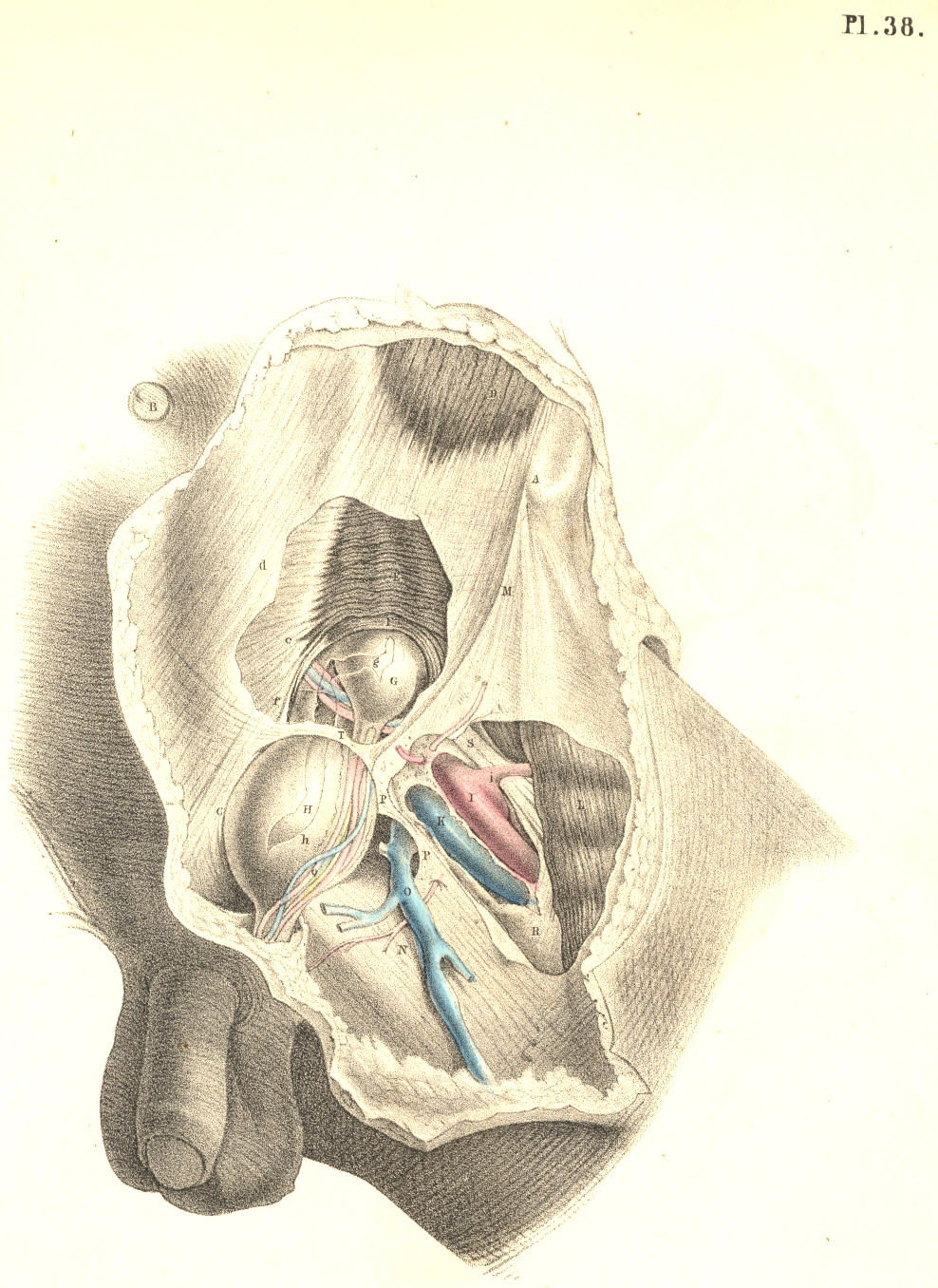
Plate 38
Plate 38
COMMENTARY ON PLATES 39 & 40.
DEMONSTRATIONS OF THE NATURE OF CONGENITAL AND INFANTILE INGUINAL HERNIAE, AND OF HYDROCELE.
DEMONSTRATIONS OF THE NATURE OF CONGENITAL AND INFANTILE INGUINAL HERNIAE, AND OF HYDROCELE.
PLATE 39. Fig. 1—The descent of the testicle from the loins to the scrotum.—The foetal abdomen and scrotum form one general cavity, and are composed of parts which are structurally identical. The cutaneous, fascial, muscular, and membranous layers of the abdominal parietes are continued into those of the scrotum. At the fifth month of foetal life, the testicle, 3, is situated in the loins beneath the kidney, 2. The testicle is then numbered amongst the abdominal viscera, and, like these, it is developed external to the peritonaeal membrane, which forms an envelope for it. At the back and sides of the testicle, where the peritonaeum is reflected from it, a small membranous fold or mesentery (mesorchium, Seiler) is formed, and between the layers of this the nerves and vessels enter the organ, the nerves being derived from the neighbouring sympathetic ganglia (aortic plexus), while the arteries and veins spring directly from the main abdominal bloodvessels. It being predetermined that the testicle, 3, should migrate from the loins to the scrotum, 6 a, 7, at a period included between the sixth and ninth month, certain structural changes are at this time already effected for its sure and easy passage. By the time that the testis, 5, is about to enter the internal inguinal ring, 6 a, (seventh or eighth month,) a process or pouch of the peritonaeal membrane (processus vaginalis) has already descended through this aperture into the scrotum, and the testicle follows it.
PLATE 39. Fig. 1—The descent of the testicle from the loins to the scrotum.—The fetal abdomen and scrotum form one continuous cavity and are made up of structurally identical parts. The skin, fascia, muscles, and membrane layers of the abdominal walls extend into those of the scrotum. At the fifth month of fetal life, the testicle, 3, is located in the loins beneath the kidney, 2. At this stage, the testicle is considered to be among the abdominal organs, and like them, it develops outside the peritoneal membrane, which surrounds it. At the back and sides of the testicle, where the peritoneum reflects away, a small membranous fold or mesentery (mesorchium, Seiler) is formed, through which the nerves and blood vessels enter the organ. The nerves come from nearby sympathetic ganglia (aortic plexus), while the arteries and veins originate directly from the main abdominal blood vessels. Since it is determined that the testicle, 3, will move from the loins to the scrotum, 6 a, 7, between the sixth and ninth months, certain structural changes are already taking place to ensure its smooth passage. By the time the testis, 5, is ready to enter the internal inguinal ring, 6 a, (in the seventh or eighth month), a pouch of the peritoneal membrane (processus vaginalis) has already descended through this opening into the scrotum, and the testicle follows it.
The descent of the testis is effected by a very slow and gradual process of change. (Tout va par degres dans la nature, et rien par sauts.—Bonnet.) But how, or by what distinct and active structural agent, this descent is effected, or whether there does exist, in fact, any such agent as that which anatomists name “gubernaculum testis,” are questions which appear to me by no means settled.[Footnote]
The descent of the testis happens through a very slow and gradual process of change. (Everything in nature happens gradually, nothing in leaps.—Bonnet.) But how, or by what specific and active structural agent this descent occurs, or whether there really is an agent that anatomists refer to as the “gubernaculum testis,” are questions that still seem to me far from settled.[Footnote]
[Footnote: Dr. Carpenter (Principles of Human Physiology) remarks, that “the cause of this descent is not very clear. It can scarcely be due merely, as some have supposed, to the contraction of the gubernaculum, since that does not contain any fibrous structure until after the lowering of the testis has commenced.” Dr. Sharpey (Quain’s Anatomy, 5th edition) observes, that “the office of the gubernaculum is yet imperfectly understood.” The opinions of these two distinguished physiologists will doubtless be regarded as an impartial estimate of the results of the researches prosecuted in reference to these questions by Haller, Camper, Hunter, Arnaud, Lobstein, Meckel, Paletta, Wrisberg, Vicq d’Azyr, Brugnone, Tumiati, Seiler, Girardi, Cooper, Bell, Weber, Carus, Cloquet, Curling, and others. From my own observations, I am led to believe that no such muscular structure as a gubernaculum exists, and therefore that the descent of the testis is the effect of another cause. Leaving these matters, however, to the consideration of the physiologist, it is sufficient for the surgeon to know that the testis in its transition derives certain coverings from the parietes of the groin, and that a communication is thereby established between the scrotal and abdominal cavities.]
[Footnote: Dr. Carpenter (Principles of Human Physiology) notes that “the cause of this descent isn’t very clear. It can hardly be just due to the contraction of the gubernaculum, as some have thought, since it doesn’t contain any fibrous structure until after the lowering of the testis has started.” Dr. Sharpey (Quain’s Anatomy, 5th edition) states that “the role of the gubernaculum is still not well understood.” The views of these two respected physiologists are likely to be seen as an unbiased assessment of the findings from research conducted on these issues by Haller, Camper, Hunter, Arnaud, Lobstein, Meckel, Paletta, Wrisberg, Vicq d’Azyr, Brugnone, Tumiati, Seiler, Girardi, Cooper, Bell, Weber, Carus, Cloquet, Curling, and others. From my observations, I believe that no muscular structure known as a gubernaculum exists, and thus the descent of the testis is caused by something else. However, setting these issues aside for the physiologists to consider, it suffices for the surgeon to know that as the testis moves down, it takes on certain coverings from the walls of the groin, and this creates a connection between the scrotal and abdominal cavities.]
The general lining membrane of the foetal abdomen is composed of two layers—an outer one of fibrous, and an inner one of serous structure. Of these two layers, the abdominal viscera form for themselves a double envelope. [Footnote] The testis in the loins has a covering from both membranes, and is still found to be enclosed by both, even when it has descended to the scrotum. The two coverings of fibro-serous structure which surrounded the testis in the loins become respectively the tunica albuginea and tunica vaginalis when the gland occupies the scrotal cavity.
The general lining membrane of the fetal abdomen has two layers—an outer fibrous layer and an inner serous layer. The abdominal organs create a double layer around themselves from these two layers. [Footnote] The testis in the lower back is covered by both membranes and remains encased by both even after it descends into the scrotum. The two fibro-serous coverings that surrounded the testis in the lower back become the tunica albuginea and tunica vaginalis when the gland is in the scrotal cavity.
[Footnote: Langenbeck describes the peritonaeum as consisting of two layers; one external and fibrous, another internal and serous. By the first, he means, I presume, that membrane of which the transversalis and iliac fasciae are parts. (See Comment. de Periton. Structura, &c.) ]
[Footnote: Langenbeck describes the peritoneum as having two layers; one is external and fibrous, while the other is internal and serous. By the first, I assume he refers to the membrane that includes the transversalis and iliac fasciae. (See Comment. de Periton. Structura, &c.) ]

Plate 39—Figure 1
Plate 39—Fig 1
PLATE 39, Fig. 2.—The testicle in the scrotum.—When the testicle, 5, descends into the scrotum, 7, which happens in general at the time of birth, the abdomino-scrotal fibro-serous membrane, 6 a, 6 d, is still continuous at the internal ring, 6 b. From this point downwards, to a level with the upper border of the testicle, the canal of communication between the scrotal cavity and the abdomen becomes elongated and somewhat constricted. At this part, the canal itself consists, like the abdominal membrane above and the scrotal membrane below, of a fibrous and serous layer, the latter enclosed within the former. The serous lining of this canal is destined to be obliterated, while the outer fibrous membrane is designed to remain in its primitive condition. When the serous canal contracts and degenerates to the form of a simple cord, it leaves the fibrous canal still continuous above with the fibrous membrane (transversalis fascia) of the abdomen, and below with the fibrous envelope (tunica albuginea) of the testis; and at the adult period, this fibrous canal is known as the internal spermatic sheath, or infundibuliform fascia enclosing the remains of the serous canal, together with the spermatic vessels, &c.
PLATE 39, Fig. 2.—The testicle in the scrotum.—When the testicle, 5, drops into the scrotum, 7, which usually happens at birth, the abdomino-scrotal fibro-serous membrane, 6 a, 6 d, is still connected at the internal ring, 6 b. From this point down to the upper edge of the testicle, the channel between the scrotal cavity and the abdomen becomes longer and a bit narrower. In this section, the channel itself is made up, like the abdominal membrane above and the scrotal membrane below, of a fibrous layer and a serous layer, the latter contained within the former. The serous lining of this channel is meant to be eliminated, while the outer fibrous membrane is expected to stay in its original form. When the serous channel narrows and shrinks down to a simple cord, it leaves the fibrous channel still connected above to the fibrous membrane (transversalis fascia) of the abdomen, and below to the fibrous covering (tunica albuginea) of the testis; and in adulthood, this fibrous channel is referred to as the internal spermatic sheath or infundibuliform fascia, which encloses the remnants of the serous channel along with the spermatic vessels, etc.

Plate 39—Figure 2
Plate 39—Fig. 2
PLATE 39, Fig. 3.—The serous tunica vaginalis is separated from the peritonaeum.—When the testicle, 7, has descended to the scrotum, the serous tube or lining of the inguinal canal and cord, 6 b, 6 c, closes and degenerates into a simple cord, (infantile spermatic cord,) and thereby the peritonaeal sac, 6 a, becomes distinct from the serous tunica vaginalis, 6 d. But the fibrous tube, or outer envelope of the inguinal canal, remains still pervious, and continues in this condition throughout life. In the adult, we recognise this fibrous tube as the infundibuliform fascia of the cord, or as forming the fascia propria of an external inguinal hernia. The anterior part of the fibrous spermatic tube descends from the fascia transversalis; the posterior part is continuous with the fascia iliaca. In relation to the testicle, the posterior part will be seen to be reflected over the body of the gland as the tunica albuginea, while the anterior part blends with the cellular tissue of the front wall of the scrotum. The tunica vaginalis, 6 d, is now traceable as a distinct sac,[Footnote] closed on all sides, and reflected from the fore part of the testicle, above and below, to the posterior aspect of the front wall of the scrotum.
PLATE 39, Fig. 3.—The serous tunica vaginalis is separated from the peritoneum.—When the testicle, 7, has descended to the scrotum, the serous tube or lining of the inguinal canal and cord, 6 b, 6 c, closes and turns into a simple cord (infantile spermatic cord), which makes the peritoneal sac, 6 a, distinct from the serous tunica vaginalis, 6 d. However, the fibrous tube, or outer layer of the inguinal canal, remains open and stays that way throughout life. In adults, we recognize this fibrous tube as the infundibuliform fascia of the cord or as part of the fascia propria of an external inguinal hernia. The front part of the fibrous spermatic tube extends from the transversalis fascia, while the back part connects with the iliac fascia. In relation to the testicle, the back part will be seen as it wraps around the body of the gland as the tunica albuginea, while the front part merges with the tissue of the front wall of the scrotum. The tunica vaginalis, 6 d, is now identifiable as a distinct sac,[Footnote] closed on all sides, and extending from the front part of the testicle, above and below, to the back side of the front wall of the scrotum.
[Footnote: Mr. Owen states that the Chimpanzee alone, amongst brute animals, has the tunica vaginalis as a distinct sac.]
[Footnote: Mr. Owen states that the Chimpanzee, unlike other animals, has the tunica vaginalis as a separate sac.]

Plate 39—Figure 3
Plate 39—Figure 3
PLATE 40, Fig. 1.—The abdomino-scrotal serous lining remains continuous at the internal ring, and a congenital hydrocele is formed.—When the serous spermatic tube, 6 b, 6 c, remains pervious and continuous above with the peritonaeum, 6 a, and below with the serous tunica vaginalis, 6 d, the serous fluid of the abdomen will naturally gravitate to the most depending part—viz., the tunica vaginalis; and thus a hydrocele is formed. This kind of hydrocele is named congenital, owing to the circumstance that the natural process of obliteration, by which the peritonaeum becomes separated from the tunica vaginalis, has been, from some cause, arrested. [Footnote 1] As long as the canal of communication, 6 b, 6 c, between the tunica vaginalis, 6 d, and the peritonaeum 6 a, remains pervious, which it may be throughout life, this form of hydrocele is, of course, liable to occur. It may be diagnosed from diseased enlargements of the testicle, by its transparency, its fluctuation, and its smooth, uniform fulness and shape, besides its being of less weight than a diseased testis of the same size would be. It may be distinguished from the common form of hydrocele of the isolated tunica vaginalis by the fact, that pressure made on the scrotum will cause the fluid to pass freely into the general cavity of the peritonaeum. As the fluid distends the tunica vaginalis, 6 c, 6 d, in front of the testis, this organ will of course lie towards the back of the scrotum, and therefore, if it be found necessary to evacuate the fluid, the puncture may be made with most safety in front of the scrotum. If ascites should form in an adult in whom the tunica vaginalis still communicates with the peritonaeal sac, the fluid which accumulates in the latter membrane will also distend the former, and all the collected fluid may be evacuated by tapping the scrotum. When a hydrocele is found to be congenital, it must be at once obvious that to inject irritating fluids into the tunica vaginalis (the radical cure) is inadmissible. In an adult, free from all structural disease, and in whom a congenital hydrocele is occasioned by the gravitation of the ordinary serous secretion of the peritonaeum, a cure may be effected by causing the obliteration of the serous spermatic canal by the pressure of a truss. When a congenital hydrocele happens in an infant in whom the testicle, 5, Fig. 1, Plate 39, is arrested in the inguinal canal, [Footnote 2] if pressure be made on this passage with a view of causing its closure, the testicle will be prevented from descending.
PLATE 40, Fig. 1.—The abdomen-scrotum serous lining stays connected at the internal ring, resulting in a congenital hydrocele.—When the serous spermatic tube, 6 b, 6 c, remains open and linked above to the peritoneum, 6 a, and below to the serous tunica vaginalis, 6 d, the abdominal serous fluid will naturally flow to the lowest point—specifically, the tunica vaginalis; thus, a hydrocele forms. This type of hydrocele is called congenital because the natural process of closure that separates the peritoneum from the tunica vaginalis has been halted for some reason. [Footnote 1] As long as the communication channel, 6 b, 6 c, between the tunica vaginalis, 6 d, and the peritoneum 6 a, remains open—which can happen throughout life—this form of hydrocele can occur. It can be diagnosed from diseased enlargement of the testicle by its transparency, fluctuation, and smooth, even fullness and shape, in addition to being lighter than a diseased testis of the same size. It can be differentiated from the common form of hydrocele of the isolated tunica vaginalis because applying pressure to the scrotum will allow the fluid to move freely into the general cavity of the peritoneum. As the fluid fills the tunica vaginalis, 6 c, 6 d, in front of the testis, this organ will naturally sit towards the back of the scrotum, so if it becomes necessary to drain the fluid, the puncture can safely be made in front of the scrotum. If ascites develops in an adult with a tunica vaginalis still connected to the peritoneal sac, the fluid accumulating in the latter membrane will also stretch the former, and all the gathered fluid can be drained by tapping the scrotum. When a hydrocele is found to be congenital, it is clear that injecting irritating fluids into the tunica vaginalis (the radical cure) is not appropriate. In a healthy adult, where a congenital hydrocele is caused by the gravitational pull of the normal serous secretion from the peritoneum, a cure can be achieved by pressing a truss to close off the serous spermatic canal. When a congenital hydrocele occurs in an infant with the testicle, 5, Fig. 1, Plate 39, stuck in the inguinal canal, [Footnote 2] applying pressure to this passage to promote closure will prevent the testicle from descending.
[Footnote 1: The serous spermatic tube remains open in all quadrupeds; but their natural prone position renders them secure against hydrocele or hernial protrusion. It is interesting to notice how in man, and the most anthropo-morphous animals, where the erect position would subject these to the frequent accident of hydrocele or hernia, nature causes the serous spermatic tube to close.]
[Footnote 1: The serous spermatic tube stays open in all four-legged animals; but their natural lying-down position protects them from hydrocele or hernia. It’s interesting to see how in humans and the most human-like animals, where the upright position would make them more prone to issues like hydrocele or hernia, nature causes the serous spermatic tube to close.]
[Footnote 2: In many quadrupeds (the Rodentia and Monotremes) the testes remain within the abdomen. In the Elephant, the testes always occupy their original position beneath the kidneys, in the loins. Human adults are occasionally found to be “testi-conde;” the testes being situated below the kidneys, or at some part between this position and the internal inguinal ring. Sometimes only one of the testes descends to the scrotum.]
[Footnote 2: In many four-legged animals (like rodents and monotremes), the testes stay inside the abdomen. In elephants, the testes always stay in their original spot beneath the kidneys, in the lower back area. In adult humans, it's occasionally found that they have "testicular retention," meaning the testes are located below the kidneys or somewhere in between that point and the internal inguinal ring. Sometimes, only one of the testes drops down into the scrotum.]

Plate 40—Figure 1.
Plate 40—Fig. 1.
PLATE 40, Fig. 2.—The serous spermatic canal closes imperfectly, so as to become sacculated, and thus a hydrocele of the cord is formed.—After the testicle, 7, has descended to the scrotum, the sides of the serous tube, or lining of the inguinal canal and cord, 6 b, 6 c, may become adherent at intervals; and the intervening sacs of serous membrane continuing to secrete their proper fluid, will occasion a hydrocele of the cord. This form of hydrocele will differ according to the varieties in the manner of closure; and these may take place in the following modes:—1st, if the serous tube close only at the internal ring, 6 a, while the lower part of it, 6 b, 6 c, remains pervious, and communicating with the tunica vaginalis, 6 d, a hydrocele will be formed of a corresponding shape; 2nd, if the tube close at the upper part of the testicle, 6 c, thus isolating the tunica vaginalis, 6 d, while the upper part, 6 b, remains pervious, and the internal ring, 6 a, open, and communicating with the peritonaeal sac, a hydrocele of the cord will happen distinct from the tunica vaginalis; or this latter may be, at the same time, distended with fluid, if the disposition of the subject be favourable to the formation of dropsy; 3rd, the serous tube may close at the internal ring, form sacculi along the cord, and close again at the top of the testicle, thus separating the tunica vaginalis from the abdomen, and thereby several isolated hydroceles may be formed. If in this condition of the parts we puncture one of the sacs for the evacuation of its contents, the others, owing to their separation, will remain distended.
PLATE 40, Fig. 2.—The serous spermatic canal closes in an incomplete manner, creating pouches that lead to a hydrocele of the cord.—After the testicle, 7, has dropped into the scrotum, the sides of the serous tube, or lining of the inguinal canal and cord, 6 b, 6 c, may stick together at various points; the remaining sections of serous membrane will keep producing fluid, resulting in a hydrocele of the cord. This type of hydrocele will vary based on how the closure occurs; and these may happen in the following ways:—1st, if the serous tube closes only at the internal ring, 6 a, while the lower part, 6 b, 6 c, stays open and connected to the tunica vaginalis, 6 d, a hydrocele will form in a similar shape; 2nd, if the tube closes at the top of the testicle, 6 c, thus separating the tunica vaginalis, 6 d, while the upper part, 6 b, remains open and the internal ring, 6 a, stays open and connected to the peritoneal sac, a hydrocele of the cord will occur separately from the tunica vaginalis; or this latter may also be filled with fluid if the individual is prone to fluid accumulation; 3rd, the serous tube may close at the internal ring, form pouches along the cord, and then close again at the top of the testicle, thus isolating the tunica vaginalis from the abdomen, leading to several separate hydroceles. If we puncture one of the pouches to drain its contents in this situation, the others will remain swollen due to their separation.

Plate 40—Figure 2.
Plate 40—Fig. 2.
PLATE 40, Fig. 3.—Hydrocele of the isolated tunica vaginalis.—When the serous spermatic tube, 6 b, 6 c, becomes obliterated, according to the normal rule, after the descent of the testicle, 7, the tunica vaginalis, 6 d, is then a distinct serous sac. If a hydrocele form in this sac, it may be distinguished from the congenital variety by its remaining undiminished in bulk when the subject assumes the horizontal position, or when pressure is made on the tumour, for its contents cannot now be forced into the abdomen. The testicle, 7, holds the same position in this as it does in the congenital hydrocele. [Footnote] The radical cure may be performed here without endangering the peritonaeal sac. Congenital hydrocele is of a cylindrical shape; and this is mentioned as distinguishing it from isolated hydrocele of the tunica vaginalis, which is pyriform; but this mark will fail when the cord is at the same time distended, as it may be, in the latter form of the complaint.
PLATE 40, Fig. 3.—Hydrocele of the isolated tunica vaginalis.—When the serous spermatic tube, 6 b, 6 c, becomes closed off, as is normally expected after the testicle descends, 7, the tunica vaginalis, 6 d, then forms a separate serous sac. If a hydrocele develops in this sac, it can be differentiated from the congenital type by its size remaining the same when the person lies down or when pressure is applied to the tumor, since its contents cannot be pushed into the abdomen anymore. The testicle, 7, stays in the same position in this case as it does in congenital hydrocele. [Footnote] The radical cure can be conducted here without risking damage to the peritoneal sac. Congenital hydrocele has a cylindrical shape, which helps distinguish it from isolated hydrocele of the tunica vaginalis, which is pear-shaped; however, this distinction may not hold if the cord is also distended, as can happen in the latter type of the condition.
[Footnote: When a hydrocele is interposed between the eye and a strong light, the testis appears as an opaque body at the back of the tunica vaginalis. But this position of the organ is, from several causes, liable to vary. The testis may have become morbidly adherent to the front wall of the serous sac, in which case the hydrocele will distend the sac laterally. Or the testis may be so transposed in the scrotum, that, whilst the gland occupies its front part, the distended tunica vaginalis is turned behind. The tunica vaginalis, like the serous spermatic tube, may, in consequence of inflammatory fibrinous effusion, become sacculated-multilocular, in which case, if a hydrocele form, the position of the testis will vary accordingly.—See Sir Astley Cooper’s work, (“Anatomy and Diseases of the Testis;”) Morton’s “Surgical Anatomy;” Mr. Curling’s “Treatise on Diseases of the Testis;” and also his article “Testicle,” in the Cyclopaedia of Anatomy and Physiology.]
[Footnote: When a hydrocele is between the eye and a strong light, the testis looks like an opaque object at the back of the tunica vaginalis. However, this position can change for several reasons. The testis might be abnormally stuck to the front wall of the serous sac, causing the hydrocele to expand the sac sideways. Alternatively, the testis could be positioned in such a way that, while the gland is in the front part of the scrotum, the distended tunica vaginalis is located behind it. The tunica vaginalis, similar to the serous spermatic tube, can become sacculated-multilocular due to inflammatory fibrinous effusion, which means that if a hydrocele forms, the position of the testis will change accordingly.—See Sir Astley Cooper’s work, (“Anatomy and Diseases of the Testis;”) Morton’s “Surgical Anatomy;” Mr. Curling’s “Treatise on Diseases of the Testis;” and also his article “Testicle,” in the Cyclopaedia of Anatomy and Physiology.]

Plate 40—Figure 3.
Plate 40—Fig. 3.
PLATE 40, Fig. 4.—The serous spermatic tube remaining pervious, a congenital hernia is formed.—When the testicle, 7, has descended to the scrotum, if the communication between the peritonaeum, 6 a, and the tunica vaginalis, 6 c, be not obliterated, a fold of the intestine, 13, will follow the testicle, and occupy the cavity of the tunica vaginalis, 6 d. In this form of hernia (hernia tunicae vaginalis, Cooper), the intestine is in front of, and in immediate contact with, the testicle. The intestine may descend lower than the testicle, and envelope this organ so completely as to render its position very obscure to the touch. This form of hernia is named congenital, since it occurs in the same condition of the parts as is found in congenital hydrocele—viz., the inguinal ring remaining unclosed. It may occur at any period of life, so long as the original congenital defect remains. It may be distinguished from hydrocele by its want of transparency and fluctuation. The impulse which is communicated to the hand applied to the scrotum of a person affected with scrotal hernia, when he is made to cough, is also felt in the case of congenital hydrocele. But in hydrocele of the separate tunica vaginalis, such impulse is not perceived. Congenital hernia and hydrocele may co-exist; and, in this case, the diagnostic signs which are proper to each, when occurring separately, will be so mingled as to render the precise nature of the case obscure.
PLATE 40, Fig. 4.—The open spermatic tube leads to a congenital hernia.—When the testicle, 7, has moved down to the scrotum, if the connection between the peritoneum, 6 a, and the tunica vaginalis, 6 c, is not closed off, a loop of the intestine, 13, will follow the testicle and fill the space in the tunica vaginalis, 6 d. In this type of hernia (hernia tunicae vaginalis, Cooper), the intestine is in front of and in direct contact with the testicle. The intestine may move down further than the testicle and completely wrap around it, making its position hard to identify by touch. This type of hernia is called congenital because it occurs in the same state of the parts as seen in congenital hydrocele—namely, the inguinal ring remains open. It can happen at any point in life as long as the original congenital defect persists. It can be distinguished from hydrocele by its lack of transparency and fluctuation. The movement felt through the hand applied to the scrotum of someone with a scrotal hernia when they cough is also noticed in cases of congenital hydrocele. However, in the hydrocele of the separate tunica vaginalis, this movement is not felt. Congenital hernia and hydrocele can occur together; in such cases, the diagnostic signs of each, when occurring alone, will be mixed together, making the exact nature of the situation unclear.

Plate 40—Figure 4.
Plate 40—Fig. 4.
PLATE 40, Fig. 5.—Infantile hernia.—When the serous spermatic tube becomes merely closed, or obliterated at the inguinal ring, 6 b, the lower part of it, 6 c, is pervious, and communicating with the tunica vaginalis, 6 d. In consequence of the closure of the tube at the inguinal ring, if a hernia now occur, it cannot enter the tunica vaginalis, and come into actual contact with the testicle. The hernia, 13, therefore, when about to force the peritonaeum, 6 a, near the closed ring, 6 b, takes a distinct sac or investment from this membrane. This hernial sac, 6 e, will vary as to its position in regard to the tunica vaginalis, 6 d, according to the place whereat it dilates the peritonaeum at the ring. The peculiarity of this hernia, as distinguished from the congenital form, is owing to the scrotum containing two sacs,—the tunica vaginalis and the proper sac of the hernia; whereas, in the congenital variety, the tunica vaginalis itself becomes the hernial sac by a direct reception of the naked intestine. If in infantile hernia a hydrocele should form in the tunica vaginalis, the fluid will also distend the pervious serous spermatic tube, 6 c, as far up as the closed internal ring, 6 b, and will thus invest and obscure the descending herniary sac, 13. This form of hernia is named infantile (Hey), owing to the congenital defect in that process, whereby the serous tube lining the cord is normally obliterated. Such a form of hernia may occur at the adult age for the first time, but it is still the consequence of original default.
PLATE 40, Fig. 5.—Infantile hernia.—When the serous spermatic tube is only blocked or closed at the inguinal ring, 6 b, the lower part, 6 c, remains open and connects with the tunica vaginalis, 6 d. Because the tube is closed at the inguinal ring, if a hernia occurs, it can't enter the tunica vaginalis and make contact with the testicle. Therefore, the hernia, 13, as it pushes against the peritoneum, 6 a, near the closed ring, 6 b, forms a distinct sac or covering from this membrane. This hernial sac, 6 e, will vary in position relative to the tunica vaginalis, 6 d, depending on where it expands the peritoneum at the ring. The unique aspect of this hernia, unlike the congenital type, is that the scrotum contains two sacs—the tunica vaginalis and the proper sac of the hernia; whereas in the congenital type, the tunica vaginalis itself acts as the hernial sac by directly receiving the exposed intestine. If a hydrocele forms in the tunica vaginalis in cases of infantile hernia, the fluid will also stretch the open serous spermatic tube, 6 c, up to the closed internal ring, 6 b, and will thus cover and obscure the descending hernial sac, 13. This type of hernia is called infantile (Hey) due to the congenital defect in the process where the serous tube lining the cord is normally closed off. Such a hernia can appear for the first time in adults, but it still results from an original defect.

Plate 40—Figure 5.
Plate 40 - Fig. 5.
PLATE 40, Fig. 6.—Oblique inguinal hernia in the adult.—This variety of hernia occurs not in consequence of any congenital defect, except inasmuch as the natural weakness of the inguinal wall opposite the internal ring may be attributed to this cause. The serous spermatic tube has been normally obliterated for its whole length between the internal ring and the tunica vaginalis; but the fibrous tube, or spermatic fascia, is open at the internal ring where it joins the transversalis fascia, and remains pervious as far down as the testicle. The intestine, 13, forces and distends the upper end of the closed serous tube; and as this is now wholly obliterated, the herniary sac, 6 c, derived anew from the inguinal peritonaeum, enters the fibrous tube, or sheath of the cord, and descends it as far as the tunica vaginalis, 6 d, but does not enter this sac, as it is already closed. When we compare this hernia, Fig. 6, Plate 40, with the infantile variety, Fig. 5, Plate 40, we find that they agree in so far as the intestinal sac is distinct from the tunica vaginalis; whereas the difference between them is caused by the fact of the serous cord remaining in part pervious in the infantile hernia; and on comparing Fig. 6, Plate 40, with the congenital variety, Fig. 4, Plate 40, we see that the intestine has acquired a new sac in the former, whereas, in the latter, the intestine has entered the tunica vaginalis. The variable position of the testicle in Figs. 4, 5, & 6, Plate 40, is owing to the variety in the anatomical circumstances under which these herniae have happened.
PLATE 40, Fig. 6.—Oblique inguinal hernia in adults.—This type of hernia doesn’t happen due to any congenital defect, except that the natural weakness of the inguinal wall across from the internal ring can be attributed to this cause. The serous spermatic tube has completely closed along its length between the internal ring and the tunica vaginalis; however, the fibrous tube, or spermatic fascia, stays open at the internal ring where it connects with the transversalis fascia, and remains open all the way down to the testicle. The intestine, 13, pushes and stretches the upper end of the closed serous tube; and since this tube is now fully closed, the hernial sac, 6 c, forms anew from the inguinal peritoneum, enters the fibrous tube, or sheath of the cord, and moves down into it as far as the tunica vaginalis, 6 d, but doesn’t enter this sac as it is already closed. When we compare this hernia, Fig. 6, Plate 40, with the infantile type, Fig. 5, Plate 40, we find that they share the characteristic that the intestinal sac is separate from the tunica vaginalis; however, the difference between them is due to the fact that the serous cord remains partly open in the infantile hernia. Comparing Fig. 6, Plate 40, with the congenital type, Fig. 4, Plate 40, shows us that the intestine has developed a new sac in the former case, while in the latter, the intestine has entered the tunica vaginalis. The varying positions of the testicle in Figs. 4, 5, & 6, Plate 40, arise from the differences in the anatomical circumstances under which these hernias occurred.

Plate 40—Figure 6.
Plate 40—Fig. 6.
COMMENTARY ON PLATES 41 & 42.
DEMONSTRATIONS OF THE ORIGIN AND PROGRESS OF INGUINAL HERNIAE IN GENERAL.
DEMONSTRATIONS OF THE ORIGIN AND PROGRESS OF INGUINAL HERNIAE IN GENERAL.
PLATE 41, Fig. 1.—When the serous spermatic tube is obliterated for its whole length between the internal ring, 1, and the top of the testicle, 13, a hernia, in order to enter the inguinal canal, 1, 4, must either rupture the peritonaeum at the point 1, or dilate this membrane before it in the form of a sac. [Footnote] If the peritonaeum at the point 1 be ruptured by the intestine, this latter will enter the fibrous spermatic tube, 2, 3, and will pass along this tube devoid of the serous sac. If, on the other hand, the intestine dilates the serous membrane at the point, 1, where it stretches across the internal ring, it will, on entering the fibrous tube, (infundibuliform fascia,) be found invested by a sac of the peritonaeum, which it dilates and pouches before itself. As the epigastric artery, 9, bends in general along the internal border of the ring of the fibrous tube, 2, 2, the neck of the hernial sac which enters the ring at a point external to the artery must be external to it, and remain so despite all further changes in the form, position, and dimensions of the hernia. And as this hernia enters the ring at a point anterior to the spermatic vessels, its neck must be anterior to them. Again, if the bowel be invested by a serous sac, formed of the peritonaeum at the point 1, the neck of such sac must intervene between the protruding bowel and the epigastric and spermatic vessels. But if the intestine enter the ring of the fibrous tube, 2, 2, by having ruptured the peritonaeum at the point 1, then the naked intestine will lie in immediate contact with these vessels.
PLATE 41, Fig. 1.—When the serous spermatic tube is closed off completely between the internal ring, 1, and the top of the testicle, 13, a hernia wanting to enter the inguinal canal, 1, 4, must either break through the peritoneum at point 1, or stretch this membrane into a sac shape. [Footnote] If the peritoneum at point 1 is broken by the intestine, it will enter the fibrous spermatic tube, 2, 3, and travel through this tube without the serous sac. If, however, the intestine stretches the serous membrane at point 1, where it crosses the internal ring, it will enter the fibrous tube (infundibuliform fascia) covered by a peritoneal sac, which it will stretch and pouch in front of it. Since the epigastric artery, 9, generally runs along the inner edge of the fibrous tube’s ring, 2, 2, the neck of the hernial sac entering the ring at a point outside the artery must be outside of it and will remain so despite any further changes in the shape, position, and size of the hernia. Additionally, as this hernia enters the ring at a point in front of the spermatic vessels, its neck must be in front of them as well. Furthermore, if the bowel is covered by a serous sac made from the peritoneum at point 1, the neck of that sac must be between the protruding bowel and the epigastric and spermatic vessels. But if the intestine enters the ring of the fibrous tube, 2, 2, by breaking the peritoneum at point 1, then the bare intestine will be in direct contact with these vessels.
[Footnote: Mr. Lawrence (op. cit.) remarks, “When we consider the texture of the peritonaeum, and the mode of its connexion to the abdominal parietes, we cannot fancy the possibility of tearing the membrane by any attitude or motion.” Cloquet and Scarpa have also expressed themselves to the effect, that the peritonaeum suffers a gradual distention before the protruding bowel.]
[Footnote: Mr. Lawrence (op. cit.) says, “When we look at the texture of the peritoneum and how it connects to the abdominal walls, we can’t imagine that the membrane could be torn by any position or movement.” Cloquet and Scarpa have also indicated that the peritoneum experiences gradual stretching before the bowel begins to protrude.]

Plate 41—Figure 1
Plate 41—Fig 1
PLATE 41, Fig. 2—When the serous spermatic tube, 11, remains pervious between the internal ring, 1, (where it communicates with the general peritonaeal membrane,) and the top of the testicle, (where it opens into the tunica vaginalis,) the bowel enters this tube directly, without a rupture of the peritonaeum at the point 1. This tube, therefore, becomes one of the investments of the bowel. It is the serous sac, not formed by the protruding bowel, but one already open to receive the bowel. This is the condition necessary to the formation of congenital hernia. This hernia must be one of the external oblique variety, because it enters the open abdominal end of the infantile serous spermatic tube, which is always external to the epigastric artery. Its position in regard to the spermatic vessels is the same as that noticed in Fig, 1, Plate 41. But, as the serous tube through which the congenital hernia descends, still communicates with the tunica vaginalis, so will this form of hernia enter this tunic, and thereby become different to all other herniae, forasmuch as it will lie in immediate contact with the testicle. [Footnote]
PLATE 41, Fig. 2—When the thin spermatic tube, 11, stays open between the internal ring, 1, (where it connects to the general peritoneal membrane,) and the top of the testicle, (where it opens into the tunica vaginalis,) the intestine can enter this tube directly, without a rupture in the peritoneum at point 1. This tube then becomes one of the layers surrounding the intestine. It is the serous sac, not created by the protruding intestine, but one that's already open to receive it. This condition is essential for the development of a congenital hernia. This hernia must be of the external oblique type because it enters the open abdominal end of the infantile serous spermatic tube, which is always outside the epigastric artery. Its position in relation to the spermatic vessels is the same as shown in Fig. 1, Plate 41. However, since the serous tube through which the congenital hernia descends still connects with the tunica vaginalis, this type of hernia will enter this tunic, making it different from all other hernias, as it will be in direct contact with the testicle. [Footnote]
[Footnote: A hernia may be truly congenital, and yet the intestine may not enter the tunica vaginalis. Thus, if the serous spermatic tube close only at the top of the testicle, the bowel which traverses the open internal inguinal ring and pervious tube will not enter the tunica vaginalis.]
[Footnote: A hernia can be genuinely congenital, yet the intestine may not enter the tunica vaginalis. So, if the serous spermatic cord closes only at the top of the testicle, the bowel that goes through the open internal inguinal ring and open tube will not enter the tunica vaginalis.]

Plate 41—Figure 2
Plate 41—Fig. 2
PLATE 41, Fig. 3.—The infantile serous spermatic tube, 11, sometimes remains pervious in the neighbourhood of the internal ring, 1, and a narrow tapering process of the tube (the canal of Nuck) descends within the fibrous tube, 2, 3, and lies in front of the spermatic vessels and epigastric artery. Before this tube reaches the testicle, it degenerates into a mere filament, and thus the tunica vaginalis has become separated from it as a distinct sac. When the bowel enters the open abdominal end of the serous tube, this latter becomes the hernial sac. It is not possible to distinguish by any special character a hernia of this nature, when already formed, from one which occurs in the condition of parts proper to Fig. 1, Plate 41, or that which is described in the note to Fig. 2, Plate 41; for when the intestine dilates the tube, 11, into the form of a sac, this latter assumes the exact shape of the sac, as noticed in Fig. 1, Plate 41. The hernia in question cannot enter the tunica vaginalis. Its position in regard to the epigastric and spermatic vessels is the same as that mentioned above.
PLATE 41, Fig. 3.—The infant spermatic tube, 11, sometimes remains open near the internal ring, 1, and a narrow tapering part of the tube (the canal of Nuck) descends within the fibrous tube, 2, 3, lying in front of the spermatic vessels and epigastric artery. Before this tube reaches the testicle, it shrinks down to a thin filament, and the tunica vaginalis separates from it, forming a distinct sac. When the intestine enters the open abdominal end of the serous tube, this tube becomes the hernial sac. There’s no way to tell apart a hernia of this type from one that occurs in the setup shown in Fig. 1, Plate 41, or the one described in the note to Fig. 2, Plate 41; because when the intestine pushes out the tube, 11, into a sac shape, it takes on the exact form of the sac shown in Fig. 1, Plate 41. This hernia can't enter the tunica vaginalis. Its position relative to the epigastric and spermatic vessels is the same as mentioned above.

Plate 41—Figure 3
Plate 41—Fig 3
PLATE 41, Fig. 4.—If the serous spermatic tube, 11, be obliterated or closed at the internal ring, 1, thus cutting off communication with the general peritonaeal membrane; and if, at the same time, it remain pervious from this point above to the tunica vaginalis below, then the herniary bowel, when about to protrude at the point 1, must force and dilate the peritonaeum, in order to form its sac anew, as stated of Fig. 1, Plate 41. Such a hernia does not enter either the serous tube or the tunica vaginalis; but progresses from the point 1, in a distinct sac. In this case, there will be found two sacs—one enclosing the bowel; and another, consisting of the serous spermatic tube, still continuous with the tunica vaginalis. This original state of the parts may, however, suffer modification in two modes: 1st, if the bowel rupture the peritonaeum at the point 1, it will enter the serous tube 11, and descend through this into the cavity of the tunica vaginalis, as in the congenital variety. 2nd, if the bowel rupture the peritonaeum near the point 1, and does not enter the serous tube 11, nor the tunica vaginalis, then the bowel will be found devoid of a proper serous sac, while the serous tube and tunica vaginalis still exist in communication. In either case, the hernia will hold the same relative position in regard to the epigastric artery and spermatic vessels, as stated of Fig. 1, Plate 41.
PLATE 41, Fig. 4.—If the serous spermatic tube, 11, is blocked or closed at the internal ring, 1, cutting off communication with the general peritoneal membrane; and if, at the same time, it remains open from this point above to the tunica vaginalis below, then when the herniated bowel is about to push out at point 1, it must stretch and expand the peritoneum to create its sac again, as mentioned in Fig. 1, Plate 41. This type of hernia does not enter the serous tube or the tunica vaginalis but progresses from point 1 in a separate sac. In this case, there will be two sacs—one surrounding the bowel and another made up of the serous spermatic tube, which is still connected to the tunica vaginalis. However, this original state of the parts can change in two ways: 1st, if the bowel breaks through the peritoneum at point 1, it will enter the serous tube 11 and move down through it into the cavity of the tunica vaginalis, as seen in the congenital type. 2nd, if the bowel breaks the peritoneum near point 1 but does not enter the serous tube 11 or the tunica vaginalis, then the bowel will lack a proper serous sac, while the serous tube and tunica vaginalis will still be connected. In either situation, the hernia will maintain the same relative position regarding the epigastric artery and spermatic vessels, as detailed in Fig. 1, Plate 41.

Plate 41—Figure 4
Plate 41—Fig. 4
PLATE 41, Fig. 5.—Sudden rupture of the peritonaeum at the closed internal serous ring, 1, though certainly not impossible, may yet be stated as the exception to the rule in the formation of an external inguinal hernia. The aphorism, “natura non facit saltus,” is here applicable. When the peritonaeum suffers dilatation at the internal ring, 1, it advances gradatim and pari passu with the progress of the protruding bowel, and assumes the form, character, position, and dimensions of the inverted curved phases, marked 11, 11, till, from having at first been a very shallow pouch, lying external to the epigastric artery, 9, it advances through the inguinal canal to the external ring, 4, and ultimately traverses this aperture, taking the course of the fibrous tube, 3, down to the testicle in the scrotum.
PLATE 41, Fig. 5.—A sudden rupture of the peritoneum at the closed internal serous ring, 1, while certainly not impossible, can be considered an exception to the rule in the development of an external inguinal hernia. The saying, “nature does not make leaps,” applies here. When the peritoneum stretches at the internal ring, 1, it progresses gradually and in line with the protruding bowel, taking on the shape, characteristics, position, and size of the inverted curved phases marked 11, 11, until it evolves from a very shallow pouch, lying outside the epigastric artery, 9, and moves through the inguinal canal to the external ring, 4, eventually passing through this opening, following the path of the fibrous tube, 3, down to the testicle in the scrotum.

Plate 41—Figure 5
Plate 41—Fig. 5
PLATE 41, Fig. 6.—When the bowel dilates the peritonaeum opposite the internal ring, and carries a production of this membrane before it as its sac, then the hernia will occupy the inguinal canal, and become invested by all those structures which form the canal. These structures are severally infundibuliform processes, so fashioned by the original descent of the testicle; and, therefore, as the bowel follows the track of the testicle, it becomes, of course, invested by the selfsame parts in the selfsame manner. Thus, as the infundibuliform fascia, 2, 3, contains the hernia and spermatic vessels, so does the cremaster muscle, extending from the lower margins of the internal oblique and transversalis, invest them also in an infundibuliform manner. [Footnote]
PLATE 41, Fig. 6.—When the bowel expands and pushes the peritoneum in front of it at the internal ring, it creates a sac that allows the hernia to enter the inguinal canal, surrounded by all the structures that make up the canal. These structures are funnel-shaped processes, formed by the original descent of the testicle. Therefore, as the bowel follows the journey of the testicle, it naturally gets covered by the same parts in the same way. Thus, as the funnel-shaped fascia, 2, 3, contains the hernia and spermatic vessels, the cremaster muscle, which extends from the lower edges of the internal oblique and transversalis muscles, also encases them in a funnel shape. [Footnote]
[Footnote: Much difference of opinion prevails as to the true relation which the cord (and consequently the oblique hernia) bears to the lower margins of the oblique and transverse muscles, and their cremasteric prolongation. Mr. Guthrie (Inguinal and Femoral Hernia) has shown that the fibres of the transversalis, as well as those of the internal oblique, are penetrated by the cord. Albinus, Haller, Cloquet, Camper, and Scarpa, record opinions from which it may be gathered that this disposition of the parts is (with some exceptions) general. Sir Astley Cooper describes the lower edge of the transversalis as curved all round the internal ring and cord. From my own observations, coupled with these, I am inclined to the belief that, instead of viewing these facts as isolated and meaningless particulars, we should now fuse them into the one idea expressed by the philosophic Carus, and adopted by Cloquet, that the cremaster is a production of the abdominal muscles, formed mechanically by the testicle, which in its descent dilates, penetrates, and elongates their fibres.]
[Footnote: There's a lot of disagreement about the actual relationship between the cord (and therefore the oblique hernia) and the lower edges of the oblique and transverse muscles, as well as their cremasteric extension. Mr. Guthrie (Inguinal and Femoral Hernia) has shown that the fibers of the transversalis, along with those of the internal oblique, pass through the cord. Albinus, Haller, Cloquet, Camper, and Scarpa have recorded views suggesting that this arrangement is generally consistent, with some exceptions. Sir Astley Cooper describes the lower edge of the transversalis as curved around the internal ring and cord. Based on my own observations, along with these, I believe that instead of seeing these facts as isolated and meaningless details, we should unify them into a single idea expressed by the philosopher Carus, and adopted by Cloquet, that the cremaster is a product of the abdominal muscles, formed mechanically by the testicle, which, during its descent, expands, penetrates, and stretches their fibers.]

Plate 41—Figure 6
Plate 41—Fig 6
PLATE 41. Fig. 7.—When an external inguinal hernia, 11, dilates and protrudes the peritonaeum from the closed internal ring, 1, and descends the inguinal canal and fibrous tube, 3, 3, it imitates, in most respects, the original descent of the testicle. The difference between both descents attaches alone to the mode in which they become covered by the serous membrane; for the testicle passes through the internal ring behind the inguinal peritonaeum, at the same time that it takes a duplicature of this membrane; whereas the bowel encounters this part of the peritonaeum from within, and in this mode becomes invested by it on all sides. This figure also represents the form and relative position of a hernia, as occurring in Figs. 1 and 3, 5, and 6, Plate 41.
PLATE 41. Fig. 7.—When an external inguinal hernia, 11, stretches and pushes the peritoneum out from the closed internal ring, 1, and moves down the inguinal canal and fibrous tube, 3, 3, it resembles, in most ways, the original descent of the testicle. The only difference between the two descents is based on how they get covered by the serous membrane; the testicle passes through the internal ring behind the inguinal peritoneum, while taking a fold of this membrane; whereas the bowel approaches this part of the peritoneum from within, and in this way gets surrounded by it on all sides. This figure also shows the shape and relative position of a hernia, as seen in Figs. 1 and 3, 5, and 6, Plate 41.

Plate 41—Figure 7
Plate 41—Fig. 7
PLATE 41, Fig. 8.—When the serous spermatic tube only closes at the internal ring, as seen at 1, Fig. 4, Plate 41, if a hernia afterwards pouch the peritonaeum at this part, and enter the inguinal canal, we shall then have the form of hernia, Fig. 8, Plate 41, termed infantile. Two serous sacs will be here found, one within the cord, 13, and communicating with the tunica vaginalis, the other, 11, containing the bowel, and being received by inversion into the upper extremity of the first. Thus the infantile serous canal, 13, receives the hernial sac, 11. The inguinal canal and cord may become multicapsular, as in Fig. 8, from various causes, each capsule being a distinct serous membrane. First, independent of hernial formation, the original serous tube may become interruptedly obliterated, as in Plate 40, Fig. 2. Secondly, these sacs may persist to adult age, and have a hernial sac added to their number, whatever this may be. Thirdly, the original serous tube, 13, Fig. 8, may persist, and after having received the hernial sac, 11, the bowel may have been reduced, leaving its sac behind it in the inguinal canal; the neck of this sac may have been obliterated by the pressure of a truss, a second hernia may protrude at the point 1, and this may be received into the first hernial sac in the same manner as the first was received into the original serous infantile tube. The possibility of these occurrences is self-evident, even if they were never as yet experienced. [Footnote]
PLATE 41, Fig. 8.—When the serous spermatic tube only closes at the internal ring, as shown at 1, Fig. 4, Plate 41, if a hernia later bulges the peritoneum in this area and enters the inguinal canal, we will then observe the form of hernia depicted in Fig. 8, Plate 41, known as infantile. Two serous sacs will be present here: one within the cord, labeled 13, and connected to the tunica vaginalis, and the other, labeled 11, which contains the bowel and is inverted at the upper end of the first. Thus, the infantile serous canal, 13, receives the hernial sac, 11. The inguinal canal and cord can develop multiple sacs, as shown in Fig. 8, due to various causes, with each capsule representing a distinct serous membrane. First, regardless of hernia formation, the original serous tube may become intermittently blocked, as in Plate 40, Fig. 2. Secondly, these sacs may persist into adulthood and have an additional hernial sac, whatever that may be. Thirdly, the original serous tube, 13, Fig. 8, may remain, and after it has received the hernial sac, 11, the bowel might move back, leaving its sac behind in the inguinal canal; the neck of this sac may become blocked due to pressure from a truss, and a second hernia may emerge at point 1, which can be received into the first hernial sac in the same way that the first was taken up into the original serous infantile tube. The likelihood of these situations occurring is clear, even if they haven’t been experienced yet. [Footnote]
[Footnote: According to Mr. Lawrence and M. Cloquet, most of the serous cysts found around hernial tumours are ancient sacs obliterated at the neck, and adhering to the new swelling (opera cit.)]
[Footnote: According to Mr. Lawrence and M. Cloquet, most of the fluid-filled cysts found around hernial tumors are old sacs that have been sealed off at the neck and are attached to the new swelling (opera cit.)]

Plate 41—Figure 8
Plate 41—Fig 8
PLATE 42, Fig. 1.—The epigastric artery, 9, being covered by the fascia transversalis, can lend no support to the internal ring, 2, 2, nor to the tube prolonged from it. The herniary bowel may, therefore, dilate the peritonaeum immediately on the inner side of the artery, and enter the inguinal canal. In this way the hernia, 11, although situated internal to the epigastric artery, assumes an oblique course through the canal, and thus closely simulates the external variety of inguinal hernia, Fig. 7, Plate 41. If the hernia enter the canal, as represented in Fig. 1, Plate 42, it becomes invested by the same structures, and assumes the same position in respect to the spermatic vessels, as the external hernia.
PLATE 42, Fig. 1.—The epigastric artery, 9, being covered by the transversalis fascia, does not provide any support to the internal ring, 2, 2, or to the tube extending from it. Therefore, the herniated bowel can dilate the peritoneum just inside the artery and enter the inguinal canal. In this way, the hernia, 11, while located internal to the epigastric artery, follows an oblique path through the canal, closely resembling the external type of inguinal hernia, Fig. 7, Plate 41. If the hernia enters the canal, as shown in Fig. 1, Plate 42, it becomes surrounded by the same structures and takes the same position in relation to the spermatic vessels as the external hernia.

Plate 42—Figure 1
Plate 42—Fig. 1
PLATE 42, Fig. 2.—The hernial sac, 11, which entered the ring of the fibrous tube, 2, 2, at a point immediately internal to the epigastric artery, 9, may, from having been at first oblique, as in Fig. 1, Plate 42, assume a direct position. In this case, the ring of the fibrous tube, 2, 2, will be much widened; but the artery and spermatic vessels will remain in their normal position, being in no wise affected by the gravitating hernia. If the conjoined tendon, 6, be so weak as not to resist the gravitating force of the hernia, the tendon will become bent upon itself. If the umbilical cord, 10, be side by side with the epigastric artery at the time that the hernia enters the mouth of the fibrous tube, then, of course, the cord will be found external. If the cord lie towards the pubes, apart from the vessel, the hernia may enter the fibrous tube between the cord, 10, and artery, 9. [Footnote:] It is impossible for any internal hernia to assume the congenital form, because the neck of the original serous spermatic tube, 11, Fig. 2, Plate 41, being external to the epigastric artery, 9, cannot be entered by the hernia, which originates internally to this vessel.
PLATE 42, Fig. 2.—The hernial sac, 11, which passed through the opening of the fibrous tube, 2, 2, just inside the epigastric artery, 9, may, after initially being positioned obliquely like in Fig. 1, Plate 42, take on a direct position. In this situation, the opening of the fibrous tube, 2, 2, will be significantly widened; however, the artery and spermatic vessels will stay in their usual position, as they are not affected by the hernia's movement. If the conjoined tendon, 6, is weak enough to not withstand the downward pull of the hernia, it will bend upon itself. If the umbilical cord, 10, is next to the epigastric artery when the hernia enters the fibrous tube, then the cord will be located externally. If the cord is closer to the pubic area, away from the vessel, the hernia may enter the fibrous tube between the cord, 10, and the artery, 9. [Footnote:] It's impossible for any internal hernia to take on the congenital form, because the neck of the original serous spermatic tube, 11, Fig. 2, Plate 41, being outside the epigastric artery, 9, cannot be entered by the hernia that starts inside this vessel.
[Footnote: M. Cloquet states that the umbilical cord is always found on the inner side of the external hernia. Its position varies in respect to the internal hernia, (op. cit. prop. 52.)]
[Footnote: M. Cloquet states that the umbilical cord is always found on the inner side of the external hernia. Its position varies in relation to the internal hernia, (op. cit. prop. 52.)]

Plate 42—Figure 2
Plate 42—Figure 2
PLATE 42, Fig. 3.—Every internal hernia, which does not rupture the peritonaeum, carries forward a sac produced anew from this membrane, whether the hernia enter the inguinal canal or not. But this is not the case with respect to the fibrous membrane which forms the fascia propria. If the hernia enter the inguinal wall immediately on the inner side of the epigastric artery, Fig. 1, Plate 42, it passes direct into the ring of the fibrous tube, 2, 2, already prepared to receive it. But when the hernia, 11, Fig. 3, Plate 42, cleaves the conjoined tendon, 6, 6, then the artery, 9, and the tube, 2, 2, remain in their usual position, while the bowel carries forward a new investment from the transversalis fascia, 5, 5. That part of the conjoined tendon which stands external to the hernia keeps the tube, 2, 2, in its proper place, and separates it from the fold of the fascia which invests the hernial sac. This is the only form in which an internal hernia can be said to be absolutely distinct from the inguinal canal and spermatic vessels. This hernia, when passing the external ring, 4, has the spermatic cord on its outer side.
PLATE 42, Fig. 3.—Every internal hernia that doesn’t rupture the peritoneum pushes forward a sac created from this membrane, whether the hernia enters the inguinal canal or not. However, this isn't true for the fibrous membrane that makes up the fascia propria. If the hernia enters the inguinal wall just inside the epigastric artery, Fig. 1, Plate 42, it goes straight into the ring of the fibrous tube, 2, 2, which is already ready for it. But when the hernia, 11, Fig. 3, Plate 42, breaks through the conjoined tendon, 6, 6, the artery, 9, and the tube, 2, 2, stay in their normal position, while the bowel pushes forward a new covering from the transversalis fascia, 5, 5. The part of the conjoined tendon that lies outside the hernia keeps the tube, 2, 2, in its right place and separates it from the layer of fascia that covers the hernial sac. This is the only situation where an internal hernia can be considered completely distinct from the inguinal canal and spermatic vessels. This hernia, when passing the external ring, 4, has the spermatic cord on its outer side.

Plate 42—Figure 3
Plate 42—Fig 3
PLATE 42, Fig. 4.—The external hernia, from having been originally oblique, may assume the position of a hernia originally internal and direct. The change of place exhibited by this form of hernia does not imply a change either in its original investments or in its position with respect to the epigastric artery and spermatic vessels. The change is merely caused by the weight and gravitation of the hernial mass, which bends the epigastric artery, 9*, from its first position on the inner margin of the internal ring, 1, till it assumes the place 9. In consequence of this, the internal ring of the fascia transversalis, 2, 2, is considerably widened, as it is also in Fig. 2, Plate 42. It is the inner margin of the fibrous ring which has suffered the pressure; and thus the hernia now projects directly from behind forwards, through, 4, the external ring. The conjoined tendon, 6, when weak, becomes bent upon itself. The change of place performed by the gravitating hernia may disturb the order and relative position of the spermatic vessels; but these, as well as the hernia, still occupy the inguinal canal, and are invested by the spermatic fascia, 3, 3. When an internal hernia, Fig. 1, Plate 42, enters the inguinal canal, it also may descend the cord as far as the testicle, and assume in respect to this gland the same position as the external hernia. [Footnote]
PLATE 42, Fig. 4.—An external hernia, which originally might have been angled, can shift to the position of a hernia that began internally and directly. This change in location does not affect its original coverings or its position in relation to the epigastric artery and spermatic vessels. The shift is simply due to the weight and pull of the hernia, which bends the epigastric artery, 9*, from its initial position on the inner edge of the internal ring, 1, to its new location at 9. As a result, the internal ring of the transversalis fascia, 2, 2, becomes significantly wider, similar to what is shown in Fig. 2, Plate 42. The inner edge of the fibrous ring is what has been pressed, causing the hernia to now push directly forward through, 4, the external ring. When the conjoined tendon, 6, is weak, it bends in on itself. The movement of the descending hernia may disrupt the arrangement and relative position of the spermatic vessels; however, both the vessels and the hernia continue to exist within the inguinal canal, covered by the spermatic fascia, 3, 3. When an internal hernia, Fig. 1, Plate 42, enters the inguinal canal, it can also move along the cord down to the testicle, adopting a position relative to this gland similar to that of the external hernia. [Footnote]
[Footnote: As the external hernia, Fig. 4, Plate 42, may displace the epigastric artery inwards, so may the internal hernia, Fig. 1, Plate 42, displace the artery outwards. Mr. Lawrence, Sir Astley Cooper, Scarpa, Hesselbach, and Langenbeck, state, however, that the internal hernia does not disturb the artery from its usual position three-fourths of an inch from the external ring.]
[Footnote: Just like the external hernia in Fig. 4, Plate 42, can push the epigastric artery inward, the internal hernia shown in Fig. 1, Plate 42, can push the artery outward. However, Mr. Lawrence, Sir Astley Cooper, Scarpa, Hesselbach, and Langenbeck note that the internal hernia typically doesn't move the artery from its normal position, which is three-fourths of an inch from the external ring.]

Plate 42—Figure 4
Plate 42—Fig. 4
PLATE 42, Figs. 5, 6, 7.—The form and position of the inguinal canal varies according to the sex and age of the individual. In early life, Fig. 6, the internal ring is situated nearly opposite to the external ring, 4. As the pelvis widens gradually in the advance to adult age, Fig. 5, the canal becomes oblique as to position. This obliquity is caused by a change of place, performed rather by the internal than the external ring. [Footnote] The greater width of the female pelvis than of the male, renders the canal more oblique in the former; and this, combined with the circumstance that the female inguinal canal, Fig. 7, merely transmits the round ligament, 14, accounts anatomically for the fact, that this sex is less liable to the occurrence of rupture in this situation.
PLATE 42, Figs. 5, 6, 7.—The shape and position of the inguinal canal differ based on the individual's sex and age. In early life, Fig. 6, the internal ring is located almost directly across from the external ring, 4. As the pelvis gradually expands into adulthood, Fig. 5, the canal takes on an oblique position. This obliqueness is due to a shift primarily involving the internal ring rather than the external one. [Footnote] The wider female pelvis compared to the male pelvis makes the canal more oblique in females; additionally, since the female inguinal canal, Fig. 7, only carries the round ligament, 14, this anatomically explains why females are less prone to experiencing rupture in this area.
[Footnote: M. Velpeau (Nouveaux Elemens de med. Operat.) states the length of the inguinal canal in a well-formed adult, measured from the internal to the external ring, to be 1-1/2 or 2 inches, and 3 inches including the rings; but that in some individuals the rings are placed nearly opposite; whilst in young subjects the two rings nearly always correspond. When, in company with these facts, we recollect how much the parts are liable to be disturbed in ruptures, it must be evident that their relative position cannot be exactly ascertained by measurement, from any given point whatever. The judgment alone must fix the general average.]
[Footnote: M. Velpeau (Nouveaux Élément de méd. Opérée) notes that the length of the inguinal canal in a normally formed adult, measured from the internal to the external ring, is about 1.5 to 2 inches, and 3 inches when including the rings; however, in some individuals, the rings are located almost directly opposite each other, while in younger individuals, the two rings usually line up. Considering these facts and how easily the parts can be affected by hernias, it’s clear that their relative positions cannot be precisely measured from any specific point. Judgment alone must determine the general average.]

Plate 42—Figure 5
Plate 42—Fig. 5

Plate 42—Figure 6
Plate 42—Fig. 6

Plate 42—Figure 7
Plate 42—Fig. 7
COMMENTARY ON PLATES 43 & 44.
THE DISSECTION OF FEMORAL HERNIA, AND THE SEAT OF STRICTURE.
THE DISSECTION OF FEMORAL HERNIA AND THE SEAT OF STRICTURE.
Whilst all forms of inguinal herniae escape from the abdomen at places situated immediately above Poupart’s ligament, the femoral hernia, G, Fig. 1, Plate 43, is found to pass from the abdomen immediately below this structure, A I, and between it and the horizontal branch of the pubic bone. The inguinal canal and external abdominal ring are parts concerned in the passage of inguinal herniae, whether oblique or direct, external or internal; whilst the femoral canal and saphenous opening are the parts through which the femoral hernia passes. Both these orders of parts, and of the herniae connected with them respectively, are, however, in reality situated so closely to each other in the inguino-femoral region, that, in order to understand either, we should, examine both at the same time comparatively.
While all types of inguinal hernias emerge from the abdomen at locations just above Poupart’s ligament, the femoral hernia, G, Fig. 1, Plate 43, occurs below this structure, A I, and between it and the horizontal branch of the pubic bone. The inguinal canal and external abdominal ring are involved in inguinal hernias, whether oblique or direct, external or internal; whereas the femoral canal and saphenous opening are the pathways for femoral hernias. However, these two sets of structures, along with the hernias associated with them, are positioned so closely in the inguino-femoral region that to fully understand either, we should compare both simultaneously.
The structure which is named Poupart’s ligament in connexion with inguinal herniae, is named the femoral or crural arch (Gimbernat) in relation to femoral hernia. The simple line, therefore, described by this ligament explains the narrow interval which separates both varieties of the complaint. So small is the line of separation described between these herniae by the ligament, that this (so to express the idea) stands in the character of an arch, which, at the same time, supports an aqueduct (the inguinal canal) and spans a road (the femoral sheath.) The femoral arch, A I, Fig. 1, Plate 43, extends between the anterior superior iliac spinous process and the pubic spine. It connects the aponeurosis of the external oblique muscle, D d, Fig. 2, Plate 44, with F, the fascia lata. Immediately above and below its pubic extremity appear the external ring and the saphenous opening. On cutting through the falciform process, F, Fig. 1, Plate 44, we find Gimbernat’s ligament, R, a structure well known in connexion with femoral hernia. Gimbernat’s ligament consists of tendinous fibres which connect the inner end of the femoral arch with the pectineal ridge of the os pubis. The shape of the ligament is acutely triangular, corresponding to the form of the space which it occupies. Its apex is internal, and close to the pubic spine; its base is external, sharp and concave, and in apposition with the sheath of the femoral vessels. It measures an inch, more or less, in width, and it is broader in the male than in the female—a fact which is said to account for the greater frequency of femoral hernia in the latter sex than in the former, (Monro.) Its strength and density also vary in different individuals. It is covered anteriorly by, P, Fig. 1, Plate 44, the upper cornu of the falciform process; and behind, it is in connexion with, k, the conjoined tendon. This tendon is inserted with the ligament into the pectineal ridge. The falciform process also blends with the ligament; and thus it is that the femoral hernia, when constricted by either of these three structures, may well be supposed to suffer pressure from the three together.
The structure known as Poupart’s ligament, related to inguinal hernias, is referred to as the femoral or crural arch (Gimbernat) when it comes to femoral hernias. The simple line formed by this ligament highlights the narrow space that separates the two types of hernias. The gap created by the ligament is so slight that it resembles an arch, which simultaneously supports an aqueduct (the inguinal canal) and spans a pathway (the femoral sheath). The femoral arch, A I, Fig. 1, Plate 43, stretches between the anterior superior iliac spine and the pubic spine. It connects the aponeurosis of the external oblique muscle, D d, Fig. 2, Plate 44, with F, the fascia lata. Just above and below its pubic end are the external ring and the saphenous opening. When cutting through the falciform process, F, Fig. 1, Plate 44, we encounter Gimbernat’s ligament, R, a structure well known in relation to femoral hernias. Gimbernat’s ligament consists of tendon fibers that connect the inner end of the femoral arch to the pectineal ridge of the pubis. The shape of the ligament is sharply triangular, matching the space it occupies. Its apex is internal, close to the pubic spine, while its base is external, sharp, and concave, fitting against the sheath of the femoral vessels. It measures about an inch in width, being broader in males than in females; this difference is said to explain the higher incidence of femoral hernia in females compared to males (Monro). The strength and density of the ligament also vary among individuals. It is covered in front by P, Fig. 1, Plate 44, the upper cornu of the falciform process, and behind, it connects with k, the conjoined tendon. This tendon is joined with the ligament at the pectineal ridge. The falciform process also merges with the ligament, which is why a femoral hernia, when constricted by any of these three structures, may experience pressure from all three together.
A second or deep femoral arch is occasionally met with. This structure consists of tendinous fibres, lying deeper than, but parallel with, those of the superficial arch. The deep arch spans the femoral sheath more closely than the superficial arch, and occupies the interval left between the latter and the sheath of the vessels. When the deep arch exists, its inner end blends with the conjoined tendon and Gimbernat’s ligament, and with these may also constrict the femoral hernia.
A second or deep femoral arch is sometimes found. This structure is made up of tendinous fibers that lie deeper than, but parallel to, those of the superficial arch. The deep arch covers the femoral sheath more closely than the superficial arch and fills the space between the latter and the sheath of the vessels. When the deep arch is present, its inner end merges with the conjoined tendon and Gimbernat’s ligament, and can also constrict the femoral hernia.
The sheath, e f, of the femoral vessels, E F, Fig. 1, Plate 43, passes from beneath the middle of the femoral arch. In this situation, the iliac part of the fascia lata, F G, Fig. 2, Plate 44, covers the sheath. Its inner side is bounded by Gimbernat’s ligament, R, Fig. 1, Plate 44, and F, the falciform edge of the saphenous opening. On its outer side are situated the anterior crural nerve, and the femoral parts of the psoas and iliacus muscles. Of the three compartments into which the sheath is divided by two septa in its interior, the external one, E, Fig. 1, Plate 43, is occupied by the femoral artery; the middle one, F, by the femoral vein; whilst the inner one, G, gives passage to the femoral lymphatic vessels; and occasionally, also, a lymphatic body is found in it. The inner compartment, G, is the femoral canal, and through it the femoral hernia descends from the abdomen to the upper and forepart of the thigh. As the canal is the innermost of the three spaces inclosed by the sheath, it is that which lies in the immediate neighbourhood of the saphenous opening, Gimbernat’s ligament, and the conjoined tendon, and between these structures and the femoral vein.
The sheath, e f, of the femoral vessels, E F, Fig. 1, Plate 43, extends from under the center of the femoral arch. In this area, the iliac section of the fascia lata, F G, Fig. 2, Plate 44, covers the sheath. Its inner side is bordered by Gimbernat’s ligament, R, Fig. 1, Plate 44, and F, the curved edge of the saphenous opening. On the outer side are the anterior crural nerve, along with the femoral portions of the psoas and iliacus muscles. The sheath is divided into three compartments by two internal septa, with the outer compartment, E, Fig. 1, Plate 43, containing the femoral artery; the middle one, F, containing the femoral vein; and the inner one, G, allowing passage for the femoral lymphatic vessels. Sometimes, a lymphatic body can also be found in this compartment. The inner compartment, G, is the femoral canal, which is where a femoral hernia can descend from the abdomen to the front and upper part of the thigh. As the canal is the closest of the three spaces enclosed by the sheath, it is situated right next to the saphenous opening, Gimbernat’s ligament, and the conjoined tendon, and is located between these structures and the femoral vein.
The sheath of the femoral vessels, like that of the spermatic cord, is infundibuliform. Both are broader at their abdominal ends than elsewhere. The femoral sheath being broader above than below, whilst the vessels are of a uniform diameter, presents, as it were, a surplus space to receive a hernia into its upper end. This space is the femoral or crural canal. Its abdominal entrance is the femoral or crural ring.
The sheath of the femoral vessels, similar to that of the spermatic cord, has a funnel shape. Both are wider at the abdominal ends than at other points. The femoral sheath is wider at the top than at the bottom, while the vessels maintain a consistent diameter, creating extra space to allow a hernia to enter its upper end. This space is known as the femoral or crural canal. The entrance at the abdomen is referred to as the femoral or crural ring.
The femoral ring, H, Fig. 2, Plate 43, is, in the natural state of the parts, closed over by the peritonaeum, in the same manner as this membrane shuts the internal inguinal ring. There is, however, corresponding to each ring, a depression in the peritonaeal covering; and here it is that the bowel first forces the membrane and forms of this part its sac.
The femoral ring, H, Fig. 2, Plate 43, is naturally covered by the peritoneum, just like this membrane covers the internal inguinal ring. However, for each ring, there's a dip in the peritoneal layer; it's here that the bowel first pushes through the membrane and forms a sac in this area.
On removing the peritonaeum from the inguinal wall on the inner side of the iliac vessels, K L, we find the horizontal branch of the os pubis, and the parts connected with it above and below, to be still covered by what is called the subserous tissue. The femoral ring is not as yet discernible on the inner side of the iliac vein, K; for the subserous tissue being stretched across this aperture masks it. The portion of the tissue which closes the ring is named the crural septum, (Cloquet.) When we remove this part, we open the femoral ring leading to the corresponding canal. The ring is the point of union between the fibrous membrane of the canal and the general fibrous membrane which lines the abdominal walls external to the peritonaeum. This account of the continuity between the canal and abdominal fibrous membrane equally applies to the connexion existing between the general sheath of the vessels and the abdominal membrane. The difference exists in the fact, that the two outer compartments of the sheath are occupied by the vessels, whilst the inner one is vacant. The neck or inlet of the hernial sac, H, Fig. 2, Plate 43, exactly represents the natural form of the crural ring, as formed in the fibrous membrane external to, or (as seen in this view) beneath the peritonaeum.
When we remove the peritoneum from the inner side of the iliac vessels, K L, we see that the horizontal branch of the pubic bone, along with the tissues above and below it, is still covered by what’s called subserous tissue. The femoral ring isn’t visible yet on the inner side of the iliac vein, K, because the subserous tissue stretched across this opening hides it. The part of the tissue that closes the ring is known as the crural septum (Cloquet). Once we take away this part, we open the femoral ring that leads to the corresponding canal. The ring is where the fibrous membrane of the canal meets the general fibrous membrane lining the abdominal walls outside the peritoneum. This explanation about the connection between the canal and the abdominal fibrous membrane also applies to the link between the general sheath of the vessels and the abdominal membrane. The difference is that the two outer sections of the sheath are filled with vessels, while the inner section is empty. The neck or opening of the hernial sac, H, Fig. 2, Plate 43, perfectly represents the natural shape of the crural ring, as it appears in the fibrous membrane outside of, or (from this view) beneath the peritoneum.
The femoral ring, H, is girt round on all sides by a dense fibrous circle, the upper arc being formed by the two femoral arches; the outer arc is represented by the septum of the femoral sheath, which separates the femoral vein from the canal; the inner arc is formed by the united dense fibrous bands of the conjoined tendon and Gimbernat’s ligament; and the inferior arc is formed by the pelvic fascia where this passes over the pubic bone to unite with the under part of the femoral canal and sheath. The ring thus bound by dense resisting fibrous structure, is rendered sharp on its pubic and upper sides by the salient edges of the conjoined tendon and Gimbernat’s ligament, &c. From the femoral ring the canal extends down the thigh for an inch and a-half or two inches in a tapering form, supported by the pectineus muscle, and covered by the iliac part of the fascia lata. It lies side by side with the saphenous opening, but does not communicate with this place. On a level with the lower cornu of the saphenous opening, the walls of the canal become closely applied to the femoral vessels, and here it may be said to terminate.
The femoral ring, H, is surrounded on all sides by a dense fibrous circle. The upper part is formed by the two femoral arches, the outer part is made up of the septum of the femoral sheath, which separates the femoral vein from the canal, and the inner part consists of the combined dense fibrous bands of the conjoined tendon and Gimbernat’s ligament. The lower part is formed by the pelvic fascia as it travels over the pubic bone to connect with the underside of the femoral canal and sheath. This ring, reinforced by a sturdy fibrous structure, has sharp edges on its pubic and upper sides due to the prominence of the conjoined tendon and Gimbernat’s ligament, etc. From the femoral ring, the canal extends down the thigh for about one and a half to two inches in a tapered shape, supported by the pectineus muscle and covered by the iliac part of the fascia lata. It runs alongside the saphenous opening but does not connect with it. At the level of the lower corner of the saphenous opening, the walls of the canal closely adhere to the femoral vessels, and it can be said to terminate here.
The bloodvessels which pass in the neighbourhood of the femoral canal are, 1st. the femoral vein, F, Fig. 1, Plate 43, which enclosed in its proper sheath lies parallel with and close to the outer side of the passage. 2nd, Within the inguinal canal above are the spermatic vessels, resting on the upper surface of the femoral arch, which alone separates them from the upper part or entrance of the femoral canal. 3rd, The epigastric artery, F, Fig. 2, Plate 43, which passes close to the outer and upper border of, H, the femoral ring. This vessel occasionally gives off the obturator artery, which, when thus derived, will be found to pass towards the obturator foramen, in close connexion with the ring; that is, either descending by its outer border, G*, between this point and the iliac vein, K; or arching the ring, G, so as to pass down close to its inner or pubic border. In some instances, the vessel crosses the ring; a vein generally accompanies the artery. These peculiarities in the origin and course of the obturator artery, especially that of passing on the pubic side of the ring, behind Gimbernat’s ligament and the conjoined tendon, E H, are fortunately very rare.
The blood vessels that run near the femoral canal are: 1. The femoral vein, F, Fig. 1, Plate 43, which is contained in its sheath and lies parallel to and close to the outer side of the passage. 2. Above in the inguinal canal are the spermatic vessels, resting on the upper surface of the femoral arch, which is the only thing separating them from the upper part or entrance of the femoral canal. 3. The epigastric artery, F, Fig. 2, Plate 43, passes close to the outer and upper border of H, the femoral ring. This artery sometimes gives rise to the obturator artery, which, when it does, will be found heading towards the obturator foramen, closely associated with the ring; it may either drop down along its outer edge, G*, between this point and the iliac vein, K, or curve around the ring, G, to pass down near its inner or pubic border. In some cases, the vessel crosses the ring; a vein usually follows the artery. These variations in the origin and path of the obturator artery, especially the part where it passes on the pubic side of the ring, behind Gimbernat’s ligament and the conjoined tendon, E H, are thankfully quite rare.
As the course to be taken by the bowel, when a femoral hernia is being formed, is through the crural ring and canal, the structures which have just now been enumerated as bounding this passage, will, of course, hold the like relation to the hernia. The manner in which a femoral hernia is formed, and the way in which it becomes invested in its descent, may be briefly stated thus: The bowel first dilates the peritonaeum opposite the femoral ring, H, Fig. 2, Plate 43, and pushes this membrane before it into the canal. This covering is the hernial sac. The crural septum has, at the same time, entered the canal as a second investment of the bowel. The hernia is now enclosed by the sheath, G, Fig. 1, Plate 43, of the canal itself. [Footnote 1] Its further progress through the saphenous opening, B F, Fig. 1, Plate 44, must be made either by rupturing the weak inner wall of the canal, or by dilating this part; in one or other of these modes, the herniary sac emerges from the canal through the saphenous opening. In general, it dilates the side of the canal, and this becomes the fascia propria, B G. If it have ruptured the canal, the hernial sac appears devoid of this covering. In either case, the hernia, increasing in size, turns up over the margin of F, the falciform process, [Footnote 2] and ultimately rests upon the iliac fascia lata, below the pubic third of Poupart’s ligament. Sometimes the hernia rests upon this ligament, and simulates, to all outward appearance, an oblique inguinal hernia. In this course, the femoral hernia will have its three parts—neck, body, and fundus—forming nearly right angles with each other: its neck [Footnote 3] descends the crural canal, its body is directed to the pubis through the saphenous opening, and its fundus is turned upwards to the femoral arch.
As the bowel takes its path when a femoral hernia forms, it goes through the crural ring and canal, so the structures that were just listed as surrounding this passage will also relate to the hernia in the same way. The process of how a femoral hernia forms and how it gets wrapped up as it moves down can be summarized like this: The bowel first expands the peritoneum opposite the femoral ring, H, Fig. 2, Plate 43, and pushes this membrane ahead into the canal. This covering is the hernial sac. At the same time, the crural septum also enters the canal as a second layer around the bowel. The hernia is now surrounded by the sheath, G, Fig. 1, Plate 43, of the canal itself. [Footnote 1] Its further movement through the saphenous opening, B F, Fig. 1, Plate 44, has to happen either by breaking the weak inner wall of the canal or by expanding this area; in one of these ways, the hernial sac comes out of the canal through the saphenous opening. Generally, it stretches the side of the canal, which then becomes the fascia propria, B G. If it has broken the canal, the hernial sac appears without this covering. In either case, as the hernia grows in size, it rises over the edge of F, the falciform process, [Footnote 2] and eventually rests on the iliac fascia lata, below the pubic third of Poupart’s ligament. Sometimes the hernia sits on this ligament, making it look, from the outside, like an oblique inguinal hernia. In this process, the femoral hernia will have three parts—neck, body, and fundus—that form nearly right angles with each other: its neck [Footnote 3] goes down the crural canal, its body points toward the pubis through the saphenous opening, and its fundus points upward to the femoral arch.
[Footnote 1: The sheath of the canal, together with the crural septum, constitutes the “fascia propria” of the hernia (Sir Astley Cooper). Mr. Lawrence denies the existence of the crural septum.]
[Footnote 1: The covering of the canal, along with the crural septum, makes up the “fascia propria” of the hernia (Sir Astley Cooper). Mr. Lawrence disputes the existence of the crural septum.]
[Footnote 2: The “upper cornu of the saphenous opening,” the “falciform process” (Burns), and the “femoral ligament” (Hey), are names applied to the same part. With what difficulty and perplexity does this impenetrable fog of surgical nomenclature beset the progress of the learner!]
[Footnote 2: The “upper corner of the saphenous opening,” the “falciform process” (Burns), and the “femoral ligament” (Hey) are all terms used for the same part. How much difficulty and confusion this dense fog of surgical terminology creates for students!]
[Footnote 3: The neck of the sac at the femoral ring lies very deep, in the undissected state of the parts (Lawrence).]
[Footnote 3: The neck of the sac at the femoral ring is positioned quite deep in the undissected state of the parts (Lawrence).]
The crural hernia is much more liable to suffer constriction than the inguinal hernia. The peculiar sinuous course which the former takes from its point of origin, at the crural ring, to its place on Poupart’s ligament, and the unyielding fibrous structures which form the canal through which it passes, fully account for the more frequent occurrence of this casualty. The neck of the sac may, indeed, be supposed always to suffer more or less constriction at the crural ring. The part which occupies the canal is also very much compressed; and again, where the hernia turns over the falciform process, this structure likewise must cause considerable compression on the bowel in the sac. [Footnote] This hernia suffers stricture of the passive kind always; for the dense fibrous bands in its neighbourhood compress it rather by withstanding the force of the herniary mass than by reacting upon it. There are no muscular fibres crossing the course of this hernia; neither are the parts which constrict it likely to change their original position, however long it may exist. In the inguinal hernia, the weight of the mass may in process of time widen the canal by gravitating; but the crural hernia, resting on the pubic bone, cannot be supposed to dilate the crural ring, however greatly the protrusion may increase in size and weight.
The crural hernia is much more likely to get constricted than the inguinal hernia. The unique winding path that it takes from its starting point at the crural ring to its spot on Poupart’s ligament, along with the rigid fibrous structures that make up the canal it travels through, explains why this type of hernia occurs more often. The neck of the sac is usually thought to always experience some degree of constriction at the crural ring. The portion that sits in the canal is also very compressed; additionally, when the hernia rolls over the falciform process, this structure applies considerable pressure on the bowel within the sac. [Footnote] This hernia always experiences passive stricture; the dense fibrous bands nearby press on it by resisting the force of the herniated tissue rather than pushing back against it. There are no muscle fibers crossing the path of this hernia, and the structures causing the constriction are unlikely to change their initial position, no matter how long it lasts. In the case of an inguinal hernia, the weight of the tissue might eventually widen the canal due to gravity; however, the crural hernia, which rests on the pubic bone, cannot be expected to expand the crural ring, regardless of how much the bulge increases in size and weight.
[Footnote: Sir A. Cooper (Crural Hernia) is of opinion that the stricture is generally in the neck of the sheath. Mr. Lawrence remarks, “My own observations of the subject have led me to refer the cause of stricture to the thin posterior border (Gimbernat’s ligament) of the crural arch, at the part where it is connected to the falciform process.” (Op. cit.) This statement agrees also with the experience of Hey, (Practical Obs.)]
[Footnote: Sir A. Cooper (Crural Hernia) believes that the constriction is usually in the neck of the sheath. Mr. Lawrence notes, “In my own observations on the subject, I have come to attribute the cause of constriction to the thin back edge (Gimbernat’s ligament) of the crural arch, where it connects to the falciform process.” (Op. cit.) This observation is also consistent with Hey's experience, (Practical Obs.)]
DESCRIPTION OF THE FIGURES OF PLATES 43 & 44.
PLATE 43.
PLATE 43.
FIGURE 1.
FIGURE 1
A. Anterior superior iliac spine.
A. Anterior superior iliac spine.
B. Iliacus muscle, cut.
B. Iliacus muscle, severed.
C. Anterior crural nerve, cut.
C. Anterior crural nerve, severed.
D. Psoas muscle, cut.
Psoas muscle, severed.
E. Femoral artery enclosed in e, its compartment of the femoral sheath.
E. Femoral artery enclosed in e, its compartment of the femoral sheath.
F. Femoral vein in its compartment, f, of the femoral sheath.
F. Femoral vein in its section, f, of the femoral sheath.
G. The fascia propria of the hernia; g, the contained sac.
G. The fascia propria of the hernia; g, the sac that is contained.
H. Gimbernat’s ligament.
Gimbernat's ligament.
I. Round ligament of the uterus.
I. Round ligament of the uterus.

Plate 43.—Figure 1.
Plate 43.—Fig 1.
FIGURE 2.
FIGURE 2.
A. Anterior superior iliac spine.
A. Anterior superior iliac spine.
B. Symphysis pubis.
B. Pubic symphysis.
C. Rectus abdominis muscle.
C. Abs muscle.
D. Peritonaeum.
D. Peritoneum.
E. Conjoined tendon.
E. Connected tendon.
F. Epigastric artery.
F. Epigastric artery.
G* G. Positions of the obturator artery when given off from the epigastric.
G* G. Positions of the obturator artery when it branches off from the epigastric.
H. Neck of the sac of the crural hernia.
H. Neck of the sac of the thigh hernia.
I. Round ligament of the uterus.
I. Round ligament of the uterus.
K. External iliac vein.
K. External iliac vein.
L. External iliac artery.
L. External iliac artery.
M. Tendon of the psoas parvus muscle, resting on the psoas magnus.
M. tendon of the psoas minor muscle, lying on the psoas major.
N. Iliacus muscle.
Iliacus muscle.
O. Transversalis fascia.
O. Transversalis fascia.

Plate 43.—Figure 2.
Plate 43 — Fig. 2.
PLATE 44.
PLATE 44.
FIGURE 1.
FIGURE 1.
A. Anterior superior iliac spine.
A. Anterior superior iliac spine.
B. The crural hernia.
B. The groin hernia.
C. Round ligament of the uterus.
C. Round ligament of the uterus.
D. External oblique muscle; d, Fig. 2, its aponeurosis.
D. External oblique muscle; d, Fig. 2, its aponeurosis.
E. Saphaena vein.
Saphaena vein.
F. Falciform process of the saphenous opening.
F. Falciform process of the saphenous opening.
G. Femoral artery in its sheath.
G. Femoral artery in its sheath.
H. Femoral vein in its sheath.
H. Femoral vein in its sheath.
I. Sartorius muscle.
Sartorius muscle.
K. Internal oblique muscle; k, conjoined tendon.
K. Internal oblique muscle; k, combined tendon.
L L. Transversalis fascia.
L L. Transversalis fascia.
M. Epigastric artery.
M. Epigastric artery.
N. Peritonaeum.
N. Peritoneum.
O. Anterior crural nerve.
O. Anterior leg nerve.
P. The hernia within the crural canal.
P. The hernia in the groin canal.
Q Q. Femoral sheath.
Femoral sheath.
R. Gimbernat’s ligament.
Gimbernat's ligament.
FIGURE 2.
FIG 2.
The other letters refer to the same parts as seen in Fig. 1.
The other letters refer to the same sections as shown in Fig. 1.
G. Glands in the neighbourhood of Poupart’s ligament.
Glands near Poupart's ligament.
H. Glands in the neighbourhood of the saphenous opening.
H. Glands near the saphenous opening.
I. The sartorius muscle seen through its fascia.
I. The sartorius muscle viewed through its fascia.

Plate 44.—Figure 1, 2.
Plate 44.—Fig 1, 2.
COMMENTARY ON PLATES 45 & 46.
DEMONSTRATIONS OF THE ORIGIN AND PROGRESS OF FEMORAL HERNIA— ITS DIAGNOSIS, THE TAXIS, AND THE OPERATION.
DEMONSTRATIONS OF THE ORIGIN AND PROGRESS OF FEMORAL HERNIA— ITS DIAGNOSIS, THE TAXIS, AND THE OPERATION.
PLATE 45, Fig. 1.—The point, 3, from which an external inguinal hernia first progresses, and the part, 5, within which the femoral hernia begins to be formed, are very close to each other. The inguinal hernia, 3, arising above, 5, the crural arch, descends the canal, 3, 3, under cover of the aponeurosis of the external oblique muscle, obliquely downwards and inwards till it gains the external abdominal ring formed in the aponeurosis, and thence descends to the scrotum. The femoral hernia, commencing on a level with, 5, the femoral arch, descends the femoral canal, under cover of the fascia lata, and appears on the upper and forepart of the thigh at the saphenous opening, 6, 7, formed in the fascia lata; and thence, instead of descending to the scrotum, like the inguinal hernia, turns, on the contrary, up over the falciform process, 6, till its fundus rests near, 5, the very place beneath which it originated. Such are the peculiarities in the courses of these two hernial; and they are readily accounted for by the anatomical relations of the parts concerned.
PLATE 45, Fig. 1.—The point, 3, where an external inguinal hernia first develops, and the area, 5, where a femoral hernia starts to form, are very close to each other. The inguinal hernia, starting above, 5, the crural arch, moves down the canal, 3, 3, beneath the cover of the aponeurosis of the external oblique muscle, slanting downwards and inwards until it reaches the external abdominal ring created in the aponeurosis, and then descends to the scrotum. The femoral hernia, beginning at the same level as, 5, the femoral arch, moves down the femoral canal, under the fascia lata, and shows up on the upper and front part of the thigh at the saphenous opening, 6, 7, in the fascia lata; and instead of going down to the scrotum like the inguinal hernia, it moves upwards over the falciform process, 6, until its fundus rests near, 5, the exact place where it originated. These are the unique aspects of the paths of these two types of hernias, which can be easily explained by the anatomical relationships of the involved parts.

Plate 45.—Figure 1
Plate 45.—Fig. 1
PLATE 45, Fig. 2.—There exists a very evident analogy between the canals through which both herniae pass. The infundibuliform fascia, 3, 3, of the spermatic vessels is like the infundibuliform sheath, 9, 9, of the femoral vessels. Both sheaths are productions of the general fibrous membrane of the abdomen. They originate from nearly the same locality. The ring of the femoral canal, 12, is situated immediately below, but to the inner side of the internal inguinal ring, 3. The epigastric artery, 1, marks the width of the interval which separates the two rings. Poupart’s ligament, 5, being the line of union between the oblique aponeurosis of the abdominal muscle and the fascia lata, merely overarches the femoral sheath, and does not separate it absolutely from the spermatic sheath.
PLATE 45, Fig. 2.—There is a clear similarity between the canals through which both types of hernias pass. The funnel-shaped fascia, 3, 3, of the spermatic vessels is similar to the funnel-shaped sheath, 9, 9, of the femoral vessels. Both sheaths come from the same general fibrous membrane of the abdomen. They start from nearly the same area. The opening of the femoral canal, 12, is located directly below and to the inner side of the internal inguinal ring, 3. The epigastric artery, 1, indicates the space between the two rings. Poupart’s ligament, 5, which is the connection between the oblique aponeurosis of the abdominal muscle and the fascia lata, simply arches over the femoral sheath and does not completely separate it from the spermatic sheath.

Plate 45.—Figure 2
Plate 45.—Fig 2
PLATE 45, Fig. 3.—The peritonaeum, 2, 3, closes the femoral canal, 12, at the femoral ring, in the same way as this membrane closes the inguinal canal at the internal inguinal ring, 3, Fig. 2, Plate 45. The epigastric artery always holds an intermediate position between both rings. The spermatic vessels in the inguinal tube, 3, 3, Fig. 2, Plate 45, are represented by the round ligament in the female inguinal canal, Fig. 3, Plate 45. When the bowel is about to protrude at either of the rings, it first dilates the peritonaeum, which covers these openings.
PLATE 45, Fig. 3.—The peritoneum, 2, 3, closes the femoral canal, 12, at the femoral ring, just like this membrane closes the inguinal canal at the internal inguinal ring, 3, Fig. 2, Plate 45. The epigastric artery is always positioned between both rings. The spermatic vessels in the inguinal tube, 3, 3, Fig. 2, Plate 45, are represented by the round ligament in the female inguinal canal, Fig. 3, Plate 45. When the bowel is about to push through at either of the rings, it first stretches the peritoneum that covers these openings.

Plate 45.—Figure 3
Plate 45—Figure 3
PLATE 45, Fig. 4.—The place of election for the formation of any hernia is that which is structurally the weakest. As the space which the femoral arch spans external to the vessels is fully occupied by the psoas and iliacus muscles, and, moreover, as the abdominal fibrous membrane and its prolongation, the femoral sheath, closely embrace the vessels on their outer anterior and posterior sides, whilst on their inner side the membrane and sheath are removed at a considerable interval from the vessels, it is through this interval (the canal) that the hernia may more readily pass. The peritonaeum, 2, and crural septum, 13, form at this place the only barrier against the protrusion of the bowel into the canal.
PLATE 45, Fig. 4.—The site of election for the development of any hernia is the structurally weakest point. Since the space that the femoral arch covers outside the vessels is completely filled by the psoas and iliacus muscles, and since the abdominal fibrous membrane and its extension, the femoral sheath, closely surround the vessels on their outer front and back sides, while on their inner side the membrane and sheath are situated quite a distance from the vessels, it is through this space (the canal) that a hernia can more easily occur. The peritoneum, 2, and crural septum, 13, create the only barrier here against the bowel bulging into the canal.

Plate 45.—Figure 4
Plate 45.—Fig 4
PLATE 45, Fig. 5.—The hernia cannot freely enter the compartment, 10, occupied by the artery, neither can it enter the place 11, occupied as it is by the vein. It cannot readily pass through the inguinal wall at a point internal to, 9, the crural sheath, for here it is opposed by, 4, the conjoined tendon, and by, 8, Gimbernat’s ligament. Neither will the hernia force a way at a point external to the femoral vessels in preference to that of the crural canal, which is already prepared to admit it. [Footnote] The bowel, therefore, enters the femoral canal, 9, and herein it lies covered by its peritonaeal sac, derived from that part of the membrane which once masked the crural ring. The septum crurale itself, having been dilated before the sac, of course invests it also. The femoral canal forms now the third covering of the bowel. If in this stage of the hernia it should suffer constriction, Gimbernat’s ligament, 8, is the cause of it. An incipient femoral hernia of the size of 2, 12, cannot, in the undissected state of the parts, be detected by manual operation; for, being bound down by the dense fibrous structures which gird the canal, it forms no apparent tumour in the groin.
PLATE 45, Fig. 5.—The hernia cannot easily enter the compartment, 10, occupied by the artery, nor can it go into the space 11, which is occupied by the vein. It also cannot easily pass through the inguinal wall at a point inside, 9, the crural sheath, because it is blocked by, 4, the conjoined tendon, and by, 8, Gimbernat’s ligament. The hernia also cannot push through at a point outside the femoral vessels instead of going through the crural canal, which is already open for it. [Footnote] Therefore, the bowel enters the femoral canal, 9, where it is covered by its peritoneal sac, which comes from the part of the membrane that once covered the crural ring. The crural septum itself, having been stretched before the sac, naturally surrounds it as well. The femoral canal now acts as the third layer covering the bowel. If, at this stage of the hernia, it gets constricted, it is caused by Gimbernat’s ligament, 8. An early femoral hernia of the size of 2, 12, cannot be detected through physical examination in the undissected state, because it is held down by the dense fibrous structures that surround the canal, resulting in no visible lump in the groin.
[Footnote: The mode in which the femoral sheath, continued from the abdominal membrane, becomes simply applied to the sides of the vessels, renders it of course not impossible for a hernia to protrude into the sheath at any point of its abdominal entrance. Mr. Stanley and M. Cloquet have observed a femoral hernia external to the vessels. Hesselbach has also met with this variety. A hernia of this nature has come under my own observation. Cloquet has seen the hernia descend the sheath once in front of the vessels, and once behind them. These varieties, however, must be very rare. The external form has never been met with by Hey, Cooper, or Scarpa; whilst no less than six instances of it have come under the notice of Mr. Macilwain, (on Hernia, p. 293.)]
[Footnote: The way the femoral sheath, extending from the abdominal membrane, is applied to the sides of the vessels makes it possible for a hernia to push through into the sheath at any point where it enters the abdomen. Mr. Stanley and M. Cloquet have noted a femoral hernia that was outside the vessels. Hesselbach has also encountered this type. I've personally seen a hernia of this kind. Cloquet observed the hernia move down the sheath once in front of the vessels and once behind them. However, these varieties are likely very rare. The external form has never been seen by Hey, Cooper, or Scarpa, while Mr. Macilwain has noted no less than six cases of it (on Hernia, p. 293.)]

Plate 45.—Figure 5
Plate 45.—Fig. 5
PLATE 45, Fig. 6.—The hernia, 2, 12, increasing gradually in size, becomes tightly impacted in the crural canal, and being unable to dilate this tube uniformly to a size corresponding with its own volume, it at length bends towards the saphenous opening, 6, 7, this being the more easy point of egress. Still, the neck of the sac, 2, remains constricted at the ring, whilst the part which occupies the canal is also very much narrowed. The fundus of the sac, 9*, 12, alone expands, as being free of the canal; and covering this part of the hernia may be seen the fascia propria, 9*. This fascia is a production of the inner wall of the canal; and if we trace its sides, we shall find its lower part to be continuous with the femoral sheath, whilst its upper part is still continuous with the fascia transversalis. When the hernia ruptures the saphenous side of the canal, the fascia propria is, of course, absent.
PLATE 45, Fig. 6.—The hernia, 2, 12, gradually increases in size and becomes tightly trapped in the crural canal. Unable to expand this tube evenly to match its own volume, it eventually bends towards the saphenous opening, 6, 7, which is the easier exit point. Still, the neck of the sac, 2, remains constricted at the ring, while the part that occupies the canal is also significantly narrowed. The fundus of the sac, 9*, 12, expands since it’s free from the canal, and covering this part of the hernia is the fascia propria, 9*. This fascia is a structure from the inner wall of the canal; if we trace its sides, we find that its lower part connects with the femoral sheath, while its upper part is still connected to the fascia transversalis. When the hernia breaks through the saphenous side of the canal, the fascia propria is absent.

Plate 45.—Figure 6
Plate 45.—Fig. 6
PLATE 46, Fig. 1.—The anatomical circumstances which serve for the diagnosis of a femoral from an inguinal hernia may be best explained by viewing in contrast the respective positions assumed by both complaints. The direct hernia, 13, traverses the inguinal wall from behind, at a situation corresponding with the external ring; and from this latter point it descends the scrotum. An oblique external inguinal hernia enters the internal ring, 3, which exists further apart from the general median line, and, in order to gain the external ring, has to take an oblique course from without inwards through the inguinal canal. A femoral hernia enters the crural ring, 2, immediately below, but on the inner side of, the internal inguinal ring, and descends the femoral canal, 12, vertically to where it emerges through, 6, 7, the saphenous opening. The direct inguinal hernia, 13, owing to its form and position, can scarcely ever be mistaken for a femoral hernia. But in consequence of the close relationship between the internal inguinal ring, 3, and the femoral ring, 2, through which their respective herniae pass, some difficulty in distinguishing between these complaints may occur. An incipient femoral hernia, occupying the crural canal between the points, 2, 12, presents no apparent tumour in the undissected state of the parts; and a bubonocele, or incipient inguinal hernia, occupying the inguinal canal, 3, 3, where it is braced down by the external oblique aponeurosis, will thereby be also obscured in some degree. But, in most instances, the bubonocele distends the inguinal canal somewhat; and the impulse which on coughing is felt at a place above the femoral arch, will serve to indicate, by negative evidence, that it is not a femoral hernia.
PLATE 46, Fig. 1.—The anatomical differences that help diagnose a femoral hernia versus an inguinal hernia can be best understood by comparing their positions. The direct inguinal hernia, 13, moves through the inguinal wall from behind, corresponding to the external ring, and descends into the scrotum from there. An oblique external inguinal hernia enters the internal ring, 3, which is located farther from the midline, and to reach the external ring, it takes an oblique path from outside in through the inguinal canal. A femoral hernia enters the crural ring, 2, just below and on the inner side of the internal inguinal ring, and moves down the femoral canal, 12, straight to where it appears through, 6, 7, the saphenous opening. The direct inguinal hernia, 13, due to its shape and location, is rarely confused with a femoral hernia. However, because the internal inguinal ring, 3, and the femoral ring, 2, are closely related, there might be some difficulty distinguishing between these conditions. An early femoral hernia in the crural canal between points 2 and 12 doesn't show a noticeable bulge in its undissected state; similarly, a bubonocele or early inguinal hernia in the inguinal canal, 3, 3, where it is held down by the external oblique aponeurosis, may also be somewhat hidden. In most cases, however, the bubonocele will cause some enlargement of the inguinal canal, and the impulse felt when coughing above the femoral arch will help indicate, by negative evidence, that it is not a femoral hernia.

Plate 46.—Figure 1
Plate 46.—Fig 1
PLATE 46, Fig. 2.—When the inguinal and femoral herniae are fully produced, they best explain their distinctive nature. The inguinal hernia, 13, descends the scrotum, whilst the femoral hernia, 9*, turns over the falciform process, 6, and rests upon the fascia lata and femoral arch. Though in this position the fundus of a femoral hernia lies in the neighbourhood of the inguinal canal, 3, yet the swelling can scarcely be mistaken for an inguinal rupture, since, in addition to its being superficial to the aponeurosis which covers the inguinal canal, and also to the femoral arch, it may be withdrawn readily from this place, and its body, 12, traced to where it sinks into the saphenous opening, 6, 7, on the upper part of the thigh. An inguinal hernia manifests its proper character more and more plainly as it advances from its point of origin to its termination in the scrotum. A femoral hernia, on the contrary, masks its proper nature, as well at its point of origin as at its termination. But when a femoral hernia stands midway between these two, points—viz. in the saphenous opening, 6, 7, it best exhibits its special character; for here it exists below the femoral arch, and considerably apart from the external abdominal ring.
PLATE 46, Fig. 2.—When inguinal and femoral hernias are fully developed, they clearly show their different characteristics. The inguinal hernia, 13, descends into the scrotum, while the femoral hernia, 9*, crosses over the falciform process, 6, and rests on the fascia lata and femoral arch. Although the top of a femoral hernia is close to the inguinal canal, 3, it's hard to confuse this swelling with an inguinal rupture. This is because it's positioned above the aponeurosis that covers the inguinal canal and femoral arch, and it can be easily moved from this location, with its body, 12, traced to where it enters the saphenous opening, 6, 7, on the upper thigh. An inguinal hernia shows its true nature more clearly as it moves from its starting point to its end in the scrotum. In contrast, a femoral hernia hides its true nature both at its start and end. However, when a femoral hernia is located midway between these two points—in the saphenous opening, 6, 7—it best displays its unique characteristics, as it sits below the femoral arch and quite far from the external abdominal ring.

Plate 46.—Figure 2
Plate 46—Fig. 2
PLATE 46, Fig. 3.—The neck of the sac of a femoral hernia, 2, lies always close to, 3, the epigastric artery. When the obturator artery is derived from the epigastric, if the former pass internal to the neck behind, 8, Gimbernat’s ligament, it can scarcely escape being wounded when this structure is being severed by the operator’s knife. If, on the other hand, the obturator artery descend external to the neck of the sac, the vessel will be comparatively remote from danger while the ligament is being divided. In addition to the fact that the cause of stricture is always on the pubic side, 8, of the neck of the sac, 12, thereby requiring the incision to correspond with this situation only, other circumstances, such as the constant presence of the femoral vein, 11, and the epigastric artery, 1, determine the avoidance of ever incising the canal on its outer or upper side. And if the obturator artery, [Footnote] by rare occurrence, happen to loop round the inner side of the neck of the sac, supposing this to be the seat of stricture, what amount of anatomical knowledge, at the call of the most dexterous operator, can render the vessel safe from danger?
PLATE 46, Fig. 3.—The neck of a femoral hernia sac, 2, is always located close to, 3, the epigastric artery. When the obturator artery branches from the epigastric artery, if it goes behind the neck, just under 8, Gimbernat’s ligament, it’s very likely to get cut during the procedure. Conversely, if the obturator artery runs down outside the neck of the sac, it will be relatively safe from harm while the ligament is being cut. In addition to the fact that the cause of the stricture is always on the pubic side, 8, of the neck of the sac, 12, which means the incision needs to be aligned with this area, other factors, such as the constant presence of the femoral vein, 11, and the epigastric artery, 1, necessitate avoiding any cuts on the outer or upper side of the canal. And if the obturator artery, [Footnote] by rare chance, loops around the inner side of the neck of the sac, which we assume to be where the stricture is, what amount of anatomical knowledge can ensure that the vessel is safe from risk, even in the hands of the most skilled surgeon?
[Footnote: M. Velpeau (Medecine Operatoire), in reference to the relative frequency of cases in which the obturator artery is derived from the epigastric, remarks, “L’examen que j’ai pu en faire sur plusieurs milliers de cadavres, ne me permet pas de dire qu’elle se rencontre un fois sur trois, ni sur cinq, ni meme sur dix, mais bien seulement sur quinze a vingt.” Monro (Obs. on Crural Hernia) states this condition of the obturator artery to be as 1 in 20-30. Mr. Quain (Anatomy of the Arteries) gives, as the result of his observations, the proportion to be as 1 in 3-1/2, and in this estimate he agrees to a great extent with the observations of Cloquet and Hesselbach. Numerical tables have also been drawn up to show the relative frequency in which the obturator descends on the outer and inner borders of the crural ring and neck of the sac. Sir A. Cooper never met with an example where the vessel passed on the inner side of the sac, and from this alone it may be inferred that such a position of the vessel is very rare. It is generally admitted that the obturator artery, when derived from the epigastric, passes down much more frequently between the iliac vein and outer border of the ring. The researches of anatomists (Monro and others) in reference to this point have given rise to the question, “What determines the position of the obturator artery with respect to the femoral ring?” It appears to me to be one of those questions which do not admit of a precise answer by any mode of mathematical computation; and even if it did, where then is the practical inference?]
[Footnote: M. Velpeau (Surgical Medicine), regarding the relative frequency of cases where the obturator artery comes from the epigastric artery, notes, “The examination I've done on several thousand cadavers doesn't allow me to say that it occurs once in three, five, or even ten times, but rather only once in fifteen to twenty.” Monro (Observations on Crural Hernia) states this condition of the obturator artery is about 1 in 20-30. Mr. Quain (Anatomy of the Arteries) observes that the ratio is about 1 in 3.5, and this estimate largely aligns with the findings of Cloquet and Hesselbach. Numerical tables have also been created to show how often the obturator descends on the outer and inner edges of the crural ring and the neck of the sac. Sir A. Cooper never encountered a case where the vessel ran on the inner side of the sac, suggesting that this positioning is very rare. It's generally accepted that when the obturator artery comes from the epigastric artery, it more commonly passes between the iliac vein and the outer edge of the ring. The studies of anatomists (Monro and others) on this issue have led to the question, “What determines the position of the obturator artery relative to the femoral ring?” It seems to me that this question may not have a precise answer through any mathematical calculation; and even if it did, what would be the practical takeaway?]
The taxis, in a case of crural hernia, should be conducted in accordance with anatomical principles. The fascia lata, Poupart’s ligament, and the abdominal aponeurosis, are to be relaxed by bending the thigh inwards to the hypogastrium. By this measure, the falciform process, 6, is also relaxed; but I doubt whether the situation occupied by Gimbernat’s ligament allows this part to be influenced by any position of the limb or abdomen. The hernia is then to be drawn from its place above Poupart’s ligament, (if it have advanced so far,) and when brought opposite the saphenous opening, gentle pressure made outwards, upwards, and backwards, so as to slip it beneath the margin of the falciform process, will best serve for its reduction. When this cannot be effected by the taxis, and the stricture still remains, the cutting operation is required.
The taxis, in a case of groin hernia, should be performed according to anatomical principles. The fascia lata, Poupart’s ligament, and the abdominal aponeurosis should be relaxed by bending the thigh inward toward the lower abdomen. This technique also relaxes the falciform process, but I doubt that the position taken by Gimbernat’s ligament allows this part to be affected by any position of the limb or abdomen. The hernia should then be moved from its position above Poupart’s ligament (if it has advanced that far), and when it is brought opposite the saphenous opening, gentle pressure should be applied outward, upward, and backward to guide it beneath the edge of the falciform process for reduction. If this maneuver doesn’t work and the constriction persists, then surgery is necessary.
The precise seat of the stricture cannot be known except during the operation. But it is to be presumed that the sac and contained intestine suffer constriction throughout the whole length of the canal. [Footnote] Previously to the commencement of the operation, the urinary bladder should be emptied; for this organ, in its distended state, rises above the level of the pubic bone, and may thus be endangered by the incision through the stricture—especially if Gimbernat’s ligament be the structure which causes it.
The exact location of the stricture can only be determined during the surgery. However, it’s assumed that the sac and the intestinal contents are constricted along the entire length of the canal. [Footnote] Before starting the operation, the urinary bladder should be emptied, as a full bladder extends above the pubic bone and could be at risk during the incision through the stricture—especially if Gimbernat’s ligament is the structure responsible for it.
[Footnote: “The seat of the stricture is not the same in all cases, though, in by far the greater number of instances, the constriction is relieved by the division upwards and inwards of the falciform process of the fascia lata, and the lunated edge of Gimbernat’s ligament, where they join with each other. In some instances, it will be the fibres of the deep crescentic (femoral) arch; in others, again, the neck of the sac itself, and produced by a thickening and contraction of the subserous and peritonaeal membranes where they lie within the circumference of the crural ring.”—Morton (Surgical Anatomy of the Groin p. 148).]
[Footnote: “The location of the stricture varies in different cases, but in most instances, the constriction is relieved by cutting upwards and inwards through the falciform process of the fascia lata and the curved edge of Gimbernat’s ligament, where they connect. In some cases, it may involve the fibers of the deep crescentic (femoral) arch; in others, it could be the neck of the sac itself, caused by a thickening and tightening of the subserous and peritoneal membranes where they are located within the crural ring.”—Morton (Surgical Anatomy of the Groin p. 148).]
An incision commencing a little way above Poupart’s ligament, is to be carried vertically over the hernia, parallel with, but to the inner side of its median line. This incision divides the skin and subcutaneous adipose membrane, which latter varies considerably in quantity in several individuals. One or two small arteries (superficial pubic, &c.) may be divided, and some lymphatic bodies exposed. On cautiously turning aside the incised adipose membrane contained between the two layers of the superficial fascia, the fascia propria, 9, Figs. 4, 5, Plate 46, of the hernia is exposed. This envelope, besides varying in thickness in two or more cases, may be absent altogether. The fascia closely invests the sac, 12; but sometimes a layer of fatty substance interposes between the two coverings, and resembles the omentum so much, that the operator may be led to doubt whether or not the sac has been already opened. The fascia is to be cautiously slit open on a director; and now the sac comes in view. The hernia having been drawn outwards, so as to separate it from the inner wall of the crural canal, a director [Footnote] is next to be passed along the interval thus left, the groove of the instrument being turned to the pubic side. The position of the director is now between the neck of the sac and the inner wall of the canal. The extent to which the director passes up in the canal will vary according to the suspected level of the stricture. A probe-pointed bistoury is now to be slid along the director, and with its edge turned upwards and inwards, according to the seat of stricture, the following mentioned parts are to be divided—viz., the falciform process, 6; the inner wall of the canal, which is continuous with the fascia propria, 9; Gimbernat’s ligament, 8; and the conjoined tendon, 4; where this is inserted with the ligament into the pectineal ridge. By this mode of incision, which seems to be all-sufficient for the liberation of the stricture external to the neck of the sac, we avoid Poupart’s ligament; and thereby the spermatic cord, 3, and epigastric artery, 1, are not endangered. The crural canal being thus laid open on its inner side, and the constricting fibrous bands being severed, the sac may now be gently manipulated, so as to restore it and its contents to the cavity of the abdomen; but if any impediment to the reduction still remain, the cause, in all probability, arises either from the neck of the sac having become strongly adherent to the crural ring, or from the bowel being bound by bands of false membrane to the sac. In either case, it will be necessary to open the sac, and examine its contents. The neck of the sac is then to be exposed by an incision carried through the integument across the upper end of the first incision, and parallel with Poupart’s ligament. The neck is then to be divided on its inner side, and the exposed intestine may now be restored to the abdomen.
An incision starting a little above Poupart’s ligament should be made vertically over the hernia, parallel to, but on the inner side of, its midline. This incision will cut through the skin and the underlying fat layer, which can vary a lot in thickness among individuals. One or two small arteries (like the superficial pubic artery) might be cut, and some lymph nodes will be exposed. Carefully moving aside the fat layer between the two layers of the superficial fascia reveals the hernia's own covering, referred to as fascia propria. This layer can differ in thickness or might be completely absent in some cases. The fascia tightly surrounds the hernia sac, but sometimes there’s a layer of fat separating the two layers that looks a lot like omentum, which can make the surgeon unsure if the sac has already been opened. The fascia should be carefully slit open using a director, and then the sac will be visible. Once the hernia is pulled outward to separate it from the inner wall of the crural canal, a director should be passed along the gap created, with its groove facing the pubic area. The director will now be positioned between the neck of the sac and the inner wall of the canal. The depth to which the director is inserted will depend on the suspected level of the stricture. A probe-pointed bistoury should then be slid along the director, with its edge facing upward and inward, depending on where the stricture is located, to cut the following parts: the falciform process; the inner wall of the canal, which connects to the fascia propria; Gimbernat’s ligament; and the conjoined tendon, where it joins with the ligament at the pectineal ridge. This method of incision seems sufficient for freeing the stricture outside the neck of the sac, avoiding Poupart’s ligament and thus protecting the spermatic cord and epigastric artery. With the inner side of the crural canal now opened and the fibrous constricting bands severed, the sac can be gently manipulated back into the abdomen; however, if there’s still a blockage to reduction, it’s likely due to the neck of the sac being strongly stuck to the crural ring or the bowel being tethered by bands of false membrane to the sac. In either case, the sac will need to be opened to examine what’s inside. The neck of the sac is exposed by making an incision through the skin across the top of the first incision, parallel to Poupart’s ligament. The neck is then cut on its inner side, allowing the exposed intestine to be returned to the abdomen.
[Footnote: The finger is the safest director; for at the same time that it guides the knife it feels the stricture and protects the bowel. As all the structures which are liable to become the seat of stricture—viz., the falciform process, Gimbernat’s ligament, and the conjoined tendon, lie in very close apposition, a very short incision made upwards and inwards is all that is required.]
[Footnote: The finger is the best guide; it not only directs the knife but also senses any constriction and shields the bowel. Since all the structures that can become constricted—namely, the falciform ligament, Gimbernat’s ligament, and the conjoined tendon—are situated very close to each other, a small incision made upward and inward is all that's needed.]

Plate 46—Figure 3
Plate 46—Fig. 3

Plate 46—Figure 4
Plate 46—Fig. 4

Plate 46—Figure 5
Plate 46—Figure 5
COMMENTARY ON PLATE 47.
THE SURGICAL DISSECTION OF THE PRINCIPAL BLOODVESSELS AND NERVES OF THE ILIAC AND FEMORAL REGIONS.
THE SURGICAL DISSECTION OF THE MAIN BLOOD VESSELS AND NERVES OF THE ILIAC AND FEMORAL REGIONS.
Through the groin, as through the axilla, the principal blood vessels and nerves are transmitted to, the corresponding limb. The main artery of the lower limb frequently becomes the subject of a surgical operation. The vessel is usually described as divisible into parts, according to the regions which it traverses. But, as in examining any one of those parts irrespective of the others, many facts of chief surgical importance are thereby obscured and overlooked, I propose to consider the vessel as a whole, continuous from the aorta to where it enters the popliteal space. The general course and position of the main artery may be described as follows:—The abdominal aorta, A, bifurcates on the body of the fourth lumbar vertebra. The level of the aortic bifurcation corresponds with the situation of the navel in front, and the crista ilii laterally. The aorta is in this situation borne so far forwards by the lumbar spine as to occupy an almost central position in the cavity of the abdomen. If the abdomen were pierced by two lines, one extending from a little to the left side of the navel, horizontally backwards to the fourth lumbar vertebra, and the other from immediately over the middle of one crista ilii, transversely to a corresponding point in the opposite side, these lines would intersect at the aortic bifurcation. The two arteries, G G,* into which the aorta divides symmetrically at the median line, diverge from one another in their descent towards the two groins. As both vessels correspond in form and relative position, the description of one will serve for the other.
Through the groin and armpit, the main blood vessels and nerves connect to the corresponding limb. The main artery of the lower limb often becomes the focus of surgical procedures. This vessel is typically broken down into sections based on the areas it passes through. However, examining any single section on its own can obscure and overlook important surgical facts. Therefore, I intend to look at the vessel as a whole, running continuously from the aorta to where it enters the popliteal space. The general path and position of the main artery can be described as follows: The abdominal aorta, A, splits at the body of the fourth lumbar vertebra. The level where the aorta divides aligns with the navel in the front and the iliac crest on the sides. In this location, the aorta is positioned so far forward by the lumbar spine that it takes up an almost central spot in the abdominal cavity. If you were to draw two lines across the abdomen—one starting slightly to the left side of the navel and extending horizontally back to the fourth lumbar vertebra, and the other starting just above the midpoint of one iliac crest and crossing horizontally to a matching point on the opposite side—those lines would intersect at the aortic bifurcation. The two arteries, G G,* into which the aorta divides symmetrically down the center, move apart as they descend toward the two groins. Since both vessels have similar shapes and relative positions, a description of one will apply to the other.
While the thigh is abducted and rotated outwards, if a line be drawn from the navel to a point, D, of the inguinal fold, midway between B, the anterior iliac spine, and C, the symphysis pubis, and continued thence to the inner condyle of the femur, it would indicate the general course of the artery, G I W. In this course, the vessel may be regarded as a main trunk, giving off at intervals large branches for the supply of the pelvic organs, the abdominal parietes, and the thigh. From the point where the vessel leaves the aorta, A, down to the inguinal fold, D, it lies within the abdomen, and here, therefore, all operations affecting the vessel are attended with more difficulty and danger than elsewhere, in its course.
While the thigh is moved outward and rotated, if you draw a line from the navel to a point D on the inguinal fold, which is halfway between B, the front iliac spine, and C, the pubic symphysis, and extend it to the inner condyle of the femur, it would show the general path of the artery, G I W. In this path, the artery can be seen as a main trunk that periodically branches off to supply the pelvic organs, the abdominal walls, and the thigh. From where the artery leaves the aorta, A, down to the inguinal fold, D, it is located within the abdomen, making any procedures affecting the artery there more complicated and dangerous than in other parts of its route.
The artery of the lower limb, arising at the bifurcation of the aorta on the fourth lumbar vertebra, descends obliquely outwards to the sacra-iliac junction, and here it gives off its first branch, G, (internal iliac,) to the pelvic organs. The main vessel is named common iliac, at the interval between its origin from the aorta and the point where it gives off the internal iliac branch. This interval is very variable as to its length, but it is stated to be usually two inches. The artery, I, continuing to diverge in its first direction from its fellow of the opposite side, descends along the margin of the true pelvis as far as Poupart’s ligament, D, where it gives off its next principal branches,—viz., the epigastric and circumflex iliac. At the interval between the internal iliac and epigastric branches, the main artery, I, is named external iliac; and the surgical length of this part is also liable to vary, in consequence of the epigastric or circumflex iliac branches arising higher up or lower down than usual. The main vessel, after passing beneath the middle of Poupart’s ligament, D, next gives off the profundus branch, N, to supply the thigh. This branch generally arises at a point an inch and half or two inches below the fold of the groin; and between it and the epigastric above, the main artery is named common femoral. From the point where the profundus branch arises, down to the popliteal space, the vessel remains as an undivided trunk, being destined to supply the leg and foot. In this course, the artery is accompanied by the vein, H K O, which, according to the region in which it lies, assumes different names, corresponding to those applied to the artery. Both vessels may now be viewed in relation to each other, and to the several structures which lie in connexion with them.
The artery of the lower limb begins at the point where the aorta splits near the fourth lumbar vertebra. It moves diagonally outward to the sacroiliac junction and here it branches off for the first time, providing the internal iliac artery (G) to the pelvic organs. This main vessel is referred to as the common iliac artery between its origin from the aorta and the point where it gives off the internal iliac branch. The length of this section can vary, but it's generally about two inches. The artery (I) continues to diverge from its counterpart on the opposite side and travels along the edge of the true pelvis until it reaches Poupart’s ligament (D), where it branches into its next main branches: the epigastric and circumflex iliac arteries. Between the internal iliac and epigastric branches, the main artery (I) is called the external iliac. The surgical length of this section can also vary depending on whether the epigastric or circumflex iliac branches arise higher or lower than usual. After passing beneath the center of Poupart’s ligament (D), the main vessel gives off the profundus branch (N) to supply the thigh. This branch typically originates about one and a half to two inches below the groin fold, and between it and the epigastric artery above, the main artery is referred to as the common femoral. From the point where the profundus branch arises down to the popliteal space, the vessel remains an undivided trunk, intended to supply the leg and foot. Along this path, the artery is accompanied by the vein (H K O), which takes on different names based on its location, mirroring those of the artery. Both vessels can now be examined in relation to each other and to the various structures connected to them.
The two vessels above Poupart’s ligament lie behind the intestines, and are closely invested by the serous membrane. The origin of the vena cava, F, lies close to the right side of the bifurcation of the aorta, A; and here both vessels are supported by the lumbar spine. Each of the two arteries, G G,* into which the aorta divides, has its accompanying vein, H, on its inner side, but the common iliac part of the right artery is seen to lie upon the upper portions of both the veins, as these joining beneath it form the commencement of the vena cava. The external iliac part, I, of each artery has its vein, K, on its inner side. At the point, G, where the artery gives off its internal iliac branch, the ureter, g, crosses it, and thence descends to the bladder. The internal iliac branch subdivides in general so soon after its origin, that it may be regarded as for the most part an unsafe proceeding to place a ligature upon it.
The two blood vessels above Poupart’s ligament are located behind the intestines and are closely surrounded by the serous membrane. The origin of the vena cava, F, is near the right side of where the aorta splits, A; and here both vessels are supported by the lumbar spine. Each of the two arteries, G G,* that the aorta divides into has its accompanying vein, H, on its inner side, but the common iliac portion of the right artery is positioned over the upper parts of both veins, which come together underneath it to form the start of the vena cava. The external iliac part, I, of each artery has its vein, K, on its inner side. At the point, G, where the artery branches off into the internal iliac, the ureter, g, crosses it, then descends to the bladder. The internal iliac branch generally splits shortly after its origin, making it mostly an unreliable practice to place a ligature on it.
The iliac vessels, A G I, in approaching Poupart’s ligament along the border of the true pelvis, are supported by the psoas muscle, and invested and bound to their place by the peritonaeum, and a thin process of the iliac fascia. Some lymphatic glands are here found to lie over the course of the vessels. The spermatic artery and vein, together with the genito-crural nerve, descend along the outer border of the iliac artery. When arrived at Poupart’s ligament, the iliac vessels, I K, become complicated by their own branches, and also by the spermatic vessels, as these are about to pass from the abdomen through the internal inguinal ring. While passing beneath the middle of Poupart’s ligament, D, the iliac artery, I, having its vein, K, close to its inner side, rests upon the inner border of the psoas muscle, and in this place it may be effectually compressed against the os pubis. The anterior crural nerve, P, which in the iliac region lies concealed by the psoas muscle, and separated by this from the vessels, now comes into view, lying on the outer side of the artery. When the vessels have passed from beneath Poupart’s ligament, the serous membrane no longer covers them, but the fibrous membrane is seen to invest them in the form of a sheath, divided into two compartments, one of which (internal) receives the vein, the other the artery. The iliac vessels, in passing to the thigh, assume the name of femoral.
The iliac vessels, A G I, when nearing Poupart’s ligament along the edge of the true pelvis, are supported by the psoas muscle and are held in place by the peritoneum and a thin layer of iliac fascia. Some lymph nodes are located along the path of the vessels. The spermatic artery and vein, along with the genitocrural nerve, run down the outer edge of the iliac artery. Once they reach Poupart’s ligament, the iliac vessels, I K, become more complex due to their own branches and the spermatic vessels that are about to exit the abdomen through the internal inguinal ring. As they pass beneath the middle of Poupart’s ligament, D, the iliac artery, I, with its vein, K, close to its inner side, rests on the inner edge of the psoas muscle, where it can be effectively compressed against the pubic bone. The anterior crural nerve, P, which is hidden by the psoas muscle in the iliac region and separated from the vessels, now becomes visible, positioned on the outer side of the artery. After the vessels have moved past Poupart’s ligament, the serous membrane no longer covers them, but a fibrous membrane envelopes them like a sheath, divided into two compartments, with one (the internal) housing the vein and the other the artery. As the iliac vessels continue to the thigh, they are referred to as the femoral vessels.
The femoral vessels, O N W, in the upper third of the thigh traverse a triangular space, the base of which is formed by Poupart’s ligament, D, whilst the sides and apex are formed by the sartorius, Q, and adductor longus muscles, T, approaching each other. In the undissected state of the part, the structures which bound this space can in general be easily recognised. A central depression extends from the middle of its base, D, to its apex, V, and marks the course of the vessels. Near the middle of Poupart’s ligament, the vessels are comparatively superficial, and here the artery may be felt pulsating; but lower down, as they approach the apex of the triangle, the vessels become gradually deeper, till the sartorius muscle inclining from its origin obliquely inwards to the centre of the thigh, w, at length overlaps them. The inner border of the sartorius muscle at the lower part of the upper third of the thigh, W, guides to the position of the artery. Whilst traversing the femoral triangle, the vessels enclosed in their proper sheath are covered by the fascia lata, adipose membrane, and integument. In this place they lie imbedded in loose cellular and adipose tissue. The femoral vein, O, is on the same plane with the artery near Poupart’s ligament; but from this place downwards through the thigh, the vein gradually winds from the inner to the back part of the artery; and when both vessels pass under cover of the sartorius, they enter a strong fibrous sheath, V, derived from the tendons of the adductor muscles upon which they lie. The artery approaches the shaft of the femur near its middle; and in this place it may be readily compressed against the bone by the hand. The anterior crural nerve, P, dividing on the outer side of the artery, sends some of its branches coursing over the femoral sheath; and one of these—the long saphenous nerve—enters the sheath and follows the artery as far as the opening in the great adductor tendon. The femoral artery, before it passes through this opening into the popliteal space, gives off its anastomatic branch. The profundus branch, N, springs from the outer side of the femoral artery usually at a distance of from one to two inches (seldom more) below Poupart’s ligament, and soon subdivides. [Footnote] The femoral artery in a few instances has been found double.
The femoral vessels, O N W, in the upper third of the thigh pass through a triangular space, where the base is formed by Poupart’s ligament, D, and the sides and apex are formed by the sartorius, Q, and adductor longus muscles, T, as they come together. In an undissected state, the structures that outline this space are usually easy to identify. A central dip runs from the middle of the base, D, to the apex, V, marking the path of the vessels. Near the middle of Poupart’s ligament, the vessels are relatively superficial, and you can feel the artery pulsing; however, lower down, as they near the triangle's apex, the vessels become deeper until the sartorius muscle, slanting inward from its origin towards the center of the thigh, w, eventually overlaps them. The inner edge of the sartorius muscle at the lower part of the upper third of the thigh, W, indicates the artery's position. While traveling through the femoral triangle, the vessels are enclosed in their sheath and covered by the fascia lata, fat tissue, and skin. Here, they are embedded in loose connective and fat tissue. The femoral vein, O, is at the same level as the artery near Poupart’s ligament; but further down the thigh, the vein gradually moves from the inner side to the back of the artery; and when both vessels pass under the sartorius, they enter a strong fibrous sheath, V, formed from the tendons of the adductor muscles beneath them. The artery approaches the femur's shaft near its middle; at this point, it can be easily pressed against the bone with your hand. The anterior crural nerve, P, divides on the outer side of the artery, sending some branches over the femoral sheath; one of these—the long saphenous nerve—enters the sheath and follows the artery up to the opening in the great adductor tendon. Before passing through this opening into the popliteal space, the femoral artery branches off an anastomatic branch. The profundus branch, N, usually arises from the outer side of the femoral artery about one to two inches (rarely more) below Poupart’s ligament and quickly splits. [Footnote] In some cases, the femoral artery has been found to be double.
[Footnote: The ordinary length of each part of the main artery is stated on the authority of Mr. Quain. See “Anatomy of the Arteries,” &c. ]
[Footnote: The usual length of each section of the main artery is based on the authority of Mr. Quain. See “Anatomy of the Arteries,” etc.]
The main artery of the lower limb may be exposed and tied in any part of its course from the aorta to the popliteal space. But the situation most eligible for performing such an operation depends of course upon circumstances, both anatomical and pathological. If an aneurism affect the popliteal part of the vessel, or if, from whatever cause arising, it be found expedient to tie the femoral above this part, the place best suited for the operation is that where the artery, W, first passes under cover of the sartorius muscle. [Footnote] For, considering that the vessel gives off no important branch destined to supply any part of the thigh or leg between the profundus branch and those into which it divides below the popliteal space, the arrest to circulation will be the same in amount at whichever part of the vessel between these two points the ligature be applied. But since the vessel in the situation specified can be reached with greater facility here than elsewhere lower down; and since, moreover, a ligature applied to it here will be sufficiently removed from the profundus branch above, and the seat of disease below, to produce the desired result, the choice of the operator is determined accordingly. The steps of the operation performed at the situation W, where the artery is about to pass beneath the sartorius, are these: an incision of sufficient length—from two to three inches—is to be made over the course of the vessel, so as to divide the skin and adipose membrane, and expose the fascia lata, through which the inner edge of the sartorius muscle becomes now readily discernible. A vein (anterior saphena) may be found to cross in this situation, but the saphena vein proper is not met with, as this lies nearer the inner side of the thigh. The fascia having been next divided, the edge of the sartorius is to be turned aside, and now the pulsation of the artery in its sheath will indicate its exact position. The sheath is next to be opened, for an extent sufficient only to carry the point of the ligature-needle safely around the artery, care being taken not to injure the femoral vein, which lies close behind it, and also to exclude any nerve which may lie in contact with the vessel.
The main artery in the lower leg can be accessed and tied at any point along its path from the aorta to the back of the knee. However, the best place to perform this procedure depends on the specific anatomical and pathological circumstances. If an aneurysm affects the part of the artery near the knee, or if it's deemed necessary to tie the femoral artery above this section for any reason, the most suitable spot for the operation is where the artery, W, first goes beneath the sartorius muscle. [Footnote] This is because the artery does not give off any significant branches that supply the thigh or leg between the deep branch and those it splits into below the knee, so the impact on circulation will be the same no matter where along this section the ligature is applied. Since the artery can be accessed more easily at this point than further down, and since tying it here will be far enough from both the deep branch above and the area of disease below to achieve the desired outcome, the surgeon's choice is guided accordingly. The steps for the operation at point W, where the artery is about to pass under the sartorius, are as follows: an incision of adequate length—from two to three inches—should be made along the artery's path to cut through the skin and fatty tissue, exposing the fascia lata. The inner edge of the sartorius muscle should now be visible. A vein (anterior saphena) might be seen crossing in this area, but the main saphena vein is located closer to the inner thigh. After that, the fascia should be cut, the sartorius edge pushed aside, and the pulse of the artery in its sheath will indicate its exact location. Next, the sheath should be opened just enough to safely guide the point of the ligature needle around the artery, taking care not to injure the femoral vein, which is located just behind it, and also to avoid any nerve that may be in contact with the vessel.
[Footnote: This is the situation chosen by Scarpa for arresting by ligature the circulation through the femoral artery in cases of popliteal aneurism. The reasons stated in the text are those which determine the surgeon to perform the operation in this place in preference to that (the lower third of the thigh) where Mr. Hunter first proposed to tie the vessel.]
[Footnote: This is the method Scarpa chose for stopping blood flow through the femoral artery in cases of popliteal aneurism. The reasons mentioned in the text explain why the surgeon opts to perform the procedure in this location rather than the lower third of the thigh, where Mr. Hunter originally suggested tying the vessel.]
If an aneurism affect the common femoral portion of the artery, the external iliac part would require to be tied, because, between the seat of the tumour and the epigastric and circumflex ilii branches above, there would not be sufficient space to allow the ligature to rest undisturbed; and even if the aneurism arose from the femoral below the profundus branch in the upper third of the thigh, or if, after amputation of the thigh, a secondary haemorrhage took place from the femoral and the profunda arteries, a ligature would with more safety be applied to the external iliac part than to the common femoral; because of this latter, even when of its clear normal length, presenting so small an interval between the epigastric and profundus branches. In addition to this, it must be noticed, that occasionally the profundus itself, or some one of its branches, (external and internal circumflex, &c.), arises as high up as Poupart’s ligament, close to the origin of the epigastric and circumflex iliac. [Footnote]
If an aneurysm affects the common femoral part of the artery, the external iliac section would need to be tied off because there wouldn’t be enough space between the tumor's location and the epigastric and circumflex ilii branches above for the ligature to remain undisturbed. Even if the aneurysm developed from the femoral artery below the profunda branch in the upper third of the thigh, or if a secondary hemorrhage occurred from the femoral and profunda arteries after a thigh amputation, it would be safer to apply a ligature to the external iliac section rather than the common femoral. This is because the common femoral artery, even at its normal length, has a very small distance between the epigastric and profunda branches. Additionally, it should be noted that sometimes the profunda itself, or one of its branches (like the external and internal circumflex), can originate as high as Poupart’s ligament, close to where the epigastric and circumflex iliac arteries begin. [Footnote]
[Footnote: The main artery (Plate 47) has been exposed in the iliac and femoral regions with the object of showing the relation which its parts bear to each other and to the whole; all the other dissections have been made upon the same plan, the practical tendency of which will be illustrated when considering the subject of arterial anastomosis.]
[Footnote: The main artery (Plate 47) has been revealed in the iliac and femoral areas to demonstrate how its parts relate to one another and to the entire structure; all other dissections have followed the same approach, the practical implications of which will be discussed when examining arterial anastomosis.]
The external iliac part of the artery, G I, when requiring to be tied, may be reached in the following way: an incision, commencing above the anterior iliac spine, B, is to be carried inwards parallel to, and above, Poupart’s ligament, D, as far as the outer margin of the internal abdominal ring. This incision is the one best calculated for avoiding the epigastric artery, and for not disturbing the peritonaeum more than is necessary. The skin and the three abdominal muscles having been successively incised, the fibrous transversalis fascia is next to be carefully divided, so as to expose the peritonaeum. This membrane is then to be gently raised by the fingers, from off the iliacus and psoas muscles as far inwards as the margin of the true pelvis where the artery lies. On raising the peritonaeum the spermatic vessels will be found adhering to it. The iliac artery itself is liable to be displaced by adhering to the serous membrane, when this is being detached from the inner side of the psoas muscle. [Footnote] The artery having been divested of its serous covering as far up as a point midway between I G, the epigastric and internal iliac branches, the ligature is to be passed around it in this place, as being equidistant from these two sources of disturbance. As the vein, K, lies close along the inner side of the artery, the point of the instrument should first be inserted between them, and passed from within outwards, in order to avoid wounding the vein. If an aneurism affect the upper end of the external iliac artery, it is proposed to tie the common iliac; but this is an operation of so serious a nature, that it can in this respect be exceeded only by tying the aorta itself. The common iliac artery is so situated, that it can as easily be reached from the groin upwards as from the side of the abdomen inwards, and in both directions the peritonaeum would have to be disturbed to an equal extent.
The external iliac part of the artery, G I, can be tied off using the following method: start with an incision above the anterior iliac spine, B, and carry it inward, parallel to and above Poupart’s ligament, D, until you reach the outer edge of the internal abdominal ring. This incision is the best way to avoid the epigastric artery and limit disturbance to the peritoneum. After cutting through the skin and three abdominal muscles, carefully divide the transversalis fascia to expose the peritoneum. Gently lift the peritoneum away from the iliacus and psoas muscles, extending inward to the edge of the true pelvis where the artery is located. While lifting the peritoneum, you'll find the spermatic vessels attached to it. The iliac artery might be displaced by sticking to the serous membrane when detaching it from the inner side of the psoas muscle. [Footnote] Once the artery is cleared of its serous covering up to a point halfway between I G, the epigastric, and internal iliac branches, pass a ligature around it at this location, as it’s equally distanced from these two potential issues. Since the vein, K, runs close along the inner side of the artery, insert the instrument's tip between them and pass it from the inside out to avoid injuring the vein. If an aneurysm affects the upper end of the external iliac artery, it’s recommended to tie off the common iliac; however, this is a very serious procedure, second only to tying the aorta itself. The common iliac artery can be accessed easily from the groin upwards or from the side of the abdomen inwards, and in both cases, the peritoneum would be equally disturbed.
[Footnote: The student, in operating upon the dead subject, is often puzzled to find that the iliac artery does not appear in its usual situation, unaware at the time that he has lifted the vessel in connexion with the peritonaeum. I have once seen a very distinguished surgeon, whilst performing this operation on the living body, at fault owing to the same cause.]
[Footnote: The student, when working on the cadaver, often gets confused to find that the iliac artery isn’t in its usual place, not realizing at the moment that he has lifted the vessel along with the peritoneum. I once saw a well-known surgeon, while performing this operation on a living patient, make the same mistake for the same reason.]
DESCRIPTION OF PLATE 47.
A. The aorta at its point of bifurcation.
A. The aorta at the point where it splits.
B. The anterior superior iliac spine.
B. The front top part of the hip bone.
C. The symphysis pubis.
C. The pubic symphysis.
D. Poupart’s ligament, immediately above which are seen the circumflex ilii and epigastric arteries, with the vas deferens and spermatic vessels.
D. Poupart’s ligament, directly above which are the circumflex ilii and epigastric arteries, along with the vas deferens and spermatic vessels.
E E*. The right and left iliac muscles covered by the peritonaeum; the external cutaneous nerve is seen through the membrane.
E E*. The right and left iliac muscles are covered by the peritoneum; the external cutaneous nerve can be seen through the membrane.
F. The vena cava.
F. The vena cava.
G G*. The common iliac arteries giving off the internal iliac branches on the sacro-iliac symphyses; g g, the right and left ureters.
G G*. The common iliac arteries branching off into the internal iliac branches at the sacro-iliac joints; g g, the right and left ureters.
H H*. The right and left common iliac veins.
H H*. The right and left common iliac veins.
I I*. The right and left external iliac arteries, each is crossed by the circumflex ilii vein.
I I*. The right and left external iliac arteries are each crossed by the circumflex iliac vein.
K K *. The right and left external iliac veins.
K K *. The right and left external iliac veins.
L. The urinary bladder covered by the peritonaeum.
L. The urinary bladder covered by the peritoneum.
M. The rectum intestinum.
M. The rectal intestine.
N. The profundus branch of the femoral artery.
N. The deep branch of the femoral artery.
O. The femoral vein; O, the saphena vein.
O. The femoral vein; O, the saphenous vein.
P. The anterior crural nerve.
The front thigh nerve.
Q. The sartorius muscle, cut.
The sartorius muscle, severed.
S. The pectinaeus muscle.
S. The pectineus muscle.
T. The adductor longus muscle.
The adductor longus muscle.
U. The gracilis muscle.
The gracilis muscle.
V. The tendinous sheath given off from the long adductor muscle, crossing the vessels, and becoming adherent to the vastus internus muscle.
V. The tendon sheath extending from the long adductor muscle crosses the blood vessels and becomes attached to the vastus internus muscle.
W. The femoral artery. The letter is on the part where the vessel becomes first covered by the sartorius muscle.
W. The femoral artery. The letter is located on the part where the vessel is first covered by the sartorius muscle.

Plate 47.
Plate 47.
COMMENTARY ON PLATES 48 & 49.
THE RELATIVE ANATOMY OF THE MALE PELVIC ORGANS.
THE RELATIVE ANATOMY OF THE MALE PELVIC ORGANS.
As the abdomen and pelvis form one general cavity, the organs contained in both regions are thereby intimately related. The viscera of the abdomen completely fill this region, and transmit to the pelvic organs all the impressions made upon them by the diaphragm and abdominal walls. The expansion of the lungs, the descent of the diaphragm, and the contraction of the abdominal muscles, cause the abdominal viscera to descend and compress the pelvic organs; and at the same time the muscles occupying the pelvic outlet, becoming relaxed or contracted, allow the perinaeum to be protruded or sustained voluntarily according to the requirements. Thus it is that the force originated in the muscular parietes of the thorax and abdomen is, while opposed by the counterforce of the perinæal muscles, brought so to bear upon the pelvic organs as to become the principal means whereby the contents of these are evacuated. The abdominal muscles are, during this act, the antagonists of the diaphragm, while the muscles which guard the pelvic outlet become at the time the antagonists of both. As the pelvic organs appear therefore to be little more than passive recipients of their contents, the voluntary processes of defecation and micturition may with more correctness be said to be performed rather for them than by them. The relations which they bear to the abdomen and its viscera, and their dependence upon these relations for the due performance of the processes in which they serve, are sufficiently explained by pathological facts. The same system of muscles comprising those of the thorax, abdomen and perinaeum, performs consentaneously the acts of respiration, vomiting, defecation and micturition. When the spinal cord suffers injury above the origin of the phrenic nerve, immediate death supervenes, owing to a cessation of the respiratory act. Considering, however, the effect of such an injury upon the pelvic organs alone, these may be regarded as being absolutely excluded from the pale of voluntary influence in consequence of the paralysis of the diaphragm, the abdominal and perinæal muscles. The expulsory power over the bladder and rectum being due to the opposing actions of these muscles above and below, if the cord be injured in the neck below the origin of the phrenic nerve, the inferior muscles becoming paralysed, the antagonism of muscular forces is thereby interrupted, and the pelvic organs are, under such circumstances, equally withdrawn from the sphere of volition. The antagonism of the abdominal muscles to the diaphragm being necessary, in order that the pelvic viscera may be acted upon, if the cord be injured in the lower dorsal region, so as to paralyse the abdominal walls and the perinæal muscles, the downward pressure of the diaphragm alone could not evacuate the pelvic organs voluntarily, for the abdominal muscles are now incapable of deflecting the line of force backwards and downwards through the pelvic axis; and the perinæal muscles being also unable to act in agreement, the contents of the viscera pass involuntarily. Again, as the muscular apparatus which occupies the pelvic outlet acts antagonistic to the abdomen and thorax, when by an injury to the cord in the sacral spine the perinæal apparatus alone becomes paralysed, its relaxation allows the thoracic and abdominal force to evacuate the pelvic organs involuntarily. It would appear, therefore, that the term “paralysis” of the bladder or rectum, when following spinal injuries, &c. &c. means, or should mean, only a paralytic state of the abdomino-pelvic muscular apparatus, entirely or in part. For, in fact, neither the bladder nor rectum ever acts voluntarily per se any more than the stomach does, and therefore the name “detrusor” urinae, as applied to the muscular coat investing the bladder, is as much a misnomer (if it be meant that the act of voiding the organ at will be dependent upon it) as would be the name “detrusor” applied to the muscular coat of the stomach, under the meaning that this were the agent in the spasmodic effort of vomiting.
As the abdomen and pelvis form one general cavity, the organs in both areas are closely connected. The organs in the abdomen completely fill this space and transfer all the effects they experience from the diaphragm and abdominal walls to the pelvic organs. When the lungs expand, the diaphragm lowers, and the abdominal muscles contract, the abdominal organs push down and apply pressure on the pelvic organs. At the same time, the muscles at the pelvic opening can either relax or contract, allowing the perineum to either bulge or be held up as needed. This force from the muscles of the thorax and abdomen is countered by the opposing force of the perineal muscles, allowing it to effectively impact the pelvic organs, which helps in the expulsion of their contents. The abdominal muscles act against the diaphragm during this process, while the muscles that protect the pelvic opening oppose both during this time. Since the pelvic organs mainly seem to passively receive their contents, it’s more accurate to say that the voluntary processes of defecation and urination are done for them rather than by them. Their relationship with the abdomen and its organs, and their reliance on this relationship for the proper performance of their functions, can be clearly seen through pathological evidence. The same muscle system, consisting of those in the thorax, abdomen, and perineum, works together during respiration, vomiting, defecation, and urination. When the spinal cord is injured above where the phrenic nerve begins, death occurs immediately due to a halt in breathing. However, if we only consider how such an injury affects the pelvic organs, they can be seen as completely outside the realm of voluntary control due to the paralysis of the diaphragm, abdominal, and perineal muscles. The ability to expel contents from the bladder and rectum relies on the interactions of these muscles above and below; if the spinal cord is injured in the neck, disabling the lower muscles, the opposing muscular forces are disrupted, taking the pelvic organs out of voluntary control. The abdominal muscles need to act against the diaphragm for the pelvic organs to function properly; if the spinal cord is damaged in the lower back, paralyzing the abdominal wall and perineal muscles, the downward pressure from the diaphragm alone cannot voluntarily empty the pelvic organs because the abdominal muscles are incapable of directing the force downward through the pelvic axis. With the perineal muscles also unable to perform together, the contents of the organs are expelled involuntarily. Likewise, when the muscle structure at the pelvic outlet acts against the abdomen and thorax, an injury to the spinal cord in the sacral area that paralyzes the perineal muscles allows the forces from the thoracic and abdominal areas to involuntarily empty the pelvic organs. Therefore, the term “paralysis” of the bladder or rectum following spinal injuries means, or should mean, a paralysis of the abdominal and pelvic muscular system, entirely or in part. In reality, neither the bladder nor rectum functions voluntarily on its own any more than the stomach does, which is why calling the muscle layer around the bladder the “detrusor” is a misnomer if it implies that this muscle is responsible for intentionally emptying the bladder, just as it would be incorrect to call the muscle layer of the stomach a “detrusor” based on the belief that it drives the act of vomiting.
The urinary bladder, G, Plate 49, (in the adult body,) occupies the true pelvic region when the organ is collapsed, or only partly distended. It is situated behind the pubic symphysis and in front of the rectum, C,—the latter lies between it and the sacrum, A. In early infancy, when the pelvis is comparatively small, the bladder is situated in the hypogastric region, with its summit pointing towards the umbilicus; as the bladder varies in shape, according to whether it be empty or full, its relations to neighbouring parts, especially to those in connexion with its summit, vary also considerably. When empty, the back and upper surface of the bladder collapse against its forepart, and in this state the organ lies flattened against the pubic symphysis. Whether the bladder be distended or not, the small intestines lie in contact with its upper surface, and compress it in the manner of a soft elastic cushion. When distended largely, its summit is raised above the pubic symphysis, the small intestines having yielded place to it, and in this state it can be felt by the hand laid upon the hypogastrium.
The urinary bladder, G, Plate 49, (in an adult body,) is located in the true pelvic region when the organ is collapsed or only partially filled. It is positioned behind the pubic symphysis and in front of the rectum, C — which lies between it and the sacrum, A. In early infancy, when the pelvis is relatively small, the bladder is found in the hypogastric region, with its top pointing toward the belly button; as the bladder changes shape depending on whether it is empty or full, its relationship with nearby structures, especially those linked to its top, also changes significantly. When empty, the back and upper surface of the bladder collapse against its front, making the organ lie flat against the pubic symphysis. Whether the bladder is full or not, the small intestines touch its upper surface and compress it like a soft, elastic cushion. When the bladder is very full, its top rises above the pubic symphysis, allowing the small intestines to move aside, and in this condition, it can be felt by placing a hand on the hypogastrium.
The shape of the bladder varies in different individuals. In some it is rounded, in others pyriform, in others peaked towards its summit. Its capacity varies also considerably at different ages and in different sexes. When distended, its long axis will be found to coincide with a line passing from a point midway between the navel and pubes to the point of the coccyx, the obliquity of this direction being greatest when the body is in the erect posture, for the intestines now gravitate upon it. When the body is recumbent, the bladder recedes somewhat from the pubes, and as the intestines do not now press upon it from above, it allows of being distended to a much greater degree without causing uneasiness, and a desire to void its contents.
The shape of the bladder varies from person to person. For some, it is rounded, for others it is pear-shaped, and for others still, it is pointed at the top. Its capacity also changes significantly at different ages and between genders. When full, the long axis of the bladder aligns with an imaginary line from a point halfway between the belly button and pubic area to the tailbone, and this angle is greatest when the body is upright, as the intestines put pressure on it. When lying down, the bladder moves slightly away from the pubic area, and since the intestines no longer press down on it from above, it can expand much more without causing discomfort or the urge to empty itself.
The manner in which the bladder is connected to neighbouring parts is such as to admit of its full distension. Its summit, back, and upper sides are free and covered by the elastic peritonaeum, whilst its front, lower sides, and base are adherent to adjacent parts, and divested of the serous membrane. On tracing the peritonaeum from the front wall of the abdomen to its point of reflexion over the summit of the bladder, we find the membrane to be in this part so loosely adherent, that the bladder when much distended, raises the peritonaeum above the level of the upper margin of the pubic symphysis. In this state the organ may be punctured immediately above the pubic symphysis without endangering the serous sac. When the bladder is collapsed, the peritonaeum follows its summit below the level of the pubes, and in this position of the organ such an operation would be inadmissible, if indeed the necessity for it can now be conceived.
The way the bladder connects to surrounding areas allows it to expand fully. Its top, back, and upper sides are free and covered by flexible peritoneum, while its front, lower sides, and base are attached to nearby structures and lack the serous membrane. When we follow the peritoneum from the front of the abdomen to where it reflects over the top of the bladder, we see that in this area, the membrane attaches loosely enough that a fully distended bladder can lift the peritoneum above the level of the upper edge of the pubic symphysis. In this condition, the bladder can be punctured just above the pubic symphysis without risking damage to the serous sac. However, when the bladder is empty, the peritoneum follows its top below the level of the pubes, making such a procedure unadvisable, if the need for it can even be imagined.
By removing the os innominatum, A D, Plate 48, together with the internal obturator, and levator ani muscles, which arise from its inner side, we obtain a lateral view, Plate 49, of the pelvic viscera, and of the vessels &c. connected with them. Those parts of the bladder, G, and the rectum, C, which are invested by the peritonaeum, are also now fully displayed. On tracing this membrane from before backwards, over the summit of the bladder, G, we find it descending deeply upon the posterior surface of the organ, before it becomes reflected so as to ascend over the forepart of the rectum. This duplicature of the serous membrane, H H, is named the recto-vesical pouch, and it is required to ascertain with all the exactness possible the level to which it descends, so as to avoid it in the operation of puncturing the bladder through the rectum. The serous pouch descends lower in some bodies than in others; but in all there exists a space, of greater or less dimensions, between it and the prostate, V, whereat the base of the bladder is in direct apposition with the rectum, W, the serous membrane not intervening.
By removing the os innominatum, A D, Plate 48, along with the internal obturator and levator ani muscles that originate from its inner side, we get a side view, Plate 49, of the pelvic organs and the associated vessels. The parts of the bladder, G, and the rectum, C, that are covered by the peritoneum are now clearly visible. When we trace this membrane from front to back over the top of the bladder, G, we see it dropping down on the back surface of the organ before it bends to rise over the front of the rectum. This fold of the serous membrane, H H, is called the recto-vesical pouch, and it’s crucial to determine how low it goes to avoid hitting it when puncturing the bladder through the rectum. The serous pouch can drop lower in some bodies than in others; however, in all cases, there is a space of varying size between it and the prostate, V, where the base of the bladder is directly next to the rectum, W, without any intervening serous membrane.
When the peritonaeum is traced from one iliac fossa to the other, we find it sinking deeply into the hollow of the pelvis behind the bladder, so as to form the sides of the recto-vesical pouch; but when traced over the summit of the bladder, this organ is seen to have the membrane reflected upon it, almost immediately below the pelvic brim. At the situations where the peritonaeum becomes reflected in front, laterally, and behind, upon the sides of the bladder, the membrane is thrown into folds, which are named “false ligaments.” The pelvic fascia, in being reflected to the bladder from the front and sides of the pelvis, at a lower level than that of the peritonaeum, forms the “true ligaments.” In addition to these ligaments, which serve to keep the base and front of the bladder fixed in the pelvis, other structures, such as the ureters, K, the vasa deferentia, I, the hypogastric cords, the urachus, and the bloodvessels, embrace the organ in various directions, and act as bridles, to limit its expansion more or less in all directions, but least so towards its summit, which is always comparatively free.
When the peritoneum is traced from one iliac fossa to the other, it goes deep into the pelvic cavity behind the bladder, forming the sides of the recto-vesical pouch. However, when traced over the top of the bladder, this organ shows that the membrane reflects onto it, almost right below the pelvic brim. At the points where the peritoneum reflects in front, to the sides, and behind the bladder, the membrane forms folds, which are called “false ligaments.” The pelvic fascia reflects to the bladder from the front and sides of the pelvis, at a lower level than the peritoneum, forming the “true ligaments.” In addition to these ligaments, which help keep the base and front of the bladder anchored in the pelvis, other structures like the ureters, K, the vasa deferentia, I, the hypogastric cords, the urachus, and the blood vessels surround the organ in various directions, acting as supports to limit its expansion to some extent in all directions, but least towards its top, which remains relatively free.
The neck and outlet of the bladder, V, are situated at the anterior part of its base, and point towards the subpubic space. The prostate gland, V, surrounds its neck, and occupies a position behind and below the pubic arch, D, and in front of the rectum, W. The gland, V, being of a rounded form and dense structure, can be felt in this situation by the finger, passed upwards through the bowel. The prostate is suspended from the back of the pubic arch by the anterior true ligament of the bladder, and at its forepart, where the membranous portion of the urethra commences, this passes through the deep perinæal fascia, X. The anterior fibres of the levator ani muscle embrace the prostate on both its sides. Behind the base of the prostate, the ureter, K, is seen to enter the coats of the bladder obliquely, whilst the vas deferens, I, joined by the vesicula seminalis, L, penetrates the substance of the prostate, V, at its lower and back part, which lies in apposition with the rectum.
The neck and outlet of the bladder, V, are located at the front part of its base and point toward the subpubic space. The prostate gland, V, surrounds its neck and is positioned behind and below the pubic arch, D, and in front of the rectum, W. The gland, V, has a rounded shape and dense structure, and it can be felt in this position by inserting a finger upwards through the bowel. The prostate is attached to the back of the pubic arch by the anterior true ligament of the bladder, and at its front part, where the membranous part of the urethra begins, this passes through the deep perineal fascia, X. The anterior fibers of the levator ani muscle surround the prostate on both sides. Behind the base of the prostate, the ureter, K, can be seen entering the bladder walls at an angle, while the vas deferens, I, along with the seminal vesicle, L, penetrates the substance of the prostate, V, at its lower and back part, which is next to the rectum.
The rectum, W C, at its middle and upper parts, occupies the hollow of the sacrum, A Q, and is behind the bladder. The lower third of the rectum, W, not being covered by the peritonaeum, is that part on which the various surgical operations are performed. At its upper three-fifths, the rectum describes a curve corresponding to that of the sacrum; and if the bladder be full, its convex back part presses the bowel against the bone, causing its curve to be greater than if the bladder were empty and collapsed. This fact requires to be borne in mind, for, in order to introduce a bougie, or to allow a large injection to pass with freedom into the bowel, the bladder should be first evacuated. The coccygeal bones, Q, continuing in the curve of the sacrum, bear the rectum, W, forwards against the base of the bladder, and give to this part a degree of obliquity upwards and backwards, in respect to the perinaeum and anus. From the point where the prostate, V, lies in contact with the rectum, W, this latter curves downwards, and slightly backwards, to the anus, P. The prostate is situated at a distance of about an inch and a half or two inches from the anus—the distance varying according to whether the bladder and bowel be distended or not. [Footnote]
The rectum, labeled W C, at its middle and upper sections, fits into the hollow of the sacrum, labeled A Q, and is located behind the bladder. The lower third of the rectum, labeled W, is not covered by the peritoneum, making it the part where various surgical procedures are carried out. In its upper three-fifths, the rectum curves in line with the sacrum; when the bladder is full, its rounded back presses against the bowel, increasing the curve compared to when the bladder is empty and collapsed. It's important to remember this because to insert a bougie or allow for a large injection to flow freely into the bowel, the bladder should be emptied first. The coccygeal bones, labeled Q, continue the curve of the sacrum, pushing the rectum, labeled W, forward against the base of the bladder and creating an upward and backward tilt in relation to the perineum and anus. From the area where the prostate, labeled V, contacts the rectum, labeled W, the rectum curves downward and slightly backward toward the anus, labeled P. The prostate is located about an inch and a half to two inches from the anus, with the distance varying based on whether the bladder and bowel are full or not. [Footnote]
[Footnote: The distance between any two given parts is found to vary in different cases. “In subjects of an advanced age,” Mr. Stanley remarks, “a deep perinaeum, as it is termed, is frequently met with. This may be occasioned either by an unusual quantity of fat in the perinaeum, or by an enlarged prostate, or by the dilatation of that part of the rectum which is contiguous to the prostate and bladder. Under either of these circumstances, the prostate and bladder become situated higher in the pelvis than naturally, and consequently at a greater distance from the perinaeum.”—On the Lateral Operation of Lithotomy.]
[Footnote: The distance between any two specific parts can change in different cases. “In older individuals,” Mr. Stanley notes, “a deep perineum, as it is called, is often found. This can be caused by either an unusual amount of fat in the perineum, an enlarged prostate, or the expansion of the part of the rectum that is next to the prostate and bladder. In any of these situations, the prostate and bladder end up positioned higher in the pelvis than usual, thus increasing the distance from the perineum.”—On the Lateral Operation of Lithotomy.]
The arteries of the bladder are derived from the branches of the internal iliac, S. The rectum receives its arteries from the inferior mesenteric and pudic. The veins which course upwards from the rectum are large and numerous, and devoid of valves. When these veins become varicose, owing to a stagnation of their circulation, produced from whatever cause, the bowel is liable to be affected with haemorrhoids or to assume a haemorrhagic tendency.
The arteries of the bladder come from the branches of the internal iliac artery. The rectum gets its arteries from the inferior mesenteric and pudendal arteries. The veins that run upward from the rectum are large and numerous, and they lack valves. When these veins become varicose due to stagnant circulation for any reason, the bowel may be affected by hemorrhoids or can develop a tendency to bleed.
The pudic artery, S s, is a branch of the internal iliac. It passes from the pelvis by the great sciatic foramen, below the pyriformis muscle, and in company with the sciatic artery. The pudic artery and vein wind around the spine, E, of the ischium, where they are joined by the pudic nerve, derived from, T, the sacral plexus. The artery, in company with the nerve and vein, re-enters the pelvis by the small sciatic foramen, and gets under cover of a dense fibrous membrane (obturator fascia), between which and the obturator muscle, it courses obliquely downwards and forwards to the forepart of the perinaeum. At the place where the vessel re-enters the pelvis, it lies removed at an interval of an inch and a half from the perinaeum, but becomes more superficial as it approaches the subpubic space, N. The levator ani muscle separates the pudic vessels and nerves from the sides of the rectum and bladder. The principal branches given off from the pudic artery of either side, are (1st), the inferior hemorrhoidal, to supply the lower end of the rectum; (2nd), the transverse and superficial perinæal; (3rd), the artery of the bulb; (4th), that which enters the corpus cavernosum of the penis, N; and (5th), the dorsal artery of the penis. [Footnote] The branches given off from the pudic nerve correspond in number and place to those of the artery. Having now considered the relations of the pelvic organs in a lateral view, we are better prepared to understand these relations when seen at their perinæal aspect.
The pudic artery, S s, is a branch of the internal iliac artery. It travels from the pelvis through the greater sciatic foramen, beneath the piriformis muscle, and alongside the sciatic artery. The pudic artery and vein wrap around the ischium spine, E, where they connect with the pudic nerve, which comes from the sacral plexus, T. The artery, together with the nerve and vein, re-enters the pelvis through the lesser sciatic foramen and is covered by a dense fibrous membrane (obturator fascia). Between this membrane and the obturator muscle, it moves diagonally downwards and forwards towards the front of the perineum. At the point where the vessel re-enters the pelvis, it is about an inch and a half away from the perineum but becomes more superficial as it gets closer to the subpubic space, N. The levator ani muscle separates the pudic vessels and nerves from the sides of the rectum and bladder. The main branches that come off the pudic artery on either side are: (1st) the inferior hemorrhoidal artery, supplying the lower end of the rectum; (2nd) the transverse and superficial perineal arteries; (3rd) the artery of the bulb; (4th) the artery entering the corpus cavernosum of the penis, N; and (5th) the dorsal artery of the penis. [Footnote] The branches from the pudic nerve match in number and location to those of the artery. Having now looked at the relationships of the pelvic organs from the side, we are better equipped to understand these relationships when viewed from the perineal perspective.
[Footnote: The pudic artery, or some one of its branches, occasionally undergoes marked deviations from the ordinary course. In Mr. Quain’s work, (“Anatomy of the Arteries,”) a case is represented in which the artery of the bulb arose from the pudic as far back as the tuber ischii, and crossed the line of incision made in the lateral operation of lithotomy. In another figure is seen a vessel (“accessory pudic”), which, passing between the base of the bladder and the levator ani muscle, crosses in contact with the left lobe of the prostate.]
[Footnote: The pudic artery, or one of its branches, sometimes shows significant deviations from its usual path. In Mr. Quain’s work (“Anatomy of the Arteries”), there's a case illustrated where the artery of the bulb originated from the pudic as far back as the ischial tuberosity and crossed the incision line made during the lateral lithotomy procedure. Another illustration shows a vessel (“accessory pudic”) that runs between the base of the bladder and the levator ani muscle, crossing in contact with the left lobe of the prostate.]
DESCRIPTION OF PLATES 48 & 49.
PLATE 48.
PLATE 48.
A. The anterior superior iliac spine.
A. The front upper part of the pelvis.
B. The anterior inferior iliac spine.
B. The front lower part of the iliac spine.
C. The acetabulum; c, the ligamentum teres.
C. The hip socket; c, the round ligament.
D. The tuber ischii.
D. The ischial tuberosity.
E. The spine of the ischium.
E. The spine of the ischium.
F. The pubic horizontal ramus.
F. The pubic horizontal branch.
G. The summit of the bladder covered by the peritonaeum.
G. The top of the bladder covered by the peritoneum.
H. The femoral artery.
The femoral artery.
I. The femoral vein.
The femoral vein.
K. The anterior crural nerve.
K. The front thigh nerve.
L. The thyroid ligament.
L. The thyroid ligament.
M. The spermatic cord.
M. The spermatic cord.
N. The corpus cavernosum penis; n, its artery.
N. The corpus cavernosum penis; n, its artery.
O. The urethra; o, the bulbus urethrae.
O. The urethra; o, the bulbar portion of the urethra.
P. The sphincter ani muscle.
The anal sphincter muscle.
Q. The coccyx.
The tailbone.
R. The sacro-sciatic ligament.
R. The sacroiliac ligament.
S. The pudic artery and nerve.
S. The pudic artery and nerve.
T. The sacral nerves.
The sacral nerves.
U. The pyriformis muscle, cut.
U. The piriformis muscle, cut.
V. The gluteal artery.
V. The gluteal artery.
W. The small gluteus muscle.
W. The gluteus minimus muscle.

Plate 48
Plate 48
PLATE 49.
PLATE 49.
A. The part of the sacrum which joins the ilium.
A. The part of the sacrum that connects to the ilium.
B. The external iliac artery, cut across.
B. The external iliac artery, cut across.
C. The upper part of the rectum.
C. The upper part of the rectum.
D. The ascending pubic ramus.
D. The rising pubic ramus.
E. The spine of the ischium, cut.
E. The spine of the ischium, cut.
F. The horizontal pubic ramus, cut.
F. The horizontal pubic ramus, cut.
G. The summit of the bladder covered by the peritonaeum; G *, its side, not covered by the membrane.
G. The top of the bladder covered by the peritoneum; G*, its side, not covered by the membrane.
H H. The recto-vesical peritonaeal pouch,
H H. The recto-vesical peritoneal pouch,
I. The vas deferens.
The vas deferens.
K. The ureter.
K. The ureter.
L. The vesicula seminalis.
L. The seminal vesicle.
M, N, O, P, Q, R, S, T, U, refer to the same parts as in Plate 48.
M, N, O, P, Q, R, S, T, U, refer to the same parts as in Plate 48.
V. The prostate.
V. The prostate.
W. The lower part of the rectum.
W. The lower part of the rectum.
X. The deep perinæal fascia.
X. The deep perineal fascia.

Plate 49
Plate 49
COMMENTARY ON PLATES 50 & 51.
THE SURGICAL DISSECTION OF THE SUPERFICIAL STRUCTURES OF THE MALE PERINAEUM.
THE SURGICAL DISSECTION OF THE SURFACE STRUCTURES OF THE MALE PERINEUM.
The median line of the body is marked as the situation where the opposite halves unite and constitute a perfect symmetrical figure. Every structure—superficial as well as deep—which occupies the median line is either single, by the union of halves, or dual, by the cleavage and partition of halves. The two sides of the body being absolutely similar, the median line at which they unite is therefore common to both. Union along the median line is an occlusion taking place by the junction of sides; and every hiatus or opening, whether normal or abnormal, which happens at this line, signifies an omission in the process of central union. The sexual peculiarities are the results of the operation of this law, and all forms which are anomalous to either sex, may be interpreted as gradations in the same process of development; a few of these latter occasionally come under the notice of the surgeon.
The median line of the body is where the two halves come together to create a perfectly symmetrical shape. Every structure—whether superficial or deep—along this median line is either singular, resulting from the union of the halves, or dual, resulting from the splitting and separation of the halves. Because the two sides of the body are exactly alike, the median line where they join is common to both. The connection along the median line is a closure that happens when the sides meet; any openings, whether normal or abnormal, that occur at this line indicate a gap in the process of central union. The sexual characteristics are the results of this principle, and any unusual forms related to either sex can be seen as variations in the same developmental process; some of these cases occasionally come to the attention of the surgeon.
The region which extends from the umbilicus to the point of the coccyx is marked upon the cutaneous surface by a central raphe dividing the hypogastrium, the penis, the scrotum, and the perinaeum respectively into equal and similar sides. The umbilicus is a cicatrix formed after the metamorphosis of a median foetal structure—the placental cord, &c. In the normal form, the meatus urinarius and the anus coincide with the line of the median raphe, and signify omissions at stated intervals along the line of central union. When between these intervals the process of union happens likewise to be arrested, malformations are the result; and of these the following are examples:—Extrusion of the bladder at the hypogastrium is caused by a congenital hiatus at the lower part of the linea alba, which is in the median line; Epispadias, which is an urethral opening on the dorsum of the penis; and Hypospadias, which is a similar opening on its under surface, are of the same nature—namely, omissions in median union. Hermaphrodism may be interpreted simply as a structural defect, compared to the normal form of the male, and as a structural excess compared to that of the female. Spina bifida is a congenital malformation or hiatus in union along the median line of the sacrum or loins. As the process of union along the median line may err by a defect or omission, so may it, on the other hand, err by an excess of fulfilment, as, for example, when the urethra, the vagina, or the anus are found to be imperforate. As the median line of union thus seems to influence the form of the hypogastrium, the genitals, and the perinaeum, the dissection of these parts has been conducted accordingly.
The area that stretches from the belly button to the tailbone is marked on the skin by a central line that divides the lower abdomen, the penis, the scrotum, and the perineum into equal halves. The belly button is a scar formed after the transformation of a median fetal structure—the umbilical cord, etc. In a typical scenario, the urine opening and the anus line up with the median line, indicating points where the central fusion did not occur at regular intervals. When fusion is also interrupted between these intervals, it can lead to malformations, such as: the bladder protruding at the lower abdomen due to a congenital gap in the lower part of the midline; epispadias, which is an opening of the urethra on the top side of the penis; and hypospadias, which is a similar opening on the underside, all resulting from failures in median fusion. Hermaphroditism can be seen simply as a structural defect compared to the typical male anatomy, and as a structural excess compared to the female anatomy. Spina bifida is a congenital malformation or gap in the fusion along the midline of the sacrum or lower back. Just as the fusion along the midline can fail or be omitted, it can also have excess fulfillment, as seen when the urethra, vagina, or anus are found to be blocked. Since the midline of fusion appears to influence the shape of the lower abdomen, genitals, and perineum, the dissection of these areas has been carried out accordingly.
By removing the skin and subjacent adipose membrane from the hypogastrium, we expose the superficial fascia. This membrane, E E E*, Fig. 1, Plate 50, is, in the middle line, adherent to B, the linea alba, and thereby contributes to form the central depression which extends from the navel to the pubes. The adipose tissue, which in some subjects accumulates on either side of the linea alba, renders this depression more marked in them. At the folds of the groin the fascia is found adherent to Poupart’s ligament, and this also accounts for the depressions in both these localities. From the central linea alba to which the fascia adheres, outwards on either side to the folds of both groins, the membrane forms two distinct sacs, which droop down in front, so as to invest the testicles, E**, and penis in a manner similar to that of the skin covering these parts. As the two sacs of the superficial fascia join each other at the line B, coinciding with the linea alba, they form by that union the suspensory ligament of the penis, which is a structure precisely median.
By removing the skin and the underlying fat layer from the lower abdomen, we reveal the superficial fascia. This membrane, E E E*, Fig. 1, Plate 50, is attached to B, the linea alba, in the middle, which helps create the central depression that runs from the belly button to the pubic area. The fat tissue that builds up on either side of the linea alba in some individuals makes this depression more pronounced. At the groin folds, the fascia is connected to Poupart’s ligament, which also explains the depressions in these areas. From the central linea alba where the fascia is attached, moving outward to the folds of both groins, the membrane forms two distinct sacs that hang down in front, covering the testicles, E**, and the penis similarly to how the skin covers these regions. As the two sacs of the superficial fascia meet at line B, which aligns with the linea alba, they create the suspensory ligament of the penis, a structure that is exactly in the middle.
The superficial fascia having invested the testicles each in a distinct sac, the adjacent sides of both these sacs, by joining together, form the median septum scroti, E, Fig. 2, Plate 50. In the perinaeum, Fig. 1, Plate 51, the fascia, A, may be traced from the back of the scrotum to the anus. In this region the membrane is found to adhere laterally to the rami of the ischium and pubes; whilst along the median perinæal line the two sacs of which the membrane is composed unite, as in the scrotum, and form an imperfect septum. In front of the anus, beneath the sphincter ani, the fascia degenerates into cellular membrane, one layer of which is spread over the adipose tissue in the ischio-rectal space, whilst its deeper and stronger layer unites with the deep perinæal fascia, and by this connexion separates the urethral from the anal spaces. The superficial fascia of the hypogastrium, the scrotum, and the perinaeum forming a continuous membrane, and being adherent to the several parts above noticed, may be regarded as a general double sac, which isolates the inguino-perinæal region from the femoral and anal regions, and hence it happens that when the urethra becomes ruptured, the urine which is extravasated in the perinaeum, is allowed to pass over the scrotum and the abdomen, involving these parts in consequent inflammation, whilst the thighs and anal space are exempt. The tunicae vaginales, which form the immediate coverings of the testicles, cannot be entered by the urine, as they are distinct sacs originally protruded from the abdomen. It is in consequence of the imperfect state of the inguino-perinæal septum of the fascia, that urine effused into one of the sacs is allowed to enter the other.
The superficial fascia surrounds each testicle in its own separate sac, and the sides of these sacs come together to form the median septum of the scrotum, E, Fig. 2, Plate 50. In the perineum, Fig. 1, Plate 51, the fascia, A, extends from the back of the scrotum to the anus. In this area, the membrane attaches laterally to the rami of the ischium and pubes, while along the middle perineal line, the two sacs that make up the membrane join together, similar to the scrotum, forming an incomplete septum. In front of the anus, beneath the anal sphincter, the fascia changes into a cellular membrane, with one layer spread over the fatty tissue in the ischio-rectal space, while its deeper and stronger layer connects with the deep perineal fascia, effectively separating the urethral and anal spaces. The superficial fascia of the hypogastrium, scrotum, and perineum forms a continuous membrane that adheres to the various parts mentioned above, functioning as a general double sac that separates the inguinal and perineal regions from the femoral and anal areas. As a result, when the urethra is ruptured, the urine that leaks into the perineum can spread over the scrotum and abdomen, causing inflammation in these areas while leaving the thighs and anal region unaffected. The tunica vaginales, which are the coverings of the testicles, cannot be penetrated by urine as they are separate sacs originally formed from the abdomen. It is due to the incomplete state of the inguinal-perineal septum of the fascia that urine leaking into one sac can enter the other.
Like all the other structures which join on either side of the median line, the penis appears as a symmetrical organ, D D, Fig. 2, Plate 50. While viewed in section, its two corpora cavernosa are seen to unite anteriorly, and by this union to form a septum “pectiniforme;” posteriorly they remain distinct and lateral, F F, Fig. 2, Plate 51, being attached to the ischio-pubic rami as the crura penis. The urethra, B, Fig. 2, Plate 50, is also composed of two sides, united along the median line, but forming between them a canal by the cleavage and partition of the urethral septum. All the other structures of the perinaeum will be seen to be either double and lateral, or single and median, according as they stand apart from, or approach, or occupy the central line.
Like all the other structures that join on either side of the median line, the penis looks like a symmetrical organ, D D, Fig. 2, Plate 50. When viewed in cross-section, its two corpora cavernosa come together at the front, creating a “pectiniforme” septum; at the back, they remain separate and on the sides, F F, Fig. 2, Plate 51, attached to the ischio-pubic rami as the crura penis. The urethra, B, Fig. 2, Plate 50, also has two sides that are joined along the median line but form a canal between them due to the division and partition of the urethral septum. All the other structures of the perineum can be seen as either double and lateral or single and median, depending on whether they are positioned away from or closer to the central line.
The perinaeum, Figs. 1, 2, Plate 51, is that space which is bounded above by the arch of the pubes, behind by C, the os coccygis, and the lower borders of, I I, the glutaei muscles and sacro-sciatic ligaments, and laterally by D D, the ischiatic tuberosities. The osseous boundaries can be felt through the integuments. Between the back of the scrotum and the anus the perinaeum swells on both sides of the raphe, A B, Fig. 3, Plate 50, and assumes a form corresponding with the bag of the superficial fascia which encloses the structures connected with the urethra. The anus is centrally situated in the depression formed between D D, the ischiatic tuberosities, and the double folds of the nates.
The perineum, Figs. 1, 2, Plate 51, is the area that is bordered above by the arch of the pubic bones, behind by C, the coccyx, and the lower edges of I I, the gluteal muscles and sacro-sciatic ligaments, and on the sides by D D, the ischial tuberosities. You can feel the bony edges through the skin. Between the back of the scrotum and the anus, the perineum bulges on both sides of the raphe, A B, Fig. 3, Plate 50, and takes on a shape that fits the pouch of the superficial fascia which surrounds the structures connected to the urethra. The anus is centrally located in the dip created between D D, the ischial tuberosities, and the double folds of the buttocks.
The perinaeum, Fig. 3, Plate 50, is, for surgical purposes, described as divisible into two spaces (anterior and posterior) by a transverse line drawn from one tuber ischii, D, to the other, D, and crossing in front of the anus. The anterior space, A D D, contains the urethra; the posterior space, D D C, contains the rectum. The central raphe, A B C, traverses both these spaces. The anterior or urethral space is (while viewed in reference to its osseous boundaries) triangular in shape, the apex being formed by the pubic symphysis beneath A, whilst two lines drawn from A to D D, would coincide with the ischio-pubic rami which form its sides. The raphe in the anterior space indicates the central position of the urethra, as may be ascertained by passing a sound into the bladder, when the shaft of the instrument will be felt prominently between the points A B. Behind the point B, the sound or staff sinks deeper in the perinaeum as it follows the curve of the urethra towards the bladder, and becomes overlaid by the bulb, &c.
The perineum, Fig. 3, Plate 50, can be divided for surgical purposes into two areas (anterior and posterior) by drawing a horizontal line from one ischial tuberosity, D, to the other, D, crossing in front of the anus. The anterior area, A D D, contains the urethra; the posterior area, D D C, contains the rectum. The central raphe, A B C, runs through both spaces. The anterior or urethral area is triangular in shape, with the apex formed by the pubic symphysis below A, while two lines drawn from A to D D align with the ischio-pubic rami that make up its sides. The raphe in the anterior area indicates the central position of the urethra, which can be confirmed by inserting a sound into the bladder; the shaft of the instrument will be noticeably felt between points A B. Behind point B, the sound or staff goes deeper into the perineum as it follows the curve of the urethra towards the bladder and becomes covered by the bulb, etc.
The ischiatic tuberosities, D D, Fig. 3, Plate 50, are, in all subjects, sufficiently prominent to be felt through the integuments, &c.; and the line which, when drawn from one to the other, serves to divide the two perinæal spaces, forms the base of the anterior one. In well-formed subjects, the anterior space is equiangular, the base being equal to each side; but according as the tuberosities approach the median line, the base becomes narrowed, and the triangle is thereby rendered acute. These circumstances influence the direction in which the first incision in the lateral operation of lithotomy should be made. When the tuberosity of the left ischium stands well apart from the perinæal centre, the line of incision, B E, Fig. 3, Plate 50, is carried obliquely from above downwards and outwards; but in cases where the tuberosity approaches the centre, the incision must necessarily be made more vertical. The posterior perinæal space may be described on the surface by two lines drawn from D D, the ischiatic tuberosities, to C, the point of the coccyx, whilst the transverse line between D and D bounds it above.
The ischiatic tuberosities, D D, Fig. 3, Plate 50, are noticeable enough in all individuals to be felt through the skin, etc.; and the line drawn from one to the other divides the two perineal spaces, forming the base of the anterior one. In well-formed individuals, the anterior space is equiangular, with the base equal to each side; however, as the tuberosities move closer to the median line, the base narrows, and the triangle becomes acute. These factors affect the direction of the first incision in the lateral operation of lithotomy. When the left ischial tuberosity is well away from the perineal center, the incision line, B E, Fig. 3, Plate 50, runs obliquely from above downwards and outwards; but if the tuberosity is closer to the center, the incision must be more vertical. The posterior perineal space can be outlined on the surface by two lines drawn from D D, the ischiatic tuberosities, to C, the point of the coccyx, while the transverse line between D and D forms the upper boundary.
By removing the integument and superficial fascia, we expose the superficial vessels and nerves, together with the muscles in the neighbourhood of the urethra and the anus. The accelerator urinae, E, Fig. 2, Plate 51, which embraces the urethra, and the sphincter ani, B C, which surrounds the anus, H, occupy the median line, and are divided each into halves by a central tendon, E B C, which traverses the perinaeum from before backwards, to the point of the coccyx. On either side of the anus, in the ischio-rectal space, D D, Fig. 1, Plate 51, is found a considerable quantity of granular adipose tissue, traversed by the inferior haemorrhoidal arteries and nerves-branches of the pudic artery and nerve.
By removing the skin and superficial fat, we reveal the surface blood vessels and nerves, along with the muscles near the urethra and anus. The accelerator urinae, E, Fig. 2, Plate 51, which surrounds the urethra, and the sphincter ani, B C, which encircles the anus, H, are located along the midline and each is split into two halves by a central tendon, E B C, that runs through the perineum from front to back, reaching the coccyx. On either side of the anus, in the ischio-rectal space, D D, Fig. 1, Plate 51, there is a significant amount of granular fat tissue, interlaced with the inferior hemorrhoidal arteries and nerves, which are branches of the pudic artery and nerve.
In front of the anus are seen two small muscles (transversae perinaei), G G, Fig. 2, Plate 51, each arising from the tuber ischii of its own side, and the two becoming inserted into, B, the central tendon. These transverse muscles serve to mark the boundary between the anterior and posterior perinæal spaces. Behind each muscle is found a small artery, crossing to the median line. The left transverse muscle and artery are always divided in the lateral operation of lithotomy. On the outer sides of the anterior perinæal space are seen the erectores penis muscles, F F, overlaying the crura penis. Between each muscle and the accelerator urinae, the superficialis perinaei artery and nerve course forwards to the scrotum, &c.
In front of the anus are two small muscles (transversae perinaei), G G, Fig. 2, Plate 51, each coming from the ischial tuberosity of its respective side, and both connecting to, B, the central tendon. These transverse muscles help define the boundary between the front and back perineal spaces. Behind each muscle, there's a small artery that crosses to the middle line. The left transverse muscle and artery are always cut during the lateral operation of lithotomy. On the outer sides of the front perineal space are the erectores penis muscles, F F, covering the crura penis. Between each muscle and the accelerator urinae, the superficialis perinaei artery and nerve travel forward to the scrotum, etc.
The perinæal muscles having been brought fully into view, Plate 52, Fig. 1, their symmetrical arrangement on both sides of the median line at once strikes the attention. On either side of the anterior space appears a small angular interval, L, formed between B, the accelerator urinae, D, the erector penis, and E, the transverse muscle. Along the surface of this interval, the superficial perinæal artery and nerve are seen to pass forwards; and deep in it, beneath these, may also be observed, L, the artery of the bulb, arising from the pudic, and crossing inwards, under cover of the anterior layer of the membrane named the deep perinæal fascia. The first incision in the lateral operation of lithotomy is commenced over the inferior inner angle of this interval.
The perineal muscles are fully visible in Plate 52, Fig. 1, and their symmetrical alignment on both sides of the median line immediately grabs attention. On either side of the front space, there's a small angular gap, L, created between B, the accelerator urinae, D, the erector penis, and E, the transverse muscle. The superficial perineal artery and nerve can be seen running forward along the surface of this gap, while deeper within, below these structures, is L, the artery of the bulb, which comes from the pudic artery and crosses inward, covered by the front layer of the membrane known as the deep perineal fascia. The first incision in the lateral approach to lithotomy starts over the bottom inner corner of this gap.
The muscles occupying the anterior perinæal space require to be removed, Fig. 1, Plate 53, in order to expose the urethra, B M, the crus penis, D, and the deep perinæal fascia. The fascia will be now seen stretched across the subpubic triangular space, reaching from one ischio-pubic ramus to the other, whilst by its lower border, corresponding with the line of the transversae perinaei muscles, it becomes continuous with the superficial fascia, in the manner before described. The deep perinæal fascia (triangular ligament) encloses between its two layers, C E, on either side of the urethra, the pudic artery, the artery of the bulb, Cowper’s glands, and some muscular fibres occasionally to be met with, to which the name “Compressor urethrae” has been assigned. At this stage of the dissection, as the principal vessels and parts composed of erectile tissue are now in view, their relative situations should be well noticed, so as to avoid wounding them in the several cutting operations required to be performed in their vicinity.
The muscles in the front perineal space need to be removed, Fig. 1, Plate 53, to expose the urethra, B M, the crus of the penis, D, and the deep perineal fascia. The fascia is now seen stretched across the subpubic triangular space, reaching from one ischio-pubic ramus to the other. Its lower border aligns with the line of the transverse perineal muscles, transitioning into the superficial fascia as previously described. The deep perineal fascia (triangular ligament) encloses between its two layers, C E, on either side of the urethra, the pudic artery, the artery of the bulb, Cowper’s glands, and some muscle fibers occasionally found there, referred to as the “Compressor urethrae.” At this stage of the dissection, as the main vessels and erectile tissue are now visible, their relative positions should be carefully noted to avoid injuring them during the necessary surgical procedures nearby.
Along the median line (marked by the raphe) from the scrotum to the coccyx, and close to this line on either side, the vessels are unimportant as to size. The urethra lies along the middle line in the anterior perinæal space; the rectum occupies the middle in the posterior space. When either of these parts specially requires to be incised—the urethra for impassable stricture, &c., and the lower part of the rectum for fistula in ano—the operation may be performed without fear of inducing dangerous arterial haemorrhage. With the object of preserving from injury these important parts, deep incisions at, or approaching to, the middle line must be avoided. The outer (ischio-pubic) boundary of the perinaeum is the line along which the pudic artery passes. The anterior half of this boundary supports also the crus penis; hence, therefore, in order to avoid these, all deep incisions should be made parallel to, but removed to a proper distance from this situation. The structures placed at the middle line, B M F, Fig. 2, Plate 52, and those in connexion with the left perinæal boundary, D G L, require (in order to insure the safety of these parts) that the line of incision necessary to gain access to the neck of the bladder in lithotomy should be made through the left side of the perinaeum from a point midway between M, the bulb, and D, crus penis above, to a point, K, midway between the anus, F, and tuber ischii, G, below. As the upper end of this incision is commenced over the situation of the superficial perinæal artery and the artery of the bulb, the knife at this place should only divide the skin and superficial fascia. The lower end, K, just clears the outer side of the dilated lower part of the rectum. The middle of the incision is over the left lobe of the prostate gland and neck of the bladder, which parts, together with the membranous portion of the urethra, are still concealed by the deep perinæal fascia, the structures between its layers, and the anterior fibres of K, the levator ani muscle. The incision, if made in due reference to the relative situation of the parts above noticed, will leave them untouched; but when the pudic artery, or some one of its branches, deviates from its ordinary course and crosses the line of incision, a serious haemorrhage will ensue, despite the anatomical knowledge of the most experienced operator. When it is requisite to divide the superficial and deep sphincter ani as in the operation for complete fistula in ano, if the incision be made transversely in the ischio-rectal fossa, the haemorrhoidal arteries and nerves converging towards the anus will be the more likely to escape being wounded.
Along the middle line (marked by the raphe) from the scrotum to the coccyx, the vessels on either side of this line are not significant in size. The urethra runs along the middle line in the front perineal space, while the rectum is positioned in the middle of the back space. When either of these areas needs to be cut—such as the urethra for an unpassable stricture or the lower part of the rectum for a fistula—the surgery can be done without worrying about serious arterial bleeding. To protect these important areas from damage, deep cuts at or near the middle line should be avoided. The outer (ischio-pubic) edge of the perineum is where the pudic artery runs. The front half of this edge also supports the crus penis; therefore, to avoid these structures, all deep cuts should be made parallel to this area but at a safe distance away. The structures located at the middle line, B M F, Fig. 2, Plate 52, and those connected with the left perineal edge, D G L, require that the incision needed to access the neck of the bladder in lithotomy be made through the left side of the perineum from a point halfway between M, the bulb, and D, crus penis above, to a point, K, halfway between the anus, F, and tuber ischii, G, below. Since the upper part of this incision starts over where the superficial perineal artery and the artery of the bulb are located, the knife at this spot should only cut through the skin and superficial fascia. The lower end, K, just clears the outer side of the expanded lower part of the rectum. The middle of the incision is over the left lobe of the prostate gland and the neck of the bladder, which, along with the membranous part of the urethra, are still hidden by the deep perineal fascia and the anterior fibers of K, the levator ani muscle. If the incision is made carefully considering the positions of the parts mentioned, they will remain intact; however, if the pudic artery or one of its branches strays from its usual path and intersects the incision line, serious bleeding can occur, even with the anatomical knowledge of the most skilled surgeon. When it’s necessary to cut through the superficial and deep sphincter ani during the procedure for complete fistula in ano, making the incision across the ischio-rectal fossa will reduce the chance of injuring the hemorrhoidal arteries and nerves that move toward the anus.
DESCRIPTION OF THE FIGURES OF PLATES 50 & 51.
PLATE 50.
PLATE 50.
FIGURE 1.
FIGURE 1.
A. The umbilicus.
The belly button.
B. The linea alba.
B. The white line.
C. The suspensory ligament of the penis.
C. The suspensory ligament of the penis.
D D. The two corpora cavernosa penis.
D D. The two corpora cavernosa of the penis.
E E**. The hypogastric and scrotal superficial fascia.
E E**. The hypogastric and scrotal superficial fascia.
F F. The spermatic cords.
F F. The spermatic cords.
FIGURE 2.
FIGURE 2.
A. The umbilicus.
The belly button.
B. The urethra.
B. The urethra.
C*. The tunica vaginalis; c, the testicle invested by the tunic.
C*. The tunica vaginalis; c, the testicle covered by the tunic.
D D. The corpora cavernosa seen in section.
D D. The corpora cavernosa displayed in cross-section.
E. The scrotal raphe and septum scroti.
E. The scrotal raphe and scrotal septum.
FIGURE 3.
FIGURE 3.
A B. The perinæal raphè.
A B. The perineal raphe.
C. The place of the coccyx.
C. The location of the coccyx.
D D. The projections of the ischiatic tuberosities.
D D. The projections of the ischial tuberosities.
BE. The line of section in lithotomy.
BE. The line of incision in a lithotomy procedure.

Plate 50
Plate 50
PLATE 51.
PLATE 51.
FIGURE 1.
FIGURE 1.
A. The superficial fascia covering the urethral space.
A. The outer layer of tissue that covers the area around the urethra.
B. The sphincter ani.
B. The anal sphincter.
C. The coccyx.
C. The tailbone.
D D. The right and left ischiatic tuberosities.
D D. The right and left ischial tuberosities.
H. The anus.
H. The anus.
I I. The glutei muscles.
The glute muscles.
FIGURE 2.
FIGURE 2.
A, B, C, D, H, I. The same parts as in Fig. 1.
A, B, C, D, H, I. The same parts as in Fig. 1.
E. The accelerator urinae muscle.
E. The bladder sphincter muscle.
F F. Right and left erector penis muscle.
F F. Right and left erectile muscle.
G G. Right and left transverse muscle.
G G. Right and left transverse muscle.

Plate 51
Plate 51
COMMENTARY ON PLATES 52 & 53.
THE SURGICAL DISSECTION OF THE DEEP STRUCTURES OF THE MALE PERINAEUM.
THE SURGICAL DISSECTION OF THE DEEP STRUCTURES OF THE MALE PERINEUM.
THE LATERAL OPERATION OF LITHOTOMY.
Lateral lithotomy procedure.
The urethra, at its membranous part, M, Fig. 1, Plate 53, which commences behind the bulb, perforates the centre of the deep perinaeal fascia, E E, at about an inch and a half in front of F, the anus. The anterior layer of the fascia is continued forwards over the bulb, whilst the posterior layer is reflected backwards over the prostate gland.
The urethra, at its membranous section, M, Fig. 1, Plate 53, starts just behind the bulb and passes through the center of the deep perineal fascia, E E, about an inch and a half in front of F, the anus. The front layer of the fascia extends forward over the bulb, while the back layer is directed backward over the prostate gland.
Behind the deep perinaeal fascia, the anterior fibres of K, the levator ani muscle, arise from either side of the pubic symphysis posteriorly, and descend obliquely down wards and forwards, to be inserted into the sides of N N, the rectum above the anus. These fibres of the muscle, and the lower border of the fascia which covers them, lie immediately in front of the prostate, C C, Fig. 2, Plate 53, and must necessarily be divided in the operation of lithotomy. Previously to disturbing the lower end of the rectum from its natural position in the perinaeum, its close relation to the prostate and base of the bladder should be noticed. While the anus remains connected with the deep perinaeal fascia in front, the fibres of the levator ani muscle of the left side may be divided; and by now inserting the finger between them and the rectum, the left lobe of the prostate can be felt in apposition with the forepart of the bowel, an inch or two above the anus. It is owing to this connexion between these parts that the lithotomist has to depress the bowel, lest it be wounded, while the prostate is being incised. If either the bowel or the bladder, or both together, be over-distended, they are brought into closer apposition, and the rectum is consequently more exposed to danger during the latter stages of the operation. The prostate being in contact with the rectum, the surgeon is enabled to examine by the touch, per anum, the state of the gland. If the prostate be diseased and irregularly enlarged, the urethra, which passes through it, becomes, in general, so distorted, that the surgeon, after passing the catheter along the urethra as far as the prostate, will find it necessary to guide the point of the instrument into the bladder, by the finger introduced into the bowel. The middle or third lobe of the prostate being enlarged, bends the prostatic part of the urethra upwards. But when either of the lateral lobes is enlarged, the urethra becomes bent towards the opposite side.
Behind the deep perineal fascia, the front fibers of K, the levator ani muscle, originate from both sides of the pubic symphysis at the back, then slope down and forward to attach to the sides of N N, the rectum above the anus. These muscle fibers, along with the lower edge of the fascia covering them, are located directly in front of the prostate, C C, Fig. 2, Plate 53, and must be cut during a lithotomy procedure. Before moving the lower end of the rectum from its usual position in the perineum, it's important to note its close connection to the prostate and the base of the bladder. As long as the anus remains attached to the deep perineal fascia in front, the left side fibers of the levator ani muscle can be cut; then, by inserting a finger between them and the rectum, the left lobe of the prostate can be felt pressing against the front part of the bowel, one to two inches above the anus. This connection between these parts requires the lithotomist to push down the bowel to avoid injury while incising the prostate. If either the bowel or the bladder, or both, are overstretched, they come closer together, putting the rectum at greater risk during later stages of the operation. Since the prostate is in contact with the rectum, the surgeon can manually check the condition of the gland. If the prostate is diseased and irregularly enlarged, the urethra, which passes through it, often becomes so distorted that after inserting the catheter into the urethra as far as the prostate, the surgeon may need to use a finger in the bowel to help direct the catheter's tip into the bladder. Enlargement of the middle or third lobe of the prostate pushes the prostatic part of the urethra upwards. However, when either of the lateral lobes is enlarged, the urethra bends toward the opposite side.
By dividing the levator ani muscle on both sides of the rectum, F, Fig. 2, Plate 53, and detaching and depressing this from the perinaeal centre, the prostate, C C, and base of the bladder, P, are brought into view. The pelvic fascia may be now felt reflected from the inner surface of the levator ani muscle to the bladder at a level corresponding with the base of the prostate, and the neck of the bladder in front, and the vesiculae seminales, N N, laterally. In this manner the pelvic fascia serves to insulate the perinaeal space from the pelvic cavity. The prostate occupies the centre of the perinaeum. If the perinaeum were to be penetrated at a point midway between the bulb of the urethra and the anus, and to the depth of two inches straight backwards, the instrument would transfix the apex of the gland. Its left lobe lies directly under the middle of the line of incision which the lithotomist makes through the surface; a fibrous membrane forms a capsule for the gland, and renders its surface tough and unyielding, but its proper substance is friable, and may be lacerated or dilated with ease, after having partly incised its fibrous envelope. The membranous part of the urethra, M, Fig. 2, Plate 53, enters the apex of the prostate, and traverses this part in a line, nearer to the upper than to the under surface; and that portion of the canal which the gland surrounds, is named prostatic. The prostate is separated from the pudic artery by the levator ani muscle, and from the artery of the bulb, by the deep perinaeal fascia and the muscular fibres enclosed between its two layers.
By cutting the levator ani muscle on both sides of the rectum, F, Fig. 2, Plate 53, and loosening and lowering it from the perineal center, the prostate, C C, and the base of the bladder, P, become visible. You can now feel the pelvic fascia stretched from the inner surface of the levator ani muscle to the bladder at the level of the prostate's base, with the neck of the bladder in front and the seminal vesicles, N N, on the sides. This way, the pelvic fascia helps separate the perineal space from the pelvic cavity. The prostate is located in the center of the perineum. If you were to puncture the perineum at a point halfway between the bulb of the urethra and the anus, and to a depth of two inches straight back, the instrument would pierce the apex of the gland. Its left lobe is directly beneath the middle of the incision made by the lithotomist; a fibrous membrane forms a capsule for the gland, making its surface tough and resistant, but the inner substance is soft and can be torn or stretched easily after partly cutting through its fibrous covering. The membranous part of the urethra, M, Fig. 2, Plate 53, enters at the apex of the prostate and runs through this section closer to the top than the bottom surface; the section of the canal surrounded by the gland is called the prostatic portion. The prostate is separated from the pudic artery by the levator ani muscle, and from the bulb artery by the deep perineal fascia and the muscle fibers enclosed between its two layers.
The prostate being a median structure, is formed of two lobes, united at the median line. The bulbus urethrae being also a median structure, is occasionally found notched in the centre, and presenting a bifid appearance. On the base of the bladder, P, Fig. 2, Plate 53, the two vasa deferentia, Q Q, are seen to converge from behind forwards, and enter the base of the gland; a triangular interval is thus formed between the vasa, narrower before than behind, and at the middle of this place the point of the trocar is to be passed (through the rectum,) for the purpose of evacuating the contents of the bladder, when other measures fail. When this operation is required to be performed, the situation of the prostate is first to be ascertained through the bowel; and at a distance of an inch behind the posterior border of the gland, precisely in the median line, the distended base of the bladder may be safely punctured. If the trocar pierce the bladder at this point, the seminal vessels converging to the prostate from either side, and the recto-vesical serous pouch behind, will escape being wounded. If the prostate happen to be much enlarged, the relative position of the neighbouring parts will be found disturbed, and in such case the bladder can be punctured above the pubes with greater ease and safety. In cases of impassable stricture, when extravasation of urine is threatened, or has already occurred, the urethra should be opened in the perinaeum behind the place where the stricture is situated, and this (in the present instance) certainly seems to be the more effectual measure, for at the same time that the stricture is divided, the contents of the bladder may be evacuated through the perinaeum. If the membranous part of the urethra be that where the stricture exists, a staff with a central groove is to be passed as far as the strictured part, and having ascertained the position of the instrument by the finger in the bowel, the perinaeum should be incised, at the middle line, between the bulb of the urethra and the anus. The urethra in this situation will be found to curve backwards at the depth of an inch or more from the surface. The point of the staff is now to be felt for, and the urethra is to be incised upon it. The bistoury is next to be carried backwards through the stricture till it enters that part of the urethra (usually dilated in such cases) which intervenes between the seat of obstruction and the neck of the bladder.
The prostate is a central structure made up of two lobes joined at the midline. The bulbus urethrae, also a central structure, can sometimes be notched in the center, giving it a split appearance. At the base of the bladder, P, Fig. 2, Plate 53, the two vas deferens, Q Q, come together from behind and enter the base of the gland; this creates a triangular space between the vas deferens that is narrower at the front than at the back. In the middle of this space, the trocar should be inserted (through the rectum) to drain the bladder's contents when other methods are unsuccessful. When this procedure needs to be performed, the location of the prostate should first be checked through the bowel, and at a point an inch behind the back edge of the gland, exactly on the midline, the distended base of the bladder can be safely punctured. If the trocar pierces the bladder at this point, the seminal vessels coming from both sides toward the prostate and the recto-vesical pouch behind will avoid damage. If the prostate is significantly enlarged, the positioning of nearby parts may be altered, making it easier and safer to puncture the bladder above the pubic bone. In cases of impassable stricture, where urine leakage is threatened or already occurring, the urethra should be opened in the perineum behind the stricture's location. This approach seems to be more effective because, while dividing the stricture, the bladder contents can be drained through the perineum. If the stricture is in the membranous part of the urethra, a staff with a central groove should be passed up to the strictured area, and after confirming the instrument's position using a finger in the bowel, the perineum should be incised along the midline, between the bulb of the urethra and the anus. In this position, the urethra will curve backwards, about an inch or more below the surface. The tip of the staff should then be located, and the urethra should be incised on it. Next, the bistoury is to be advanced backward through the stricture until it reaches the more dilated part of the urethra that lies between the obstruction and the neck of the bladder.
The lateral operation of lithotomy is to be performed according to the above described anatomical relations of the parts concerned. The bowel being empty and the bladder moderately full, a staff with a groove in its left side is to be passed by the urethra into the bladder. The position and size of the prostate is next to be ascertained by the left fore-finger in the rectum. Having now explored the surface of the perinaeum in order to determine the situation of the left tuberosity and ischio-pubic ramus, in relation to the perinaeal middle line, the staff being held steadily against the symphysis pubis, the operator proceeds to divide the skin and superficial fascia on the left side of the perinaeum, commencing the incision on the left of the raphe about an inch in front of the anus, and carrying it downwards and outwards midway between the anus and ischiatic tuberosity, to a point below these parts. The left fore-finger is then to be passed along the incision for the purpose of parting the loose cellular tissue; and any of the more resisting structures, such as the transverse and levator ani muscles, are to be divided by the knife. Deep in the forepart of the wound, the position of the staff is now to be felt for, and the structures which cover the membranous portion of the urethra are to be cautiously divided. Recollecting now that the artery of the bulb passes anterior to the staff in the urethra on a level with the bulb, the vessel is to be avoided by inserting the point of the knife in the groove of the staff as far backwards—that is, as near the apex of the prostate—as possible. The point of the knife having been inserted in the groove of the staff, the bowel is then to be depressed by the left fore-finger; and now the knife, with its back to the staff, and its edge lateralized (towards the lower part of the left tuber ischii), is to be pushed steadily along the groove in the direction of the staff, and made to divide the membranous part of the urethra and the anterior two-thirds of the left lobe of the prostate. The gland must necessarily be divided to this extent if the part of the urethra which it surrounds be traversed by the knife. The extent to which the prostate is divided depends upon the degree of the angle which the knife, passing along the urethra, makes with the staff. The greater this angle is, the greater the extent to which the gland will be incised. The knife being next withdrawn, the left fore-finger is to be passed through the opening into the bladder, and the parts are to be dilated by the finger as it proceeds, guided by the staff. The staff is now to be removed while the point of the finger is in the neck of the bladder, and the forceps is to be passed into the bladder along the finger as a guide. The calculus, now in the grip of the forceps, is to be extracted by a slow undulating motion.
The lateral lithotomy procedure should be carried out based on the anatomical relationships described above. With the bowel empty and the bladder moderately full, a staff with a groove on its left side should be inserted through the urethra into the bladder. Next, the position and size of the prostate should be checked using the left forefinger in the rectum. After examining the perineum to locate the left tuberosity and ischio-pubic ramus in relation to the perineal midline, and while holding the staff steady against the pubic symphysis, the operator should make an incision through the skin and superficial fascia on the left side of the perineum, starting about an inch in front of the anus on the left of the raphe, and cutting downwards and outwards to a point below these areas, midway between the anus and the ischiatic tuberosity. The left forefinger should then be inserted along the incision to separate loose cellular tissue, and any tougher structures, such as the transverse and levator ani muscles, should be cut with the knife. Deep inside the front part of the wound, the position of the staff should be felt, and the tissues covering the membranous part of the urethra should be carefully cut. Remembering that the artery of the bulb runs in front of the staff in the urethra at the level of the bulb, this vessel should be avoided by placing the tip of the knife in the groove of the staff as far back as possible, close to the apex of the prostate. Once the knife's tip is in the groove of the staff, the bowel should be gently pushed down using the left forefinger, and then the knife, with its back against the staff and edge angled towards the lower part of the left ischial tuber, should be steadily pushed along the groove in the direction of the staff to cut through the membranous part of the urethra and the anterior two-thirds of the left lobe of the prostate. The gland must be cut to this extent if the urethra it surrounds is to be traversed by the knife. The extent of the prostate’s division depends on the angle formed between the knife and the staff while cutting along the urethra; a larger angle results in a greater incision of the gland. After withdrawing the knife, the left forefinger should be passed through the opening into the bladder, dilating the parts as it progresses, guided by the staff. The staff can now be removed while the fingertip is at the bladder neck, and forceps should be introduced into the bladder along the finger as a guide. The calculus caught in the forceps should be carefully extracted using a slow, smooth motion.
The general rules to be remembered and adopted in performing the operation of lithotomy are as follow:—1st, The incision through the skin and sub-cutaneous cellular membrane should be freely made, in order that the stone may be easily extracted and the urine have ready egress. The incision which (judging from the anatomical relations of the parts) appears to be best calculated to effect these objects, is one which would extend from a point an inch above the anus to a point in the posterior perinaeal space an inch or more below the anus. The wound thus made would depend in relation to the neck of the bladder; the important parts, vessels, &c., in the anterior perinaeal space would be avoided where the incision, if extended upwards, would have no effect whatever in facilitating the extraction of the stone or the egress of the urine; and what is also of prime importance, the external opening would directly correspond with the incision through the prostate and neck of the bladder. 2nd, After the incision through the skin and superficial fascia is made, the operator should separate as many of the deeper structures as will admit of it, by the finger rather than by the knife; and especially use the knife cautiously towards the extremities of the wound, so as to avoid the artery of the bulb, and the bulb itself in the upper part, and the rectum below. The pudic artery will not be endangered if the deeper parts be divided by the knife, with its edge directed downwards and outwards, while its point slides securely along the staff in the prostate. 3rd, The prostate should be incised sparingly, for, in addition to the known fact that the gland when only partly cut admits of dilatation to a degree sufficient to admit the passage of even a stone of large size, it is also stated upon high authority that by incising the prostate and neck of the bladder to a length equal to the diameter of the stone, such a proceeding is more frequently followed with disastrous results, owing to the circumstance that the pelvic fascia being divided at the place where it is reflected upon the base of the gland and the side and neck of the bladder, allows the urine to infiltrate the cellular tissue of the pelvis. [Footnote]
The general rules to remember and follow when performing a lithotomy procedure are as follows: 1st, The incision through the skin and subcutaneous tissue should be made generously so that the stone can be easily removed and the urine can flow freely. The best incision, based on the anatomical relationships of the parts, should extend from a point about an inch above the anus to a point in the posterior perineal area at least an inch below the anus. This incision would depend in relation to the neck of the bladder; it would avoid important structures like vessels in the anterior perineal space, as extending the incision upward wouldn't help in extracting the stone or allowing urine to exit. Additionally, the external opening would directly match the incision made through the prostate and neck of the bladder. 2nd, Once the incision through the skin and superficial fascia is made, the operator should separate as many of the deeper structures as possible using their finger rather than a knife; they should also use the knife carefully at the wound's edges to avoid damaging the bulbar artery, the bulb itself at the top, and the rectum below. The pudic artery will remain safe if the deeper layers are cut with the knife's edge directed downwards and outwards, while the tip slides securely along the staff in the prostate. 3rd, The prostate should be cut only sparingly, as it is known that the gland can dilate enough to allow even a large stone to pass when only partially cut. High authority also notes that making an incision through the prostate and neck of the bladder equal to the diameter of the stone often leads to serious complications, because dividing the pelvic fascia where it reflects on the base of the gland and the sides and neck of the bladder allows urine to seep into the pelvic tissue. [Footnote]
[Footnote: “The object in following this method,” Mr. Liston observes, “is to avoid all interference with the reflexion of the ilio-vesical fascia from the sides of the pelvic cavity over the base of the gland and side of the bladder. If this natural boundary betwixt the external and internal cellular tissue is broken up, there is scarcely a possibility of preventing infiltration of the urine, which must almost certainly prove fatal. The prostate and other parts around the neck of the bladder are very elastic and yielding, so that without much solution of their continuity, and without the least laceration, the opening can be so dilated as to admit the fore-finger readily through the same wound; the forceps can be introduced upon this as a guide, and they can also be removed along with a stone of considerable dimensions, say from three to nearly five inches in circumference, in one direction, and from four to six in the largest.”—Practical Surgery, page 510. This doctrine (founded, no doubt, on Mr. Liston’s own great experience) coincides with that first expressed by Scarpa, Le Cat, and others. Sir Benjamin Brodie, Mr. Stanley, and Mr. Syme are also advocates for limited incisions, extending no farther than a partial division of the prostate, the rest being effected by dilatation. The experience, however, of Cheselden, Martineau, and Mr. S. Cooper, inclined them in favour of a rather free incision of the prostate and neck of the bladder proportioned to the size of the calculus, so that this may be extracted freely, without lacerating or contusing the parts, “and,” says the distinguished lithotomist Klein, “upon this basis rests the success of my operations; and hence I invariably make it a rule to let the incision be rather too large than too small, and never to dilate it with any blunt instrument when it happens to be too diminutive, but to enlarge it with a knife, introduced, if necessary, several times.”—Practische Ansichten der Bedeutendsten Chirurgische Operationen. Opinions of the highest authority being thus opposed, in reference to the question whether free or limited incisions in the neck of the bladder are followed respectively by the greater number of fatal or favourable results, and these being thought mainly to depend upon whether the pelvic fascia be opened or not, one need not hesitate to conclude, that since facts seem to be noticed in support of both modes of practice equally, the issue of the cases themselves must really be dependent upon other circumstances, such as the state of the constitution, the state of the bladder, and the relative position of the internal and external incisions. “Some individuals (observes Sir B. Brodie) are good subjects for the operation, and recover perhaps without a bad symptom, although the operation may have been very indifferently performed. Others may be truly said to be bad subjects, and die, even though the operation be performed in the most perfect manner. What is it that constitutes the essential difference between these two classes of cases? It is, according to my experience, the presence or absence of organic disease.”—Diseases of the Urinary Organs.]
[Footnote: “The purpose of using this approach,” Mr. Liston notes, “is to prevent any disruption of the reflection of the ilio-vesical fascia from the sides of the pelvic cavity over the base of the gland and the side of the bladder. If this natural boundary between the external and internal cellular tissue is disrupted, it’s nearly impossible to prevent urine infiltration, which will almost certainly be fatal. The prostate and surrounding areas at the neck of the bladder are quite elastic and flexible, allowing for dilation without significant damage, so that the forefinger can easily pass through the same wound; forceps can be used as a guide and can also remove a stone of considerable size, ranging from three to nearly five inches in circumference in one direction and from four to six inches at its largest.” —Practical Surgery, page 510. This principle (based, no doubt, on Mr. Liston’s extensive experience) aligns with ideas first put forth by Scarpa, Le Cat, and others. Sir Benjamin Brodie, Mr. Stanley, and Mr. Syme are also proponents of limited incisions, which extend only to a partial division of the prostate, with the remainder achieved through dilation. However, the experiences of Cheselden, Martineau, and Mr. S. Cooper led them to support more extensive incisions of the prostate and neck of the bladder in proportion to the size of the stone, allowing for its free extraction without damaging or bruising the surrounding tissues. “And,” says the notable lithotomist Klein, “the success of my operations is based on this; therefore, I always prefer to make the incision slightly larger than necessary and never to dilate it with a blunt instrument if it turns out to be too small, but to enlarge it with a knife, introducing it several times if needed.” —Practische Ansichten der Bedeutendsten Chirurgische Operationen. With opinions from leading authorities differing on whether more extensive or limited incisions in the neck of the bladder lead to more fatal or favorable outcomes, and these being thought to hinge largely on whether the pelvic fascia is cut, one can reasonably conclude that since evidence supports both practices equally, the outcome of cases must depend on other factors, such as the patient's constitution, the condition of the bladder, and the relative positions of the internal and external incisions. “Some individuals (says Sir B. Brodie) are suitable candidates for the operation, recovering perhaps without complications, even if the procedure was not performed very well. Others can truly be considered poor candidates and might die, even with a perfectly executed operation. What defines the critical difference between these two groups? In my experience, it is the presence or absence of organic disease.” —Diseases of the Urinary Organs.]
The position in which the staff is held while the membranous urethra and prostate are being divided should be regulated by the operator himself. If he requires the perinaeum to be protruded and the urethra directed towards the place of the incision, he can effect this by depressing the handle of the instrument a little towards the right groin, taking care at the same time that the point is kept beyond the prostate in the interior of the bladder.
The position of the staff during the division of the membranous urethra and prostate should be managed by the operator. If the operator needs the perineum to be pushed out and the urethra aimed toward the incision site, he can achieve this by slightly lowering the handle of the instrument toward the right groin, ensuring that the tip remains past the prostate and inside the bladder.
DESCRIPTION OF THE FIGURES OF PLATES 52 & 53.
PLATE 52.
PLATE 52.
FIGURE 1.
FIG. 1.
A. The urethra.
The urethra.
B. Accelerator urinae muscle.
B. Accelerator of urine muscle.
C. Central perinaeal tendon.
C. Central perineal tendon.
D D. Right and left erector penis muscle.
D D. Right and left erectile muscle.
E E. The transverse muscles.
E E. The transverse muscles.
F. The anus.
F. The rectum.
G G. The ischiatic tuberosities.
G G. The ischial tuberosities.
H. The coccyx.
H. The tailbone.
I I. The glutei muscles.
I I. The glute muscles.
K K. The levator ani muscle.
K K. The levator ani muscle.
L. The left artery of the bulb.
L. The left artery of the bulb.

Plate 52.—Figure 1
Plate 52.—Fig. 1
FIGURE 2.
FIG 2.
A, D, F, G, H, I, K, L refer to the same parts as in Fig. 1, Plate 52.
A, D, F, G, H, I, K, L refer to the same parts as in Fig. 1, Plate 52.
B. The urethra.
B. The urethra.
C. Cowper’s glands between the two layers of—
C. Cowper's glands are located between the two layers of—
E. The deep perinaeal fascia.
E. The deep perineal fascia.
M. The bulb of the urethra.
M. The bulb of the urethra.

Plate 52.—Figure 2
Plate 52 - Figure 2
PLATE 53.
PLATE 53.
FIGURE 1.
FIGURE 1.
A, B, C, E, F, G, H, I, K, L refer to the same parts as in Fig. 2, Plate 52.
A, B, C, E, F, G, H, I, K, L refer to the same parts as shown in Fig. 2, Plate 52.
D D. The two crura penis.
D D. The two crura of the penis.
M. The urethra in section
M. The urethra in section
N N. The rectum.
N N. The rectum.
O. The sacro-sciatic ligament.
O. The sacroiliac ligament.

Plate 53.—Figure 1
Plate 53 — Fig. 1
FIGURE 2.
FIG 2.
A, B, D, G, H, I, K, L, O refer to the same parts as in Fig. 1, Plate 53.
A, B, D, G, H, I, K, L, and O refer to the same parts as in Fig. 1, Plate 53.
C C. The two lobes of the prostate.
C C. The two lobes of the prostate.
F. The rectum turned down.
F. The anus faced downward.
M. The membranous part of the urethra.
M. The membranous section of the urethra.
N N. The vesiculae seminales.
N N. The seminal vesicles.
P. The base of the bladder.
P. The bottom of the bladder.
Q Q. The two vasa deferentia.
Q Q. The two vas deferens.

Plate 53.—Figure 2.
Plate 53—Fig. 2.
COMMENTARY ON PLATES 54, 55, & 56.
THE SURGICAL DISSECTION OF THE MALE BLADDER AND URETHRA.—LATERAL AND BILATERAL LITHOTOMY COMPARED.
THE SURGICAL DISSECTION OF THE MALE BLADDER AND URETHRA.—LATERAL AND BILATERAL LITHOTOMY COMPARED.
Having examined the surgical relations of the bladder and adjacent structures, in reference to the lateral operation of lithotomy, it remains to reconsider these same parts as they are concerned in the bilateral operation and in catheterism.
Having looked at the surgical connections of the bladder and nearby structures in relation to the lateral approach for lithotomy, we now need to review these same areas as they relate to the bilateral approach and catheterization.
Fig. 1, Plate 54, represents the normal relations of the more important parts concerned in lithotomy as performed at the perinaeal region. The median line, AA, drawn from the symphysis pubis above, to the point of the coccyx below, is seen to traverse vertically the centres of the urethra, the prostate, the base of the bladder, the anus, and the rectum. These several parts are situated at different depths from the perinaeal surface. The bulb of the urethra and the lower end of the bowel are on the same plane comparatively superficial. The prostate lies between these two parts, and on a plane deeper than they. The base of the bladder is still more deeply situated than the prostate; and hence it is that the end of the bowel is allowed to advance so near the pendent bulb, that those parts are in a great measure concealed by these. As the apex of the prostate lies an inch (more or less) deeper than the bulb, so the direction of the membranous urethra, which intervenes between the two, is according to the axis of the pelvic outlet; the prostatic end of the membranous urethra being deeper than the part near the bulb. The scalpel of the lithotomist, guided by the staff in this part of the urethra, is made to enter the neck of the bladder deeply in the same direction. On comparing the course of the pudic arteries with the median line, A A, we find that they are removed from it at a wider interval below than above; and also that where the vessels first enter the perinaeal space, winding around the spines of the ischia, they are much deeper in this situation (on a level with the base of the bladder) than they are when arrived opposite the bulb of the urethra. The transverse line B B, drawn in front of the anus from one tuber ischii to the other, is seen to divide the perinaeum into the anterior and posterior spaces, and to intersect at right angles the median line A A. In the same way the line B B divides transversely both pudic arteries, the front of the bowel, the base of the prostate, and the sides of the neck of the bladder. Lateral lithotomy is performed in reference to the line A A; the bilateral operation in regard to the line B B. In order to avoid the bulb and rectum at the median line, and the pudic artery at the outer side of the perinaeum, the lateral incisions are made obliquely in the direction of the lines CD. In the bilateral operation the incision necessary to avoid the bulb of the urethra in front, the rectum behind, and the pudic arteries laterally, is required to be made of a semicircular form, corresponding with the forepart of the bowel; the cornua of the incision being directed behind. In the lateral operation, the incision C through the integument, crosses at an acute angle the deeper incision D, which divides the neck of the bladder, the prostate, &c. The left lobe of the prostate is divided obliquely in the lateral operation; both lobes transversely in the bilateral.
Fig. 1, Plate 54, illustrates the normal relationships of the key parts involved in lithotomy performed in the perineal region. The median line, AA, drawn from the pubic symphysis above to the coccyx below, vertically passes through the centers of the urethra, prostate, base of the bladder, anus, and rectum. These parts are located at different depths from the perineal surface. The bulb of the urethra and the lower end of the bowel are on the same relatively superficial plane. The prostate is situated between these two and is on a deeper plane. The base of the bladder is even deeper than the prostate, which allows the end of the bowel to come close to the hanging bulb, largely concealing these parts. The apex of the prostate is about an inch (more or less) deeper than the bulb, so the direction of the membranous urethra, which lies between the two, follows the axis of the pelvic outlet; the prostatic end of the membranous urethra being deeper than the part near the bulb. The scalpel of the lithotomist, guided by the staff in this part of the urethra, is directed to enter the neck of the bladder deeply in the same direction. Comparing the path of the pudic arteries with the median line, AA, we see that they are further away from it below than above; also, where the vessels first enter the perineal space, winding around the ischial spines, they are much deeper at this level (at the base of the bladder) than when they reach the area opposite the bulb of the urethra. The transverse line BB, drawn in front of the anus from one ischial tuberosity to the other, divides the perineum into anterior and posterior spaces and intersects the median line AA at right angles. Similarly, line BB transversely divides the pudic arteries, the front of the bowel, the base of the prostate, and the sides of the neck of the bladder. Lateral lithotomy takes place concerning line AA; the bilateral operation takes place with regards to line BB. To avoid the bulb and rectum at the median line, as well as the pudic artery on the outer side of the perineum, the lateral incisions are made obliquely in the direction of lines CD. In the bilateral operation, the incision made to avoid the bulb of the urethra in front, the rectum behind, and the pudic arteries on the sides, is required to be semicircular, following the front of the bowel; the ends of the incision pointing backward. In the lateral operation, incision C through the skin crosses at an acute angle with the deeper incision D, which divides the neck of the bladder, prostate, etc. The left lobe of the prostate is cut obliquely in the lateral operation; both lobes are cut transversely in the bilateral.

Plate 54, Figure 1.
Plate 54, Fig 1.
Fig. 2, Plate 54.—If the artery of the bulb happen to arise from the pudic opposite the tuber ischii, or if the inferior hemorrhoidal arteries be larger than usual, these vessels crossing the lines of incision in both operations will be divided. If the superficial lateral incision C, Fig. 1, be made too deeply at its forepart, the artery of the bulb, even when in its usual place, will be wounded; and if the deep lateral incision D be carried too far outwards, the trunk of the pudic artery will be severed. These accidents are incidental in the bilateral operation also, in performing which it should be remembered that the bulb is in some instances so large and pendulous, as to lie in contact with the front of the rectum.
Fig. 2, Plate 54.—If the artery of the bulb happens to come from the pudic near the ischial tuberosity, or if the inferior hemorrhoidal arteries are larger than normal, these vessels will be cut during the incisions made in both procedures. If the superficial lateral incision C, Fig. 1, is made too deeply at the front, the artery of the bulb, even when in its usual location, may be injured; and if the deep lateral incision D is extended too far outward, the main trunk of the pudic artery will be cut. These complications can also occur in the bilateral operation, where it should be noted that the bulb can sometimes be so large and drooping that it touches the front of the rectum.

Plate 54, Figure 2.
Plate 54, Fig. 2.
Fig. 1, Plate 55.—When the pudic artery crosses in contact with the prostate, F, it must inevitably be divided in either mode of operation. Judging from the shape of the prostate, I am of opinion that this part, whether incised transversely in the line B B, or laterally in the line D, will exhibit a wound in the neck of the bladder of equal dimensions. When the calculus is large, it is recommended to divide the neck of the bladder by an incision, combined of the transverse and the lateral. The advantages gained by such a combination are, that while the surface of the section made in the line D is increased by “notching” the right lobe of the prostate in the direction of the line B, the sides of both sections are thereby rendered more readily separable, so as to suit with the rounded form of the calculus to be extracted. These remarks are equally applicable as to the mode in which the superficial perinaeal incision should be made under the like necessity. If the prostate be wholly divided in either line of section, the pelvic fascia adhering to the base of this body will be equally subject to danger. By incising the prostate transversely, B B, the seminal ducts, G H, which enter the base of this body, are likewise divided; but by the simple lateral incision D being made through the forepart of the left lobe, F, these ducts will escape injury. [Footnote] On the whole, therefore, the lateral operation appears preferable to the bilateral one.
Fig. 1, Plate 55.—When the pudic artery crosses the prostate, F, it will definitely need to be cut in either type of surgery. Based on the shape of the prostate, I believe that whether it is cut transversely along line B B or laterally along line D, it will result in a wound in the neck of the bladder that is the same size. When the stone is large, it is advised to cut the neck of the bladder using a combination of both transverse and lateral incisions. The benefits of this combination are that while increasing the surface area of the cut made along line D by "notching" the right lobe of the prostate in the direction of line B, the sides of both cuts become easier to separate, fitting the rounded shape of the stone that needs to be removed. These observations also apply to how the superficial perineal incision should be made under similar circumstances. If the prostate is completely cut along either line, the pelvic fascia attached to the base of this organ will be equally at risk. By making a transverse incision along B B, the seminal ducts, G H, which enter the base of the prostate, will also be cut; however, if the simple lateral incision D is made through the front part of the left lobe, F, these ducts will not be harmed. [Footnote] Therefore, overall, the lateral approach seems better than the bilateral one.
[Footnote: As to the mode in which the superficial and deep incisions in lateral lithotomy should be made, a very eminent operating surgeon remarks—“a free incision of the skin I consider a most important feature in the operation; but beyond this the application of the knife should, in my opinion, be extremely limited. In so far as I can perceive, there should be no hesitation in cutting any part of the gland which seems to offer resistance, with the exception, perhaps, of its under surface, where the position of the seminal ducts, and other circumstances, should deter the surgeon from using a cutting instrument.”—Wm. Fergusson, Practical Surgery, 3d Am. Ed., p. 610.]
[Footnote: Regarding how the superficial and deep cuts in lateral lithotomy should be made, a highly respected surgeon states—“A thorough incision of the skin is, in my view, a crucial aspect of the procedure; however, beyond that, the use of the knife should be very limited. As far as I can see, there should be no reluctance in cutting any part of the gland that appears to resist, except perhaps for the underside, where the placement of the seminal ducts and other factors should prevent the surgeon from using a cutting tool.”—Wm. Fergusson, Practical Surgery, 3d Am. Ed., p. 610.]

Plate 55—Figure 1.
Plate 55—Fig. 1.
Fig. 2, Plate 55.—The muscular structures surrounding the membranous urethra and the neck of the bladder, and which are divided in lithotomy, have been examined from time to time by anatomists with more than ordinary painstaking, owing to the circumstance that they are found occasionally to offer, by spasmodic contraction, an obstacle to the passage of the catheter along the urethral canal. These muscles do not appear to exist in all subjects alike. In some, they are altogether wanting; in others, a few of them only appear; in others, they seem to be not naturally separable from the larger muscles which are always present. Hence it is that the opinions of anatomists respecting their form, character, and even their actual existence, are so conflicting, not only against each other, but against nature. In Fig. 2, Plate 55, I have summed together all the facts recorded concerning them, [Footnote] and on comparing these facts with what I have myself observed, the muscles seem to me to assume originally the form and relative position of the parts B C D E F viewed in their totality. Each of these parts of muscular structure arises from the ischio-pubic ramus, and is inserted at the median line A A. They appear to me, therefore, to be muscles of the same category, which, if all were present, would assume the serial order of B C D E F. When one or more of them are omitted from the series, there occurs anatomical variety, which of course occasions variety in opinion, fruitless though never ending. By that interpretation of the parts which I here venture to offer, and to which I am guided by considerations of a higher law of formation, I encompass and bind together, as with a belt, all the dismembered parts of variety, and of these I construct a uniform whole. Forms become, when not viewed under comparison, as meaningless hieroglyphics, as the algebraic symbols a + c - d = 11 are when the mind is devoid of the power of calculation.
Fig. 2, Plate 55.—The muscle structures around the membranous urethra and the neck of the bladder, which are cut during lithotomy, have been examined by anatomists with extra care because they sometimes cause spasmodic contractions that block the catheter's passage through the urethra. These muscles don't seem to be the same in everyone. In some people, they are completely absent; in others, only a few are present; and in some cases, they seem to blend with the larger muscles that are always there. This is why anatomists have such conflicting views about their shape, nature, and even their existence, both among themselves and in relation to nature. In Fig. 2, Plate 55, I’ve compiled all the documented facts about them, [Footnote] and when I compare these facts with my own observations, the muscles seem to originally take on the form and position of parts B C D E F viewed as a whole. Each of these muscle components originates from the ischio-pubic ramus and connects at the midline A A. Therefore, they seem to belong to the same category of muscles, which, if all were present, would be arranged in the order of B C D E F. When one or more of them are missing from the series, it leads to anatomical variation, which naturally results in differing opinions that are endless yet unproductive. By the interpretation of the parts that I offer here, guided by a broader principle of formation, I connect and unify all the disparate elements of variety, creating a cohesive whole. When not compared, forms become like meaningless hieroglyphics, just as the algebraic symbols a + c - d = 11 are meaningless when one lacks the ability to calculate.
[Footnote: The part C is that alone described by Santorini, who named it “elevator urethrae,” as passing beneath the urethra. The part B is that first observed and described by Mr. Guthrie as passing above the urethra. The part F represents the well-known “transversalis perinaei,” between which and the part C there occasionally appears the part E, supposed to be the “transversalis alter” of Albinus, and also the part D, which is the “ischio bulbosus” of Cruveilhier. It is possible that I may not have given one or other of these parts its proper name, but this will not affect their anatomy.]
[Footnote: Part C is the one described by Santorini, who called it “elevator urethrae,” as it runs beneath the urethra. Part B was first observed and described by Mr. Guthrie, as it goes above the urethra. Part F represents the well-known “transversalis perinaei,” and between it and part C, there may sometimes be part E, thought to be the “transversalis alter” of Albinus, as well as part D, which is the “ischio bulbosus” of Cruveilhier. It's possible that I may not have given one or more of these parts its correct name, but this won't affect their anatomy.]

Plate 55—Figure 2
Plate 55—Fig 2
Fig. 3, Plate 55.—The membranous urethra A is also in some instances embraced by two symmetrical fasciculi of muscular fibres B B, which arising from the posterior and lower part of the symphysis pubis, descend on either side of the canal and join beneath it. The muscles B C, Fig. 2, Plate 55, are between the two layers of the deep perinaeal fascia, while the muscle B B, Fig. 3, Plate 55, lies like the forepart of the levator ani, C C, behind this structure and between it and the anterior ligaments of the bladder. [Footnote] As to the interpretation of the muscle, I, myself, am inclined to believe that it is simply a part of the levator ani, and for these reasons—1st, it arises from the pubic symphysis, and is inserted into the perinaeal median line with the levator ani; 2nd, the fibres of both muscles overlie the forepart of the prostate, and present the same arrangement in parallel order; 3rd, the one is not naturally separable from the other.
Fig. 3, Plate 55.—The membranous urethra A is sometimes surrounded by two symmetrical bundles of muscle fibers B B, which come from the back and bottom part of the pubic symphysis, descend on either side of the canal, and connect beneath it. The muscles B C, Fig. 2, Plate 55, are located between the two layers of the deep perineal fascia, while the muscle B B, Fig. 3, Plate 55, is positioned like the front part of the levator ani, C C, behind this structure and between it and the anterior ligaments of the bladder. [Footnote] Regarding the interpretation of the muscle, I personally believe it is simply a portion of the levator ani for the following reasons—1st, it originates from the pubic symphysis and connects to the median line of the perineum with the levator ani; 2nd, the fibers of both muscles overlap the front part of the prostate and show the same parallel arrangement; 3rd, one cannot be naturally separated from the other.
[Footnote: This is the muscle, B B, which is described by Santorini as the “levator prostatae;” by Winslow as “le prostatique superieur;” by Wilson as the “pubo-urethrales;” by Muller as not existing; by Mr. Guthrie as forming (when existing), with the parts B C, Fig. 2, Plate 55, his “compressor isthmi urethrae;” and by M. Cruveilhier as being part of the levator ani muscle. “As in one case,” (observes Mr. Quain,) “I myself saw a few vertical muscular fibres connected with the transverse compressor, it has been thought best to retain the muscle in the text.”—Dr. Quain’s Anat., Am. Ed. vol. ii. p. 539.]
[Footnote: This is the muscle, B B, which Santorini refers to as the “levator prostatae;” Winslow calls it “le prostatique supérieur;” Wilson names it the “pubo-urethrales;” Muller claims it doesn't exist; Mr. Guthrie states that when it does exist, it forms, together with parts B C, Fig. 2, Plate 55, his “compressor isthmi urethrae;” and M. Cruveilhier describes it as part of the levator ani muscle. “As in one case,” (notes Mr. Quain,) “I personally observed a few vertical muscular fibers connected with the transverse compressor, it has been considered best to keep the muscle in the text.” —Dr. Quain’s Anat., Am. Ed. vol. ii. p. 539.]

Plate 55—Figure 3
Plate 55—Fig 3
Fig. 1, Plate 56, represents by section the natural forms of the urethra and bladder. The general direction of the urethra measured during its relaxed state from the vesical orifice to the glans is usually described as having the form of the letter S laid procumbent to the right side [capital S rotated 90 degrees right] or to the left [capital S rotated 90 degrees left]. But as the anterior half of the canal is moveable, and liable thereby to obliterate the general form, while the posterior half is fixed, I shall direct attention to the latter half chiefly, since upon its peculiar form and relative position depends most of the difficulty in the performance of catheterism. The portion of the urethra which intervenes between the neck of the bladder, K, and the point E, where the penis is suspended from the front of the symphysis pubis by the suspensory ligament, assumes very nearly the form of a semicircle, whose anterior half looks towards the forepart, and whose posterior half is turned to the back of the pubis. The pubic arch, A, spans crossways, the middle of this part of the urethra, G, opposite the bulb H. The two extremes, F K, of this curve, and the lower part of the symphysis pubis, occupy in the adult the same antero-posterior level; and it follows, therefore, that the distance to which the urethra near its bulb, H, is removed from the pubic symphysis above must equal the depth of its own curve, which measures about an inch perpendicularly. The urethral aperture of the triangular ligament appears removed at this distance below the pubic symphysis, and that portion of the canal which lies behind the ligament, and ascends obliquely backwards and upwards to the vesical orifice on a level with the symphysis pubis in the adult should be remembered, as varying both in direction and length in individuals of the extremes of age. In the young, this variation is owing to the usual high position of the bladder in the pelvis, whilst in the old it may be caused by an enlarged state of the prostate. The curve of the urethra now described is permanent in all positions of the body, while that portion of the canal anterior to the point F, which is free, relaxed, and moveable, can by traction towards the umbilicus be made to continue in the direction of the fixed curve F K, and this is the general form which the urethra assumes when a bent catheter of ordinary shape is passed along the canal into the bladder. The length of the urethra varies at different ages and in different individuals, and its structure in the relaxed state is so very dilatable that it is not possible to estimate the width of its canal with fixed accuracy. As a general rule, the urethra is much more dilatable, and capable consequently of receiving an instrument of much larger bore in the aged than in the adult.
Fig. 1, Plate 56, shows a cross-section of the natural shapes of the urethra and bladder. The overall direction of the urethra, when measured in its relaxed state from the bladder opening to the glans, is often described as resembling a sideways 'S' [capital S rotated 90 degrees right] or [capital S rotated 90 degrees left]. However, since the front half of the canal is movable and can change its overall shape, while the back half is fixed, I will focus primarily on the posterior portion. This is because the specific shape and position of this part are crucial for understanding the challenges of catheterization. The section of the urethra between the neck of the bladder, K, and point E, where the penis connects to the front of the pubic symphysis via the suspensory ligament, roughly forms a semicircle, with the front half facing forward and the back half facing backward toward the pubis. The pubic arch, A, spans horizontally across the middle of this section of the urethra, G, opposite the bulb H. The two ends, F K, of this curve, and the lower part of the pubic symphysis, are at the same anteroposterior level in adults. Consequently, the distance from the urethra near its bulb, H, to the pubic symphysis above must equal the depth of its curve, which is about an inch vertically. The urethral opening of the triangular ligament is found at this distance below the pubic symphysis, and it’s important to note that the part of the canal behind the ligament, which moves diagonally backward and upward to the bladder opening at the same level as the pubic symphysis in adults, varies in both direction and length in individuals at different ages. In younger people, this variation is due to the bladder's generally higher position in the pelvis, while in older individuals, it may be due to an enlarged prostate. The described curve of the urethra is consistent in all body positions, while the part of the canal in front of point F, which is free, relaxed, and mobile, can be pulled toward the navel to align with the fixed curve F K. This is the typical shape that the urethra takes when a bent catheter of standard shape is inserted into the bladder. The length of the urethra varies with age and among different individuals, and its structure in a relaxed state is so elastic that it’s impossible to measure the canal's width with exact precision. Generally, the urethra is much more flexible and can thus accommodate a larger instrument in older adults than in younger ones.
The three portions into which the urethra is described as being divisible, are the spongy, the membranous, and the prostatic. These names indicate the difference in the structure of each part. The spongy portion is the longest of the three, and extending from the glans to the bulb may be said on a rough, but for practical purposes, a sufficiently accurate estimate to comprise seven parts of the whole urethra, which measures nine. The membranous and prostatic portions measure respectively one part of the whole. These relative proportions of the three parts are maintained in different individuals of the same age, and in the same individual at different ages. The spongy part occupies the inferior groove formed between the two united corpora cavernosa of the penis, and is subcutaneous as far back as the scrotum under the pubes, between which point and the bulb it becomes embraced by the accelerator urinae muscle. The bulb and glans are expansions or enlargements of the spongy texture, and do not affect the calibre of the canal. When the spongy texture becomes injected with blood, the canal is rendered much narrower than otherwise. The canal of the urethra is uniform-cylindrical. The meatus is the narrowest part of it, and the prostatic part is the widest. At the point of junction between the membranous and spongy portions behind the bulb, the canal is described as being naturally constricted. Behind the meatus exists a dilatation (fossa navicularis), and opposite the bulb another (sinus of the bulb). Muscular fibres are said to enter into the structure of the urethra, but whether such be the case or not, it is at least very certain that they never prove an obstacle to the passage of instruments, or form the variety of stricture known as spasmodic. The urethra is lined by a delicate mucous membrane presenting longitudinal folds, which become obliterated by distention; and its entire surface is numerously studded with the orifices of mucous cells (lacunae), one of which, larger than the rest, appears on the upper side of the canal near the meatus. Some of these lacunae are nearly an inch long, and all of them open in an oblique direction forwards. Instruments having very narrow apices are liable to enter these ducts and to make false passages. The ducts of Cowper’s glands open by very minute orifices on the sides of the spongy urethra anterior to and near the bulb. On the floor of the prostatic urethra appears the crest of the veru montanum, upon which the two seminal ducts open by orifices directed forwards. On either side of the veru montanum the floor of the prostate may be seen perforated by the “excretory ducts” of this so-called gland. The part K, which is here represented as projecting from the floor of the bladder, near its neck, is named the “uvula vesicae,” (Lieutaud.) It is the same as that which is named the “third lobe of the prostate,” (Home.) The part does not appear as proper to the bladder in the healthy condition, Fig. 2, Plate 56. On either side of the point K may be seen the orifices, M M, of the ureters, opening upon two ridges of fibrous substance directed towards the uvula. These are the fibres which have been named by Sir Charles Bell as “the muscles of the ureters;” but as they do not appear in the bladder when in a state of health, I do not believe that nature ever intended them to perform the function assigned to them by this anatomist. And the same may be said of the fibres, which surrounding the vesical orifice, are supposed to act as the “sphincter vesicae.” The form of that portion of the base of the bladder which is named “trigone vesical” constitutes an equilateral triangle, and may be described by two lines drawn from the vesical orifice to both openings of the ureters, and another line reaching transversely between the latter. Behind the trigone a depression called “bas fond” is formed in the base of the bladder. Fig. 2, Plate 56, represents the prostate of a boy nine years of age. Fig. 3, Plate 56, represents that of a man aged forty years. A difference as to form and size, &c., is observable between both.
The urethra is divided into three sections: the spongy, the membranous, and the prostatic. These names reflect the structural differences of each part. The spongy section is the longest, extending from the glans to the bulb, and can be roughly estimated to make up seven parts of the total urethra, which measures nine parts in total. The membranous and prostatic sections each account for one part of the whole. These relative sizes of the three sections remain consistent among individuals of the same age and within the same individual at different ages. The spongy part occupies the lower groove formed between the two joined corpora cavernosa of the penis and is beneath the skin as far back as the scrotum, lying under the pubes. Between this point and the bulb, it is surrounded by the accelerator urinae muscle. The bulb and glans are expansions of the spongy tissue and do not change the diameter of the canal. When the spongy tissue fills with blood, the canal becomes much narrower. The urethra itself is uniformly cylindrical. The meatus is the narrowest part, while the prostatic portion is the widest. Just behind the bulb, where the membranous and spongy sections meet, the canal is naturally constricted. There is a widening behind the meatus known as the fossa navicularis, and another opposite the bulb called the sinus of the bulb. It’s believed that muscular fibers are part of the urethra's structure; however, they rarely obstruct the passage of instruments or create the type of stricture called spasmodic. The urethra is lined with a delicate mucous membrane that has longitudinal folds, which flatten out when stretched. Its surface is dotted with openings of mucous cells (lacunae), with one larger opening located on the upper side of the canal near the meatus. Some of these lacunae can be nearly an inch long and all open forwards at an angle. Instruments with narrow tips often accidentally enter these ducts, creating false passages. The ducts of Cowper’s glands open through tiny openings along the sides of the spongy urethra, just in front of the bulb. On the floor of the prostatic urethra is the crest of the veru montanum, where the two seminal ducts open forward. On either side of the veru montanum, the floor of the prostate has perforations from the “excretory ducts” of this so-called gland. The part labeled K, depicted as projecting from the bladder floor near the neck, is known as the “uvula vesicae” (Lieutaud). This is the same as what is referred to as the “third lobe of the prostate” (Home). Typically, this portion does not appear in a healthy bladder, as shown in Fig. 2, Plate 56. On either side of point K, you can see the openings M M of the ureters, which open onto two fibrous ridges facing the uvula. These are the fibers Sir Charles Bell referred to as “the muscles of the ureters”; however, since they do not appear in a healthy bladder, I doubt that nature intended them to function as this anatomist suggested. The same applies to the fibers around the bladder opening, which are thought to act as the “sphincter vesicae.” The base of the bladder portion called “trigone vesical” forms an equilateral triangle, described by two lines drawn from the bladder opening to both ureter openings, and a third line connecting those two. Behind the trigone is a depression known as “bas fond” at the bladder's base. Fig. 2, Plate 56 shows the prostate of a nine-year-old boy, while Fig. 3, Plate 56 depicts that of a forty-year-old man. Differences in shape, size, etc., can be observed between the two.

Plate 56—Figure 1, 2, 3
Plate 56—Fig. 1, 2, 3
COMMENTARY ON PLATES 57 & 58.
CONGENITAL AND PATHOLOGICAL DEFORMITIES OF THE PREPUCE AND URETHRA.—STRICTURE AND MECHANICAL OBSTRUCTIONS OF THE URETHRA.
CONGENITAL AND PATHOLOGICAL DEFORMITIES OF THE PREPUCE AND URETHRA.—STRICTURE AND MECHANICAL OBSTRUCTIONS OF THE URETHRA.
When any of the central organs of the body presents in a form differing from that which we term natural, or structurally perfect and efficient, if the deformity be one which results as a malformation, ascribable to an error in the law of development, it is always characterized as an excess or defect of the substance of the organ at, and in reference to, the median line. And when any of the canals which naturally open upon the external surface at the median line happens to deviate from its proper position, such deviation, if it be the result of an error in the law of development, always occurs, by an actual necessity, at the median line. On the contrary, though deformities which are the results of diseased action in a central organ may and do, in some instances, simulate those which occur by an error in the process of development, the former cannot bear a like interpretation with the latter, for those are the effects of ever-varying circumstances, whereas these are the effects of certain deviations in a natural process—a law, whose course is serial, gradational, and in the sequent order of a continuous chain of cause and effect.
When any of the main organs of the body shows up in a way that's different from what we consider normal, or structurally perfect and efficient, if the deformity is due to a malformation resulting from a mistake in the development process, it's always marked by an excess or deficiency of the organ's substance in relation to the median line. Similarly, when any of the canals that normally open on the outer surface at the median line deviates from its correct position, if that deviation is due to a mistake in the development process, it will necessarily occur at the median line. On the other hand, while deformities caused by disease in a central organ may look similar to those resulting from developmental errors, they can't be interpreted in the same way. The former are influenced by constantly changing circumstances, while the latter are caused by specific deviations in a natural process — a law that follows a sequential, gradual course in an ongoing chain of cause and effect.
Fig. 1, Plate 57, represents the prepuce in a state of congenital phymosis. The part hypertrophied and pendent projects nearly an inch in front of the meatus, and forms a canal, continued forwards from this orifice. As the prepuce in such a state becomes devoid of its proper function, and hence must be regarded, not only as a mere superfluity, but as a cause of impediment to the generative function of the whole organ, it should be removed by an operation.
Fig. 1, Plate 57, shows the foreskin in a state of congenital phimosis. The enlarged and hanging part extends nearly an inch in front of the opening and creates a channel that continues forward from this orifice. Since the foreskin in this condition loses its normal function and is not just an excess but also a hindrance to the reproductive function of the entire organ, it should be surgically removed.

Plate 57.—Figure 1.
Plate 57—Figure 1.
Fig. 2, Plate 57, represents the prepuce in the condition of paraphymosis following gonorrhoeal inflammation. The part appears constricting the penis and urethra behind the corona glandis. This state of the organ is produced in the following-mentioned way:—the prepuce, naturally very extensible, becomes, while covering the glans, inflamed, thickened, and its orifice contracted. It is during this state withdrawn forcibly backwards over the glans, and in this situation, while being itself the first cause of constriction, it induces another—namely, an arrest to the venous circulation, which is followed by a turgescence of the glans. In the treatment of such a case, the indication is, first, to reduce by gradual pressure the size of the glans, so that the prepuce may be replaced over it; secondly, to lessen the inflammation by the ordinary means.
Fig. 2, Plate 57, shows the foreskin in a state of paraphimosis after gonorrheal inflammation. It appears to constrict the penis and urethra just behind the corona glandis. This condition occurs as follows: the foreskin, which is normally quite stretchy, becomes inflamed, thickened, and its opening tightens while covering the glans. It is during this state that it is forcefully pulled back over the glans, and in this position, while causing constriction itself, it leads to another issue—specifically, a blockage in the venous circulation, resulting in swelling of the glans. In treating such a case, the first step is to gradually apply pressure to reduce the size of the glans so that the foreskin can be repositioned over it; the second step is to reduce the inflammation using standard methods.

Plate 57.—Figure 2.
Plate 57—Fig. 2.
Fig. 3, Plate 57, exhibits the form of a gonorrhoeal phymosis. The orifice of the prepuce is contracted, and the tissue of it infiltrated. If in this state of the part, consequent upon diseased action, or in that of Fig. 1, which is congenital, the foreskin be retracted over the glans, a paraphymosis, like Fig. 2, will be produced.
Fig. 3, Plate 57, shows the form of a gonorrheal phimosis. The opening of the foreskin is narrowed, and its tissue is swollen. If in this condition, as a result of disease, or in the case of Fig. 1, which is congenital, the foreskin is pulled back over the glans, a paraphimosis, like in Fig. 2, will occur.

Plate 57.—Figure 3.
Plate 57—Fig. 3.
Fig. 4, Plate 57, shows a form of phymosis in which the prepuce during inflammation has become adherent to the whole surface of the glans. The orifice of the prepuce being directly opposite the meatus, and the parts offering no obstruction to the flow of urine, an operation for separating the prepuce from the glans would not be required.
Fig. 4, Plate 57, shows a type of phimosis where the foreskin has become stuck to the entire surface of the glans during inflammation. Since the opening of the foreskin is directly opposite the urethra, and there’s no blockage to the flow of urine, surgery to separate the foreskin from the glans isn’t necessary.

Plate 57.—Figure 4.
Plate 57.—Fig. 4.
Fig. 5, Plate 57.—In this figure is represented the form of the penis of an adult, in whom the prepuce was removed by circumcision at an early age. The membrane covering the glans and the part which is cicatrised becomes in these cases dry, indurated, and deprived of its special sense.
Fig. 5, Plate 57.—This figure shows the shape of an adult's penis, in which the foreskin was removed through circumcision at a young age. The membrane covering the glans and the scarred area in these cases becomes dry, hardened, and loses its sensitivity.

Plate 57.—Figure 5.
Plate 57—Fig. 5.
Fig. 6, Plate 57.—In this figure the glans appears protruding through the upper surface of the prepuce, which is thickened and corrugated. This state of the parts was caused by a venereal ulceration of the upper part of the prepuce, sufficient to allow the glans to press through the aperture. The prepuce in this condition being superfluous, and acting as an impediment, should be removed by operation.
Fig. 6, Plate 57.—In this figure, the glans is protruding through the upper surface of the prepuce, which is thickened and wrinkled. This condition was caused by a sexually transmitted ulceration of the upper part of the prepuce, which allowed the glans to push through the opening. The prepuce in this state is excessive and acts as a hindrance, so it should be removed surgically.

Plate 57.—Figure 6.
Plate 57—Fig. 6.
Fig. 7, Plate 57.—In this figure is shown a condition of the glans and prepuce resembling that last mentioned, and the effect of a similar cause. By the removal of the prepuce when in the position here represented, or in that of Fig. 6, the organ may be made to assume the appearance of Fig. 5.
Fig. 7, Plate 57.—This figure shows a condition of the glans and prepuce similar to the one mentioned before, caused by a comparable factor. By removing the prepuce in the position shown here, or in that of Fig. 6, the organ can take on the appearance seen in Fig. 5.

Plate 57.—Figure 7.
Plate 57.—Fig. 7.
Fig. 8, Plate 57, represents the form of a congenital hypospadias. The corpus spongiosum does not continue the canal of the urethra as far forwards as the usual position of the meatus, but has become defective behind the fraenum praeputii, leaving the canal open at this place. In a case of this kind an operation on the taliacotian principle might be tried in order to close the urethra where it presents abnormally patent.
Fig. 8, Plate 57, shows the shape of a congenital hypospadias. The corpus spongiosum doesn't extend the urethra canal as far forward as the typical position of the meatus, but has become faulty behind the frenum of the foreskin, leaving the canal open at this point. In such cases, a surgery using the taliacotian method might be attempted to close the urethra where it is abnormally open.

Plate 57.—Figure 8.
Plate 57 — Fig. 8.
Fig. 9, Plate 57, represents a congenital hypospadias, in which the canal of the urethra opens by two distinct apertures along the under surface of the corpus spongiosum at the middle line. A probe traverses both apertures. In such a case, if the canal of the urethra were perforate as far forwards as the meatus, and this latter in its normal position, the two false openings should be closed by an operation.
Fig. 9, Plate 57, shows a case of congenital hypospadias, where the urethra opens through two separate openings along the underside of the corpus spongiosum in the middle area. A probe passes through both openings. In this situation, if the urethra is intact up to the meatus, which is in its normal position, a surgical procedure should be done to close the two abnormal openings.

Plate 57.—Figure 9.
Plate 57 — Fig. 9.
Fig. 10, Plate 57.—The urethra is here represented as having a false opening on its under surface behind the fraenum. The perforation was caused by a venereal ulcer. The meatus and urethra anterior to the false aperture remained perforate. Part of a bougie appears traversing the false opening and the meatus. In this state of the organ an attempt should be made to close the false aperture permanently.
Fig. 10, Plate 57.—The urethra is shown here with a false opening on its underside, just behind the frenum. This perforation was caused by a venereal ulcer. The meatus and the urethra in front of the false opening remain intact. Part of a bougie is seen passing through the false opening and the meatus. In this condition, an effort should be made to permanently close the false opening.

Plate 57.—Figure 10.
Plate 57 — Figure 10.
Fig. 11, Plate 57, shows a state of the urethra similar to that of Fig. 10, and the effect of the same cause. Part of a bougie is seen traversing the false aperture from the meatus before to the urethra behind. In this case, as the whole substance of the corpus spongiosum was destroyed for half an inch in extent, the taliacotian operation, by which lost quantity is supplied, is the measure most likely to succeed in closing the canal.
Fig. 11, Plate 57, shows a condition of the urethra similar to Fig. 10, and the effect of the same cause. Part of a bougie is seen passing through the false opening from the meatus in front to the urethra behind. In this case, since the entire substance of the corpus spongiosum was destroyed over half an inch, the taliacotian operation, which supplies the lost tissue, is the method most likely to successfully close the canal.

Plate 57.—Figure 11
Plate 57.—Fig. 11
Fig. 12, Plate 57.—Behind the meatus, and on the right of the fraenum, is represented a perforation in the urethra, caused by a venereal ulcer. The meatus and the false opening have approached by the contraction of the cicatrix; in consequence of which, also, the apex of the glans is distorted towards the urethra; a bougie introduced by the meatus occupies the urethral canal.
Fig. 12, Plate 57.—Behind the opening and to the right of the frenulum, there’s a hole in the urethra caused by a venereal ulcer. The opening and the false passage have come closer together due to the narrowing from the scar tissue; as a result, the tip of the glans is bent towards the urethra. A catheter inserted through the opening fills the urethral canal.

Plate 57.—Figure 12.
Plate 57 — Figure 12.
Fig. 13, Plate 57.—In this figure the canal of the urethra appears turning upwards and opening at the median line behind the corona glandis. This state of the urethra was caused by a venereal ulcer penetrating the canal from the dorsum of the penis. The proper direction of the canal might be restored by obliterating the false passage, provided the urethra remained perforate in the direction of the meatus.
Fig. 13, Plate 57.—In this figure, the canal of the urethra seems to curve upward and open at the center behind the corona glandis. This condition of the urethra was caused by a venereal ulcer that penetrated the canal from the top of the penis. The correct direction of the canal could be restored by closing off the false passage, as long as the urethra remained open in the direction of the meatus.

Plate 57.—Figure 13.
Plate 57.—Fig. 13.
Fig. 14, Plate 57, exhibits the form of a congenital epispadias, in which the urethra is seen to open on the dorsal surface of the prepuce at the median line. The glans appears cleft and deformed. The meatus is deficient at its usual place. The prepuce at the dorsum is in part deficient, and bound to the glans around the abnormal orifice.
Fig. 14, Plate 57, shows the shape of a congenital epispadias, where the urethra opens on the upper surface of the foreskin along the center line. The glans looks split and misshapen. The opening is missing from its usual spot. The foreskin on the upper side is partly missing and attached to the glans around the unusual opening.
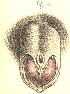
Plate 57.—Figure 14.
Plate 57 – Fig. 14.
Fig. 15, Plate 57, represents in section a state of the parts in which the urethra opened externally by one fistulous aperture, a, behind the scrotum; and by another, b, in front of the scrotum. At the latter place the canal beneath the penis became imperforate for an inch in extent. Parts of catheters are seen to enter the urethra through the fistulous openings a b; and another instrument, c, is seen to pass by the proper meatus into the urethra as far as the point where this portion of the canal fails to communicate with the other. The under part of the scrotum presents a cleft corresponding with the situation of the scrotal septum. This state of the urinary passage may be the effect either of congenital deficiency or of disease. When caused by disease, the chief features in its history, taking these in the order of their occurrence, are, 1st, a stricture in the anterior part of the urethra; 2ndly, a rupture of this canal behind the stricture; 3rdly, the formation (on an abscess opening externally) of a fistulous communication between the canal and the surface of some part of the perinaeum; 4thly, the habitual escape of the urine by the false aperture; 5thly, the obliteration of the canal to a greater or less extent anterior to the stricture; 6thly, the parts situated near the urethral fistula become so consolidated and confused that it is difficult in some and impossible in many cases to find the situation of the urethra, either by external examination or by means of the catheter passed into the canal. The original seat of the stricture becomes so masked by the surrounding disease, and the stricture itself, even if found by any chance, is generally of so impassable a kind, that it must be confessed there are few operations in surgery more irksome to a looker-on than is the fruitless effort made, in such a state of the parts, by a hand without a guide, to pass perforce a blunt pointed instrument like a catheter into the bladder. In some instances the stricture is slightly pervious, the urine passing in small quantity by the meatus. In others, the stricture is rendered wholly imperforate, and the canal either contracted or nearly obliterated anteriorly through disuse. Of these two conditions, the first is that in which catheterism may be tried with any reasonable hope of passing the instrument into the bladder. In the latter state, catheterism is useless, and the only means whereby the urethra may be rendered pervious in the proper direction is that of incising the stricture from the perinaeum, and after passing a catheter across the divided part into the bladder, to retain the instrument in this situation till the wound and the fistulae heal and close under the treatment proper for this end. (Mr. Syme.)
Fig. 15, Plate 57, shows in section a case where the urethra opens externally through one fistulous opening, a, behind the scrotum, and another, b, in front of the scrotum. At the latter spot, the canal beneath the penis is blocked for an inch. Parts of catheters can be seen entering the urethra through the fistulous openings a b; and another instrument, c, is shown passing through the normal opening into the urethra up to the point where this part of the canal does not connect with the other. The underside of the scrotum shows a cleft corresponding to where the scrotal septum is located. This condition of the urinary passage could result from either a congenital issue or a disease. When caused by disease, the main aspects in its timeline, in the order they occur, are: 1st, a stricture in the front part of the urethra; 2nd, a rupture of this canal behind the stricture; 3rd, the formation (due to an abscess opening externally) of a fistulous connection between the canal and the surface of some part of the perineum; 4th, the regular escape of urine through the false opening; 5th, the blocking of the canal to a greater or lesser extent in front of the stricture; 6th, the areas around the urethral fistula become so consolidated and confused that it is difficult in some cases and impossible in many to locate the urethra, either through external examination or by using a catheter passed into the canal. The original location of the stricture becomes so obscured by the surrounding disease, and the stricture itself, even if discovered by chance, is usually of such a severe type that it must be acknowledged that few surgical procedures are more frustrating for an observer than the fruitless attempts made, in such a situation, by a hand without guidance to force a blunt-tipped instrument like a catheter into the bladder. In some cases, the stricture is slightly passable, allowing small amounts of urine to flow through the normal opening. In others, the stricture is completely blocked, and the canal either shrinks or is nearly obliterated in front due to lack of use. Of these two situations, the first is one where catheterization can be attempted with a reasonable chance of passing the instrument into the bladder. In the latter situation, catheterization is futile, and the only way to make the urethra passable in the correct direction is to cut the stricture from the perineum, and after inserting a catheter across the opened part into the bladder, keep the instrument in place until the wound and the fistulas heal and close properly. (Mr. Syme.)

Plate 57.—Figure 15.
Plate 57.—Fig. 15.
Fig. 1, Plate 58.—In this figure the urethra appears communicating with a sac like a scrotum. A bougie is represented entering by the meatus, traversing the upper part of the sac, and passing into the membranous part of the urethra beyond. This case which was owing to a congenital malformation of the urethra, exhibits a dilatation of the canal such as might be produced behind a stricture wherever situated. The urine impelled forcibly by the whole action of the abdominal muscles against the obstructing part dilates the urethra behind the stricture, and by a repetition of such force the part gradually yields more and more, till it attains a very large size, and protrudes at the perinaeum as a distinct fluctuating tumour, every time that an effort is made to void the bladder. If the stricture in such a case happen to cause a complete retention of urine, and that a catheter cannot be passed into the bladder, the tumour should be punctured prior to taking measures for the removal of the stricture. (Sir B. Brodie.)
Fig. 1, Plate 58.—In this figure, the urethra is shown connecting with a sac resembling a scrotum. A bougie is depicted entering through the meatus, moving through the upper part of the sac, and then into the membranous section of the urethra beyond. This case, resulting from a congenital malformation of the urethra, shows a widening of the canal similar to what might occur behind a stricture, regardless of its location. The urine, pushed forcefully by the entire action of the abdominal muscles against the blocked area, expands the urethra behind the stricture, and with repeated force, the area gradually gives way more and more until it reaches a very large size, protruding at the perineum as a distinct, fluctuating mass every time there is an attempt to empty the bladder. If the stricture causes complete retention of urine and a catheter cannot be inserted into the bladder, the mass should be punctured before any attempts to remove the stricture are made. (Sir B. Brodie.)

Plate 58.—Figure 1.
Plate 58.—Fig. 1.
Fig. 2, Plate 58, represents two close strictures of the urethra, one of which is situated at the bulb, and the other at the adjoining membranous part. These are the two situations in which strictures of the organic kind are said most frequently to occur, (Hunter, Home, Cooper, Brodie, Phillips, Velpeau.) False passages likewise are mentioned as more liable to be made in these places than elsewhere in the urethral canal. These occurrences—the disease and the accident—would seem to follow each other closely, like cause and consequence. The frequency with which false passages occur in this situation appears to me to be chiefly owing to the anatomical fact, that the urethra at and close to the bulb is the most dependent part of the curve, F K, Fig. 1, Plate 56; and hence, that instruments descending to this part from before push forcibly against the urethra, and are more apt to protrude through it than to have their points turned so as to ascend the curve towards the neck of the bladder. If it be also true that strictures happen here more frequently than elsewhere, this circumstance will of course favour the accident. An additional cause why the catheter happens to be frequently arrested at this situation and to perforate the canal, is owing to the fact, that the triangular ligament is liable to oppose it, the urethral opening in this structure not happening to coincide with the direction of the point of the instrument. In the figure, part of a bougie traverses the urethra through both strictures and lodges upon the enlarged prostate. Another instrument, after entering the first stricture, occupies a false passage which was made in the canal between the two constricted parts.
Fig. 2, Plate 58, shows two close strictures of the urethra, one located at the bulb and the other at the nearby membranous area. These are the two spots where organic strictures are most commonly reported to occur (Hunter, Home, Cooper, Brodie, Phillips, Velpeau). False passages are also noted to be more likely in these areas than anywhere else in the urethral canal. These issues—the disease and the mishap—seem to be closely linked, like cause and effect. The reason false passages happen so frequently here is mainly due to the anatomical fact that the urethra near the bulb is the lowest point of the curve, F K, Fig. 1, Plate 56. Consequently, instruments that come down to this area from above push against the urethra more forcefully and are more likely to push through it rather than redirect their points to follow the curve up towards the bladder neck. If it’s also true that strictures occur more often here than elsewhere, this situation will naturally increase the risk of accidents. Another reason why the catheter often gets stuck here and can puncture the canal is that the triangular ligament can obstruct it, as the urethral opening in this structure does not align with the direction of the instrument’s tip. In the figure, part of a bougie passes through the urethra, navigating both strictures and resting on the enlarged prostate. Another instrument, after entering the first stricture, occupies a false passage that developed in the canal between the two narrowed sections.

Plate 58.—Figure 2.
Plate 58.—Fig. 2.
Fig. 3, Plate 58.—A calculus is here represented lodging in the urethra at the bulb. The walls of the urethra around the calculus appear thickened. Behind the obstructing body the canal has become dilated, and, in front of it, contracted. In some instances the calculus presents a perforation through its centre, by which the urine escapes. In others, the urine makes its exit between the calculus and the side of the urethra, which it dilates. In this latter way the foreign body becomes loosened in the canal and gradually pushed forwards as far as the meatus, within which, owing to the narrowness of this aperture, it lodges permanently. If the calculus forms a complete obstruction to the passage of the urine, and its removal cannot be effected by other means, an incision should be made to effect this object.
Fig. 3, Plate 58.—This illustration shows a stone lodged in the urethra at the bulb. The walls of the urethra around the stone appear thickened. Behind the obstructing stone, the canal has widened, and in front of it, it has narrowed. In some cases, the stone has a hole in the center, allowing urine to escape. In other instances, urine exits between the stone and the side of the urethra, causing it to expand. In this way, the foreign object becomes loosened in the canal and is gradually pushed forward until it gets stuck at the meatus, where it lodges permanently due to the tightness of this opening. If the stone completely blocks the urine flow and cannot be removed by other means, an incision should be made to address this problem.

Plate 58.—Figure 3.
Plate 58—Fig. 3.
Fig. 4, Plate 58, represents the neck of the bladder and neighbouring part of the urethra of an ox, in which a polypous growth is seen attached by a long pedicle to the veru montanum and blocking up the neck of the bladder. Small irregular tubercles of organized lymph, and tumours formed by the lacunae distended by their own secretion, their orifices being closed by inflammation, are also found to obstruct the urethral canal.
Fig. 4, Plate 58, shows the neck of the bladder and nearby part of the urethra of an ox, where a polyp-like growth is attached by a long stalk to the veru montanum, blocking the neck of the bladder. Small, irregular lumps of organized lymph and tumors formed by the lacunae swollen with their own secretion, with their openings closed due to inflammation, are also present, obstructing the urethral canal.

Plate 58.—Figure 4.
Plate 58—Fig. 4.
Fig. 5, Plate 58.—In this figure is represented a small calculus impacted in and dilating the membranous part of the urethra.
Fig. 5, Plate 58.—This figure shows a small stone lodged in and expanding the membranous section of the urethra.

Plate 58.—Figure 5.
Plate 58.—Fig. 5.
Fig. 6, Plate 58.—Two strictures are here shown to exist in the urethra, one of which is situated immediately in front of the bulb, and the other at a point midway between the bulb and the meatus.
Fig. 6, Plate 58.—Two blockages are shown in the urethra, one located just in front of the bulb, and the other halfway between the bulb and the meatus.

Plate 58.—Figure 6.
Plate 58.—Fig. 6.
Fig. 7, Plate 58.—A stricture is here shown situated at the bulb.
Fig. 7, Plate 58.—A constriction is shown here located at the bulb.

Plate 58.—Figure 7.
Plate 58.—Figure 7.
Fig. 8, Plate 58, represents a stricture of the canal in front of the bulb.
Fig. 8, Plate 58, shows a narrowing of the canal in front of the bulb.

Plate 58.—Figure 8.
Plate 58 — Fig. 8.
Fig. 9, Plate 58, represents the form of an old callous stricture half an inch long, situated midway between the bulb and the meatus. This is perhaps the most common site in which a stricture of this kind is found to exist. In some instances of old neglected cases the corpus spongiosum appears converted into a thick gristly cartilaginous mass, several inches in extent, the passage here being very much contracted, and chiefly so at the middle of the stricture. When it becomes impossible to dilate or pass the canal of such a stricture by the ordinary means, it is recommended to divide the part by the lancetted stilette. (Stafford.) Division of the stricture, by any means, is no doubt the readiest and most effectual measure that can be adopted, provided we know clearly that the cutting instrument engages fairly the part to be divided. But this is a knowledge less likely to be attained if the stricture be situated behind than in front of the triangular ligament.
Fig. 9, Plate 58, shows the shape of an old callous stricture that is half an inch long, located midway between the bulb and the meatus. This is likely the most common spot where this kind of stricture is found. In some neglected cases, the corpus spongiosum can turn into a thick, gristly cartilaginous mass, several inches long, with the passage being significantly narrowed, particularly at the middle of the stricture. When it becomes impossible to dilate or navigate the canal of such a stricture using standard methods, it is suggested to cut the area with a lancetted stiletto. (Stafford.) Dividing the stricture, by any means, is definitely the quickest and most effective approach that can be taken, as long as we clearly know that the cutting instrument properly engages the area to be divided. But this understanding is less likely to be achieved if the stricture is located behind the triangular ligament rather than in front of it.

Plate 58.—Figure 9.
Plate 58.—Fig. 9.
Fig. 10, Plate 58, exhibits a lateral view of the muscular parts which surround the membranous portion of the urethra and the prostate; a, the membranous urethra embraced by the compressor urethrae muscle; b, the levator prostatae muscle; c, the prostate; d, the anterior ligament of the bladder.
Fig. 10, Plate 58, shows a side view of the muscle structures that surround the soft part of the urethra and the prostate; a, the membranous urethra surrounded by the compressor urethrae muscle; b, the levator prostatae muscle; c, the prostate; d, the front ligament of the bladder.

Plate 58.—Figure 10.
Plate 58 — Fig. 10.
Fig. 11, Plate 58.—A posterior view of the parts seen in Fig. 10; a, the urethra divided in front of the prostate; b b, the levator prostatae muscle; c c, the compressor urethrae; d d, parts of the obturator muscles; e e, the anterior fibres of the levator ani muscle; f g, the triangular ligament enclosing between its layers the artery of the bulb, Cowper’s glands, the membranous urethra, and the muscular parts surrounding this portion of the canal. The fact that the flow of urine through the urethra happens occasionally to be suddenly arrested, and this circumstance contrasted with the opposite fact that the organic stricture is of slow formation, originated the idea that the former occurrence arose from a spasmodic muscular contraction. By many this spasm was supposed to be due to the urethra being itself muscular. By others, it was demonstrated as being dependent upon the muscles which surround the membranous part of the urethra, and which act upon this part and constrict it. From my own observations I have formed the settled opinion that the urethra itself is not muscular. And though, on the one hand, I believe that this canal, per se, never causes by active contraction the spasmodic form of stricture, I am far from supposing, on the other, that all sudden arrests to the passage of urine through the urethra are solely attributable to spasm of the muscles which embrace this canal.
Fig. 11, Plate 58.—A view from the back of the parts shown in Fig. 10; a, the urethra cut in front of the prostate; b b, the levator prostatae muscle; c c, the compressor urethrae; d d, parts of the obturator muscles; e e, the front fibers of the levator ani muscle; f g, the triangular ligament that encloses the artery of the bulb, Cowper’s glands, the membranous urethra, and the muscle around this section of the canal. The fact that the flow of urine through the urethra can sometimes be suddenly stopped, alongside the fact that an organic stricture forms slowly, led to the idea that the sudden blockage is caused by a spasm of the muscles. Many believed this spasm was because the urethra itself is muscular. Others demonstrated that it depended on the muscles surrounding the membranous part of the urethra that act on and constrict this area. Based on my own observations, I've come to the clear opinion that the urethra itself is not muscular. While I believe that this canal, per se, does not actively contract to cause the spasmodic form of stricture, I do not think that all sudden stoppages of urine flow through the urethra are solely due to spasms of the muscles surrounding this canal.

Plate 58.—Figure 11.
Plate 58.—Fig. 11.
COMMENTARY ON PLATES 59 & 60.
THE VARIOUS FORMS AND POSITIONS OF STRICTURES AND OTHER OBSTRUCTIONS OF THE URETHRA.—FALSE PASSAGES.—ENLARGEMENTS AND DEFORMITIES OF THE PROSTATE.
THE VARIOUS FORMS AND POSITIONS OF STRICTURES AND OTHER OBSTRUCTIONS OF THE URETHRA.—FALSE PASSAGES.—ENLARGEMENTS AND DEFORMITIES OF THE PROSTATE.
Impediments to the passage of the urine through the urethra may arise from different causes, such as the impaction of a small calculus in the canal, or any morbid growth (a polypus, &c.) being situated therein, or from an abscess which, though forming externally to the urethra, may press upon this tube so as either to obstruct it partially, by bending one of its sides towards the other, or completely, by surrounding the canal on all sides. These causes of obstruction may happen in any part of the urethra, but there are two others (the prostatic and the spasmodic) which are, owing to anatomical circumstances, necessarily confined to the posterior two-thirds of the urethra. The portion of the urethra surrounded by the prostate can alone be obstructed by this body when it has become irregularly enlarged, while the spasmodic stricture can only happen to the membranous portion of the urethra, and to an inch or two of the canal anterior to the bulb, these being the parts which are embraced by muscular structures. The urethra itself not being muscular, cannot give rise to the spasmodic form of stricture. But that kind of obstruction which is common to all parts of the urethra, and which is dependent, as well upon the structures of which the canal is uniformly composed, as upon the circumstance that inflammation may attack these in any situation and produce the same effect, is the permanent or organic stricture. Of this disease the forms are as various as the situations are, for as certainly as it may reasonably be supposed that the plastic lymph, effused in an inflamed state of the urethra from any cause, does not give rise to stricture of any special or particular form, exclusive of all others; so as certainly may it be inferred that, in a structurally uniform canal, inflammation points to no one particular place of it, whereat by preference to establish the organic stricture. The membranous part of the canal is, however, mentioned as being the situation most prone to the disease; but I have little doubt, nevertheless, that owing to general rules of this kind being taken for granted, upon imposing authority, many more serious evils (false passages, &c.) have been effected by catheterism than existed previous to the performance of this operation.[Footnote]
Obstacles to the flow of urine through the urethra can come from various sources, like a small stone stuck in the canal, or the presence of abnormal growths (like a polyp), or from an abscess that, despite forming outside the urethra, can press on this tube and either partially bend it or completely surround it. These types of blockages can occur anywhere in the urethra, but there are two others (prostatic and spasmodic) that, due to anatomical reasons, are limited to the back two-thirds of the urethra. The part of the urethra that is surrounded by the prostate can only be blocked by an irregularly enlarged prostate, while spasmodic stricture can only occur in the membranous segment of the urethra and in the inch or two of the canal right before the bulb, as these are the areas surrounded by muscle. The urethra itself is not made of muscle, so it cannot cause spasmodic stricture. The type of blockage that can occur in any part of the urethra, which is caused by both the uniform structures of the canal and the fact that inflammation can attack these in any location and produce a similar effect, is known as permanent or organic stricture. The variations of this condition are as diverse as the locations it can affect, because just as it can be reasonably assumed that the fibrous tissue that forms during inflammation of the urethra doesn’t create a specific or particular type of stricture alone, it can also be inferred that, in a structurally uniform canal, inflammation does not target any one specific area for establishing the organic stricture. The membranous part of the canal is noted as being the area most susceptible to this condition; however, I believe that due to general assumptions like these being accepted from authoritative sources, many more serious complications (like false passages) have been caused by catheterization than existed before the procedure was performed.[Footnote]
[Footnote: Home describes “a natural constriction of the urethra, directly behind the bulb, which is probably formed with a power of contraction to prevent,” &c. This is the part which he says is “most liable to the disease of stricture.” (Strictures of the Urethra.) Now, if anyone, even among the acute observing microscopists, can discern the structure to which Home alludes, he will certainly prove this anatomist to be a marked exception amongst those who, for the enforcement of any doctrine, can see any thing or phenomenon they wish to see. And, if Hunter were as the mirror from which Home’s mind was reflected, then the observation must be imputed to the Great Original. Upon the question, however, as to which is the most frequent seat of stricture, I find that both these anatomists do not agree, Hunter stating that its usual seat is just in front of the bulb, while Home regrets, as it were, to be obliged to differ from “his immortal friend,” and avers its seat to be an infinitesimal degree behind the bulb. Sir A. Cooper again, though arguing that the most usual situation of stricture is that mentioned by Hunter, names, as next in order of frequency, strictures of the membranous and prostatic parts of the urethra. Does it not appear strange now, how questions of this import should have occupied so much of the serious attention of our great predecessors, and of those, too, who at the present time form the vanguard of the ranks of science? Upon what circumstance, either anatomical or pathological, can one part of the urethra be more liable to the organic stricture than another?]
[Footnote: Home describes “a natural narrowing of the urethra, just behind the bulb, which likely forms with a contraction capability to prevent it,” &c. This is the section he claims is “most prone to the disease of stricture.” (Strictures of the Urethra.) Now, if anyone, even among the sharp-eyed microscopists, can identify the structure Home refers to, they will certainly show that this anatomist is a notable exception among those who can see whatever they want to support any theory. And if Hunter served as the mirror reflecting Home’s thoughts, then the observation must be attributed to the Great Original. However, regarding which area is most commonly affected by stricture, I find that these two anatomists disagree; Hunter claims its usual location is just in front of the bulb, while Home seems to regret having to differ from “his immortal friend,” asserting its location to be slightly behind the bulb. Sir A. Cooper, again, while arguing that the most common site of stricture is the one mentioned by Hunter, identifies the membranous and prostatic sections of the urethra as the next most frequent areas. Doesn’t it seem strange how such important questions have commanded so much serious attention from our great predecessors and from those who, even today, lead the forefront of science? On what anatomical or pathological basis can one area of the urethra be considered more susceptible to organic stricture than any other?]
Figs. 1 and 2, Plate 59.—In these figures are presented seven forms of organic stricture occurring, in different parts of the urethra. In a, Fig. 1, the mucous membrane is thrown into a sharp circular fold, in the centre of which the canal, appears much contracted; a section of this stricture appears in b, Fig. 2. In b, Fig. 1, the canal is contracted laterally by a prominent fold of the mucous membrane at the opposite side. In c, Fig. 1, an organized band of lymph is stretched across the canal; this stricture is seen in section in c, Fig. 2. In e, Fig. 1, a stellate band of organized lymph, attached by pedicles to three sides of the urethra, divides the canal into three passages. In d, Fig. 1, the canal is seen to be much contracted towards the left side by a crescentic fold of the lining membrane projecting from the right. In f, the canal appears contracted by a circular membrane, perforated in the centre; a section of which is seen at a, Fig. 2. The form of the organic stricture varies therefore according to the three following circumstances:—1st. When lymph becomes effused within the canal upon the surface of the lining mucous membrane, and contracts adhesions across the canal. 2ndly. When lymph is effused external to the lining membrane, and projects this inwards, thereby narrowing the diameter of the canal. 3rdly. When the outer and inner walls of a part of the urethra are involved in the effused organizable matter, and on contracting towards each other, encroach at the same time upon the area of the canal. This latter state presents the form, which is known as the old callous tough stricture, extending in many instances for an inch or more along the canal. In cases where the urethra becomes obstructed by tough bands of substance, c e, which cross the canal directly, the points of flexible catheters, especially if these be of slender shape, are apt to be bent upon the resisting part, and on pressure being continued, the operator may be led to suppose that the instrument traverses the stricture, while it is most probably perforating the wall of the urethra. But in those cases where the diameter of the canal is circularly contracted, the stricture generally presents a conical depression in front, which, receiving the point of the instrument, allows this to enter the central passage unerringly. A stricture formed by a crescentic septum, such as is seen in b d, Fig. 1, offers a more effectual obstacle to the passage of a catheter than the circular septum like a f.
Figs. 1 and 2, Plate 59.—These figures show seven types of organic strictures found in different parts of the urethra. In a, Fig. 1, the mucous membrane bends sharply in a circular fold, where the canal appears to be significantly narrowed; a cross-section of this stricture is shown in b, Fig. 2. In b, Fig. 1, the canal is narrowed laterally by a noticeable fold of the mucous membrane on the opposite side. In c, Fig. 1, an organized band of lymph stretches across the canal; this stricture is illustrated in section in c, Fig. 2. In e, Fig. 1, a star-shaped band of organized lymph is attached by pedicles to three sides of the urethra, creating three separate passages in the canal. In d, Fig. 1, the canal is significantly narrowed on the left side by a crescent-shaped fold of the lining membrane that protrudes from the right. In f, the canal appears narrowed by a circular membrane with a hole in the center; a cross-section of this is shown at a, Fig. 2. The shape of the organic stricture varies based on three main factors: 1st. When lymph seeps into the canal onto the surface of the lining mucous membrane and creates adhesions across the canal. 2nd. When lymph seeps outside the lining membrane and pushes it inward, which narrows the canal's diameter. 3rd. When both the outer and inner walls of a part of the urethra are affected by the effused organizable material, and as they contract toward each other, they also encroach on the area of the canal. This last condition is what is referred to as an old callous tough stricture, which can extend for an inch or more along the canal. In cases where the urethra is blocked by tough bands of material, c e, that cross directly over the canal, the ends of flexible catheters, especially if they are slender, can bend against the resistant area. If pressure continues, the operator may mistakenly think that the instrument is passing through the stricture, when it is likely perforating the wall of the urethra instead. However, in cases where the canal's diameter is circularly narrowed, the stricture usually has a conical depression at the front, allowing the point of the instrument to enter the central passage smoothly. A stricture formed by a crescent-shaped septum, like the one seen in b d, Fig. 1, provides a more effective barrier to the passage of a catheter than the circular septum shown in a f.

Plate 59. Figure 1
Plate 59, Fig. 1

Plate 59. Figure 2
Plate 59. Fig. 2
Fig. 3, Plate 59.—In this there are seen three separate strictures, a, b, c, situated in the urethra, anterior to the bulb. In some cases there are many more strictures (even to the number of six or seven) situated in various parts of the urethra; and it is observed that when one stricture exists, other slight tightnesses in different parts of the canal frequently attend it. (Hunter.) When several strictures occur in various parts of the urethra, they may occasion as much difficulty in passing an instrument as if the whole canal between the extreme constrictions were uniformly narrowed.
Fig. 3, Plate 59.—This shows three separate strictures, a, b, c, located in the urethra, before the bulb. In some cases, there can be many more strictures (even up to six or seven) found in different areas of the urethra; and it’s noticed that when one stricture is present, other minor tight spots in various parts of the canal often accompany it. (Hunter.) When multiple strictures occur in different areas of the urethra, they can create as much difficulty in passing an instrument as if the entire canal between the most extreme constrictions was consistently narrowed.

Plate 59.—Figure 3.
Plate 59.—Fig. 3.
Fig. 4, Plate 59.—In this the canal is constricted at the point a, midway between the bulb and glans. A false passage has been made under the urethra by an instrument which passed out of the canal at the point f, anterior to the stricture a, and re-entered the canal at the point c, anterior to the bulb. When a false passage of this kind happens to be made, it will become a permanent outlet for the urine, so long as the stricture remains. For it can be of no avail that we avoid re-opening the anterior perforation by the catheter, so long as the urine prevented from flowing by the natural canal enters the posterior perforation. Measures should be at once taken to remove the stricture.
Fig. 4, Plate 59.—In this, the canal is narrowed at the point a, halfway between the bulb and glans. A false passage has been created beneath the urethra by an instrument that exited the canal at the point f, in front of the stricture a, and re-entered the canal at the point c, also in front of the bulb. When this kind of false passage occurs, it will become a permanent outlet for urine as long as the stricture stays in place. It doesn't help to avoid reopening the anterior perforation with a catheter if the urine that can't flow through the natural canal goes into the posterior perforation instead. Steps should be taken immediately to address the stricture.

Plate 59.—Figure 4.
Plate 59—Fig. 4.
Fig. 5, Plate 59.—The stricture a appears midway between the bulb and glans, the area of the passage through the stricture being sufficient only to admit a bristle to pass. It would seem almost impossible to pass a catheter through a stricture so close as this, unless by a laceration of the part, combined with dilatation.
Fig. 5, Plate 59.—The stricture a is located halfway between the bulb and glans, with the opening through the stricture barely large enough to allow a bristle to pass through. It seems nearly impossible to insert a catheter through such a tight stricture without tearing the tissue and using dilation.

Plate 59.—Figure 5.
Plate 59.—Fig. 5.
Fig. 6, Plate 59.—Two instruments, a, b, have made false passages beneath the mucous membrane, in a case where no stricture at all existed. The resistance which the instruments encountered in passing out of the canal having been mistaken, no doubt, for that of passing through a close stricture.
Fig. 6, Plate 59.—Two instruments, a, b, created false passages under the mucous membrane in a case where there was no stricture present at all. The resistance the instruments felt when exiting the canal was likely mistaken for the resistance encountered when passing through a tight stricture.

Plate 59.—Figure 6.
Plate 59—Fig. 6.
Fig. 7, Plate 59.—A bougie, b b, is seen to perforate the urethra anterior to the stricture c, situated an inch behind the glans, and after traversing the substance of the right corpus cavernosum d, for its whole length, re-enters the neck of the bladder through the body of the prostate.
Fig. 7, Plate 59.—A bougie, b b, is seen to pierce the urethra just before the stricture c, located an inch behind the glans, and after passing through the entire length of the right corpus cavernosum d, re-enters the neck of the bladder via the prostate.

Plate 59.—Figure 7.
Plate 59 — Fig. 7.
Fig. 8, Plate 59.—A bougie, c c, appears tearing and passing beneath the lining membrane, d d, of the prostatic urethra. It is remarked that the origin of a false passage is in general anterior to the stricture. It may, however, occur at any part of the canal in which no stricture exists, if the hand that impels the instrument be not guided by a true knowledge of the form of the urethra; and perhaps the accident happening from this cause is the more general rule of the two.
Fig. 8, Plate 59.—A bougie, c c, is shown tearing and passing underneath the lining membrane, d d, of the prostatic urethra. It's noted that a false passage usually starts in front of the stricture. However, it can occur anywhere in the canal where there isn't a stricture if the hand pushing the instrument isn't guided by a proper understanding of the urethra's shape; and this type of mistake might actually be the more common scenario of the two.

Plate 59.—Figure 8.
Plate 59—Fig. 8.
Fig. 9, Plate 59.—Two strictures are represented here, the one, e, close to the bulb d, the other, f, an inch anterior to this part. In the prostate, a b, are seen irregularly shaped abscess pits, communicating with each other, and projecting upwards the floor of this body to such a degree, that the prostatic canal appears nearly obliterated.
Fig. 9, Plate 59.—This shows two strictures: one, e, near the bulb d, and the other, f, one inch in front of this area. In the prostate, a b, there are irregularly shaped abscess pits that connect with each other, causing the floor of this organ to bulge upwards so much that the prostatic canal looks almost blocked.

Plate 59.—Figure 9.
Plate 59 — Fig. 9.
Fig. 10, Plate 59.—Two bougies, d e, are seen to enter the upper wall of the urethra, c, anterior to the prostate, a b. This accident happens when the handle of a rigid instrument is depressed too soon, with the object of raising its point over the enlarged third lobe of the prostate.
Fig. 10, Plate 59.—Two bougies, d e, are seen entering the upper wall of the urethra, c, in front of the prostate, a b. This incident occurs when the handle of a rigid instrument is pushed down too early, with the aim of lifting its tip over the enlarged third lobe of the prostate.

Plate 59.—Figure 10.
Plate 59 — Fig. 10.
Fig. 11, Plate 59.—Two instruments appear transfixing the prostate, of which body the three lobes, a, b, c, are much enlarged. The instrument d perforates the third lobe, a; while the instrument e penetrates the right lobe, c, and the third lobe, a. This accident occurs when instruments not possessing the proper prostatic bend are forcibly pushed forwards against the resistance at the neck of the bladder.
Fig. 11, Plate 59.—Two instruments are shown piercing the prostate, which has three lobes, a, b, c, that are significantly enlarged. The instrument d goes through the third lobe, a; while the instrument e enters the right lobe, c, and the third lobe, a. This issue happens when instruments that don't have the correct prostatic curve are forcibly pushed forward against the blockage at the bladder neck.

Plate 59.—Figure 11.
Plate 59—Fig. 11.
Fig. 12, Plate 59.—In this case an instrument, d d, after passing beneath part of the lining membrane, e e, anterior to the bulb, penetrates b, the right lobe of the prostate. A second instrument, c c, penetrates the left lobe. A third smaller instrument, f f, is seen to pass out of the urethra anterior to the prostate, and after transfixing the right vesicula seminalis external to the neck of the bladder, enters this viscus at a point behind the prostate. The resistance which the two larger instruments met with in penetrating the prostate, made it seem, perhaps, that a tight stricture existed in this situation, to match which the smaller instrument, f f, was afterwards passed in the course marked out.
Fig. 12, Plate 59.—In this case, an instrument, d d, passes underneath part of the lining membrane, e e, in front of the bulb and penetrates b, the right lobe of the prostate. A second instrument, c c, enters the left lobe. A third, smaller instrument, f f, is seen exiting the urethra in front of the prostate, and after piercing the right seminal vesicle outside the neck of the bladder, it enters this organ at a point behind the prostate. The resistance encountered by the two larger instruments when penetrating the prostate might have made it seem like there was a tight stricture in this area, leading to the subsequent passage of the smaller instrument, f f, along the indicated path.

Plate 59.—Figure 12.
Plate 59.—Fig. 12.
Figs. 1 to 5, Plate 60, represent a series of prostates, in which the third lobe gradually increases in size. In Fig. 1, which shows the healthy state of the neck of the bladder, unmarked by the prominent lines which are said to bound the space named “trigone vesical,” or by those which indicate the position of the “muscles of the ureters,” the third lobe does not exist. In Fig. 2 it appears as the uvula vesicae, a. In Fig. 3 the part a is increased, and under the name now of third lobe is seen to contract and bend upwards the prostatic canal. In Fig. 4 the effect which the growth of the lobe, a, produces upon the form of the neck of the bladder becomes more marked, and the part presenting perforations, e e, produced by instruments, indicates that by its shape it became an obstacle to the egress of the urine as well as to the entrance of instruments. A calculus of irregular form is seen to lodge behind the third lobe, and to be out of the reach of the point of a sound, supposing this to enter the bladder over the apex of the lobe. In Fig. 5 the three lobes are enlarged, but the third is most so, and while standing on a narrow pedicle attached to the floor of the prostate, completely blocks up the neck of the bladder. [Footnote]
Figs. 1 to 5, Plate 60, show a series of prostates where the third lobe gradually gets bigger. In Fig. 1, which depicts a healthy bladder neck, there are no noticeable lines marking the area called the “trigone vesical,” nor those showing the position of the “muscles of the ureters,” so the third lobe is absent. In Fig. 2, it appears as the uvula vesicae, a. Fig. 3 shows that part a has grown, now recognized as the third lobe, which narrows and bends the prostatic canal upwards. In Fig. 4, the impact of the lobe’s growth, a, on the bladder neck shape becomes clearer, and the area with perforations, e e, created by instruments, suggests that its shape obstructs both urine flow and the entrance of instruments. An oddly shaped stone is shown resting behind the third lobe, situated out of reach of a sound assuming it enters the bladder above the apex of the lobe. In Fig. 5, all three lobes are enlarged, with the third lobe being the most prominent, standing on a narrow stalk attached to the prostate floor, completely blocking the bladder neck. [Footnote]

Plate 60.—Figure 1
Plate 60.—Fig. 1

Plate 60.—Figure 2
Plate 60—Fig. 2

Plate 60.—Figure 3
Plate 60.—Fig. 3

Plate 60.—Figure 4
Plate 60.—Fig. 4

Plate 60.—Figure 5
Plate 60.—Fig. 5
[Footnote: On comparing this series of figures, it must appear that the third lobe of the prostate is the product of diseased action, in so far at least as an unnatural hypertrophy of a part may be so designated. It is not proper to the bladder in the healthy state of this organ, and where it does manifest itself by increase it performs no healthy function in the economy. When Home, therefore, described this part as a new fact in anatomy, he had in reality as little reason for so doing as he would have had in naming any other tumour, a thing unknown to normal anatomy. Langenbeck (Neue Bibl. b. i. p. 360) denies its existence in the healthy state. Cruveilhier (Anat. Pathog. liv. xxvii.) deems it incorrect to reckon a third lobe as proper to the healthy bladder.]
[Footnote: When comparing these figures, it's clear that the third lobe of the prostate results from disease, at least to the extent that an abnormal enlargement of a part can be described this way. It doesn't belong to the bladder when this organ is healthy, and when it does appear through enlargement, it doesn't serve any healthy function in the body. So, when Home described this part as a new anatomical feature, he had just as little reason to do so as he would have had for naming any other tumor, something that doesn’t exist in normal anatomy. Langenbeck (Neue Bibl. b. i. p. 360) argues that it doesn’t exist in a healthy state. Cruveilhier (Anat. Pathog. liv. xxvii.) considers it incorrect to categorize a third lobe as part of a healthy bladder.]
Fig. 6, Plate 60.—The prostatic canal is bent upwards by the enlarged third lobe to such a degree as to form a right angle with the membranous part of the canal. A bougie is seen to perforate the third lobe, and this is the most frequent mode in which, under such circumstances, and with instruments of the usual imperfect form, access may be gained to the bladder for the relief of retention of urine. “The new passage may in every respect be as efficient as one formed by puncture or incision in any other way.” (Fergusson.)
Fig. 6, Plate 60.—The prostatic canal is angled upward by the enlarged third lobe to the point that it forms a right angle with the membranous part of the canal. A bougie can be seen penetrating the third lobe, which is the most common way, under these circumstances and with typically imperfect instruments, to access the bladder for relieving urine retention. “The new passage may be just as effective as one created by puncture or incision in any other way.” (Fergusson.)

Plate 60.—Figure 6
Plate 60.—Fig 6
Fig. 7, Plate 60.—The three lobes of the prostate, a, b, c, are equally enlarged. The prostatic canal is consequently much contracted and distorted, so that an instrument on being passed into the bladder has made a false passage through the third lobe. When a catheter is suspected to have entered the bladder by perforating the prostate, the instrument should be retained in the newly made passage till such time as this has assumed the cylindrical form of the instrument. If this be done, the new passage will be the more likely to become permanent. It is ascertained that all false passages and fistulae by which the urine escapes, become after a time lined with a membrane similar to that of the urethra. (Stafford.)
Fig. 7, Plate 60.—The three lobes of the prostate, a, b, c, are all enlarged. As a result, the prostatic canal is significantly narrowed and distorted, causing an instrument to create a false passage through the third lobe when inserted into the bladder. If a catheter is thought to have entered the bladder by perforating the prostate, it should stay in the newly created passage until that passage takes on the cylindrical shape of the instrument. Doing this increases the chances that the new passage will become permanent. It's been observed that all false passages and fistulas that allow urine to leak eventually develop a membrane lining similar to that of the urethra. (Stafford.)

Plate 60.—Figure 7
Plate 60—Figure 7
Fig. 8, Plate 60.—The three lobes, a, b, c, of the prostate are irregularly enlarged. The third lobe, a a, projecting from below, distorts the prostatic canal upwards and to the right side.
Fig. 8, Plate 60.—The three lobes, a, b, c, of the prostate are irregularly enlarged. The third lobe, a a, extending from below, distorts the prostatic canal upward and to the right side.

Plate 60.—Figure 8.
Plate 60 — Fig. 8.
Fig. 9, Plate 60.—The right lobe, a c c, of the prostate appears hollowed out so as to form the sac of an abscess which, by its projection behind, pressed upon the forepart of the rectum, and by its projection in front, contracted the area of the prostatic canal, and thereby caused an obstruction in this part. Not unfrequently when a catheter is passed along the urethra, for the relief of a retention of urine caused by the swell of an abscess in this situation, the sac becomes penetrated by the instrument, and, instead of urine, pus flows. The sac of a prostatic abscess frequently opens of its own accord into the neighbouring part of the urethra, and when this occurs it becomes necessary to retain a catheter in the neck of the bladder, so as to prevent the urine entering the sac.
Fig. 9, Plate 60.—The right lobe, a c c, of the prostate seems hollowed out to create the cavity of an abscess that, due to its projection backward, puts pressure on the front of the rectum, and its projection forward narrows the prostatic canal, resulting in an obstruction in that area. Often, when a catheter is inserted into the urethra to relieve urine retention caused by the swelling of an abscess in this area, the sac can be punctured by the instrument, and instead of urine, pus flows out. The sac of a prostatic abscess often opens on its own into the nearby part of the urethra, and when this happens, it becomes necessary to keep a catheter in the neck of the bladder to prevent urine from entering the sac.

Plate 60.—Figure 9.
Plate 60 - Fig. 9.
Fig. 10, Plate 60.—The prostate presents four lobes of equal size, and all projecting largely around the neck of the bladder. The prostatic canal is almost completely obstructed, and an instrument has made a false passage through the lobe a.
Fig. 10, Plate 60.—The prostate has four lobes of equal size, all significantly extending around the neck of the bladder. The prostatic canal is nearly completely blocked, and an instrument has created a false passage through the lobe a.

Plate 60.—Figure 10.
Plate 60 - Fig. 10.
Fig. 11, Plate 60.—The third lobe of the prostate is viewed in section, and shows the track of the false passage made by the catheter, d, through it, from its apex to its base. The proper canal is bent upwards from its usual position, which is that at present marked by the instrument in the false passage.
Fig. 11, Plate 60.—The third lobe of the prostate is shown in section and reveals the path of the false passage created by the catheter, d, running from its apex to its base. The proper canal is curved upward from its typical position, which is currently indicated by the instrument in the false passage.

Plate 60.—Figure 11.
Plate 60 — Fig. 11
Fig. 12, Plate 60.—The prostatic lobes are uniformly enlarged, and cause the corresponding part of the urethra to be uniformly contracted, so as closely to embrace the catheter, d d, occupying it, and to offer considerable resistance to the passage of the instrument.
Fig. 12, Plate 60.—The prostatic lobes are consistently enlarged, which causes the corresponding section of the urethra to be uniformly narrowed, tightly surrounding the catheter, d d, that is in place, and creating significant resistance to the movement of the instrument.

Plate 60.—Figure 12.
Plate 60—Fig. 12.
Fig. 13, Plate 60.—The prostate, bc, is considerably enlarged anteriorly, b, in consequence of which the prostatic canal appears more horizontal even than natural. The catheter, d, occupying the canal lies nearly straight. The lower wall, c, of the prostate is much diminished in thickness. A nipple-shaped process, a, is seen to be attached by a pedicle to the back of the upper part, b, of the prostate, and to act like a stopper to the neck of the bladder. The body a being moveable, it will be perceived how, while the bladder is distended with urine, the pressure from above may block up the neck of the organ with this part, and thus cause complete retention, which, on the introduction of a catheter, becomes readily relieved by the instrument pushing the obstructing body aside.
Fig. 13, Plate 60.—The prostate, bc, is significantly enlarged at the front, b, which makes the prostatic canal appear more horizontal than usual. The catheter, d, positioned in the canal, lies nearly straight. The lower wall, c, of the prostate is much thinner. A nipple-shaped projection, a, is attached by a stalk to the back of the upper part, b, of the prostate, acting like a stopper for the bladder's neck. The body a is movable, so when the bladder is filled with urine, pressure from above can compress the bladder neck with this part, leading to complete retention. This retention is quickly relieved when a catheter is inserted, as it pushes the obstructing body aside.

Plate 60.—Figure 13.
Plate 60—Fig. 13.
COMMENTARY ON PLATES 61 & 62.
DEFORMITIES OF THE PROSTATE.—DISTORTIONS AND OBSTRUCTIONS OF THE PROSTATIC URETHRA.
DEFORMITIES OF THE PROSTATE.—DISTORTIONS AND OBSTRUCTIONS OF THE PROSTATIC URETHRA.
The prostate is liable to such frequent and varied deformities, the consequence of diseased action, whilst, at the same time, its healthy function (if it have any) in the male body is unknown, that it admits at least of one interpretation which may, according to fact, be given of it—namely, that of playing a principal part in effecting some of the most distressing of “the thousand natural ills that flesh is heir to.” But heedless of such a singular explanation of a final cause, the practical surgeon will readily confess the fitting application of the interpretation, such as it is, and rest contented with the proximate facts and proofs. As physiologists, however, it behooves us to look further into nature, and search for the ultimate fact in her prime moving law. The prostate is peculiar to the male body, the uterus to the female. With the exception of these two organs there is not another which appears in the one sex but has its analogue in the opposite sex; and thus these two organs, the prostate and the uterus, appear by exclusion of the rest to approach the test of comparison, by which their analogy becomes as fully manifested as that between the two quantities, a-b, and a+b the only difference which exists depends upon the subtraction or the addition of the quantity, b. The difference between a prostate and a uterus is simply one of quantity, such as we see existing between the male and the female breast. The prostate is to the uterus absolutely what a rudimentary organ is to its fully developed analogue. The one, as being superfluous, is in accordance with nature’s law of nihil supervacaneum nihil frutra, arrested in its development, and in such a character appears the prostate. This body is not a gland any more than is the uterus, but both organs being quantitatively, and hence functionally different, I here once more venture to call down an interpretation of the part from the unfrequented bourne of comparative anatomy, and turning it to lend an interest to the accompanying figures even with a surgical bearing, I remark that the prostatic or rudimentary uterus, like a germ not wholly blighted, is prone to an occasional sprouting or increase beyond its prescribed dimensions—a hypertrophy in barren imitation, as it were, of gestation. [Footnote]
The prostate is prone to a range of deformities due to disease, and its healthy function—if it has one—in the male body is unknown. This allows for at least one interpretation: that it plays a key role in contributing to some of the most distressing “natural ills that flesh is heir to.” However, the practical surgeon is likely to acknowledge the practical application of this interpretation, even if it’s imperfect, and focus on the immediate facts and evidence. As physiologists, though, we need to delve deeper into nature to find the ultimate fact behind her fundamental laws. The prostate is exclusive to the male body, while the uterus belongs to the female. Besides these two organs, every other organ appears in one sex and has an equivalent in the other; hence, the prostate and the uterus uniquely stand out for comparison. Their similarity is as clear as that between a-b and a+b, where the only difference lies in the subtraction or addition of b. The difference between a prostate and a uterus is just a matter of quantity, similar to the difference between male and female breasts. In this sense, the prostate is to the uterus what a rudimentary organ is to its fully developed counterpart. One is superfluous and reflects nature’s law of nihil supervacaneum nihil frustra, being halted in its development, and that’s how the prostate appears. This body is not a gland any more than the uterus is, but both organs are quantitatively and functionally different. I again propose to draw an interpretation from the lesser-explored realm of comparative anatomy, making it relevant to the accompanying figures and their surgical implications. I note that the prostatic or rudimentary uterus, like a partially damaged germ, can occasionally grow or expand beyond its normal limits—a hypertrophy in a barren attempt to mimic gestation. [Footnote]
[Footnote: This expression of the fact to which I allude will not, I trust, be extended beyond the limits I assign to it. Though I have every reason to believe, that between the prostate of the male and the uterus of the female, the same amount of analogy exists, as between a coccygeal ossicle and the complete vertebral form elsewhere situated in the spinal series, I am as far from regarding the two former to be in all respects structurally or functionally alike, as I am from entertaining the like idea in respect to the two latter. But still I maintain that between a prostate and a uterus, as between a coccygeal bone and a vertebra, the only difference which exists is one of quantity, and that hence arises the functional difference. A prostate is part of a uterus, just as a coccygeal bone is part (the centrum) of a vertebra. That this is the absolute signification of the prostate I firmly believe, and were this the proper place, I could prove it in detail, by the infallible rule of analogical reasoning. John Hunter has observed that the use of the prostate was not sufficiently known to enable us to form a judgment of the bad consequences of its diseased state. When the part becomes morbidly enlarged, it acts as a mechanical impediment to the passage of urine from the bladder, but from this circumstance we cannot reasonably infer, that while of its normal healthy proportions, its special function is to facilitate the egress of the urine, for the female bladder, though wholly devoid of the prostate, performs its own function perfectly. It appears to me, therefore, that the real question should be, not what is the use of the prostate? but has it any proper function? If the former question puzzled even the philosophy of Hunter, it was because the latter question must be answered in the negative. The prostate has no function proper to itself per se. It is a thing distinct from the urinary apparatus, and distinct likewise from the generative organs. It may be hypertrophied or atrophied, or changed in texture, or wholly destroyed by abscess, and yet neither of the functions of these two systems of organs will be impaired, if the part while diseased act not as an obstruction to them. In texture the prostate is similar to an unimpregnated uterus. In form it is, like the uterus, symmetrical. In position it corresponds to the uterus. The prostate has no ducts proper to itself. Those ducts which are said to belong to it (prostatic ducts) are merely mucous cells, similar to those in other parts of the urethral lining membrane. The seminal ducts evidently do not belong to it. The texture of the prostate is not such as appears in glandular bodies generally. In short, the facts which prove what it is not, prove what it actually is—namely, a uterus arrested in its development, and as a sign of that all-encompassing law in nature, which science expresses by the term “unity in variety.” This interpretation of the prostate, which I believe to be true to nature, will last perhaps till such time as the microscopists shall discover in its “secretion” some species of mannikins, such as may pair with those which they term spermatozoa.]
[Footnote: I hope this explanation stays within the boundaries I've set. While I strongly believe that the similarities between the male prostate and the female uterus are as significant as those between a coccygeal ossicle and a vertebra found elsewhere in the spine, I don't consider these two pairs to be identical in structure or function. However, I argue that the only difference between a prostate and a uterus, as well as between a coccygeal bone and a vertebra, is a matter of quantity, which leads to their functional differences. A prostate is part of a uterus, just like a coccygeal bone is part of a vertebra. I wholeheartedly believe that this is the correct meaning of the prostate, and if this were the right context, I could prove it through the reliable method of analogical reasoning. John Hunter noted that the function of the prostate wasn't well understood, making it hard to judge the negative effects of its disease. When this part becomes abnormally enlarged, it obstructs urine flow from the bladder, but this doesn't allow us to reasonably conclude that when it is healthy, its specific role is to help urine exit. The female bladder, which has no prostate, functions perfectly well on its own. Thus, I believe the real question should be, not what is the purpose of the prostate? but does it have any unique function? If the first question puzzled even someone as philosophical as Hunter, it was because the second should be answered with a no. The prostate doesn't have a function of its own per se. It is separate from the urinary system and also distinct from the reproductive organs. It can become enlarged, shrunken, altered in texture, or completely destroyed by an abscess, yet neither of the functions of these two systems of organs will be affected as long as the diseased part doesn’t obstruct them. The prostate's texture is similar to that of an unpregnant uterus. In shape, it is symmetrical like the uterus. In location, it corresponds to the uterus. The prostate has no ducts of its own. Those ducts that are said to belong to it (prostatic ducts) are merely mucous cells, like those in other parts of the urethral lining. The seminal ducts clearly do not belong to it. The texture of the prostate does not resemble that of typical glandular tissues. In short, the facts that show what it is not help establish what it actually is—namely, a uterus that has stalled in its development, reflecting that overarching law of nature that science labels as “unity in variety.” I believe this understanding of the prostate aligns with nature and may endure until microscopists find some sort of beings in its “secretion,” similar to what they call spermatozoa.]
Fig. 1, Plate 61.—The prostate, a b, is here represented thinned in its walls above and below. The lower wall is dilated into a pouch caused by the points of misdirected instruments in catheterism having been rashly forced against it.
Fig. 1, Plate 61.—The prostate, a b, is shown here with thinner walls at the top and bottom. The lower wall has expanded into a pouch due to the tips of improperly directed instruments during catheterization being recklessly pushed against it.

Plate 61.—Figure 1.
Plate 61.—Fig. 1.
Fig. 2, Plate 61.—The prostate, a b, is here seen to be somewhat more enlarged than is natural. A tubercle, b, surmounts the lower part, c, of the prostate, and blocks up the vesical orifice. Catheters introduced by the urethra for retention of urine which existed in this case, have had their points arrested at the bulb, and on being pushed forwards in this direction, have dilated the bulb into the form of a pouch, seen at d. The sinus of the bulb, being the lowest part of the urethral canal, is very liable to be distorted or perforated by the points of instruments descending upon it from above and before. [Footnote]
Fig. 2, Plate 61.—The prostate, a b, appears to be somewhat larger than normal. A bump, b, is located at the lower part, c, of the prostate, blocking the opening to the bladder. Catheters inserted through the urethra for urine retention in this case have been stopped at the bulb, and when pushed forward, they have stretched the bulb into a pouch shape, as seen at d. The sinus of the bulb, being the lowest section of the urethral canal, is very susceptible to distortion or perforation by the tips of instruments coming down from above and in front. [Footnote]
[Footnote: When a stricture exists immediately behind the bulb, this circumstance will, of course, favour the occurrence of the accident. “False passages (observes Mr. Benjamin Phillips) are less frequent here (in the membranous part of the urethra) than in the bulbous portion of the canal. The reason of this must be immediately evident: false passages are ordinarily made in consequence of the difficulty experienced in the endeavour to pass an instrument through the strictured portion of the tube. Stricture is most frequently seated at the point of junction between the bulbous and membranous portions of the canal; consequently, the false passage will be usually anterior to this latter point.”—(On the Urethra, its Diseases. &c., p. 15.) ]
[Footnote: When a stricture is located right behind the bulb, this situation will, of course, increase the likelihood of the accident happening. “False passages (observes Mr. Benjamin Phillips) are less common here (in the membranous part of the urethra) than in the bulbous section of the canal. The reason for this is obvious: false passages are usually created due to the difficulty encountered when trying to insert an instrument through the strictured area of the tube. Stricture most often occurs at the junction point between the bulbous and membranous sections of the canal; therefore, the false passage will typically be located just before this latter point.”—(On the Urethra, its Diseases. &c., p. 15.) ]

Plate 61.—Figure 2
Plate 61.—Fig. 2
Fig. 3, Plate 61.—A cyst, c, is seen to grow from the left side of the base of the prostate, a b, and to form an obstruction at the vesical orifice.
Fig. 3, Plate 61.—A cyst, c, is observed growing from the left side of the base of the prostate, a b, and it creates a blockage at the bladder opening.

Plate 61.—Figure 3.
Plate 61.—Fig. 3.
Fig. 4, Plate 61.—A globular excrescence, a, appears blocking up the vesical orifice, and giving to this the appearance of a crescentic slit, corresponding to the shape of the obstructing body. The prostate, b b, is enlarged in both its lateral lobes. A small bougie, c, is placed in the prostatic canal and vesical opening.
Fig. 4, Plate 61.—A round growth, a, looks like it's blocking the bladder opening, making it seem like a crescent-shaped slit that matches the shape of the obstruction. The prostate, b b, is swollen in both side lobes. A small catheter, c, is positioned in the prostatic canal and bladder opening.

Plate 61.—Figure 4
Plate 61.—Fig. 4
Fig. 5, Plate 61.—The prostate, d, is considerably enlarged, and the vesical orifice is girt by a prominent ring, b b, from the right border of which the nipple-shaped body, a, projects and occupies the outlet. Owing to the retention of urine caused by this state of the prostate, the ureters, c c, have become very much dilated.
Fig. 5, Plate 61.—The prostate, d, is significantly enlarged, and the opening to the bladder is surrounded by a noticeable ring, b b, from which the nipple-like structure, a, extends and occupies the outlet. Due to the urine retention caused by this condition of the prostate, the ureters, c c, have become greatly dilated.

Plate 61.—Figure 5.
Plate 61—Fig. 5.
Fig. 6, Plate 61.—The lateral lobes of the prostate, c c, are seen enlarged, and from the inner side and base of each, irregularly shaped masses, a, b, d, project, and bend the prostatic urethra first to the right side, then to the left. The part, a, resting upon the part, b, acts like a valve against the vesical outlet, which would become closed the tighter according to the degree of superincumbent pressure. A flexible catheter would, in such a case as this, be more likely, perhaps, to follow the sinuous course of the prostatic passage than a rigid instrument of metal.
Fig. 6, Plate 61.—The lateral lobes of the prostate, c c, are enlarged, and from the inner side and base of each, irregularly shaped masses, a, b, d, protrude, bending the prostatic urethra first to the right and then to the left. The part, a, resting on the part, b, functions like a valve against the bladder outlet, which would become tighter depending on the amount of pressure above it. In this situation, a flexible catheter would likely follow the winding path of the prostatic passage better than a rigid metal instrument.

Plate 61.—Figure 6.
Plate 61.—Fig. 6.
Fig. 7, Plate 61.—A globular mass, a, of large size, occupies the neck of the bladder, and gives the vesical orifice, c, a crescentic shape, convex towards the right side. The two lobes of the prostate, b, are much enlarged.
Fig. 7, Plate 61.—A large spherical mass, a, is located at the neck of the bladder, giving the vesical opening, c, a crescent shape that curves to the right. The two lobes of the prostate, b, are significantly enlarged.

Plate 61.—Figure 7.
Plate 61.—Fig. 7.
Fig. 8, Plate 61.—The lateral lobes, b b, of the prostate are irregularly enlarged, and the urinary passage is bent towards the right side, c, from the membranous portion, which is central. Surmounting the vesical orifice, c, is seen the tuberculated mass, a, which being moveable, can be forced against the vesical orifice and thus produce complete retention of urine. In this case, also, a flexible catheter would be more suitable than a metallic one.
Fig. 8, Plate 61.—The side lobes, b b, of the prostate are unevenly enlarged, and the urinary passage turns toward the right side, c, from the central membranous part. Above the bladder opening, c, is the raised mass, a, which is movable and can press against the bladder opening, causing complete urine retention. In this situation, a flexible catheter would be better than a metal one.

Plate 61.—Figure 8.
Plate 61—Fig. 8.
Fig. 9, Plate 61.—The lateral lobes, b b, of the prostate are enlarged. The third lobe, a, projects at the neck of the bladder, distorting the vesical outlet. A small calculus occupies the prostatic urethra, and being closely impacted in this part of the canal, would arrest the progress of a catheter, and probably lead to the supposition that the instrument grated against a stone in the interior of the bladder, in which case it would be inferred that since the urine did not flow through the catheter no retention existed.
Fig. 9, Plate 61.—The side lobes, b b, of the prostate are enlarged. The third lobe, a, extends at the neck of the bladder, disturbing the flow of urine. A small stone is lodged in the prostatic urethra, and since it's tightly stuck in this section of the canal, it would block the progress of a catheter, likely leading to the assumption that the instrument is rubbing against a stone inside the bladder. In this situation, it would be assumed that since urine isn’t flowing through the catheter, there’s no retention occurring.

Plate 61.—Figure 9.
Plate 61.—Fig. 9.

Plate 61.—Figure 10.
Plate 61.—Fig. 10.
Fig. 10, Plate 61.—Both lateral lobes, b c, of the prostate appear much increased in size. A large irregular shaped mass, a, grows from the base of the right lobe, and distorts the prostatic canal and vesical orifice. When the lobes of the prostate increase in size in this direction, the prostatic canal becomes much more elongated than natural, and hence the instrument which is to be passed for relieving the existing retention of urine should have a wide and long curve to correspond with the form of this part of the urethra. [Footnote]
Fig. 10, Plate 61.—Both side lobes, b c, of the prostate seem significantly enlarged. A large, irregularly shaped mass, a, protrudes from the base of the right lobe, distorting the prostatic canal and urinary opening. When the prostate lobes enlarge in this way, the prostatic canal becomes much longer than usual, so the instrument used to relieve the current urine retention should have a wide and long curve to match the shape of this part of the urethra. [Footnote]
[Footnote: Both lobes of the prostate are equally liable to chronic enlargement. Home believed the left lobe to be oftener increased in size than the right. Wilson (on the Male Urinary and Genital Organs) mentions several instances of the enlargement of the right lobe. No reason can be assigned why one lobe should be more prone to hypertrophy than the other, even supposing it to be matter of fact, which it is not. But the observations made by Cruveilhier (Anat. Pathol.), that the lobulated projections of the prostate always take place internally at its vesical aspect, is as true as the manner in which he accounts for the fact is plausible. The dense fibrous envelope of the prostate is sufficient to repress its irregular growth externally.]
[Footnote: Both lobes of the prostate are equally prone to chronic enlargement. Home thought the left lobe was more often larger than the right. Wilson (on the Male Urinary and Genital Organs) notes several cases of enlargement in the right lobe. There's no clear reason why one lobe would be more likely to hypertrophy than the other, even if that were the case, which it isn’t. However, the observations made by Cruveilhier (Anat. Pathol.) about the lobulated projections of the prostate always occurring internally at its bladder side are accurate, and his explanation for this is quite plausible. The dense fibrous covering of the prostate is enough to limit its irregular growth on the outside.]
Fig. 11, Plate 61.—Both lobes of the prostate are enlarged, and from the base of each a mass projects prominently around the vesical orifice, a b. The prostatic urethra has been moulded to the shape of the instrument, which was retained in it for a considerable time.
Fig. 11, Plate 61.—Both lobes of the prostate are enlarged, and from the base of each, a mass protrudes prominently around the bladder opening, a b. The prostatic urethra has taken on the shape of the instrument, which was held in place for quite a while.

Plate 61.—Figure 11.
Plate 61 — Fig. 11.
Fig. 12, Plate 61.—The prostate, c b, is enlarged and dilated, like a sac. Across the neck, a, of the bladder the prostate projects in an arched form, and is transfixed by the instrument, d. The prostate may assume this appearance, as well from instruments having been forced against it, as from an abscess cavity formed in its substance having received, from time to time, a certain amount of the urine, and retained this fluid under the pressure of strong efforts, made to void the bladder while the vesical orifice was closed above.
Fig. 12, Plate 61.—The prostate, c b, is swollen and stretched, resembling a sac. It arches across the neck, a, of the bladder and is pierced by the instrument, d. The prostate can look like this either from instruments being forced against it or from an abscess formed within it that has, over time, received a certain amount of urine and retained this fluid due to strong efforts to empty the bladder while the opening above was closed.

Plate 61.—Figure 12.
Plate 61—Fig. 12.
Fig. 13, Plate 61.—The lateral lobes, d e, of the prostate are enlarged; and, occupying the position of the third lobe, appear as three masses, a b c, plicated upon each other, and directed towards the vesical orifice, which they close like valves. The prostatic urethra branches upwards into three canals, formed by the relative position of the parts, e, c, b, a, d, at the neck of the bladder. The ureters are dilated, in consequence of the regurgitation of the contents of the bladder during the retention which existed ..
Fig. 13, Plate 61.—The side lobes, d e, of the prostate are enlarged; and, taking the place of the third lobe, appear as three overlapping masses, a b c, stacked on top of each other and directed towards the opening of the bladder, which they close like valves. The prostatic urethra branches upwards into three canals, shaped by the arrangement of the parts, e, c, b, a, d, at the neck of the bladder. The ureters are widened due to the backup of the bladder contents during the existing retention...

Plate 61.—Figure 13.
Plate 61 — Fig. 13.
Fig. 1, Plate 62, exhibits the lobes of the prostate greatly increased in size. The part, a b, girds irregularly, and obstructs the vesical outlet, while the lateral lobes, c d, encroach upon the space of the prostatic canal. The walls of the bladder are much thickened.
Fig. 1, Plate 62, shows the lobes of the prostate significantly enlarged. The section, a b, wraps irregularly and blocks the outlet of the bladder, while the lateral lobes, c d, intrude on the space of the prostatic canal. The walls of the bladder are considerably thickened.

Plate 62.—Figure 1.
Plate 62—Fig. 1.
Fig. 2, Plate 62.—The three lobes, a, d, c, of the prostate are enlarged and of equal size, moulded against each other in such a way that the prostatic canal and vesical orifice appear as mere clefts between them. The three lobes are encrusted on their vesical surfaces with a thick calcareous deposit. The surface of the third lobe, a, which has been half denuded of the calcareous crust, b, in order to show its real character, appeared at first to be a stone impacted in the neck of the bladder, and of such a nature it certainly would seem to the touch, on striking it with the point of a sound or other instrument.
Fig. 2, Plate 62.—The three lobes, a, d, c, of the prostate are enlarged and equal in size, pressing against each other so that the prostatic canal and vesical opening seem like narrow slits between them. The surfaces of the three lobes that face the bladder are covered with a thick layer of calcium deposits. The surface of the third lobe, a, which has had part of the calcium layer removed, b, to reveal its true nature, initially looked like a stone lodged in the bladder neck, and it certainly feels like that when touched with the point of a sound or another instrument.

Plate 62.—Figure 2.
Plate 62 - Fig. 2.
Fig. 3, Plate 62, represents the prostate with its three lobes enlarged, and the prostatic canal and vesical orifice narrowed. The walls of the bladder are thickened, fasciculated, and sacculated; the two former appearances being caused by a hypertrophy of the vesical fibres, while the latter is in general owing to a protrusion of the mucous membrane between the fasciculi.
Fig. 3, Plate 62, shows the prostate with its three lobes enlarged, and the prostatic canal and bladder opening narrowed. The bladder walls are thickened, banded, and pouch-like; the first two conditions are due to an increase in the bladder muscle fibers, while the pouch-like appearance is usually caused by a bulging of the mucous membrane between the muscle bundles.

Plate 62.—Figure 3.
Plate 62 - Fig. 3.
Fig. 4, Plate 62.—The prostate presents four lobes, a, b, c, d, each being of large size, and projecting far into the interior of the bladder, from around the vesical orifice which they obstruct. The bladder is thickened, and the prostatic canal is elongated. The urethra and the lobes of the prostate have been perforated by instruments, passed for the retention of urine which existed. A stricturing band, e, is seen to cross the membranous part of the canal.
Fig. 4, Plate 62.—The prostate has four lobes, a, b, c, d, each large and extending deep into the bladder, around the bladder opening that they block. The bladder is thickened, and the prostatic canal is elongated. Instruments used to relieve the retained urine have pierced the urethra and the prostate lobes. A constricting band, e, is observed across the membranous section of the canal.

Plate 62.—Figure 4.
Plate 62—Fig. 4.
Fig. 5, Plate 62.—The prostate, a a, is greatly enlarged, and projects high in the bladder, the walls of the latter, b b, being very much thickened. The ureters, c, are dilated, and perforations made by instruments are seen in the prostate. The prostatic canal being directed almost vertically, and the neck of the bladder being raised nearly as high as the upper border of the pubic symphysis, it must appear that if a stone rest in the bas fond of the bladder, a sound or staff cannot reach the stone, unless by perforating the prostate; and if, while the staff occupies this position, lithotomy be performed, the incisions will not be required to be made of a greater depth than if the prostate were of its ordinary proportions. On the contrary, if the staff happen to have surmounted the prostate, the incision, in order to divide the whole vertical thickness of this body, will require to be made very deeply from the perinaeal surface, and this circumstance occasions what is termed a “deep perinaeum.”
Fig. 5, Plate 62.—The prostate, a a, is significantly enlarged and extends high into the bladder, whose walls, b b, are notably thickened. The ureters, c, are enlarged, and there are perforations from instruments visible in the prostate. Since the prostatic canal is almost vertical, and the bladder neck is raised nearly as high as the upper border of the pubic symphysis, it seems that if a stone is located in the base of the bladder, a sound or staff won’t be able to reach the stone without perforating the prostate. Moreover, if the staff is positioned this way and lithotomy is performed, the incisions won’t need to be deeper than they would be if the prostate were of normal size. On the other hand, if the staff has cleared the prostate, the incision will need to be made very deep from the perineal surface to cut through the full vertical thickness of this tissue, which results in what is called a “deep perineum.”

Plate 62.—Figure 5.
Plate 62.—Fig. 5.
Fig. 6, Plate 62.—The lower half, c, b, f, of the prostate, having become the seat of abscess, appears hollowed out in the form of a sac. This sac is separated from the bladder by a horizontal septum, e e, the proper base of the bladder, g g. The prostatic urethra, between a e, has become vertical in respect to the membranous part of the canal, in consequence of the upward pressure of the abscess. The sac opens into the urethra, near the apex of the prostate, at the point c; and a catheter passed along the urethra has entered the orifice of the sac, the interior of which the instrument traverses, and the posterior wall of which it perforates. The bladder contains a large calculus, i. The bladder and sac do not communicate, but the urethra is a canal common to both. In a case of this sort it becomes evident that, although symptoms may strongly indicate either a retention of urine, or the presence of a stone in the bladder, any instrument taking the position and direction of d d, cannot relieve the one or detect the other; and such is the direction in which the instrument must of necessity pass, while the sac presents its orifice more in a line with the membranous part of the urethra than the neck of the bladder is. The sac will intervene between the rectum and the bladder; and on examination of the parts through the bowel, an instrument in the sac will readily be mistaken for being in the bladder, while neither a calculus in the bladder, nor this organ in a state of even extreme distention, can be detected by the touch any more than by the sound or catheter. If, while performing lithotomy in such a state of the parts, the staff occupy the situation of d d d, then the knife, following the staff, will open, not the bladder which contains the stone, but the sac, which, moreover, if it happen to be filled with urine regurgigated from the urethra, will render the deception more complete.
Fig. 6, Plate 62.—The lower half, c, b, f, of the prostate, has developed an abscess, creating a hollow sac. This sac is separated from the bladder by a horizontal wall, e e, which is the actual base of the bladder, g g. The prostatic urethra, between a e, has become vertical relative to the membranous part of the canal due to the upward pressure from the abscess. The sac opens into the urethra near the tip of the prostate, at point c; and a catheter inserted into the urethra enters the opening of the sac, passing through its interior and puncturing the back wall. The bladder contains a large stone, i. There is no direct connection between the bladder and the sac, but the urethra serves as a shared passage for both. In such a case, it's clear that despite symptoms suggesting either urine retention or a stone in the bladder, any instrument positioned as d d, cannot relieve one condition or locate the other; the instrument must necessarily follow that direction, while the opening of the sac aligns more with the membranous part of the urethra than with the neck of the bladder. The sac will sit between the rectum and the bladder; during a rectal examination, an instrument within the sac could easily be mistaken for being in the bladder, while neither a stone in the bladder nor significant bladder distension can be detected by touch, sound, or catheter. If, while performing lithotomy in this condition, the staff is positioned as d d d, the knife that follows will open the sac instead of the bladder containing the stone. If the sac happens to be filled with urine backing up from the urethra, it will further complicate the situation.

Plate 62.—Figure 6.
Plate 62—Fig. 6.
Fig. 7, Plate 62.—The walls, a a, of the bladder, appear greatly thickened, and the ureters, b, dilated. The sides, c c c, of the prostate are thinned; and in the prostatic canal are two calculi, d d, closely impacted. In such a state of the parts it would be impossible to pass a catheter into the bladder for the relief of a retention of urine, or to introduce a staff as a guide to the knife in lithotomy. If, however, the staff can be passed as far as the situation of the stone, the parts may be held with a sufficient degree of steadiness to enable the operator to incise the prostate upon the stone.
Fig. 7, Plate 62.—The walls, a a, of the bladder look significantly thickened, and the ureters, b, are enlarged. The sides, c c c, of the prostate are thinned; and in the prostatic canal are two stones, d d, tightly packed together. In this condition, it would be impossible to insert a catheter into the bladder to relieve urinary retention, or to place a staff as a guide for the knife in lithotomy. However, if the staff can be inserted as far as the location of the stone, the surrounding areas may be held steady enough for the surgeon to cut into the prostate over the stone.

Plate 62.—Figure 7.
Plate 62 — Fig. 7.
COMMENTARY ON PLATES 63 & 64.
DEFORMITIES OF THE URINARY BLADDER.—THE OPERATIONS OF SOUNDING FOR STONE, OF CATHETERISM AND OF PUNCTURING THE BLADDER ABOVE THE PUBES.
DEFORMITIES OF THE URINARY BLADDER.—THE PROCEDURES OF SOUNDING FOR STONE, CATHETERIZATION, AND PUNCTURING THE BLADDER ABOVE THE PUBIS.
The urinary bladder presents two kinds of deformity—viz., congenital and pathological. As examples of the former may be mentioned that in which the organ is deficient in front, and has become everted and protruded like a fungous mass through an opening at the median line of the hypogastrium; that in which the rectum terminates in the bladder posteriorly; and that in which the foetal urachus remains pervious as a uniform canal, or assumes a sacculated shape between the summit of the bladder and the umbilicus. The pathological deformities are, those in which vesical fistulae, opening either above the pubes, at the perinaeum, or into the rectum, have followed abscesses or the operation of puncturing the bladder in these situations, and those in which the walls of the organ appear thickened and contracted, or thinned and expanded, or sacculated externally, or ridged internally, in consequence of its having been subjected to abdominal pressure while overdistended with its contents, and while incapable of voiding these from some permanent obstruction in the urethral canal.[Footnote] The bladder is liable to become sacculated from two causes—from a hernial protrusion of its mucous membrane through the separated fasciculi of its fibrous coat, or from the cyst of an abscess which has formed a communication with the bladder, and received the contents of this organ. Sacs, when produced in the former way, may be of any number, or size, or in any situation; when caused by an abscess, the sac is single, is generally formed in the prostate, or corresponds to the base of the bladder, and may attain to a size equalling, or even exceeding, that of the bladder itself. The sac, however formed, will be found lined by mucous membrane. The cyst of an abscess, when become a recipient for the urine, assumes after a time a lining membrane similar to that of the bladder. If the sac be situated at the summit or back of the bladder, it will be found invested by peritonaeum; but, whatever be its size, structure, or position, it may be always distinguished from the bladder by being devoid of the fibrous tunic, and by having but an indirect relation to the vesical orifice.
The urinary bladder can have two types of deformities: congenital and pathological. Examples of congenital deformities include a bladder that is missing part of its front and has turned inside out, protruding like a growth through an opening in the center of the lower abdomen; a situation where the rectum ends in the bladder; and a case where the fetal urachus remains open as a continuous tube or takes on a pouch-like shape between the top of the bladder and the navel. Pathological deformities include the presence of bladder fistulas that open either above the pubic bone, at the perineum, or into the rectum, usually resulting from abscesses or procedures to drain the bladder in those areas. There are also cases where the bladder walls are thickened and contracted, or thin and expanded, or have pouches on the outside or ridges on the inside due to the bladder being under pressure from being overly stretched while unable to empty due to a permanent blockage in the urethra. The bladder can develop pouches for two main reasons: from a hernia of its mucous membrane pushing through the layers of its fibrous covering, or from an abscess forming a connection with the bladder and collecting its contents. Pouches that arise from the first cause can vary in number, size, and location; whereas those caused by an abscess are usually single, forming in the prostate or at the base of the bladder, and can be as large as or even larger than the bladder itself. Regardless of how the pouch forms, it will be lined with mucous membrane. If an abscess pouch becomes a reservoir for urine, it will eventually develop a lining similar to that of the bladder. If the pouch is located at the top or back of the bladder, it will be surrounded by peritoneum. However, no matter its size, structure, or location, it can always be identified as separate from the bladder because it lacks the fibrous covering and has only an indirect connection to the bladder opening.
[Footnote: On considering these cases of physical impediments to the passage of urine from the vesical reservoir through the urethral conduit, it seems to me as if these were sufficient to account for the formation of stone in the bladder, or any other part of the urinary apparatus, without the necessity of ascribing it to a constitutional disease, such as that named the lithic diathesis by the humoral pathologists.
[Footnote: When looking at these cases of physical barriers that prevent urine from flowing from the bladder through the urethra, it appears to me that these obstacles are enough to explain the development of stones in the bladder or anywhere else in the urinary system, without having to attribute it to a systemic illness, like what the humoral pathologists referred to as the lithic diathesis.]
The urinary apparatus (consisting of the kidneys, ureters, bladder, and urethra) is known to be the principal emunctory for eliminating and voiding the detritus formed by the continual decay of the parts comprising the animal economy. The urine is this detritus in a state of solution. The components of urine are chemically similar to those of calculi, and as the components of the one vary according to the disintegration occurring at the time in the vital alembic, so do those of the other. While, therefore, a calculus is only as urine precipitated and solidified, and this fluid only as calculous matter suspended in a menstruum, it must appear that the lithic diathesis is as natural and universal as structural disintegration is constant and general in operation. As every individual, therefore, may be said to void day by day a dissolved calculus, it must follow that its form of precipitation within some part of the urinary apparatus alone constitutes the disease, since in this form it cannot be passed. On viewing the subject in this light, the question that springs directly is, (while the lithic diathesis is common to individuals of all ages and both sexes,) why the lithic sediment should present in the form of concrement in some and not in others? The principal, if not the sole, cause of this seems to me to be obstruction to the free egress of the urine along the natural passage. Aged individuals of the male sex, in whom the prostate is prone to enlargement, and the urethra to organic stricture, are hence more subject to the formation of stone in the bladder, than youths, in whom these causes of obstruction are less frequent, or than females of any age, in whom the prostate is absent, and the urethra simple, short, readily dilatable, and seldom or never strictured. When an obstruction exists, lithic concretions take place in the urinary apparatus in the same manner as sedimentary particles cohere or crystallize elsewhere. The urine becoming pent up and stagnant while charged with saline matter, either deposits this around a nucleus introduced into it, or as a surplus when the menstruum is insufficient to suspend it. The most depending part of the bladder is that where lithic concretions take place; and if a sacculus exist here, this, becoming a recipient for the matter, will favour the formation of stone.] [End Footnote]
The urinary system (which includes the kidneys, ureters, bladder, and urethra) is the main channel for eliminating and getting rid of the waste produced by the constant breakdown of the body's components. The urine represents this waste in a dissolved form. The chemical makeup of urine is similar to that of kidney stones, and just as the components of one change based on what’s breaking down in the body at the time, so do those of the other. Thus, a kidney stone is essentially urine that has precipitated and hardened, while urine can be seen as solid matter suspended in a liquid. This suggests that the tendency to form stones is as natural and widespread as the ongoing breakdown of body structures. Since every person releases what could be considered a dissolved stone every day, it follows that the way it forms into a solid piece in part of the urinary system represents the actual disease, as this solid form cannot be passed naturally. Viewing the situation this way raises a question: while the tendency to form stones is common in people of all ages and genders, why do some people develop solid deposits while others do not? The main, if not the only, reason for this seems to be a blockage preventing urine from freely flowing through the normal passage. Older men, where prostate enlargement and urethra narrowing are more likely, are more prone to stone formation in the bladder than younger males or females of any age, where the prostate isn’t present, and the urethra is simple, short, easily stretched, and usually not narrowed. When a blockage occurs, stones form in the urinary system in the same way that sediments bond or crystallize elsewhere. The urine, when trapped and stagnant while containing salt, either deposits this around a particle introduced into it or as excess when there isn’t enough liquid to keep it suspended. The lowest part of the bladder is where these stones usually form; and if a pouch exists there, it will collect this material, promoting stone formation.
FIG. 1, Plate 63.—The lateral lobes of the prostate, 3, 4, are enlarged, and contract the prostatic canal. Behind them the third lobe of smaller size occupies the vesical orifice, and completes the obstruction. The walls of the bladder have hence become fasciculated and sacculated. One sac, 1, projects from the summit of the bladder; another, 2, containing a stone, projects laterally. When a stone occupies a sac, it does not give rise to the usual symptoms as indicating its presence, nor can it be always detected by the sound.
FIG. 1, Plate 63.—The lateral lobes of the prostate, 3, 4, are enlarged, which narrows the prostatic canal. Behind them, the third lobe, which is smaller, takes up space at the bladder opening and completes the blockage. As a result, the walls of the bladder have become thickened and pouch-like. One pouch, 1, sticks out from the top of the bladder; another, 2, which contains a stone, extends to the side. When a stone is in a pouch, it doesn't produce the usual symptoms that indicate its presence, nor can it always be found using a sound.

Plate 63,—Figure 1.
Plate 63, Figure 1.
FIG. 2, Plate 63.—The prostate, 2, 3, is enlarged, and the middle lobe, 2, appears bending the prostatic canal to an almost vertical position, and obstructing the vesical orifice. The bladder, 1, 1, 1, is thickened; the ureters, 7, are dilated; and a large sac, 6, 6, projects from the base of the bladder backwards, and occupies the recto-vesical fossa. The sac, equal in size to the bladder, communicates with this organ by a small circular opening, 8, situated between the orifices of the ureters. The peritonaeum is reflected from the summit of the bladder to that of the sac. A catheter, 4, appears perforating the third lobe of the prostate, 2, and entering the sac, 5, through the base of the bladder, below the opening, 8. In a case of this kind, a catheter occupying the position 4, 5, would, while voiding the bladder through the sac, make it seem as if it really traversed the vesical orifice. If a stone occupied the bladder, the point of the instrument in the sac could not detect it, whereas, if a stone lay within the sac, the instrument, on striking it here, would give the impression as if it lay within the bladder.
FIG. 2, Plate 63.—The prostate, 2, 3, is enlarged, and the middle lobe, 2, seems to bend the prostatic canal to an almost vertical position, obstructing the bladder opening. The bladder, 1, 1, 1, is thickened; the ureters, 7, are widened; and a large sac, 6, 6, projects from the base of the bladder backwards, occupying the recto-vesical space. The sac, which is about the same size as the bladder, connects to it through a small circular opening, 8, located between the ureter openings. The peritoneum reflects from the top of the bladder to the top of the sac. A catheter, 4, is seen puncturing the third lobe of the prostate, 2, and entering the sac, 5, through the base of the bladder, below the opening, 8. In a situation like this, a catheter positioned at 4, 5, would make it look like it is passing through the bladder while voiding into the sac. If there is a stone in the bladder, the instrument's tip in the sac wouldn't be able to detect it; however, if a stone is inside the sac, when the instrument hits it, it would seem as if it were in the bladder.

Plate 63,—Figure 2.
Plate 63 — Fig. 2.
FIG. 3, Plate 63.—The urethra being strictured, the bladder has become sacculated. In the bas fond of the bladder appears a circular opening, 2, leading to a sac of large dimensions, which rested against the rectum. In such a case as this, the sac, occupying a lower position than the base of the bladder, must first become the recipient of the urine, and retain this fluid even after the bladder has been evacuated, either voluntarily or by means of instruments. If, in such a state of the parts, retention of urine called for puncturation, it is evident that this operation would be performed with greater effect by opening the depending sac through the bowel, than by entering the summit of the bladder above the pubes.
FIG. 3, Plate 63.—Due to a narrow urethra, the bladder has developed sacs. At the bottom of the bladder, there's a circular opening, 2, that leads to a large sac pressing against the rectum. In this situation, since the sac is positioned lower than the bladder's base, it will first collect urine and hold onto it even after the bladder is emptied, either voluntarily or through instruments. If the condition requires draining the urine, it's clear that doing so by accessing the lower sac through the rectum would be more effective than entering through the top of the bladder above the pubic area.

Plate 63,—Figure 3.
Plate 63, Figure 3.
FIG. 4, Plate 63.—The vesical orifice is obstructed by two portions, 3, 4, of the prostate, projecting upwards, one from each of its lateral lobes, 6, 6. The bladder is thickened and fasciculated, and from its summit projects a double sac, 1, 2, which is invested by the peritonaeum.
FIG. 4, Plate 63.—The opening of the bladder is blocked by two parts, 3, 4, of the prostate, sticking up from each of its side lobes, 6, 6. The bladder walls are thickened and have a banded appearance, and from its top juts out a double pouch, 1, 2, which is covered by the peritoneum.

Plate 63,—Figure 4.
Plate 63, Figure 4.
FIG. 5, Plate 63.—The prostatic canal is constricted and bent upwards by the third lobe. The bladder is thickened, and its base is dilated in the form of a sac, which is dependent, and upon which rests a calculus. An instrument enters the bladder by perforating the third lobe, but does not come into contact with the calculus, owing to the low position occupied by this body.
FIG. 5, Plate 63.—The prostatic canal is narrowed and angled upward by the third lobe. The bladder is thickened, and its base is expanded like a sac, which hangs down and supports a stone. An instrument enters the bladder by piercing the third lobe but doesn't touch the stone because of its low position.

Plate 63,—Figure 5.
Plate 63, Figure 5.
FIG. 6, Plate 63.—Two sacs appear projecting on either side of the base of the bladder. The right one, 5, contains a calculus, 6; the left one, of larger dimensions, is empty. The rectum lay in contact with the base of the bladder between the two sacs.
FIG. 6, Plate 63.—Two sacs are visible extending on either side of the base of the bladder. The right one, 5, contains a stone, 6; the left one, which is larger, is empty. The rectum was in contact with the base of the bladder between the two sacs.

Plate 63,—Figure 6.
Plate 63, Figure 6.
FIG. 7, Plate 63.—Four calculi are contained in the bladder. This organ is divided by two septa, 2, 4, into three compartments, each of which, 1, 3, 5, gives lodgment to a calculus; and another, 6, of these bodies lies impacted in the prostatic canal, and becomes a complete bar to the passage of a catheter. Supposing lithotomy to be performed in an instance of this kind, it is probable that, after the extraction of the calculi, 6, 5, the two upper ones, 3, 1, would, owing to their being embedded in the walls of the bladder, escape the forceps.
FIG. 7, Plate 63.—Four stones are present in the bladder. This organ is separated by two partitions, 2, 4, into three sections, each of which, 1, 3, 5, holds a stone; and another, 6, of these stones is stuck in the prostatic canal, completely blocking the passage of a catheter. If surgery is conducted in a case like this, it's likely that, after removing the stones, 6, 5, the two upper ones, 3, 1, would, due to being embedded in the bladder walls, avoid being grasped by the forceps.

Plate 63,—Figure 7.
Plate 63, Figure 7.
FIG. 8, Plate 63.-Two large polypi, and many smaller ones, appear growing from the mucous membrane of the prostatic urethra and vesical orifice, and obstructing these parts. In examining this case during life by the sound, the two larger growths, 1, 2, were mistaken by the surgeon for calculi. Such a mistake might well be excused if they happened to be encrusted with lithic matter.
FIG. 8, Plate 63.-Two large polyps and many smaller ones are found growing from the mucous membrane of the prostatic urethra and bladder opening, blocking these areas. When examining this case while the patient was alive with a sound, the surgeon misidentified the two larger growths, 1, 2, as stones. This misunderstanding could easily be forgiven if the growths were covered with stone-like material.

Plate 63,—Figure 8.
Plate 63, Figure 8.
FIG. 9, Plate 63.—The base of the bladder, 8, 8, appears dilated into a large uniform sac, and separated from the upper part of the organ by a circular horizontal fold, 2, 2. The ureters are also dilated. The left ureter, 3, 4, opens into the sac below this fold, while the right ureter opens above it into the bladder. In all cases of retention of urine from permanent obstruction of the urethra, the ureters are generally found more or less dilated. Two circumstances combine to this effect—while the renal secretion continues to pass into the ureters from above, the contents of the bladder under abdominal pressure are forced regurgitating into them from below, through their orifices.
FIG. 9, Plate 63.—The base of the bladder, 8, 8, looks enlarged into a large, uniform sac, and is separated from the upper part of the organ by a circular horizontal fold, 2, 2. The ureters are also enlarged. The left ureter, 3, 4, enters the sac below this fold, while the right ureter opens above it into the bladder. In cases of urine retention caused by a permanent blockage of the urethra, the ureters are usually found to be somewhat enlarged. Two factors contribute to this: while the kidneys continue to send their secretions into the ureters from above, the pressure in the abdomen forces the contents of the bladder back into the ureters from below, through their openings.

Plate 63,—Figure 9.
Plate 63, Figure 9.
FIG. 1, Plate 64.—The bladder, 6, appears symmetrically sacculated. One sac, 1, is formed at its summit, others, 3, 2, project laterally, and two more, 5, 4, from its base. The ureters, 7, 7, are dilated, and enter the bladder between the lateral and inferior sacs.
FIG. 1, Plate 64.—The bladder, 6, looks evenly shaped with pouches. One pouch, 1, is located at the top, while two others, 3, 2, stick out to the sides, and two more, 5, 4, extend from the bottom. The ureters, 7, 7, are widened and connect to the bladder between the side and bottom pouches.

Plate 64,—Figure 1.
Plate 64, Figure 1.
Fig. 2, Plate 64.—The prostate is greatly enlarged, and forms a narrow ring around the vesical orifice. Through this an instrument, 12, enters the bladder. The walls of the bladder are thickened and sacculated. On its left side appear numerous sacs, 2, 3, 4, 5, 6, 7, 8, and on the inner surface of its right side appear the orifices of as many more. On its summit another sac is formed. The ureters, 9, are dilated.
Fig. 2, Plate 64.—The prostate is significantly enlarged and creates a narrow ring around the urinary opening. An instrument, 12, enters the bladder through this ring. The bladder walls are thickened and have pouch-like areas. On the left side, there are several sacs, labeled 2, 3, 4, 5, 6, 7, 8, and on the inner surface of the right side, there are openings for just as many more. At the top, another sac has formed. The ureters, 9, are enlarged.

Plate 64,—Figure 2.
Plate 64, Figure 2.
FIG. 3, Plate 64.—The prostate is enlarged, its canal is narrowed, and the bladder is thickened and contracted. A calculus, 1, 2, appears occupying nearly the whole vesical interior. The incision in the neck of the bladder in lithotomy must necessarily be extensive, to admit of the extraction of a stone of this size.
FIG. 3, Plate 64.—The prostate is enlarged, its canal is narrowed, and the bladder is thickened and contracted. A stone, 1, 2, is taking up almost the entire inside of the bladder. The incision in the neck of the bladder during lithotomy needs to be large enough to allow for the removal of a stone this size.

Plate 64,—Figure 3.
Plate 64, Figure 3.
FIG. 4, Plate 64.—The prostatic canal is contracted by the lateral lobes, 4, 5; resting upon these, appear three calculi, 1, 2, 3, which nearly fill the bladder. This organ is thickened and fasciculated. In cases of this kind, and that last mentioned, the presence of stone is readily ascertainable by the sound.
FIG. 4, Plate 64.—The prostatic canal is narrowed by the lateral lobes, 4, 5; resting on these are three stones, 1, 2, 3, that almost fill the bladder. This organ is thickened and banded. In these cases, as well as the one previously mentioned, the presence of a stone can be easily detected by using a sound.

Plate 64,—Figure 4.
Plate 64—Figure 4.
FIG. 5, Plate 64.—The three prostatic lobes are enlarged, and appear contracting the vesical orifice. In the walls of the bladder are embedded several small calculi, 2, 2, 2, 2, which, on being struck with the convex side of a sound, might give the impression as though a single stone of large size existed. In performing lithotomy, these calculi would not be within reach of the forceps.
FIG. 5, Plate 64.—The three prostatic lobes are enlarged and seem to be pressing against the bladder opening. In the walls of the bladder, there are several small stones, 2, 2, 2, 2, which, when tapped with the rounded side of a tool, might give the illusion of a single large stone. During lithotomy, these stones would not be accessible with the forceps.

Plate 64,—Figure 5.
Plate 64 — Fig. 5.
FIG. 6, Plate 64.—Two sacculi, 4, 5, appear projecting at the middle line of the base of the bladder, between the vasa deferentia, 7, 7, and behind the prostate, in the situation where the operation of puncturing the bladder per anum is recommended to be performed in retention of urine.
FIG. 6, Plate 64.—Two sacs, 4, 5, are seen protruding at the midline of the bladder's base, situated between the vas deferens, 7, 7, and behind the prostate, where it is advised to carry out the procedure of puncturing the bladder through the anus in cases of urinary retention.

Plate 64,—Figure 6.
Plate 64, Figure 6.
FIG. 7, Plate 64.—A sac, 4, is situated on the left side of the bladder, 3, 3, immediately above the orifice of the ureter. In the sac was contained a mass of phosphatic calculus. This substance is said to be secreted by the mucous lining of the bladder, while in a state of chronic inflammation, but there seems nevertheless very good reason for us to believe that it is, like all other calculous matter, a deposit from the urine.
FIG. 7, Plate 64.—A sac, 4, is located on the left side of the bladder, 3, 3, directly above the opening of the ureter. Inside the sac was a mass of phosphatic stone. This substance is said to be produced by the mucous lining of the bladder during chronic inflammation, but there are strong reasons to believe that, like all other types of stones, it forms as a deposit from the urine.

Plate 64,—Figure 7.
Plate 64, Figure 7.
FIG. 8, Plate 64, represents, in section, the relative position of the parts concerned in catheterism. [Footnote] In performing this operation, the patient is to be laid supine; his loins are to be supported on a pillow; and his thighs are to be flexed and drawn apart from each other. By this means the perinaeum is brought fully into view, and its structures are made to assume a fixed relative position. The operator, standing on the patient’s left side, is now to raise the penis so as to render the urethra, 8, 8, 8, as straight as possible between the meatus, a, and the bulb, 7. The instrument (the concavity of its curve being turned to the left groin) is now to be inserted into the meatus, and while being gently impelled through the canal, the urethra is to be drawn forwards, by the left hand, over the instrument. By stretching the urethra, we render its sides sufficiently tense for facilitating the passage of the instrument, and the orifices of the lacunae become closed. While the instrument is being passed along this part of the canal, its point should be directed fairly towards the urethral opening, 6*, of the triangular ligament, which is situated an inch or so below the pubic symphysis, 11. With this object in view, we should avoid depressing its handle as yet, lest its point be prematurely tilted up, and rupture the upper side of the urethra anterior to the ligament. As soon as the instrument has arrived at the bulb, its further progress is liable to be arrested, from these causes:—1st, This portion of the canal is the lowest part of its perinaeal curve, 3, 6, 8, and is closely embraced by the middle fibres of the accelerator urinae muscle. 2nd, It is immediately succeeded by the commencement of the membranous urethra, which, while being naturally narrower than other parts, is also the more usual seat of organic stricture, and is subject to spasmodic constriction by the fibres of the compressor urethrae. 3d, The triangular ligament is behind it, and if the urethral opening of the ligament be not directly entered by the instrument, this will bend the urethra against the front of that dense structure. On ascertaining these to be the causes of resistance, the instrument is to be withdrawn a little in the canal, so as to admit of its being readjusted for engaging precisely the opening in the triangular ligament. As this structure, 6, is attached to the membranous urethra, 6*, which perforates it, both these parts may be rendered tense, by drawing the penis forwards, and thereby the instrument may be guided towards and through the aperture. The instrument having passed the ligament, regard is now to be paid to the direction of the pelvic portion of the canal, which is upwards and backwards to the vesical orifice, 3, d, 3. In order that the point of the instrument may freely traverse the urethra in this direction, its handle, a, requires to be depressed, b c, slowly towards the perinaeum, and at the same time to be impelled steadily back in the line d, d, through the pubic arch, 11. If the third lobe of the prostate happen to be enlarged, the vesical orifice will accordingly be more elevated than usual. In this case, it becomes necessary to depress the instrument to a greater extent than is otherwise required, so that its point may surmount the obstacle. But since the suspensory ligament of the penis, 10, and the perinaeal structures prevent the handle being depressed beyond a certain degree, which is insufficient for the object to be attained, the instrument should possess the prostatic curve, c c, compared with c b.
FIG. 8, Plate 64, shows, in section, the relative position of the parts involved in catheterization. [Footnote] To perform this procedure, the patient should be positioned lying on their back, with their lower back supported on a pillow and their thighs bent and separated. This arrangement makes the perineum fully visible and stabilizes its structures. The operator, standing to the patient's left, should lift the penis to straighten the urethra, 8, 8, 8, as much as possible between the opening, a, and the bulb, 7. The instrument (with the curve facing the left groin) should be inserted into the opening, and while gently pushing it through the canal, the urethra should be pulled forward with the left hand over the instrument. By stretching the urethra, we make its sides tense enough to ease the instrument's passage, causing the openings of the lacunae to close. While moving the instrument along this part of the canal, its tip should be aimed fairly at the urethral opening, 6*, of the triangular ligament, which is located about an inch below the pubic symphysis, 11. To achieve this, we should avoid pushing down on the handle too soon to prevent the tip from tilting up and possibly tearing the upper part of the urethra in front of the ligament. Once the instrument reaches the bulb, its movement might be blocked for these reasons: 1st, this area is the lowest part of the perineal curve, 3, 6, 8, and is tightly surrounded by the middle fibers of the accelerator urinae muscle. 2nd, it is immediately followed by the start of the membranous urethra, which is naturally narrower than other sections and is more likely to have strictures or experience spasms from the compressor urethrae fibers. 3rd, the triangular ligament is behind it, and if the instrument doesn’t enter the urethral opening of the ligament directly, it may bend the urethra against the front of that dense structure. When these resistance causes are identified, the instrument should be withdrawn slightly within the canal to allow for readjustment aimed directly at the opening in the triangular ligament. Since this structure, 6, is connected to the membranous urethra, 6*, which passes through it, both parts can be made tense by pulling the penis forward, guiding the instrument toward and through the opening. Once the instrument has passed the ligament, attention should be given to the direction of the pelvic portion of the canal, which goes upwards and backwards towards the vesical opening, 3, d, 3. For the tip of the instrument to move freely through the urethra in this direction, its handle, a, needs to be lowered, b c, slowly towards the perineum, while steadily pushing it back along the line d, d, through the pubic arch, 11. If the third lobe of the prostate is enlarged, the vesical opening will be higher than usual. In this situation, it's necessary to lower the instrument more than usual so that its tip can get past the obstruction. However, because the suspensory ligament of the penis, 10, and the perineal structures limit the handle's downward movement, which may not be sufficient to achieve the goal, the instrument should feature the prostatic curve, c c, compared to c b.
[Footnote: It may be necessary for me to state that, with the exception of this figure (which is obviously a plan, but sufficiently accurate for the purposes it is intended to serve) all the others representing pathological conditions and congenital deformities of the urethra, the prostate, and the bladder, have been made by myself from natural specimens in the museums and hospitals of London and Paris.]
[Footnote: I should mention that, aside from this figure (which is clearly a plan but accurate enough for the intended purposes), all the other figures depicting pathological conditions and congenital deformities of the urethra, prostate, and bladder were created by me from real specimens in the museums and hospitals of London and Paris.]

Plate 64,—Figure 8.
Plate 64, Figure 8.
In the event of its being impossible to pass a catheter by the urethra, in cases of retention of urine threatening rupture, the base or the summit of the bladder, according as either part may be reached with the greater safety to the peritonaeal sac, will require to be punctured. If the prostate be greatly and irregularly enlarged, it will be safer to puncture the bladder above the pubes, and here the position of the organ in regard to the peritonaeum, 1, becomes the chief consideration. The shape of the bladder varies very considerably from its state of collapse, 3, 3, 5, to those of mediate, 3, 3, 2, 1, and extreme distention, 3, 3, 4. This change of form is chiefly effected by the expansive elevation of its upper half, which is invested by the peritonaeum. As the summit of the bladder falls below, and rises above the level of the upper margin of the pubic symphysis, it carries the peritonaeum with it in either direction. While the bladder is fully expanded, 4, there occurs an interval between the margin of the symphysis pubis and the point of reflexion of the peritonaeum, from the recti muscles, to the summit of the viscus. At this interval, close to the pubes, and in the median line, the trocar may be safely passed through the front wall of the bladder. The instrument should, in all cases, be directed downwards and backwards, h, h, in a line pointing to the hollow of the sacrum.
If it's impossible to pass a catheter through the urethra in cases of urine retention that could lead to rupture, you will need to puncture either the base or the top of the bladder, depending on which area can be safely accessed to avoid damaging the peritoneal sac. If the prostate is significantly and irregularly enlarged, it's safer to puncture the bladder above the pubic bone, and here the position of the organ relative to the peritoneum becomes the main concern. The shape of the bladder changes quite a bit from being collapsed to being moderately or extremely distended. This change in shape is mainly caused by the expansive rise of its upper half, which is covered by the peritoneum. As the top of the bladder moves below and above the upper edge of the pubic symphysis, it takes the peritoneum with it in either direction. When the bladder is fully expanded, there's a space between the edge of the pubic symphysis and the point where the peritoneum reflects from the rectus muscles to the top of the bladder. At this space, close to the pubes and in the midline, the trocar can be safely inserted through the front wall of the bladder. The instrument should always be directed downwards and backwards, in a line pointing towards the hollow of the sacrum.
COMMENTARY ON PLATES 65 & 66.
THE SURGICAL DISSECTION OF THE POPLITEAL SPACE AND THE POSTERIOR CRURAL REGION.
THE SURGICAL DISSECTION OF THE POPLITEAL SPACE AND THE BACK OF THE LOWER LEG.
On comparing the bend of the knee with the bend of the elbow, as evident a correspondence can be discerned between these two regions, as exists between the groin and the axilla.
When you compare the bend of the knee to the bend of the elbow, you can see a clear similarity between these two areas, just like there is between the groin and the armpit.
Behind the knee-joint, the muscles which connect the leg with the thigh enclose the space named popliteal. When the integuments and subcutaneous substance are removed from this place, the dense fascia lata may be seen binding these muscles so closely together as to leave but a very narrow interval between them at the mesial line. On removing this fascia, B B M M, Plate 65, the muscles part asunder, and the popliteal space as usually described is thereby formed. This region now presents of a lozenge-shaped form, B J D K, of which the widest diameter, D J, is opposite the knee-joint. The flexor muscles, C D J, in diverging from each other as they pass down from the sides of the thigh to those of the upper part of the leg, form the upper angle of this space; whilst its lower angle is described by the two heads of the gastrocnemius muscle, E E, arising inside the flexors, from the condyles of the femur. The popliteal space is filled with adipose substance, in which are embedded several lymphatic bodies and through which pass the principal vessels and nerves to the leg.
Behind the knee joint, the muscles that connect the leg to the thigh create an area called the popliteal space. When the skin and the layer of fat underneath are removed, you can see the thick fascia lata that tightly holds these muscles together, leaving only a very narrow gap between them along the middle. Once this fascia is taken away, the muscles separate, forming the popliteal space as it is commonly described. This area takes on a diamond shape, where the widest part is across from the knee joint. The flexor muscles, branching out as they move down from the thighs to the upper part of the legs, form the upper angle of this space, while the lower angle is made by the two heads of the gastrocnemius muscle, which arise from the femur's condyles inside the flexors. The popliteal space is filled with fat tissue that contains several lymph nodes and through which the main blood vessels and nerves to the leg pass.
In the dissection of the popliteal space, the more important parts first met with are the branches of the great sciatic nerve. In the upper angle of the space, this nerve will be found dividing into the peronaeal, I, and posterior tibial branches, H K. The peronaeal nerve descends close to the inner margin of the tendon, J, of the biceps muscle; and, having reached the outer side of the knee, I*, Plate 66, below the insertion of the tendon into the head of the fibula, winds round the neck of this bone under cover of the peronaeus longus muscle, S, to join the anterior tibial artery. The posterior tibial nerve, H K, Plate 65, descends the popliteal space midway to the cleft between the heads of the gastrocnemius; and, after passing beneath this muscle, to gain the inner side of the vessels, H*, Plate 66, it then accompanies the posterior tibial artery. On the same plane with and close to the posterior tibial nerve in the popliteal space, will be seen the terminal branch of the lesser sciatic nerve, together with a small artery and vein destined for distribution to the skin and other superficial parts on the back of the knee. Opposite the heads of the gastrocnemius, the peronaeal and posterior tibial nerves give off each a branch, both of which descend along the mesial line of the calf, and joining near the upper end of the tendo Achillis, the single nerve here, N, Plate 65, becomes superficial to the fascia, and thence descends behind the outer ankle to gain the external border of the foot, where it divides into cutaneous branches and others to be distributed to the three or four outer toes. In company with this nerve will be seen the posterior saphena vein, L, which, commencing behind the outer ankle, ascends the mesial line of the calf to join the popliteal vein, G, in the cleft between the heads of the gastrocnemius.
In examining the popliteal space, the first significant structures encountered are the branches of the great sciatic nerve. In the upper part of the space, this nerve divides into the peroneal and posterior tibial branches. The peroneal nerve runs close to the inner edge of the tendon of the biceps muscle and, after reaching the outer side of the knee, below where the tendon inserts into the head of the fibula, wraps around the neck of the fibula underneath the peroneus longus muscle to connect with the anterior tibial artery. The posterior tibial nerve descends through the popliteal space, located between the two heads of the gastrocnemius muscle, and after passing below this muscle to reach the inner side of the vessels, it accompanies the posterior tibial artery. Close to the posterior tibial nerve in the popliteal space is the terminal branch of the lesser sciatic nerve, along with a small artery and vein that supply the skin and other superficial areas at the back of the knee. At the level of the heads of the gastrocnemius, both the peroneal and posterior tibial nerves give off a branch that travels along the inner line of the calf. These branches eventually meet near the top of the Achilles tendon, forming a single nerve that becomes superficial to the fascia and continues behind the outer ankle to reach the outer edge of the foot, where it splits into cutaneous branches serving the three or four outer toes. This nerve is accompanied by the posterior saphenous vein, which starts behind the outer ankle and runs up the inner line of the calf to connect with the popliteal vein in the space between the gastrocnemius heads.
On removing next the adipose substance and lymphatic glands, we expose the popliteal vein and artery. The relative position of these vessels and the posterior tibial nerve, may now be seen. Between the heads of the gastrocnemius, the nerve, H, giving off large branches to this muscle, lies upon the popliteal vein, G, where this is joined by the posterior saphena vein. Beneath the veins lies the popliteal artery, F. On tracing the vessels and nerve from this point upwards through the popliteal space, we find the nerve occupying a comparatively superficial position at the mesial line, while the vessels are directed upwards, forwards, and inwards, passing deeply, as they become covered by the inner flexor muscles, C D, to the place where they perforate the tendon of the adductor magnus on the inner side of the lower third of the femur.
After removing the fat tissue and lymph nodes, we reveal the popliteal vein and artery. You can now see the relative positions of these vessels and the posterior tibial nerve. Between the heads of the gastrocnemius muscle, the nerve, H, which branches significantly to this muscle, lies on the popliteal vein, G, where it joins the posterior saphena vein. Beneath the veins is the popliteal artery, F. As we move the vessels and nerve upward through the popliteal space, we find the nerve positioned relatively close to the surface at the midline, while the vessels move upward, forward, and inward, going deeper as they get covered by the inner flexor muscles, C D, until they pierce the tendon of the adductor magnus on the inner side of the lower third of the femur.
The popliteal artery, F, Plate 66, being the continuation of the femoral, extends from the opening in the great adductor tendon at the junction of the middle and lower third of the thigh, to the point where it divides, in the upper, and back part of the leg, at the lower border of the popliteus muscle, L, into the anterior and posterior tibial branches. In order to expose the vessel through this extent, we have to divide and reflect the heads of the gastrocnemius muscle, E E, and to retract the inner flexors. The popliteal artery will now be seen lying obliquely over the middle of the back of the joint. It is deeply placed in its whole course. Its upper and lower thirds are covered by large muscles; whilst the fascia and a quantity of adipose tissue overlies its middle. The upper part of the artery rests upon the femur, its middle part upon the posterior ligament of the joint, and its lower part upon the popliteus muscle. The popliteal vein, G; adheres to the artery in its whole course, being situated on its outer side above, and posterior to it below. The vein is not unfrequently found to be double; one vein lying to either side of the artery, and both having branches of communication with each other, which cross behind the artery. In some instances the posterior saphena vein, instead of joining the popliteal vein, ascends superficially to terminate in some of the large veins of the thigh. Numerous lymphatic vessels accompany the superficial and deep veins into the popliteal space, where they join the lymphatic bodies, which here lie in the course of the artery.
The popliteal artery, F, Plate 66, is a continuation of the femoral artery. It runs from the opening in the great adductor tendon at the junction of the middle and lower third of the thigh to the point where it splits in the upper and back part of the leg at the lower edge of the popliteus muscle, L, into the anterior and posterior tibial branches. To expose the vessel along this length, we need to cut and move aside the heads of the gastrocnemius muscle, E E, and pull back the inner flexors. The popliteal artery will then be visible, lying diagonally over the middle of the back of the joint. It is located deeply throughout its entire course. Its upper and lower thirds are covered by large muscles, while the fascia and a layer of fat cover the middle section. The upper part of the artery rests on the femur, the middle part is positioned on the posterior ligament of the joint, and the lower part sits on the popliteus muscle. The popliteal vein, G, runs alongside the artery throughout its length, sitting on its outer side above and behind it below. The vein is often found in a double configuration, with one vein on each side of the artery, and both having connecting branches that cross behind the artery. In some cases, the posterior saphena vein, instead of connecting to the popliteal vein, runs superficially to drain into some of the large veins of the thigh. Numerous lymphatic vessels accompany both the superficial and deep veins into the popliteal space, where they join the lymphatic nodes situated along the artery.
The branches derived from the popliteal artery are the muscular and the articular. The former spring from the vessel opposite those parts of the several muscles which lie in contact with it; the latter are generally five in number—two superior, two inferior, and one median. The two superior articular branches arise from either side of the artery, and pass, the one beneath the outer, the other beneath the inner flexors, above the knee-joint; and the two inferior pass off from it, the one internally, the other externally, beneath the heads of the gastrocnemius below the joint; while the middle articular enters the joint through the posterior ligament. The two superior and inferior articular branches anastomose freely around the knee behind, laterally, and in front, where they are joined by the terminal branches of the anastomotic, from the femoral, and by those of the recurrent, from the anterior tibial. The main vessel, having arrived at the lower border of the popliteus muscle, divides into two branches, of which one passes through the interosseous ligament to become the anterior tibial; while the other, after descending a short way between the bones of the leg, separates into the peronaeal and posterior tibial arteries. In some rare instances the popliteal artery is found to divide above the popliteus muscle into the anterior, or the posterior tibial, or the peronaeal.
The branches that come from the popliteal artery include the muscular and the articular ones. The muscular branches originate from the artery near the parts of the various muscles that are in contact with it; the articular branches are usually five in total—two superior, two inferior, and one median. The two superior articular branches come from either side of the artery and extend, one beneath the outer flexors and the other beneath the inner flexors, above the knee joint; the two inferior branches originate from it, one internally and the other externally, beneath the heads of the gastrocnemius below the joint; meanwhile, the middle articular branch enters the joint through the posterior ligament. The two superior and inferior articular branches connect freely around the back, sides, and front of the knee, where they are joined by the terminal branches of the anastomotic from the femoral artery and those of the recurrent from the anterior tibial. As the main artery reaches the lower edge of the popliteus muscle, it splits into two branches: one goes through the interosseous ligament to become the anterior tibial artery, while the other, after descending a little bit between the bones of the leg, divides into the peroneal and posterior tibial arteries. In rare cases, the popliteal artery may divide above the popliteus muscle into either the anterior tibial, the posterior tibial, or the peroneal artery.
The two large muscles, (gastrocnemius and soleus,) forming the calf of the leg, have to be removed together with the deep fascia in order to expose the posterior tibial, and peronaeal vessels and nerves. The fascia forms a sheath for the vessels, and binds them close to the deep layer of muscles in their whole course down the back of the leg. The point at which the main artery, F, Plate 66, gives off the anterior tibial, is at the lower border of the popliteus muscle, on a level with N, the neck of the fibula; that at which the artery again subdivides into the peronaeal, P, and posterior tibial branches, O, is in the mesial line of the leg, and generally on a level with the junction of its upper and middle thirds. From this place the two arteries diverge in their descent; the peronaeal being directed along the inner border of the fibula towards the back of the outer ankle; while the posterior tibial, approaching the inner side of the tibia, courses towards the back of the inner ankle. The gastrocnemius and soleus muscles overlie both arteries in their upper two thirds; but as these muscles taper towards the mesial line where they end in the tendo Achillis, V V, Plate 65, they leave the posterior tibial artery, O, with its accompanying nerve and vein, uncovered in the lower part of the leg, except by the skin and the superficial and deep layers of fasciae. The peronaeal artery is deeply situated in its whole course. Soon after its origin, it passes under cover of the flexor longus pollicis, R, a muscle of large size arising from the lower three fourths of the fibula, N, and will be found overlapped by this muscle on the outer border of the tendo Achillis, as low down as the outer ankle. The two arteries are accompanied by venae comites, which, with the short saphena vein, form the popliteal vein. The posterior tibial artery is closely followed by the posterior tibial nerve. In the popliteal space, this nerve crosses to the inner side of the posterior tibial artery, where both are about to pass under the gastrocnemius muscle, to which they give large branches. Near the middle of the leg, the nerve recrosses the artery to its outer side and in this relative position both descend to a point about midway between the inner ankle and calcaneum, where they appear having the tendons of the tibialis posticus and flexor longus digitorum to their inner side and the tendon of the flexor longus pollicis on their outer side. Numerous branches are given off from the nerve and artery to the neighbouring parts in their course.
The two large muscles, gastrocnemius and soleus, making up the calf of the leg, need to be removed along with the deep fascia to expose the posterior tibial and peroneal vessels and nerves. The fascia acts as a sheath for the vessels and keeps them close to the deeper layer of muscles all the way down the back of the leg. The spot where the main artery, F, Plate 66, branches into the anterior tibial artery is at the lower border of the popliteus muscle, aligned with N, the neck of the fibula; where the artery further splits into the peroneal, P, and posterior tibial branches, O, is in the middle of the leg, generally at the junction of its upper and middle thirds. From there, the two arteries spread apart as they descend; the peroneal artery follows the inner edge of the fibula toward the back of the outer ankle, while the posterior tibial artery moves toward the back of the inner ankle near the inner side of the tibia. The gastrocnemius and soleus muscles cover both arteries in their upper two-thirds; however, as these muscles taper toward the middle line and end in the Achilles tendon, V V, Plate 65, they leave the posterior tibial artery, O, along with its accompanying nerve and vein, exposed in the lower part of the leg, covered only by skin and superficial and deep layers of fascia. The peroneal artery runs deeply throughout its path. Shortly after it begins, it travels underneath the flexor longus pollicis, R, a large muscle originating from the lower three-fourths of the fibula, N, and will be found overlapping this muscle on the outer edge of the Achilles tendon, extending down to the outer ankle. The two arteries are accompanied by venae comites, which, along with the short saphenous vein, create the popliteal vein. The posterior tibial artery is closely followed by the posterior tibial nerve. In the popliteal space, this nerve crosses to the inner side of the posterior tibial artery, where both are about to pass under the gastrocnemius muscle, from which they give off large branches. Near the middle of the leg, the nerve crosses back to the outer side of the artery, and both continue downward to a point roughly midway between the inner ankle and the heel, where they appear with the tendons of the tibialis posterior and flexor digitorum longus on their inner side and the tendon of the flexor longus pollicis on their outer side. Numerous branches extend from both the nerve and artery to the surrounding areas along their route.
The varieties of the posterior crural arteries are these—the tibial vessel, in some instances, is larger than usual, while the peronaeal is small, or absent; and, in others, the peronaeal supplies the place of the posterior tibial, when the latter is diminished in size. The peronaeal has been known to take the position of the posterior tibial in the lower part of the leg, and to supply the plantar arteries. In whatever condition the two vessels may be found, there will always be seen ramifying around the ankle-joint, articular branches, which anastomose freely with each other and with those of the anterior tibial.
The different types of the posterior crural arteries are as follows: sometimes, the tibial artery is larger than normal, while the peroneal artery is small or even missing; in other cases, the peroneal artery takes the place of the posterior tibial artery when the latter is smaller. The peroneal artery has been known to take the position of the posterior tibial artery in the lower leg and to supply the plantar arteries. Regardless of the state of these two vessels, there will always be branches around the ankle joint that connect freely with each other and with those of the anterior tibial artery.
The popliteal artery is unfavourably circumstanced for the application of a ligature. It is very deeply situated, and the vein adheres closely to its posterior surface. Numerous branches (articular and muscular) arise from it at short intervals; and these, besides being a source of disturbance to a ligature, are liable to be injured in the operation, in which case the collateral circulation cannot be maintained after the main vessel is tied. There is a danger, too, of injuring the middle branch of the sciatic nerve, in the incisions required to reach the artery; and, lastly, there is a possibility of this vessel dividing higher up than usual. Considering these facts in reference to those cases in which it might be supposed necessary to tie the popliteal artery—such cases, for example, as aneurism of either of the crural arteries, or secondary haemorrhages occurring after amputations of the leg at a time when the healing process was far advanced and the bleeding vessels inaccessible,—it becomes a question whether it would not be preferable to tie the femoral, rather than the popliteal artery. But when the popliteal artery itself becomes affected with aneurism, and when, in addition to the anatomical circumstances which forbid the application of a ligature to this vessel, we consider those which are pathological,—such as the coats of the artery being here diseased, the relative position of the neighbouring parts being disturbed by the tumour, and the large irregular wound which would be required to isolate the disease, at the risk of danger to the health from profuse suppuration, to the limb from destruction of the collateral branches, or to the joint from cicatrization, rendering it permanently bent,—we must acknowledge at once the necessity for tying the femoral part of the main vessel.
The popliteal artery is not in a great position for applying a ligature. It is located quite deeply, and the vein is closely attached to its back surface. There are many branches (articular and muscular) that come off it at short intervals; these not only interfere with the ligature but can also be damaged during the procedure, which would prevent proper collateral circulation after tying the main vessel. There’s also a risk of damaging the middle branch of the sciatic nerve when making incisions to access the artery; furthermore, this artery might divide higher up than usual. Given these points, especially in cases where tying the popliteal artery seems necessary—like aneurysm of the leg arteries or secondary hemorrhages after leg amputations when healing is well underway and the bleeding vessels are hard to reach—one might argue that it’s better to tie the femoral artery instead. However, when the popliteal artery itself is affected by aneurysm, and taking into account the anatomical issues that make ligation difficult, along with pathological factors—like the disease affecting the artery walls, the displacement of surrounding structures due to the tumor, and the large, irregular cut needed to isolate the disease, which could lead to significant health risks from excessive suppuration, damage to the limb from loss of collateral branches, or potential permanent bending of the joint due to scarring—it's clear that tying off the femoral part of the main vessel is necessary.
When the popliteal artery happens to be divided in a wound, it will be required to expose its bleeding orifices, and tie both these in the wound. For this purpose, the following operation usually recommended for reaching the vessel may be necessary. The skin and fascia lata are to be incised in a direction corresponding to that of the vessel. The extent of the incision must be considerable, (about three inches,) so as the more conveniently to expose the artery in its deep situation. On laying bare the outer margin of the semi-membranosus muscle, while the knee is straight, it now becomes necessary to flex the joint, in order that this muscle may admit of being pressed inwards from over the vessel. The external margin of the wound, including the middle branch of the sciatic nerve, should be retracted outwards, so as to ensure the safety of that nerve, while room is gained for making the deeper incisions. The adipose substance, which is here generally abundant, should now be divided, between the mesial line and the semimembranosus, till the sheath of the vessels be exposed. The sheath should be incised at its inner side, to avoid wounding the popliteal vein. The pulsation of the artery will now indicate its exact position. As the vein adheres firmly to the coats of the artery, some care is required to separate the two vessels, so as to pass the ligature around each end of the artery from without inwards, while excluding the vein. While this operation is being performed in a case of wound of the popliteal artery, the haemorrhage may be arrested by compressing the femoral vessel, either against the femur or the os pubis.
When the popliteal artery is cut in a wound, it's necessary to expose its bleeding ends and tie them off. To do this, the following operation is generally recommended to access the vessel. The skin and fascia lata should be cut in the direction of the artery. The incision needs to be significant, around three inches, to better expose the artery in its deeper position. After exposing the outer edge of the semimembranosus muscle with the knee straight, you need to bend the joint so that this muscle can be pushed aside from over the vessel. The outer side of the wound, which includes the middle branch of the sciatic nerve, should be pulled outward to protect that nerve while creating space for deeper cuts. The fatty tissue, typically abundant in this area, should be cut between the middle line and the semimembranosus until the vessel sheath is visible. The sheath should be cut on its inner side to avoid injuring the popliteal vein. The artery's pulsation will now show you its exact location. Since the vein sticks closely to the layers of the artery, you should be careful to separate the two vessels so you can place a ligature around each end of the artery from the outside in, while keeping the vein out of the way. While this procedure is happening for a wound of the popliteal artery, you can stop the bleeding by applying pressure to the femoral vessel against the femur or the pubic bone.
In the operation for tying the posterior tibial artery near its middle, an incision of three or four inches in extent is to be made through the skin and fascia, in a line corresponding with the inner posterior margin of the tibia and the great muscles of the calf. The long saphena vein should be here avoided. The origins of the gastrocnemius and soleus muscles require to be detached from the tibia, and then the knee is to be flexed and the foot extended, so as to allow these muscles to be retracted from the plane of the vessels. This being done, the deep fascia which covers the artery and its accompanying nerve is next to be divided. The artery will now appear pulsating at a situation an inch from the edge of the tibia. While the ligature is being passed around the artery, due care should be taken to exclude the venae comites and the nerve.
In the procedure to tie off the posterior tibial artery approximately in the middle, make an incision three to four inches long through the skin and fascia, following the inner back edge of the tibia and along the major calf muscles. Be careful not to injure the long saphena vein in this area. The origins of the gastrocnemius and soleus muscles need to be detached from the tibia, and then the knee should be bent while the foot is extended, allowing these muscles to move away from the vessels. Once that's done, the deep fascia covering the artery and its accompanying nerve should be cut. The artery will now be visible and pulsing about an inch from the edge of the tibia. As the ligature is placed around the artery, it’s important to avoid including the accompanying veins and the nerve.
DESCRIPTION OF PLATES 65 & 66.
PLATE 65.
PLATE 65.
A. Tendon of the gracilis muscle.
A. Tendon of the gracilis muscle.
B B. The fascia lata.
The fascia lata.
C C. Tendon of the semimembranosus muscle.
C C. Tendon of the semimembranosus muscle.
D. Tendon of the semitendinosus muscle.
D. Tendon of the semitendinosus muscle.
E E. The two heads of the gastrocnemius muscle.
E E. The two parts of the gastrocnemius muscle.
F. The popliteal artery.
F. The popliteal artery.
G. The popliteal vein joined by the short saphena vein.
G. The popliteal vein is joined by the short saphenous vein.
H. The middle branch of the sciatic nerve.
H. The middle branch of the sciatic nerve.
I. The outer (peronaeal) branch of the sciatic nerve.
I. The outer (peroneal) branch of the sciatic nerve.
K. The posterior tibial nerve continued from the middle branch of the sciatic, and extending to K, behind the inner ankle.
K. The posterior tibial nerve continued from the middle branch of the sciatic nerve and extended to K, behind the inner ankle.
L. The posterior (short) saphena vein.
L. The short saphenous vein.
M M. The fascia covering the gastrocnemius muscle.
M M. The fascia covering the calf muscle.
N. The short (posterior) saphena nerve, formed by the union of branches from the peronaeal and posterior tibial nerves.
N. The short (posterior) saphenous nerve, created by the joining of branches from the peroneal and posterior tibial nerves.
O. The posterior tibial artery appearing from beneath the soleus muscle in the lower part of the leg.
O. The posterior tibial artery emerging from under the soleus muscle in the lower part of the leg.
P. The soleus muscle joining the tendo Achillis.
P. The soleus muscle connected to the Achilles tendon.
Q. The tendon of the flexor longus communis digitorum muscle.
Q. The tendon of the long flexor muscle of the common toes.
R. The tendon of the flexor longus pollicis muscle.
R. The tendon of the flexor longus pollicis muscle.
S. The tendon of the peronaeus longus muscle.
S. The tendon of the peroneus longus muscle.
T. The peronaeus brevis muscle.
The peroneus brevis muscle.
U U. The internal annular ligament binding down the vessels, nerves, and tendons in the hollow behind the inner ankle.
U U. The internal annular ligament secures the vessels, nerves, and tendons in the space behind the inner ankle.
V V. The tendo Achillis.
Achilles tendon.
W. The tendon of the tibialis posticus muscle.
W. The tendon of the posterior tibial muscle.
X. The venae comites of the posterior tibial artery.
X. The accompanying veins of the posterior tibial artery.
PLATE 66.
PLATE 66.
A C D E F G H I indicate the same parts as in Plate 65.
A C D E F G H I indicate the same parts as in Plate 65.
B. The inner condyle of the femur.
B. The inner condyle of the femur.
K. The plantaris muscle lying upon the popliteal artery.
K. The plantaris muscle resting on the popliteal artery.
L. The popliteus muscle.
L. The popliteus muscle.
M M M. The tibia.
M M M. The shinbone.
N N. The fibula.
N N. The fibula.
O O. The posterior tibial artery.
O O. The posterior tibial artery.
P. The peronaeal artery.
The peroneal artery.
Q R S T U V W. The parts shown in Plate 65.
Q R S T U V W. The parts shown in Plate 65.
X. The astragalus.
X. The ankle bone.

Plates 65, 66
Plates 65, 66
COMMENTARY ON PLATES 67 & 68.
THE SURGICAL DISSECTION OF THE ANTERIOR CRURAL REGION, THE ANKLES, AND THE FOOT.
THE SURGICAL DISSECTION OF THE FRONT OF THE LEG, THE ANKLES, AND THE FOOT.
Beneath the integuments and subcutaneous adipose tissue on the fore part of the leg and foot, the fascia H H, Plate 67, Figure 2, is to be seen stretched over the muscles and sending processes between them, thus encasing each of these in a special sheath.
Beneath the skin and the fat tissue on the front of the leg and foot, the fascia H H, Plate 67, Figure 2, is visible, stretching over the muscles and sending extensions between them, creating a unique sheath for each one.
The fascia is here of considerable density. It is attached on the inner side of the leg to the spine of the tibia, D, Plate 67, Figure 2, and on the outer side it passes over the peronaeal muscles to those forming the calf. Between the extensor communis digitorum, B b, and the peronaeus longus, F, it sends in a strong process to be attached to the fibula, E. In front of the ankle joint, the fascia is increased in density, constituting a band (anterior annular ligament) which extends between the malleoli, forms sheaths for the several extensor tendons, and binds these down in front of the joint. From the lower border of the annular ligament, the fascia is continued over the dorsum of the foot, forming sheaths for the tendons and muscles of this part. Behind the inner malleolus, d, Plate 67, Figure 1, the fascia attached to this process and to the inner side of the os calcis appears as the internal annular ligament, which being broad and strong, forms a kind of arch, beneath which in special sheaths the flexor tendons, and the posterior tibial vessel and nerve, pass to the sole of the foot. On tracing the fascia from the front to the back of the leg, it will be seen to divide into two layers—superficial and deep; the former passes over the muscles of the calf and their common tendon (tendo Achillis) to which it adheres, while the latter passes between these muscles and the deep flexors. The deep layer is that which immediately overlies the posterior tibial and peronaeal vessels and nerves. While exposing the fascia on the forepart of the leg and dorsum of the foot, we meet with the musculo-cutaneous branch of the peronaeal nerve, which pierces the fascia at about the middle of the limb, and descends superficially in a direction between the fibula, and the extensor longus digitorum muscle, and after dividing into branches a little above the outer ankle, these traverse in two groups the dorsum of the foot, to be distributed to the integuments of the five toes. On the inner side of the tibia, D, Plate 67, Figure 1, will be seen the internal or long saphena vein, B B, which commencing by numerous branches on the dorsal surface of the foot ascends in front of the inner ankle, d, to gain the inner side of the leg, after which it ascends behind the inner side of the knee and thigh, till it terminates at the saphenous opening, where it joins the femoral vein. In its course along the lower part of the thigh, the leg and the foot, this vein is closely accompanied by the long saphenous nerve, derived from the anterior crural, and also by a group of lymphatics.
The fascia here is quite dense. It connects on the inner side of the leg to the spine of the tibia, D, Plate 67, Figure 2, and on the outer side, it runs over the peroneal muscles to those making up the calf. Between the extensor digitorum longus, B b, and the peroneus longus, F, there’s a strong extension that attaches to the fibula, E. In front of the ankle joint, the fascia gets thicker, forming a band (anterior annular ligament) that stretches between the malleoli, creates sheaths for the various extensor tendons, and keeps these in place in front of the joint. Below the annular ligament, the fascia continues over the top of the foot, creating sheaths for the tendons and muscles in this area. Behind the inner malleolus, d, Plate 67, Figure 1, the fascia connects to this process and to the inner side of the calcaneus, appearing as the internal annular ligament, which is broad and strong, forming an arch under which the flexor tendons, along with the posterior tibial vessel and nerve, travel to the sole of the foot in special sheaths. When tracing the fascia from the front to the back of the leg, it divides into two layers—superficial and deep; the superficial layer covers the calf muscles and their common tendon (tendo Achillis), to which it adheres, while the deep layer runs between these muscles and the deep flexors. The deep layer is the one that directly overlays the posterior tibial and peroneal vessels and nerves. While revealing the fascia on the front of the leg and the top of the foot, we encounter the musculo-cutaneous branch of the peroneal nerve, which penetrates the fascia about the middle of the limb and travels just below it between the fibula and the extensor digitorum longus muscle. After branching a little above the outer ankle, these branches form two groups that spread over the top of the foot, supplying the skin of the five toes. On the inner side of the tibia, D, Plate 67, Figure 1, you can see the internal or long saphenous vein, B B, which starts with numerous branches on the top of the foot and rises in front of the inner ankle, d, to reach the inner side of the leg. From there, it continues behind the inner knee and thigh, eventually ending at the saphenous opening, where it connects with the femoral vein. Along its path through the lower thigh, leg, and foot, this vein runs closely alongside the long saphenous nerve, which comes from the anterior crural, as well as a group of lymphatic vessels.
By removing the fascia from the front of the leg and foot, we expose the several muscles and tendons which are situated in these parts. In the upper part of the leg the tibialis anticus, A, Plate 67, Figure 2, and extensor-communis muscle, B, are adherent to the fascia which covers them, and to the intermuscular septum which divides them. In the lower part of the leg where these muscles and the extensor pollicis, C, terminate in tendons, a b c, they are readily separable from one another. The tibialis anticus lies along the outer side of the tibia, from which, and from the head of the fibula and interosseous ligament, it arises tendinous and fleshy. This muscle is superficial in its whole length; its tendon commencing about the middle of the leg, passes in a separate loose sheath of the annular ligament in front of the inner ankle, to be inserted into the inner side of the cuneiform bone and base of the metatarsal bone of the great toe. The extensor communis digitorum lies close to the outer side of the anterior tibial muscle, and arises from the upper three-fourths of the fibula, from the interosseous ligament and intermuscular septum. At the lower part of the leg, this muscle ends in three or four flat tendons, which pass through a ring of the annular ligament, and extending forwards, b b b b, over the dorsum of the foot, become inserted into the four outer toes. The peronaeus tertius or anterior, is that part of the common extensor muscle which is inserted into the base of the fifth metatarsal bone. On separating the anterior tibial and common extensor muscles, we find the extensor pollicis, C c, which, concealed between the two, arises from the middle of the fibula, and the interosseous ligament; its tendon passes beneath the annular ligament in front of the ankle joint, and after traversing the inner part of the dorsum of the foot, becomes inserted into the three phalanges of the great toe. Beneath the tendons of the extensor communis on the instep, will be seen the extensor digitorum brevis, K K, lying in an oblique direction, between the upper and outer part of the os calcis, from which it arises, and the four inner toes, into each of which it is inserted by a small flat tendon, which joins the corresponding tendon of the long common extensor.
By taking off the fascia from the front of the leg and foot, we reveal several muscles and tendons located in these areas. In the upper part of the leg, the tibialis anterior, A, Plate 67, Figure 2, and the extensor digitorum, B, are closely attached to the fascia covering them and the intermuscular septum that separates them. In the lower part of the leg, where these muscles and the extensor hallucis, C, end in tendons, a b c, they can be easily separated from one another. The tibialis anterior runs along the outer side of the tibia, from which it originates, as well as from the head of the fibula and the interosseous ligament, both tendinous and fleshy. This muscle is superficial along its entire length; its tendon starts about the middle of the leg and passes in a loose sheath of the annular ligament in front of the inner ankle, eventually attaching to the inner side of the cuneiform bone and the base of the great toe's metatarsal bone. The extensor digitorum is close to the outer side of the anterior tibial muscle and arises from the upper three-fourths of the fibula, along with the interosseous ligament and intermuscular septum. At the lower part of the leg, this muscle ends in three or four flat tendons that go through a ring of the annular ligament and extend forward, b b b b, over the top of the foot, where they attach to the four outer toes. The peroneus tertius or anterior is a part of the common extensor muscle that connects to the base of the fifth metatarsal bone. When we separate the anterior tibial and common extensor muscles, we find the extensor hallucis, C c, which is hidden between the two, arising from the middle of the fibula and the interosseous ligament; its tendon passes beneath the annular ligament in front of the ankle joint and, after traveling along the inner part of the top of the foot, attaches to the three phalanges of the great toe. Beneath the tendons of the extensor digitorum on the instep, we can see the extensor digitorum brevis, K K, positioned at an angle between the upper and outer part of the calcaneus, from which it originates, and the four inner toes, into each of which it is inserted by a small flat tendon that connects to the corresponding tendon of the long common extensor.
The anterior tibial artery, L, Plate 67, Figure 2, extends from the upper part of the interosseous ligament which it perforates, to the bend of the ankle, whence it is continued over the dorsum of the foot. In the upper third of the leg, the anterior tibial artery lies deeply situated between the tibialis anticus, and flexor communis muscles. Here it will be found, close in front of the interosseous ligament, at about an inch and-a-half in depth from the anterior surface, and removed from the spine of the tibia at an interval equal to the width of the tibialis anticus muscle. In its course down the leg, the vessel passes obliquely from a point close to the inner side of the neck of the fibula, to midway between the ankles. In its descent, it becomes gradually more superficial. In the middle of the leg, the vessel passes between the extensor longus pollicis, and the tibialis anticus muscles. Above, beneath, and below the annular ligament, this artery will be found to pass midway between the extensor pollicis tendon, and those of the extensor communis, and to hold the same relation to these parts in traversing the dorsum of the foot, till it gains the interval between the two inner metatarsal bones, where it divides into two branches, one of which passes forwards in the first interdigital space, while the other sinks between the bones, to inosculate with the plantar arteries. The innermost tendon of the short common extensor crosses in front of the dorsal artery of the foot near its termination. Between the ankle and the first interosseous space the artery lies comparatively superficial, being here covered only by the skin and fascia and cellular membrane. Two veins accompany the anterior tibial artery and its continuation on the dorsum of the foot. The anterior tibial nerve, a branch of the peronaeal, joins the outer side of the artery, about the middle of the leg, and accompanies it closely in this position, till both have passed beneath the annular ligament. On the dorsum of the foot the nerve will be found to the inner side of the artery.
The anterior tibial artery, L, Plate 67, Figure 2, runs from the upper part of the interosseous ligament, which it goes through, to the ankle bend, and then continues over the top of the foot. In the top third of the leg, the anterior tibial artery is located deep between the tibialis anterior and flexor communis muscles. It can be found just in front of the interosseous ligament, approximately an inch and a half deep from the front surface, and at a distance from the spine of the tibia equal to the width of the tibialis anterior muscle. As it travels down the leg, the vessel moves obliquely from a spot near the inner side of the neck of the fibula to the midpoint between the ankles. During its descent, it gradually gets more superficial. In the middle of the leg, the vessel is positioned between the extensor longus pollicis and the tibialis anterior muscles. Above, beneath, and below the annular ligament, this artery can be found halfway between the extensor pollicis tendon and the tendons of the extensor communis, maintaining the same relationship as it crosses the top of the foot until it reaches the space between the two inner metatarsal bones, where it splits into two branches—one goes forward in the first interdigital space, while the other sinks between the bones to connect with the plantar arteries. The innermost tendon of the short common extensor crosses over the dorsal artery of the foot near its end. Between the ankle and the first interosseous space, the artery lies relatively close to the surface, being covered only by skin, fascia, and connective tissue. Two veins run alongside the anterior tibial artery and its continuation on the top of the foot. The anterior tibial nerve, a branch of the peroneal nerve, connects to the outer side of the artery around the middle of the leg and follows it closely in this location until both pass beneath the annular ligament. On the top of the foot, the nerve will be found on the inner side of the artery.
The branches of the anterior tibial artery are articular and muscular. From its upper end arises the recurrent branch which anastomoses in front of the knee with the articular branches of the popliteal artery. Near the ankle, arise on either side of the vessel two malleolar branches, internal and external, the former communicating with branches of the posterior tibial, the latter with those of the peronaeal. Numerous muscular branches arise, at short intervals, from the vessel in its passage down the leg. Tarsal, metatarsal, and small digital branches spring from the dorsal artery of the foot. The anterior tibial artery is rarely found to deviate from its usual course; in some cases it appears of less or of greater size than usual. When this vessel appears deficient, its place is usually supplied by some branch of the peronaeal or posterior tibial, which pierces the interosseous ligament from behind.
The branches of the anterior tibial artery are articular and muscular. From its upper end, a recurrent branch arises that connects in front of the knee with the articular branches of the popliteal artery. Near the ankle, two malleolar branches emerge on either side of the vessel: the internal one connects with branches of the posterior tibial, while the external one connects with branches of the peroneal. Numerous muscular branches arise at short intervals from the vessel as it travels down the leg. Tarsal, metatarsal, and small digital branches come from the dorsal artery of the foot. The anterior tibial artery rarely deviates from its normal course; in some cases, it may appear smaller or larger than usual. When this artery seems inadequate, its place is usually taken by a branch of the peroneal or posterior tibial artery that perforates the interosseous ligament from behind.
The anterior tibial artery when requiring a ligature to be applied to it in any part of its course, may be exposed by an incision, extending for three or four inches, (more or less, according to the depth of the vessel) along the outer border of the tibialis anticus muscle. The fibrous septum between this muscle and the extensor communis, will serve as a guide to the vessel in the upper third of the leg, where it lies deeply on the interosseous ligament. In the middle of the leg, the vessel is to be sought for between the anterior tibial and extensor longus pollicis muscles. In the lower part of the leg, and on the dorsum of the foot, it will be found between the extensor longus pollicis, and extensor communis tendons, the former being taken as a guide for the incision. In passing the ligature around this vessel at either of these situations, care is required to avoid including the venae comites and the accompanying nerve.
The anterior tibial artery, when it needs a ligature applied to it at any point along its path, can be exposed with an incision about three or four inches long (more or less, depending on how deep the vessel is) along the outer edge of the tibialis anterior muscle. The fibrous septum between this muscle and the extensor digitorum acts as a guide to the artery in the upper third of the leg, where it is positioned deep on the interosseous ligament. In the middle of the leg, you should look for the vessel between the anterior tibial muscle and the extensor hallucis longus muscle. In the lower leg and on the top of the foot, it can be found between the extensor hallucis longus and the extensor digitorum tendons, with the former used as a guide for the incision. When passing the ligature around this vessel in any of these areas, it’s important to be careful not to include the accompanying veins and nerve.
The sole of the foot is covered by a hard and thick integument, beneath which will be seen a large quantity of granulated adipose tissue so intersected by bands of fibrous structure as to form a firm, but elastic cushion, in the situations particularly of the heel and joints of the toes. On removing this structure, we expose the plantar fascia, B, Plate 68, Figure 1, extending from the os calcis, A, to the toes. This fascia is remarkably strong, especially its middle and outer parts, which serve to retain the arched form of the foot, and thereby to protect the plantar structures from superincumbent pressure during the erect posture. The superficial plantar muscles become exposed on removing the plantar fascia, to which they adhere. In the centre will be seen the thick fleshy flexor digitorum brevis muscle, B, arising from the inferior part of the os calcis, and passing forwards to divide into four small tendons, b b b b, for the four outer toes. On the inner side of the foot appears the abductor pollicis, D, arising from the inner side of the os calcis and internal annular ligament, and passing to be inserted with the flexor pollicis brevis, H, into the sesamoid bones and base of the first phalanx of the great toe. On the external border of the foot is situated the abductor minimi digiti, C, arising from the outer side of the os calcis, and passing to be inserted with the flexor brevis minimi digiti into the base of the first phalanx of the little toe. When the flexor brevis digitorum muscle is removed, the plantar arteries, L M, and nerves, are brought partially into view; and by further dividing the abductor pollicis, D, their continuity with the posterior tibial artery and nerves, K L, Plate 67, Figure 1, behind the inner ankle may be seen.
The sole of the foot has a tough, thick outer layer, under which there's a lot of fatty tissue shaped into a firm but flexible cushion, especially in the heel and toes. When this layer is removed, we reveal the plantar fascia, B, Plate 68, Figure 1, which stretches from the heel bone, A, to the toes. This fascia is particularly strong, especially in the middle and outer sections, helping to maintain the foot's arch and protect the underlying structures from pressure when standing. When the plantar fascia is taken off, the superficial plantar muscles, which are attached to it, become visible. In the middle, you'll see the thick, fleshy flexor digitorum brevis muscle, B, which originates from the underside of the heel bone and splits into four small tendons, b b b b, for the four outer toes. On the inner side of the foot, the abductor pollicis, D, begins from the inner part of the heel bone and the internal ligament, inserting along with the flexor pollicis brevis, H, into the sesamoid bones and base of the first phalanx of the big toe. On the outer edge of the foot, the abductor minimi digiti, C, originates from the outer side of the heel bone and inserts with the flexor brevis minimi digiti into the base of the first phalanx of the little toe. Once the flexor brevis digitorum muscle is removed, the plantar arteries, L M, and nerves become partly visible; and by cutting the abductor pollicis, D, we can see their connection to the posterior tibial artery and nerves, K L, Plate 67, Figure 1, behind the inner ankle.
The plantar branches of the posterior tibial artery are the internal and external, both of which are deeply placed between the superficial and deep plantar muscles. The internal plantar artery is much the smaller of the two. The external plantar artery, L, Plate 68, Figure 1, is large, and seems to be the proper continuation of the posterior tibial. It corresponds, in the foot, to the deep palmar arch in the hand. Placed at first between the origin of the abductor pollicis and the calcaneum, the external plantar artery passes outwards between the short common flexor, B, and the flexor accessorius, E, to gain the inner borders of the muscles of the little toe; from this place it curves deeply inwards between the tendons of the long common flexor of the toes, F f f, and the tarso-metatarsal joints, to gain the outer side of the first metatarsal bone, H, Plate 68, Figure 2. In this course it is covered in its posterior half by the flexor brevis digitorum, and in its anterior half by this muscle, together with the tendons of the long flexor, F, Plate 68, Figure 1, of the toes and the lumbricales muscles, i i i i. From the external plantar artery are derived the principal branches for supplying the structures in the sole of the foot. The internal plantar nerve divides into four branches, for the supply of the four inner toes, to which they pass between the superficial and deep flexors. The external plantar nerve, passing along the inner side of the corresponding artery, sends branches to supply the outer toe and adjacent side of the next, and then passes, with the artery, between the deep common flexor tendon and the metatarsus, to be distributed to the deep plantar muscles.
The plantar branches of the posterior tibial artery are the internal and external, both located deep between the superficial and deep plantar muscles. The internal plantar artery is significantly smaller than the external. The external plantar artery, L, Plate 68, Figure 1, is larger and appears to be the true continuation of the posterior tibial artery. In the foot, it corresponds to the deep palmar arch in the hand. Initially situated between the origin of the abductor pollicis and the calcaneus, the external plantar artery moves outward between the short common flexor, B, and the flexor accessorius, E, reaching the inner borders of the muscles of the little toe. From there, it curves inward between the tendons of the long common flexor of the toes, F f f, and the tarso-metatarsal joints, to reach the outer side of the first metatarsal bone, H, Plate 68, Figure 2. Along this path, it is covered in the back half by the flexor brevis digitorum and in the front half by this muscle, along with the tendons of the long flexor, F, Plate 68, Figure 1, of the toes and the lumbrical muscles, i i i i. The external plantar artery gives rise to the main branches that supply the structures in the sole of the foot. The internal plantar nerve divides into four branches to supply the four inner toes, passing between the superficial and deep flexors. The external plantar nerve runs along the inner side of the corresponding artery, sending branches to supply the outer toe and the adjacent side of the next toe, and then it follows the artery between the deep common flexor tendon and the metatarsus to be distributed to the deep plantar muscles.
The posterior tibial artery may be tied behind the inner ankle, on being laid bare in the following way:—A curved incision (the concavity forwards) of two inches in length, is to be made midway between the tendo Achillis and the ankle. The skin and superficial fascia having been divided, we expose the inner annular ligament, which will be found enclosing the vessels and nerve in a canal distinct from that of the tendons. Their fibrous sheath having been slit open, the artery will be seen between the venae comites, and with the nerve, in general, behind it.
The posterior tibial artery can be tied behind the inner ankle using the following method: A curved incision (with the curve facing forward) about two inches long should be made halfway between the Achilles tendon and the ankle. After cutting through the skin and superficial fascia, we reveal the inner annular ligament, which surrounds the blood vessels and nerve in a separate canal from the tendons. Once their fibrous sheath is opened, the artery will be visible between the accompanying veins, generally located behind the nerve.
When any of the arteries of the leg or the foot are wounded, and the haemorrhage cannot be commanded by compression, it will be necessary to search for the divided ends of the vessel in the wound, and to apply a ligature to both. The expediency of this measure must become fully apparent when we consider the frequent anastomoses existing between the collateral branches of the crural arteries, and that a ligature applied to any one of these above the seat of injury will not arrest the recurrent circulation through the vessels of the foot.
When any of the arteries in the leg or foot are injured, and the bleeding cannot be stopped with pressure, it’s important to locate the cut ends of the vessel in the wound and tie off both ends with a ligature. The need for this action is clear when we think about the common connections between the collateral branches of the leg arteries, and that tying off any one of these above the injury won't stop the blood flow that keeps circulating through the vessels in the foot.
DESCRIPTION OF PLATES 67 & 68.
PLATE 67.
PLATE 67.
FIGURE 1. A. The tendon of the tibialis anticus muscle.
FIGURE 1. A. The tendon of the tibialis anterior muscle.
B B. The long saphena vein.
B B. The long saphenous vein.
C C. The tendon of the tibialis posticus muscle.
C C. The tendon of the tibialis posterior muscle.
D. The tibia; d, the inner malleolus.
D. The tibia; d, the inner malleolus.
E E. The tendon of the flexor longus digitorum muscle.
E E. The tendon of the flexor longus digitorum muscle.
F. The gastrocnemius muscle; f, the tendo Achillis.
F. The calf muscle; f, the Achilles tendon.
G. The soleus muscle.
Soleus muscle.
H. The tendon of the plantaris muscle.
H. The tendon of the plantaris muscle.
I I. The venae comites.
I I. The accompanying veins.
K K. The posterior tibial artery.
K K. The posterior tibial artery.
L L. The posterior tibial nerve.
L L. The posterior tibial nerve.
FIGURE 2.
FIG 2.
A. The tibialis anticus muscle; a, its tendon.
A. The tibialis anterior muscle; a, its tendon.
B. The extensor longus digitorum muscle; b b b b, its four tendons.
B. The extensor longus digitorum muscle; b b b b, its four tendons.
C C. The extensor longus pollicis muscle.
C C. The extensor longus pollicis muscle.
D D. The tibia.
The tibia.
E. The fibula; e, the outer malleolus.
E. The fibula; e, the outer ankle bone.
F F. The tendon of the peronaeus longus muscle.
F F. The tendon of the peroneus longus muscle.
G G. The peronaeus brevis muscle; i, the peronaeus tertius.
G G. The peroneus brevis muscle; i, the peroneus tertius.
H H. The fascia.
H H. The fascia.
K. The extensor brevis digitorum muscle; k k, its tendons.
K. The extensor brevis digitorum muscle; k k, its tendons.
L L. The anterior tibial artery and nerve descending to the dorsum of the foot.
L L. The front tibial artery and nerve moving down to the top of the foot.

Plate 67, Figures 1, 2
Plate 67, Figures 1, 2
PLATE 68.
PLATE 68.
FIGURE 1.
FIGURE 1.
A. The calcaneum.
The heel bone.
B. The plantar fascia and flexor brevis digitorum muscle cut; b b b, its tendons.
B. The plantar fascia and flexor brevis digitorum muscle cut; b b b, its tendons.
C. The abductor minimi digiti muscle.
C. The abductor digiti minimi muscle.
D. The abductor pollicis muscle.
D. The abductor pollicis muscle.
E. The flexor accessorius muscle.
E. The accessory flexor muscle.
F. The tendon of the flexor longus digitorum muscle, subdividing into f f f f, tendons for the four outer toes.
F. The tendon of the flexor longus digitorum muscle divides into f f f f, tendons for the four outer toes.
G. The tendon of the flexor pollicis longus muscle.
G. The tendon of the long flexor muscle of the thumb.
H. The flexor pollicis brevis muscle.
H. The flexor pollicis brevis muscle.
i i i i. The four lumbricales muscles.
i i i i. The four lumbrical muscles.
K. The external plantar nerve.
K. The lateral plantar nerve.
L. The external plantar artery.
L. The lateral plantar artery.
M. The internal plantar nerve and artery.
M. The internal plantar nerve and artery.

Plate 68, Figure 1
Plate 68, Fig. 1
FIGURE 2.
FIG 2.
A. The heel covered by the integument.
A. The heel covered by the skin.
B. The plantar fascia and flexor brevis digitorum muscle cut; b b b, the tendons of the muscle.
B. The plantar fascia and flexor brevis digitorum muscle cut; b b b, the tendons of the muscle.
C. The abductor minimi digiti.
C. The abductor of the pinky.
D. The abductor pollicis.
D. The abductor pollicis.
E. The flexor accessorius cut.
E. The flexor accessorius cut.
F. The tendon of the flexor digitorum longus cut; f f f, its digital ends.
F. The tendon of the flexor digitorum longus cut; f f f, its digital ends.
G. The tendon of the flexor pollicis.
G. The tendon of the thumb flexor.
H. The head of the first metatarsal bone.
H. The head of the first metatarsal bone.
I. The tendon of the tibialis posticus.
I. The tendon of the tibialis posterior.
K. The external plantar nerve.
K. The lateral plantar nerve.
L L. The arch of the external plantar artery.
L L. The arch of the outer plantar artery.
M M M M. The four interosseous muscles.
M M M M. The four interosseous muscles.
N. The external plantar nerve and artery cut.
N. The external plantar nerve and artery are cut.

Plate 68, Figure 2
Plate 68, Fig. 2
CONCLUDING COMMENTARY. ON THE FORM AND DISTRIBUTION OF THE VASCULAR SYSTEM AS A WHOLE. ANOMALIES.—RAMIFICATION.—ANASTOMOSIS.
I.—The heart, in all stages of its development, is to the vascular system what the point of a circle is to the circumference—namely, at once the beginning and the end. The heart, occupying, it may be said, the centre of the thorax, circulates the blood in the same way, by similar channels, to an equal extent, in equal pace, and at the same period of time, through both sides of the body. In its adult normal condition, the heart presents itself as a double or symmetrical organ. The two hearts, though united and appearing single, are nevertheless, as to their respective cavities, absolutely distinct. Each heart consists again of two compartments—an auricle and a ventricle. The two auricles are similar in structure and form. The two ventricles are similar in the same respects. A septum divides the two auricles, and another—the two ventricles. Between the right auricle and ventricle, forming the right heart, there exists a valvular apparatus (tricuspid), by which these two compartments communicate; and a similar valve (bicuspid) admits of communication between the left auricle and ventricle. The two hearts being distinct, and the main vessels arising from each respectively being distinct likewise, it follows that the capillary peripheries of these vessels form the only channels through which the blood issuing from one heart can enter the other.
I.—The heart, in all stages of its development, is to the vascular system what the point of a circle is to the circumference—namely, at once the beginning and the end. The heart, placed at the center of the thorax, circulates blood in the same way, through similar channels, to an equal extent, at the same speed, and in the same timeframe through both sides of the body. In its normal adult condition, the heart appears as a double or symmetrical organ. The two halves, while united and appearing as one, are completely distinct in terms of their cavities. Each half consists of two parts—an atrium and a ventricle. The two atria are similar in structure and shape. The two ventricles are also similar in the same ways. A septum divides the two atria, and another separates the two ventricles. Between the right atrium and ventricle, forming the right heart, there’s a set of valves (tricuspid) that allows these two parts to communicate; a similar valve (bicuspid) enables communication between the left atrium and ventricle. Since the two halves of the heart are distinct and the main vessels stemming from each are also distinct, the capillary ends of these vessels form the only channels through which blood leaving one heart can enter the other.
II.—As the aorta of the left heart ramifies throughout all parts of the body, and as the countless ramifications of this vessel terminate in an equal number of ramifications of the principal veins of the right heart, it will appear that between the systemic vessels of the two hearts respectively, the capillary anastomotic circulation reigns universal.
II.—As the aorta of the left heart branches out into all parts of the body, and as the countless branches of this vessel end in an equal number of branches of the main veins of the right heart, it becomes clear that, between the systemic vessels of the two hearts, the capillary anastomotic circulation is universal.
III.—The body generally is marked by the median line, from the vertex to the perinaeum, into corresponding halves. All parts excepting the main bloodvessels in the neighbourhood of the heart are naturally divisible by this line into equals. The vessels of each heart, in being distributed to both sides of the body alike, cross each other at the median line, and hence they are inseparable according to this line, unless by section. If the vessels proper to each heart, right and left, ramified alone within the limits of their respective sides of the body, then their capillary anastomosis could only take place along the median line, and here in such case they might be separated by median section into two distinct systems. But as each system is itself double in branching into both sides of the body, the two would be at the same time equally divided by vertical section. From this it will appear that the vessels belonging to each heart form a symmetrical system, corresponding to the sides of the body, and that the capillary anastomosis of these systemic veins and arteries is divisible into two great fields, one situated on either side of the median line, and touching at this line.
III.—The body is generally divided by a median line, stretching from the top of the head to the perineum, creating two corresponding halves. All parts, except for the major blood vessels near the heart, can naturally be split into equal halves along this line. The blood vessels from each heart distribute evenly to both sides of the body, crossing each other at the median line, making them inseparable along this line unless cut. If the blood vessels connected to each heart—right and left—only branched out within their respective sides of the body, their capillary connections would only occur along the median line, and they could then be separated by a median cut into two distinct systems. However, since each system branches into both sides of the body, they would also be equally divided by a vertical cut. This shows that the vessels linked to each heart create a symmetrical system that corresponds to the body's sides, and that the capillary connections of these systemic veins and arteries can be split into two major fields, one on each side of the median line, touching at this line.
IV.—The vessels of the right heart do not communicate at their capillary peripheries, for its veins are systemic, and its arteries are pulmonary. The vessels of the left heart do not anastomose, for its veins are pulmonary, and its arteries are systemic. The arteries of the right and left hearts cannot anastomose, for the former are pulmonary, and the latter are systemic; and neither can the veins of the right and left hearts, for a similar reason. Hence, therefore, there can be, between the vessels of both hearts, but two provinces of anastomosis—viz., that of the lungs, and that of the system. In the lungs, the arteries of the right heart and the veins of the left anastomose. In the body generally (not excepting the lungs), the arteries of the left heart, and the veins of the right, anastomose; and thus in the pulmonary and the systemic circulation, each heart plays an equal part through the medium of its proper vessels. The pulmonary bear to the systemic vessels the same relation as a lesser circle contained within a greater; and the vessels of each heart form the half of each circle, the arteries of the one being opposite the veins of the other.
IV.—The vessels of the right heart do not connect at their capillary ends, because its veins are systemic and its arteries are pulmonary. The vessels of the left heart also do not connect, since its veins are pulmonary and its arteries are systemic. The arteries of the right and left hearts cannot connect, as the former are pulmonary and the latter are systemic; likewise, the veins of the right and left hearts cannot connect for the same reason. Therefore, there are only two areas of connection between the vessels of both hearts—namely, that of the lungs and that of the body. In the lungs, the arteries from the right heart and the veins from the left connect. In the body overall (including the lungs), the arteries from the left heart and the veins from the right connect; thus, in both pulmonary and systemic circulation, each heart plays an equal role through its respective vessels. The pulmonary vessels relate to the systemic vessels like a smaller circle within a larger one, with the vessels of each heart forming half of each circle, the arteries of one being opposite the veins of the other.
V.—The two hearts being, by the union of their similar forms, as one organ in regard to place, act, by an agreement of their corresponding functions, as one organ in respect to time. The action of the auricles is synchronous; that of the ventricles is the same; that of the auricles and ventricles is consentaneous; and that of the whole heart is rhythmical, or harmonious—the diastole of the auricles occurring in harmonical time with the systole of the ventricles, and vice versa. By this correlative action of both hearts, the pulmonary and systemic circulations take place synchronously; and the phenomena resulting in both reciprocate and balance each other. In the pulmonary circulation, the blood is aerated, decarbonized, and otherwise depurated; whilst in the systemic circulation, it is carbonized and otherwise deteriorated.
V.—The two hearts, by joining their similar shapes, function as one organ in terms of location and, through a mutual agreement of their functions, act as one organ with respect to time. The auricles work simultaneously; the ventricles do the same; the action of both the auricles and ventricles corresponds; and the whole heart functions rhythmically or harmoniously—the diastole of the auricles occurs in harmony with the systole of the ventricles, and vice versa. This coordinated action of both hearts allows the pulmonary and systemic circulations to occur at the same time, with the resulting phenomena reciprocating and balancing each other. In the pulmonary circulation, blood gets oxygenated, cleansed of carbon dioxide, and purified; while in the systemic circulation, it becomes carbon-rich and otherwise degraded.
VI.—The circulation through the lungs and the system is carried on through vessels having the following form and relative position, which, as being most usual, is accounted normal. The two brachio-cephalic veins joining at the root of the neck, and the two common iliac veins joining in front of the lumbar vertebrae, form the superior and inferior venae cavae, by which the blood is returned from the upper and lower parts of the body to the right auricle, and thence it enters the right ventricle, by which it is impelled through the pulmonary artery into the two lungs; and from these it is returned (aerated) by the pulmonary veins to the left auricle, which passes it into the left ventricle, and by this it is impelled through the systemic aorta, which branches throughout the body in a similar way to the systemic veins, with which the aortic branches anastomose generally. On viewing together the system of vessels proper to each heart, they will be seen to exhibit in respect to the body a figure in doubly symmetrical arrangement, of which the united hearts form a duplex centre. At this centre, which is the theatre of metamorphosis, the principal abnormal conditions of the bloodvessels appear; and in order to find the signification of these, we must retrace the stages of development.
VI.—The circulation through the lungs and the body happens through vessels that have the following typical form and positioning, which is considered normal. The two brachiocephalic veins merge at the base of the neck, and the two common iliac veins converge in front of the lumbar vertebrae, forming the superior and inferior venae cavae. These veins return blood from the upper and lower parts of the body to the right atrium, from which it enters the right ventricle. The right ventricle then pumps the blood through the pulmonary artery into the two lungs; the oxygenated blood is returned by the pulmonary veins to the left atrium, which passes it into the left ventricle. The left ventricle then pushes it through the systemic aorta, which branches throughout the body similarly to the systemic veins, with which the aortic branches generally connect. When looking at the systems of vessels associated with each heart, they display a doubly symmetrical arrangement in relation to the body, with the combined hearts forming a dual center. This center, where transformation occurs, shows the main abnormal conditions of the blood vessels; to understand these, we need to trace the stages of development.
VII.—From the first appearance of an individualized centre in the vascular area of the human embryo, that centre (punctum saliens) and the vessels immediately connected with it, undergo a phaseal metamorphosis, till such time after birth as they assume their permanent character. In each stage of metamorphosis, the embryo heart and vessels typify the normal condition of the organ in one of the lower classes of animals. The several species of the organ in these classes are parallel to the various stages of change in the human organ. In its earliest condition, the human heart presents the form of a simple canal, similar to that of the lower Invertebrata, the veins being connected with its posterior end, while from its anterior end a single artery emanates. The canal next assumes a bent shape, and the vessels of both its ends become thereby approximated. The canal now being folded upon itself in heart-shape, next becomes constricted in situations, marking out the future auricle and ventricle and arterial bulb, which still communicate with each other. From the artery are given off on either side symmetrically five branches (branchial arches), which arch laterally from before, outwards and backwards, and unite in front of the vertebrae, forming the future descending aorta. In this condition, the human heart and vessels resemble the Piscean pipe. The next changes which take place consist in the gradual subdivision, by means of septa, of the auricle and ventricle respectively into two cavities. On the separation of the single auricle into two, while the ventricle as yet remains single, the heart presents that condition which is proper to the Reptilian class. The interauricular and interventricular septa, by gradual development from without inwards, at length meet and coalesce, thereby dividing the two cavities into four—two auricles and two ventricles—a condition proper to the Avian and Mammalian classes generally. In the centre of the interauricular septum of the human heart, an aperture (foramen ovale) is left as being necessary to the foetal circulation. While the septa are being completed, the arterial bulb also becomes divided by a partition formed in its interior in such a manner as to adjust the two resulting arteries, the one in connexion with the right, the other with the left ventricle. The right ventricular artery (pulmonary aorta) so formed, has assigned to it the fifth (posterior) opposite pair of arches, and of these the right one remaining pervious to the point where it gives off the right pulmonary branch, becomes obliterated beyond this point to that where it joins the descending aorta, while the left arch remains pervious during foetal life, as the ductus arteriosus still communicating with the descending aorta, and giving off at its middle the left pulmonary branch. The left ventricular artery (systemic aorta) is formed of the fourth arch of the left side, while the opposite arch (fourth right) is altogether obliterated. The third and second arches remain pervious on both sides, afterwards to become the right and left brachio-cephalic arteries. The first pair of arches, if not converted into the vertebral arteries, or the thyroid axes, are altogether metamorphosed. By these changes the heart and primary arteries assume the character in which they usually present themselves at birth, and in all probability the primary veins corresponded in form, number, and distribution with the arterial vessels, and underwent, at the same time, a similar mode of metamorphosis. One point in respect to the original symmetrical character of the primary veins is demonstrable—namely, that in front of the aortic branches the right and left brachio-cephalic veins, after joining by a cross branch, descend separately on either side of the heart, and enter (as two superior venae cavae) the right auricle by distinct orifices. In some of the lower animals, this double condition of the superior veins is constant, but in the human species the left vein below the cross branch (left brachio-cephalic) becomes obliterated, whilst the right vein (vena cava superior) receives the two brachio-cephalic veins, and in this condition remains throughout life. After birth, on the commencement of respiration, the foramen ovale of the interauricular septum closes, and the ductus arteriosus becomes impervious. This completes the stages of metamorphosis, and changes the course of the simple foetal circulation to one of a more complex order—viz., the systemic-pulmonary characteristic of the normal state in the adult body.
VII.—From the first appearance of a distinct center in the vascular area of the human embryo, that center (punctum saliens) and the vessels directly connected to it undergo a series of transformations until they take on their permanent form after birth. At each stage of development, the embryonic heart and vessels reflect the normal structure of the organ found in lower classes of animals. The various types of this organ in those classes correspond to the different stages of change in the human organ. In its earliest form, the human heart is like a simple canal, similar to that of lower invertebrates, with veins connecting at the back and a single artery emerging at the front. The canal then bends, causing the vessels at both ends to come closer together. The canal, now shaped like a heart, becomes constricted at key points, outlining the future atrium and ventricle and the arterial bulb, which are still connected. From the artery, five symmetrical branches (branchial arches) extend on either side, arching laterally from the front, outwards, and backwards, converging in front of the vertebrae to form the future descending aorta. In this stage, the human heart and vessels resemble a fish's pipe. The following changes involve gradually dividing the atrium and ventricle into two separate chambers through the development of septa. When the single atrium divides into two while the ventricle remains one, the heart resembles that of reptiles. The interatrial and interventricular septa gradually develop from the outside in, eventually merging to create four chambers: two atria and two ventricles, a configuration typical of birds and mammals. In the center of the interatrial septum of the human heart, an opening (foramen ovale) is left, essential for fetal circulation. As the septa finish forming, the arterial bulb also splits by a partition that organizes the resulting two arteries, one linked to the right ventricle and the other to the left. The artery from the right ventricle (pulmonary aorta) is associated with the fifth (posterior) opposite pair of arches, with the right one remaining open at the point where it branches off to the right lung, but becoming closed beyond this point, connecting to the descending aorta. The left arch stays open during fetal life, as the ductus arteriosus still connects to the descending aorta and sends a branch to the left lung. The left ventricular artery (systemic aorta) develops from the fourth arch on the left side, while the opposite fourth arch (on the right) disappears completely. The third and second arches remain open on both sides, eventually becoming the right and left brachiocephalic arteries. The first pair of arches, if not transformed into the vertebral arteries or the thyroid axes, undergoes complete metamorphosis. Through these changes, the heart and primary arteries acquire the characteristics they typically have at birth, and it is likely that the primary veins matched the arteries in shape, number, and distribution, also undergoing similar transformations. One notable aspect of the original symmetrical structure of the primary veins is that in front of the aortic branches, the right and left brachiocephalic veins, after joining by a cross branch, descend separately on either side of the heart and enter (as two superior venae cavae) the right atrium through separate openings. In some lower animals, this dual configuration of the superior veins is constant, but in humans, the left vein beneath the cross branch (left brachiocephalic) becomes obliterated, while the right vein (vena cava superior) receives both brachiocephalic veins and remains this way throughout life. After birth, when respiration begins, the foramen ovale in the interatrial septum closes, and the ductus arteriosus becomes blocked. This completes the stages of transformation and changes the pathway of simple fetal circulation to a more complex arrangement—specifically, the systemic-pulmonary pattern typical of the adult body.
VIII.—Such being the phases of metamorphosis of the primary (branchial) arches which yield the vessels in their normal adult condition, we obtain in this history an explanation of the signification not only of such of their anomalies as are on record, but of such also as are potential in the law of development; a few of them will suffice to illustrate the meaning of the whole number:—lst, The interventricular as well as the interauricular septum may be arrested in growth, leaving an aperture in the centre of each; the former condition is natural to the human foetus, the latter to the reptilian class, while both would be abnormal in the human adult. 2nd. The heart may be cleft at its apex in the situation of the interventricular septum—a condition natural to the Dugong, A similar cleavage may divide the base of the heart in the situation of the interauricular septum. 3rd. The partitioning of the bulbus arteriosus may occur in such a manner as to assign to the two aortae a relative position, the reverse of that which they normally occupy—the pulmonary aorta springing from the left ventricle and the systemic aorta arising from the right, and giving off from its arch the primary branches in the usual order. [Footnote 1] 4th. As the two aortae result from a division of the common primary vessel (bulbus arteriosus), an arrest in the growth of the partition would leave them still as one vessel, which (supposing the ventricular septum remained also incomplete) would then arise from a single ventricle. 5th. The ductus arteriosus may remain pervious, and while co-existing with the proper aortic arch, two arches would then appear on the left side. 6th. The systemic normal aortic arch may be obliterated as far up as the innominate branch, and while the ductus arteriosus remains pervious, and leading from the pulmonary artery to the descending part of the aortic arch, this vessel would then present the appearance of a branch ascending from the left side and giving off the brachio-cephalic arteries. The right ventricular artery would then, through the medium of the ductus arteriosus, supply both the lungs and the system. Such a state of the vessels would require (in order that the circulation of a mixed blood might be carried on) that the two ventricles freely communicate. 7th. If the fourth arch of the right side remained pervious opposite the proper aortic arch, there would exist two aortic arches placed symmetrically, one on either side of the vertebral column, and, joining below, would include in their circle the trachea and oesophagus. 8th. If the fifth arch of the right side remained pervious opposite the open ductus arteriosus, both vessels would present a similar arrangement, as two symmetrical ducti arteriosi co-existing with symmetrical aortic arches. 9th. If the vessels appeared co-existing in the two conditions last mentioned, they would represent four aortic arches, two on either side of the vertebral column. 10th. If the fourth right arch, instead of the fourth left (aorta), remained pervious, the systemic aortic arch would then be turned to the right side of the vertebral column, and have the trachea and oesophagus on its left. 11th. When the bulbus arteriosus divides itself into three parts, the two lateral parts, in becoming connected with the left ventricle, will represent a double ascending systemic aorta, and having the pulmonary artery passing between them to the lungs. 12th. When of the two original superior venae cavae the right one instead of the left suffers metamorphosis, the vena cava superior will then appear on the left side of the normal aortic arch. [Footnote 2] Of these malformations, some are rather frequently met with, others very seldom, and others cannot exist compatible with life after birth. Those which involve a more or less imperfect discharge of the blood-aerating functions of the lungs, are in those degrees more or less fatal, and thus nature aborting as to the fitness of her creation, cancels it.
VIII.—Given the stages of transformation of the primary (branchial) arches that lead to the vessels in their typical adult form, this account provides an explanation not only for the recorded anomalies but also for those that could potentially arise according to developmental laws. A few examples will illustrate the meaning of all of them: 1st. The interventricular and interauricular septa may not develop fully, resulting in openings in the center of each; the former condition is normal for the human fetus, and the latter is common in reptiles, while both would be abnormal in adult humans. 2nd. The heart may have a cleft at its apex in the area of the interventricular septum—this condition is natural to the Dugong. A similar split may occur at the base of the heart at the interauricular septum. 3rd. The partitioning of the bulbus arteriosus may happen in such a way that the two aortae have a relative position that is the opposite of what is normal—where the pulmonary aorta springs from the left ventricle and the systemic aorta arises from the right, giving off its primary branches in the usual sequence. [Footnote 1] 4th. Since the two aortae originate from a division of the common primary vessel (bulbus arteriosus), a halt in the growth of the partition would leave them as one vessel, which, assuming the ventricular septum also remained incomplete, would then stem from a single ventricle. 5th. The ductus arteriosus may remain open, and if it exists alongside the proper aortic arch, two arches would then appear on the left side. 6th. The normal systemic aortic arch may be blocked as far up as the innominate branch. If the ductus arteriosus remains open and connects the pulmonary artery to the descending part of the aortic arch, this vessel would look like a branch rising from the left side and giving off the brachiocephalic arteries. The right ventricular artery would then supply both the lungs and the system through the ductus arteriosus. Such a configuration of the vessels would require that the two ventricles communicate freely for a mixed blood circulation. 7th. If the fourth arch on the right side remains open opposite the proper aortic arch, there would then be two aortic arches placed symmetrically, one on each side of the vertebral column, joining below to contain the trachea and esophagus. 8th. If the fifth arch on the right side remains open opposite the open ductus arteriosus, both vessels would have a similar arrangement, existing as two symmetrical ducti arteriosi alongside symmetrical aortic arches. 9th. If the vessels were co-existing in the two conditions just mentioned, they would represent four aortic arches, two on either side of the vertebral column. 10th. If the fourth right arch, instead of the fourth left (aorta), remained open, the systemic aortic arch would then be directed towards the right side of the vertebral column, with the trachea and esophagus on the left. 11th. When the bulbus arteriosus divides into three parts, the two lateral sections, connecting with the left ventricle, will form a double ascending systemic aorta, with the pulmonary artery passing between them to the lungs. 12th. When of the two original superior venae cavae the right one instead of the left undergoes transformation, the vena cava superior will then appear on the left side of the normal aortic arch. [Footnote 2] Of these malformations, some are encountered quite frequently, others very rarely, and some are incompatible with life after birth. Those that involve a more or less impaired functioning of the blood-aerating process in the lungs are more or less fatal, and thus nature self-corrects with respect to the viability of her creations.
[Footnote 1: This physiological truth has, I find, been applied by Dr. R. Quain to the explanation of a numerous class of malformations connected with the origins of the great vessels from the heart, and of their primary branches. See The Lancet, vol. I. 1842.]
[Footnote 1: This physiological fact has, I see, been used by Dr. R. Quain to explain a large number of malformations related to the origins of the major vessels from the heart and their main branches. See The Lancet, vol. I. 1842.]
[Footnote 2: For an analysis of the occasional peculiarities of these primary veins in the human subject, see an able and original monograph in the Philosophical Transactions, Part 1., 1850, entitled, “On the Development of the Great Anterior Veins in Man and Mammalia.” By John Marshall, F.R.C.S., &c. ]
[Footnote 2: For an analysis of the occasional oddities of these primary veins in humans, see an insightful and original monograph in the Philosophical Transactions, Part 1., 1850, titled “On the Development of the Great Anterior Veins in Man and Mammals.” By John Marshall, F.R.C.S., & c.]
IX.—The portal system of veins passing to the liver, and the hepatic veins passing from this organ to join the inferior vena cava, exhibit in respect to the median line of the body an example of a-symmetry, since appearing on the right side, they have no counterparts on the left. As the law of symmetry seems to prevail universally in the development of organized beings, forasmuch as every lateral organ or part has its counterpart, while every central organ is double or complete, in having two similar sides, then the portal system, as being an exception to this law, is as a natural note of interrogation questioning the signification of that fact, and in the following observations, it appears to me, the answer may be found. Every artery in the body has its companion vein or veins. The inferior vena cava passes sidelong with the aorta in the abdomen. Every branch of the aorta which ramifies upon the abdominal parietes has its accompanying vein returning either to the vena cava or the vena azygos, and entering either of these vessels at a point on the same level as that at which itself arises. The renal vessels also have this arrangement. But all the other veins of the abdominal viscera, instead of entering the vena cava opposite their corresponding arteries, unite into a single trunk (vena portae), which enters the liver. The special purpose of this destination of the portal system is obvious, but the function of a part gives no explanation of its form or relative position, whether singular or otherwise. On viewing the vessels in presence of the general law of symmetrical development, it occurs to me that the portal and hepatic veins form one continuous system, which taken in the totality, represents the companion veins of the arteries of the abdominal viscera. The liver under this interpretation appears as a gland developed midway upon these veins, and dismembering them into a mesh of countless capillary vessels, (a condition necessary for all processes of secretion,) for the special purpose of decarbonizing the blood. In this great function the liver is an organ correlative or compensative to the lungs, whose office is similar. The secretion of the liver (bile) is fluidform; that of the lungs is aeriform. The bile being necessary to the digestive process, the liver has a duct to convey that product of its secretion to the intestines. The trachea is as it were the duct of the lungs. In the liver, then, the portal and hepatic veins being continuous as veins, the two systems, notwithstanding their apparent distinctness, caused by the intervention of the hepatic lobules, may be regarded as the veins corresponding with the arteries of the coeliac axis, and the two mesenteric. The hepatic artery and the hepatic veins evidently do not pair in the sense of afferent and efferent, with respect to the liver, both these vessels having destinations as different as those of the bronchial artery and the pulmonary veins in the lungs. The bronchial artery is attended by its vein proper, while the vein which corresponds to the hepatic artery joins either the hepatic or portal veins traversing the liver, and in this position escapes notice.[Footnote]
IX.—The portal system of veins leading to the liver, and the hepatic veins leading from this organ to merge with the inferior vena cava, show an example of asymmetry in relation to the body's midline. While they appear on the right side, there are no counterparts on the left. Since symmetry generally seems to govern the development of organized beings—where every lateral organ or part has its counterpart, and central organs are double or complete with two similar sides—the portal system stands out as an exception to this rule, raising a natural question about its significance. In the observations that follow, I believe the answer can be found. Every artery in the body has its companion vein or veins. The inferior vena cava runs alongside the aorta in the abdomen. Every branch of the aorta that branches off on the abdominal walls has an accompanying vein returning to either the vena cava or the vena azygos, entering these vessels at the same level from which they arise. The renal vessels follow this pattern as well. However, the other veins from the abdominal organs, instead of connecting to the vena cava directly opposite their corresponding arteries, converge into a single trunk (vena portae), which enters the liver. The purpose behind this arrangement of the portal system is clear, but the function of a part does not explain its form or relative position, whether unique or otherwise. Considering the vessels in light of the general law of symmetric development, it seems to me that the portal and hepatic veins form one continuous system, which, when viewed in totality, represents the companion veins of the arteries of the abdominal organs. In this interpretation, the liver appears as a gland developed midway along these veins, branching them into a network of countless capillary vessels, which is essential for all secretion processes, specifically for decarbonizing the blood. In this significant function, the liver acts in relation or compensation to the lungs, whose role is similar. The secretion from the liver (bile) is in liquid form, while the secretion from the lungs is gaseous. The bile, necessary for digestion, is carried from the liver to the intestines through a duct. The trachea functions as the duct for the lungs. Therefore, in the liver, the portal and hepatic veins are continuous as veins. Despite their apparent separation caused by the hepatic lobules, these two systems can be seen as the veins corresponding to the arteries of the celiac axis and the two mesenteric arteries. The hepatic artery and hepatic veins clearly do not pair in the sense of afferent and efferent regarding the liver, as both vessels serve very different purposes much like the bronchial artery and the pulmonary veins in the lungs. The bronchial artery has its own vein, while the vein that corresponds to the hepatic artery connects with either the hepatic or portal veins passing through the liver, and this relationship tends to go unnoticed.[Footnote]
[Footnote: In instancing these facts, as serving under comparison to explain how the hepatic vessels constitute no radical exception to the law of symmetry which presides over the development and distribution of the vascular system as a whole, I am led to inquire in what respect (if in any) the liver as an organ forms an exception to this general law either in shape, in function, or in relative position. While seeing that every central organ is single and symmetrical by the union of two absolutely similar sides, and that each lateral pair of organs is double by the disunion of sides so similar to each other in all respects that the description of either side serves for the other opposite, it has long since seemed to me a reasonable inference that, since the liver on the right has no counterpart as a liver on the left, and that, since the spleen on the left has no counterpart as a spleen on the right, so these two organs (the liver and spleen) must themselves correspond to each other, and as such, express their respective significations. Under the belief that every exception (even though it be normal) to a general law or rule, is, like the anomaly itself, alone explicable according to such law, and expressing a fact not more singular or isolated from other parallel facts than is one form from another, or from all others constituting the graduated scale of being, I would, according to the light of this evidence alone, have no hesitation in stating that the liver and spleen, as opposites, represent corresponding organs, even though they appeared at first view more dissimilar than they really are. In support of this analogy of both organs, which is here, so far as I am aware, originally enunciated for anatomical science, I record the following observations:—1st. Between the opposite parts of the same organic entity (between the opposite leaves of the same plant, for example), nature manifests no such absolute difference in any case as exists between the leaf of a plant and of a book. 2ndly. When between two opposite parts of the same organic form there appears any differential character, this is simply the result of a modification or metamorphosis of one of the two perfectly similar originals or archetypes, but never carried out to such an extreme degree as to annihilate all trace of their analogy. 3rdly. The liver and the spleen are opposite parts; and as such, they are associated by arteries which arise by a single trunk (coeliac axis) from the aorta, and branch right and left, like indices pointing to the relationship between both these organs, in the same manner as the two emulgent arteries point to the opposite renal organs. 4thly. The liver is divided into two lobes, right and left; the left is less than the right; that quantity which is wanting to the left lobe is equal to the quantity of a spleen; and if in idea we add the spleen to the left lobe of the liver, both lobes of this organ become quantitatively equal, and the whole liver symmetrical; hence, as the liver plus the spleen represents the whole structural quantity, so the liver minus the spleen signifies that the two organs now dissevered still relate to each other as parts of the same whole. 5thly. The liver, as being three-fourths of the whole, possesses the duct which emanates at the centre of all glandular bodies. The spleen, as being one-fourth of the whole, is devoid of the duct. The liver having the duct, is functional as a gland, while the spleen having no duct, cannot serve any such function. If, in thus indicating the function which the spleen does not possess, there appears no proof positive of the function which it does, perhaps the truth is, that as being the ductless portion of the whole original hepatic quantity, it exists as a thing degenerate and functionless, for it seems that the animal economy suffers no loss of function when deprived of it. 6thly. In early foetal life, the left lobe of the liver touches the spleen on the left side; but in the process of abdominal development, the two organs become separated from each other right and left. 7thly. In animals devoid of the spleen, the liver appears of a symmetrical shape, both its lobes being equal; for that quantity which in other animals has become splenic, is in the former still hepatic. 8thly. In cases of transposition of both organs, it is the right lobe of the liver—that nearest the spleen, now on the right side—which is the smaller of the two lobes, proving that whichever lobe be in this condition, the spleen, as being opposite to it, represents the minus hepatic quantity. From these, among other facts, I infer that the spleen is the representative of the liver on the left side, and that as such, its signification being manifest, there exists no exception to the law of animal symmetry. “Tam miram uniformitatem in planetarum systemate, necessario fatendum est intelligentia et concilio fuisse effectam. Idemque dici possit de uniformitate illa quae est in corporibus animalium. Habent videlicet animalia pleraque omnia, bina latera, dextrum et sinistrum, forma consimili: et in lateribus illis, a posteriore quidem corporis sui parte, pedes binos; ab anteriori autem parte, binos armos, vel pedes, vel alas, humeris affixos: interque humeros collum, in spinam excurrens, cui affixum est caput; in eoque capite binas aures, binos oculos, nasum, os et linguam; similiter posita omnia, in omnibus fere animalibus.”—Newton, Optices, sive de reflex, &c. p. 411.]
[Footnote: By citing these facts to illustrate how the hepatic vessels are not a radical exception to the symmetry law that governs the development and layout of the vascular system, I am prompted to ask how (if at all) the liver as an organ differs from this general law in terms of shape, function, or relative position. It's evident that every central organ is singular and symmetrical due to the unification of two identical halves, while each paired lateral organ is duplicated but separated so much that a description of one side applies to the opposite. It has long seemed logical to me that, since the liver on the right lacks a counterpart as a liver on the left, and the spleen on the left has no counterpart as a spleen on the right, these two organs (the liver and spleen) must correspond to one another, reflecting their respective functions. I believe that any exception (even a normal one) to a general law is just as explicable under that law, representing a fact that is not more unique or isolated from other parallel facts than one form is compared to another in the spectrum of existence. Based on this evidence alone, I would confidently assert that the liver and spleen, as opposites, represent corresponding organs, even though they initially appear more different than they actually are. To support this analogy of both organs, which I believe is presented here for the first time in anatomical science, I offer the following observations: 1st. Between the opposite parts of the same organic entity (like the opposite leaves of a single plant), nature shows no absolute difference as stark as that between a plant's leaf and a book's page. 2ndly. When differences arise between two opposite parts of the same organic structure, they are merely the result of modifications or changes to one of the two perfectly similar originals, without eliminating all evidence of their similarity. 3rdly. The liver and spleen are opposite parts and are connected by arteries that branch out from a common source (the celiac axis) from the aorta, pointing to their relationship much like the two emulgent arteries connect to the opposite renal organs. 4thly. The liver is divided into two lobes, right and left, with the left being smaller than the right; the difference in volume in the left lobe is equivalent to the size of a spleen. Conceptually adding the spleen to the left lobe of the liver makes both lobes equal in volume, giving the liver a symmetrical appearance. Therefore, as the liver plus the spleen represents the complete structural quantity, the liver minus the spleen signifies that the two organs, though separated, still relate as parts of the same whole. 5thly. The liver comprises three-fourths of the total, possessing the duct that originates at the center of all glandular bodies. The spleen, making up one-fourth of the total, lacks this duct. Because the liver has the duct, it functions as a gland, while the spleen, having no duct, cannot fulfill any such role. If in highlighting the functions that the spleen does not have, I do not provide clear evidence of what it does have, it may be true that as the ductless part of the original liver volume, it exists in a degenerate and functionless state, suggesting that the animal body does not lose functionality when it is absent. 6thly. During early fetal development, the left lobe of the liver contacts the spleen on the left side, but as abdominal growth occurs, the two organs become separated left and right. 7thly. In animals without a spleen, the liver appears symmetrical in shape, with both lobes being equal; the part that becomes splenic in other animals remains hepatic in these. 8thly. In cases where both organs are transposed, it is the right lobe of the liver—that closest to the spleen, now on the right side—that is the smaller of the two lobes, demonstrating that whichever lobe is in this condition, the spleen, being opposite, represents the lesser hepatic quantity. From these and other facts, I conclude that the spleen symbolizes the liver on the left side, and thus, its role is clear; there are no exceptions to the law of animal symmetry. “Tam miram uniformitatem in planetarum systemate, necessario fatendum est intelligentia et concilio fuisse effectam. Idemque dici possit de uniformitate illa quae est in corporibus animalium. Habent videlicet animalia pleraque omnia, bina latera, dextrum et sinistrum, forma consimili: et in lateribus illis, a posteriore quidem corporis sui parte, pedes binos; ab anteriori autem parte, binos armos, vel pedes, vel alas, humeris affixos: interque humeros collum, in spinam excurrens, cui affixum est caput; in eoque capite binas aures, binos oculos, nasum, os et linguam; similiter posita omnia, in omnibus fere animalibus.”—Newton, Optices, sive de reflex, &c. p. 411.]
X.—The heart, though being itself the recipient, the prime mover, and the dispenser of the blood, does not depend either for its growth, vitality, or stimulus to action, upon the blood under these uses, but upon the blood circulating through vessels which are derived from its main systemic artery, and disposed in capillary ramifications through its substance, in the manner of the nutrient vessels of all other organs. The two coronary arteries of the heart arise from the systemic aorta immediately outside the semilunar valves, situated in the root of this vessel, and in passing right and left along the auriculo-ventricular furrows, they send off some branches for the supply of the organ itself, and others by which both vessels anastomose freely around its base and apex. The vasa cordis form an anastomotic circulation altogether isolated from the vessels of the other thoracic organs, and also from those distributed to the thoracic parietes. The coronary arteries are accompanied by veins which open by distinct orifices (foramina Thebesii) into the right auricle. Like the heart itself, its main vessels do not depend for their support upon the blood conveyed by them, but upon that circulated by the small arteries (vasa vasorum) derived either from the vessel upon which they are distributed, or from some others in the neighbourhood. These little arteries are attended by veins of a corresponding size (venules) which enter the venae comites, thus carrying out the general order of vascular distribution to the minutest particular. Besides the larger nerves which accompany the main vessels, there are delicate filaments of the cerebro-spinal and sympathetic system distributed to their coats, for the purpose, as it is supposed, of governing their “contractile movements.” The vasa vasorum form an anastomosis as well upon the inner surface of the sheath as upon the artery contained in this part; and hence in the operation for tying the vessel, the rule should be to disturb its connexions as little as possible, otherwise its vitality, which depends upon these minute branches, will, by their rupture, be destroyed in the situation of the ligature, where it is most needed.
X.—The heart, while being the receiver, the primary driver, and the distributor of blood, doesn't rely for its growth, vitality, or stimulation to act on the blood it handles in these roles, but rather on the blood circulating through vessels that come from its main systemic artery, spread out in capillary branches throughout its tissue, similar to the nutrient vessels of all other organs. The two coronary arteries of the heart originate from the systemic aorta just outside the semilunar valves, found at the base of this vessel, and as they extend right and left along the auriculo-ventricular grooves, they branch out to supply the heart itself and connect freely around its base and apex. The vasa cordis create a unique anastomotic circulation that is completely separate from the vessels of the other thoracic organs and from those serving the chest walls. The coronary arteries run alongside veins that open through distinct openings (foramina Thebesii) into the right atrium. Like the heart itself, its main vessels aren't sustained by the blood they carry but by the blood that flows through the small arteries (vasa vasorum) that come from either the vessel they are a part of or from nearby vessels. These small arteries are accompanied by corresponding-sized veins (venules) that enter the venae comites, thus maintaining the overall order of vascular distribution down to the tiniest detail. Besides the larger nerves that accompany the main vessels, there are fine fibers from the cerebro-spinal and sympathetic systems distributed to their walls, presumably to control their “contractile movements.” The vasa vasorum also form connections on both the inner surface of the sheath and on the artery contained within it; therefore, when tying the vessel, the guideline should be to minimize disturbance to its connections—otherwise, its vitality, which relies on these tiny branches, could be compromised in the area of the ligature, where it's most critical.
XI.—The branches of the systemic aorta form frequent anastomoses with each other in all parts of the body. This anastomosis occurs chiefly amongst the branches of the main arteries proper to either side. Those branches of the opposite vessels which join at the median line are generally of very small size. There are but few instances in which a large blood vessel crosses the central line from its own side to the other. Anastomosis at the median line between opposite vessels happens either by a fusion of their sides lying parallel, as for example (and the only one) that of the two vertebral arteries on the basilar process of the occipital bone; or else by a direct end-to-end union, of which the lateral pair of cerebral arteries, forming the circle of Willis, and the two labial arteries, forming the coronary, are examples. The branches of the main arteries of one side form numerous anastomoses in the muscles and in the cellular and adipose tissue generally. Other special branches derived from the parent vessel above and below the several joints ramify and anastomose so very freely over the surfaces of these parts, and seem to pass in reference to them out of their direct course, that to effect this mode of distribution appears to be no less immediate a design than to support the structures of which the joints are composed.
XI.—The branches of the systemic aorta frequently connect with each other throughout the body. This connection mainly occurs among the branches of the main arteries on either side. The branches from opposite vessels that meet at the center are usually very small. There are only a few cases where a large blood vessel crosses over from one side to the other. The connection at the center between opposite vessels occurs either by a fusion of their sides lying parallel, such as the only example of the two vertebral arteries on the basilar process of the occipital bone; or by a direct end-to-end union, like the lateral pair of cerebral arteries that form the circle of Willis and the two labial arteries that create the coronary. The branches of the main arteries on one side form many connections within the muscles and in the surrounding cellular and fatty tissue. Additional specific branches from the parent vessel, located above and below the various joints, spread and connect freely over these areas, appearing to divert from their direct path. This mode of distribution seems to be as deliberate a design as supporting the structures that make up the joints.
XII.—The innominate artery. When this vessel is tied, the free direct circulation through the principal arteries of the right arm, and the right side of the neck, head, and brain, becomes arrested; and the degree of strength of the recurrent circulation depends solely upon the amount of anastomosing points between the following arteries of the opposite sides. The small terminal branches of the two occipital, the two auricular, the two superficial temporal, and the two frontal, inosculate with each other upon the sides, and over the vertex of the head; the two vertebral, and the branches of the internal carotid, at the base and over the surface of the brain; the two facial with each other, and with the frontal above and mental below, at the median line of the face; the two internal maxillary by their palatine, pharyngeal, meningeal, and various other branches upon the surface of the parts to which they are distributed; and lastly, the two superior thyroid arteries inosculate around the larynx and in the thyroid body. By these anastomoses, it will be seen that the circulation is restored to the branches of the common carotid almost solely. In regard to the subclavian artery, the circulation would be carried on through the anastomosing branches of the two inferior thyroid in the thyroid body; of the two vertebral, in the cranium and upon the cervical vertebrae; of the two internal mammary, with each other behind the sternum, and with the thoracic branches of the axillary and the superior intercostal laterally; lastly, through the anastomosis of the ascending cervical with the descending branch of the occipital, and with the small lateral offsets of the vertebral.
XII.—The innominate artery. When this artery is clamped, blood flow through the main arteries of the right arm, and the right side of the neck, head, and brain is stopped. The strength of the alternate circulation relies entirely on the connections between the following arteries on opposite sides. The small terminal branches of the two occipital, two auricular, two superficial temporal, and two frontal arteries connect with one another on the sides and over the top of the head; the two vertebral arteries and the branches of the internal carotid connect at the base and surface of the brain; the two facial arteries connect with each other, and with the frontal above and mental below, along the center of the face; the two internal maxillary arteries connect through their palatine, pharyngeal, meningeal, and various other branches on the surface of the areas they supply; and finally, the two superior thyroid arteries connect around the larynx and in the thyroid gland. Through these connections, we can see that blood flow is mostly restored to the branches of the common carotid. Regarding the subclavian artery, blood circulation would continue through the connecting branches of the two inferior thyroid arteries in the thyroid gland; of the two vertebral arteries within the skull and on the cervical vertebrae; of the two internal mammary arteries, connecting with each other behind the sternum and with the thoracic branches of the axillary and the superior intercostal arteries laterally; and finally, through the connection of the ascending cervical artery with the descending branch of the occipital artery and with the small lateral branches of the vertebral artery.
XIII.—The common carotid arteries, Of these two vessels, the left one arising, in general, from the arch of the aorta, is longer than the right one by the measure of the innominate artery from which the right arises. When either of the common carotids is tied, the circulation will be maintained through the anastomosing branches of the opposite vessels as above specified. When the vertebral or the inferior thyroid branch arises from the middle of the common carotid, this vessel will have an additional source of supply if the ligature be applied to it below the origin of such branch. In the absence of the innominate artery, the right as well as the left carotid will be found to spring directly from the aortic arch.
XIII.—The common carotid arteries, Of these two vessels, the left one usually comes from the arch of the aorta and is longer than the right because of the distance of the innominate artery, from which the right arises. If either of the common carotids is tied off, blood flow will continue through the connecting branches of the opposite vessels as mentioned earlier. If the vertebral or the inferior thyroid branch comes from the middle of the common carotid, that vessel will have an extra source of blood supply if the ligature is placed below the origin of that branch. If the innominate artery is absent, both the right and left carotid will originate directly from the aortic arch.
XIV.—The subclavian arteries. When a ligature is applied to the inner third of this vessel within its primary branches, the collateral circulation is carried on by the anastomoses of the arteries above mentioned; but if the vertebral or the inferior thyroid arises either from the aorta or the common carotid, the sources of arterial supply in respect to the arm will, of course, be less numerous. When the outer portion of the subclavian is tied between the scalenus and the clavicle, while the branches arise from its inner part in their usual position and number, the collateral circulation in reference to the arm is maintained by the following anastomosing branches:—viz., those of the superficialis colli, and the supra and posterior scapular, with those of the acromial thoracic; the subscapular, and the anterior and posterior circumflex around the shoulder-joint, and over the dorsal surface of the scapula; and those of the internal mammary and superior intercostal, with those of the thoracic arteries arising from the axillary. Whatever be the variety as to their mode or place of origin, the branches emanating from the subclavian artery are constant as to their destination. The length of the inner portion of the right subclavian will vary according to the place at which it arises, whether from the innominate artery, from the ascending, or from the descending part of the aortic arch.
XIV.—The subclavian arteries. When a ligature is placed on the inner third of this vessel within its main branches, the collateral circulation is maintained by the connections of the arteries mentioned earlier; however, if the vertebral or the inferior thyroid originates from either the aorta or the common carotid, the options for arterial supply to the arm will naturally be fewer. When the outer section of the subclavian is tied between the scalenus and the clavicle, while the branches arise from its inner part in their typical position and number, the collateral circulation for the arm is supported by the following connecting branches:—specifically, those from the superficialis colli, as well as the supra and posterior scapular branches, along with the acromial thoracic branches; the subscapular branch, and the anterior and posterior circumflex branches around the shoulder joint, and over the back surface of the scapula; and those from the internal mammary and superior intercostal, along with the thoracic arteries arising from the axillary. Regardless of their varying origins, the branches coming from the subclavian artery consistently reach the same destinations. The length of the inner section of the right subclavian will differ based on where it originates, whether from the innominate artery, from the ascending, or from the descending part of the aortic arch.
XV.—The axillary artery. As this vessel gives off throughout its whole length, numerous branches which inosculate principally with the scapular, mammary, and superior intercostal branches of the subclavian, it will be evident that, in tying it above its own branches, the anastomotic circulation will with much greater freedom be maintained in respect to the arm, than if the ligature be applied below those branches. Hence, therefore, when the axillary artery is affected with aneurism, thereby rendering it unsafe to apply a ligature to this vessel, it becomes not only pathologically, but anatomically, the more prudent measure to tie the subclavian immediately above the clavicle.
XV.—The axillary artery. Since this artery has many branches along its entire length that connect mainly with the scapular, mammary, and upper intercostal branches of the subclavian, it’s clear that if you tie it above its branches, the blood flow to the arm will be much better maintained than if you tie it below those branches. Therefore, when the axillary artery is affected by an aneurysm, making it unsafe to tie this vessel, it is not only a better medical choice, but also an anatomically wiser one, to tie the subclavian artery just above the clavicle.
XVI.—The brachial artery, When this artery is tied immediately below the axilla, the collateral circulation will be weakly maintained, in consequence of the small number of anastomosing branches arising from it above and below the seat of the ligature. The two circumflex humeri alone send down branches to inosculate with the small muscular offsets from the middle of the brachial artery. When tied in the middle of the arm between the origins of the superior and inferior profunda arteries, the collateral circulation will depend chiefly upon the anastomosis of the former vessel with the recurrent branch of the radial, and of muscular branches with each other. When the ligature is applied to the lower third of the vessel, the collateral circulation will be comparatively free through the anastomoses of the two profundi and anastomotic branches with the radial, interosseous, and ulnar recurrent branches. If the artery happen to divide in the upper part of the arm into either of the branches of the forearm, or into all three, a ligature applied to any one of them will, of course, be insufficient to arrest the direct circulation through the forearm, if this be the object in view.
XVI.—The brachial artery, When this artery is tied just below the armpit, the collateral circulation will be weak because there are few connecting branches above and below where the ligature is placed. Only the two circumflex humeri send down branches to connect with the small muscular branches from the middle of the brachial artery. If tied in the middle of the arm between where the superior and inferior profunda arteries branch off, the collateral circulation will mainly rely on the connection of the former vessel with the recurrent branch of the radial artery and the connections between the muscular branches themselves. When the ligature is placed on the lower third of the vessel, the collateral circulation will be relatively free through the connections of the two profundi and the connecting branches with the radial, interosseous, and ulnar recurrent branches. If the artery divides in the upper part of the arm into either of the branches leading to the forearm, or into all three, tying off any one of them will not effectively stop the direct circulation through the forearm if that is the goal.
XVII.—The radial artery. If this vessel be tied in any part of its course, the collateral circulation will depend principally upon the free communications between it and the ulnar, through the medium of the superficial and deep palmar arches and those of the branches derived from both vessels, and from the two interossei distributed to the fingers and back of the hand.
XVII.—The radial artery. If this blood vessel is clamped at any point along its route, the collateral circulation will largely rely on the open connections between it and the ulnar artery, facilitated by the superficial and deep palmar arches, as well as the branches that come from both arteries and the two interossei that supply the fingers and back of the hand.
XVIII.—The ulnar artery. When this vessel is tied, the collateral circulation will depend upon the anastomosis of the palmar arches, as in the case last mentioned. While the radial, ulnar, and interosseous arteries spring from the same main vessel, and are continuous with each other in the hand, they represent the condition of a circle of which, when either side is tied, the blood will pass in a current of almost equal strength towards the seat of the ligature from above and below—a circumstance which renders it necessary to tie both ends of the vessel in cases of wounds.
XVIII.—The ulnar artery. When this vessel is clamped, the collateral circulation will rely on the connection of the palmar arches, as mentioned earlier. Since the radial, ulnar, and interosseous arteries branch off from the same main vessel and connect in the hand, they form a circular system. When one side is clamped, the blood will flow with almost equal strength from both above and below the ligature—this is why it’s essential to clamp both ends of the vessel in wound cases.
XIX.—The common iliac artery. When a ligature is applied to the middle of this artery, the direct circulation becomes arrested in the lower limb and side of the pelvis corresponding to the vessel operated on. The collateral circulation will then be carried on by the anastomosis of the following branches—viz., those of the lumbar, the internal mammary, and the epigastric arteries of that side with each other, and with their fellows in the anterior abdominal parietes; those of the middle and lateral sacral; those of the superior with the middle and inferior haemorrhoidal; those of the aortic and internal iliac uterine branches in the female; and of the aortic and external iliac spermatic branches in the male. The anastomoses of these arteries with their opposite fellows along the median line, are much less frequent than those of the arteries of the neck and head.
XIX.—The common iliac artery. When a ligature is placed on the middle of this artery, blood flow stops in the lower limb and the corresponding side of the pelvis. The collateral circulation then continues through connections between the following branches: the lumbar, internal mammary, and epigastric arteries on that side with each other and with their counterparts in the front abdominal wall; the middle and lateral sacral arteries; the superior with the middle and inferior hemorrhoidal arteries; the aortic and internal iliac uterine branches in females; and the aortic and external iliac spermatic branches in males. The connections between these arteries and their opposite counterparts along the median line are much less common than those of the arteries in the neck and head.
XX.—The external iliac artery. This vessel, when tied at its middle, will have its collateral circulation carried on by the anastomoses of the internal mammary with the epigastric; by those of the ilio-lumbar with the circumflex ilii; those of the internal circumflex femoris, and superior perforating arteries of the profunda femoris, with the obturator, when this branch arises from the internal iliac; those of the gluteal with the external circumflex; those of the latter with the sciatic; and those of both obturators, with each other, when arising—the one from the internal, the other from the external iliac. Not unfrequently either the epigastric, obturator, ilio-lumbar, or circumflex ilii, arises from the middle of the external iliac, in which case the ligature should be placed above such branch.
XX.—The external iliac artery. If this artery is tied in the middle, its collateral circulation will continue through the connections of the internal mammary with the epigastric; from the ilio-lumbar to the circumflex ilii; from the internal circumflex femoris and the superior perforating arteries of the profunda femoris to the obturator when this branch comes off the internal iliac; from the gluteal to the external circumflex; from the latter to the sciatic; and among both obturators with each other when one branches from the internal and the other from the external iliac. Often, the epigastric, obturator, ilio-lumbar, or circumflex ilii may also arise from the middle of the external iliac, in which case the ligature should be applied above that branch.
XXI.—The common femoral artery. On considering the circles of inosculation formed around the innominate bone between the branches derived from the iliac arteries near the sacro-iliac junction, and those emanating from the common femoral, above and below Poupart’s ligament, it will at once appear that, in respect to the lower limb, the collateral circulation will occur more freely if the ligature be applied to the main vessel (external iliac) than if to the common femoral below its branches.
XXI.—The common femoral artery. When looking at the connections formed around the pelvis between the branches from the iliac arteries near the sacro-iliac joint and those coming from the common femoral artery, above and below Poupart’s ligament, it becomes clear that, for the lower limb, collateral circulation will flow more easily if the ligature is applied to the main vessel (external iliac) rather than to the common femoral artery below its branches.
XXII.—The superficial femoral artery. When a ligature is applied to this vessel at the situation where it is overlapped by the sartorius muscle, the collateral circulation will be maintained by the following arteries:—the long descending branches of the external circumflex beneath the rectus muscle, inosculate with the muscular branches of the anastomotica magna springing from the lower third of the main vessel; the three perforating branches of the profunda inosculate with the latter vessel, with the sciatic, and with the articular and muscular branches around the knee-joint.
XXII.—The superficial femoral artery. When a ligature is applied to this vessel where it is crossed by the sartorius muscle, the collateral circulation will be maintained by the following arteries:—the long descending branches of the external circumflex below the rectus muscle connect with the muscular branches of the anastomotica magna coming from the lower third of the main vessel; the three perforating branches of the profunda connect with the latter vessel, with the sciatic, and with the articular and muscular branches around the knee joint.
XXIII.—The popliteal artery. When any circumstance renders it necessary to tie this vessel in preference to the femoral, the ligature should be placed above its upper pair of articular branches; for by so doing a freer collateral circulation will take place in reference to the leg. The ligature in this situation will lie between the anastomotic and articular arteries, which freely communicate with each other.
XXIII.—The popliteal artery. When it's necessary to tie this blood vessel instead of the femoral artery, the ligature should be placed above its upper pair of joint branches. This way, a better collateral circulation will occur in the leg. The ligature in this position will sit between the connecting and joint arteries, which communicate freely with one another.
XXIV.—The anterior and posterior tibial and peronoeal arteries. As these vessels correspond to the arteries of the forearm, the observations which apply to the one set apply also to the other. [Footnote]
XXIV.—The anterior and posterior tibial and peroneal arteries. Since these vessels are similar to the arteries in the forearm, the observations that apply to one set are also relevant to the other. [Footnote]
[Footnote: For a complete history of the general vascular system, see The Anatomy of the Arteries of the Human Body, by Richard Quain, F.R.S., &c., in which work, besides the results of the author’s own great experience and original observations, will be found those of Haller’s, Scarpa’s, Tiedemann’s, &c., systematically arranged with a view to operative surgery.]
[Footnote: For a complete history of the general vascular system, see The Anatomy of the Arteries of the Human Body, by Richard Quain, F.R.S., etc., which includes not only the author's extensive experience and original observations but also those of Haller, Scarpa, Tiedemann, etc., organized systematically for the purpose of operative surgery.]
THE END.
THE END.
Download ePUB
If you like this ebook, consider a donation!