This is a modern-English version of Text-book of forensic medicine and toxicology, originally written by Buchanan, R. J. M. (Robert James McLean).
It has been thoroughly updated, including changes to sentence structure, words, spelling,
and grammar—to ensure clarity for contemporary readers, while preserving the original spirit and nuance. If
you click on a paragraph, you will see the original text that we modified, and you can toggle between the two versions.
Scroll to the bottom of this page and you will find a free ePUB download link for this book.
Textbook of
FORENSIC MEDICINE
AND TOXICOLOGY
BY
BY
R. J. M. BUCHANAN, M.D., F.R.C.P.LOND., &c.
R. J. M. BUCHANAN, M.D., F.R.C.P. Lond., etc.
PROFESSOR IN FORENSIC MEDICINE AND TOXICOLOGY,
UNIVERSITY OF LIVERPOOL;
PROFESSOR OF FORENSIC MEDICINE AND TOXICOLOGY,
UNIVERSITY OF LIVERPOOL;
HONORARY PHYSICIAN, ROYAL INFIRMARY,
LIVERPOOL;
Honorary Physician, Royal Infirmary, Liverpool;
FORMERLY HONORARY PHYSICIAN,
STANLEY HOSPITAL;
EX-HONORARY PHYSICIAN,
STANLEY HOSPITAL;
ASSISTANT HONORARY PHYSICIAN,
LIVERPOOL CHEST HOSPITAL, ETC.
ASSISTANT HONORARY PHYSICIAN,
LIVERPOOL CHEST HOSPITAL, ETC.
EIGHTH EDITION,
REVISED AND ENLARGED
Eighth Edition,
Revised and Expanded
NEW YORK
NYC
WILLIAM WOOD AND COMPANY
WILLIAM WOOD & COMPANY
MDCCCCXV
1915
PRINTED IN GREAT BRITAIN
PRINTED IN THE UK
PREFACE
The present edition of Forensic Medicine, Toxicology, and Public Health has been issued in two volumes; the first, on Public Health, written by Professor Hope, has been already published separately. Hitherto the subjects have been dealt with in a single volume under the title of Husband‘s Forensic Medicine, but as they are now being taught by different lecturers and in separate classes in most of the medical schools, it has been thought advisable to issue the work in two parts. This volume on Forensic Medicine and Toxicology has been revised throughout, and certain alterations and additions have been made, whilst at the same time the view that the work is intended for students and junior practitioners has not been lost sight of.
The current edition of Forensic Medicine, Toxicology, and Public Health is released in two volumes; the first, on Public Health, written by Professor Hope, has already been published separately. Previously, these topics were covered in a single volume under the title of Husband's Forensic Medicine, but as they are now being taught by different instructors and in separate classes at most medical schools, it seemed better to divide the work into two parts. This volume on Forensic Medicine and Toxicology has been thoroughly revised, with several changes and additions made, while still keeping in mind that the work is meant for students and early-career practitioners.
The author expresses his indebtedness to Dr. M‘Fall, Demonstrator of Toxicology in the University of Liverpool, for his assistance in revising the section on “Toxicology,” and also to the publishers for the compilation of the index.
The author expresses his gratitude to Dr. M‘Fall, the Demonstrator of Toxicology at the University of Liverpool, for his help in revising the section on “Toxicology,” and also to the publishers for putting together the index.
Apart from the general bibliography mentioned in the text, the works of Taylor and Stevenson, Dickson, Mann, Glaister, Petersen and Haynes, have been consulted.
Apart from the general bibliography mentioned in the text, the works of Taylor and Stevenson, Dickson, Mann, Glaister, Petersen, and Haynes have been reviewed.
A plate, showing the centre of ossification in the lower epiphysis of the femur in a full time fœtus, has been introduced at the last moment, and will be found opposite page 64. References to the subject may also be found on pages 33 and 174.
A plate showing the center of bone formation in the lower epiphysis of the femur in a full-term fetus has been added at the last moment and will be found opposite page 64. References on the topic can also be found on pages 33 and 174.
CONTENTS
TABLE OF CONTENTS
| SECTION I | ||
| FORENSIC MEDICINE | ||
| CHAP. | PAGE | |
| Introduction | 1 | |
| I. | Legal Criminal Procedure | 2 |
| II. | Medical Evidence Generally, Identity | 11 |
| III. | Modes of Dying, Sudden Death, Signs of Death | 38 |
| IV. | Post-mortem Examinations and Exhumations, | |
| Instructions of the Crown Office in Scotland | 56 | |
| V. | Assaults, Homicide, and Wounds | 68 |
| VI. | Blood Stains, Spectra, and Biological Tests | 89 |
| VII. | Burns and Scalds, Contusions and Bruises | 110 |
| VIII. | Suffocation, Hanging, Strangling, and Throttling | 118 |
| IX. | Drowning | 127 |
| X. | Death from Starvation, Cold and Heat, | |
| Lightning and Electricity | 132 | |
| XI. | Offences against Chastity | 140 |
| XII. | Pregnancy and Delivery | 150 |
| XIII. | Fœticide, or Criminal Abortion | 159 |
| XIV. | Infanticide, Live Birth, Cause of Death to the Fœtus | 165 |
| XV. | Inheritance, Legitimacy, Impotence and Sterility, | |
| Survivorship, Malpraxis and Neglect of Duty, Feigned | ||
| Diseases, Exemption from Public Duties, Wills | 184 | |
| XVI. | Mental unsoundness, General Symptoms of Insanity, | |
| Mania, Melancholia, Dementia, Restraint of the Insane, | ||
| Forms of Medical Certificates, Testamentary Capacity | 192 | |
| SECTION II | ||
| TOXICOLOGY | ||
| I. | Definition of a Poison, Sale of Poisons, Classification | |
| of Poisons, Action of Poisons, General Evidence | ||
| of Poisoning, General Treatment in Cases of Poisoning, | ||
| General Methods of Examination for Poison | 227 | |
| II. | Division 1: Chemical—Corrosive Poisons | 246 |
| III. | Division 2: Vital—Metalloid Irritants | 267 |
| IV. | Metallic Irritants | 274 |
| V. | Vegetable and Animal Irritants | 317 |
| VI. | Food Poisoning (Bromatotoxismus | 328 |
| VII. | Vegetable Alkaloids | 335 |
| VIII. | Narcotic Poisons | 343 |
| IX. | Deliriant Poisons | 349 |
| X. | Inebriant Poisons | 354 |
| XI. | Sedative Poisons | 364 |
| XII. | Cerebral Poisons | 377 |
| XIII. | Neural Poisons | 385 |
| XIV. | Excitomotory Poisons | 388 |
| XV. | Irrespirable Gases | 397 |
| INDEX |
405 |
|
LIST OF ILLUSTRATIONS
ILLUSTRATION LIST
| SECTION I | ||
| FORENSIC MEDICINE | ||
| Plate showing Centre of Ossification in the Lower Epiphysi of | PAGE | |
| Femur in full time Fœtus | To face 64 | |
| FIG. | ||
| 1. | Finger Prints | 24 |
| 2. | Finger Prints | 25 |
| 3. | Photo-micrograph of transverse section of Normal Hair Follicle | 27 |
| 4. | Photo-micrograph of Wool Fibres | 90 |
| 5. | Photo-micrograph of Flax Fibres | 91 |
| 6. | Photo-micrograph of Silk Fibres | 92 |
| 7. | Photo-micrograph of Cotton Fibres | 93 |
| 8. | Measurement of Blood Corpuscles (human) | 97 |
| 9. | Measurement of Blood Corpuscles (sheep) | 97 |
| 10. | Photo-micrograph of Red Blood Corpuscles from Domestic Fowl | 99 |
| 11. | Photo-micrograph of Blood Corpuscles of Fish | 99 |
| 12. | Photo-micrograph of Blood Corpuscles from a Dried Stain of the Blood of a Cod-fish | 100 |
| 13. | Photo-micrograph of a Frog‘s Blood showing oval nucleated Red Corpuscles | 101 |
| 14. | Photo-micrograph of Crystals of Hæmin | 102 |
| 15. | Blood Spectra | 104 |
| 16. | ||
| 17. | ||
| 18. | opposite 121 | |
| 19. | Hymen of Child of Four Years—Annular Type | 144 |
| 20. | Virgin Hymen, with Central Slit | 144 |
| 21. | Photo-micrograph of Human Spermatozoa | 147 |
| 22. | Deflorated Hymen, after Parturition, in Adult Woman | 149 |
| 23. | Abortion at Fourth Week | 159 |
| 24. | Abortion between Sixth and Eighth Week | 160 |
| 25. | Abortion at Tenth Week | 160 |
| 26. | Photo-micrograph of Human Milk | 177 |
| 27. | Photo-micrograph of Starch Granules | 179 |
| SECTION II | ||
| TOXICOLOGY | ||
| 28. | Photo-micrograph of Crystals of Oxalic Acid | 258 |
| 29. | Photo-micrograph of Crystals of Oxalic Acid | 259 |
| 30. | Photo-micrograph of Sublimate of Arsenious Acid obtained by Reinsch‘s Process | 284 |
| 31. | Dowzard‘s Apparatus for Gutzeit‘s Test for Arsenic | 285 |
| 32. | Photo-micrograph of Crystals of Tartarated Antimony | 292 |
| 33. | Photo-micrograph of Crystals of Tartarated Antimony | 293 |
| 34. | Photo-micrograph of Crystals of Corrosive Sublimate | 298 |
| 35. | Photo-micrograph of Globules of Mercury obtained by Reinsch‘s Process | 303 |
| 36. | Photo-micrograph of Crystals of Hydrochloride of Morphine | 339 |
| 37. | Photo-micrograph of Meconic Acid crystallised from Aqueous Solution | 340 |
| 38. | Photo-micrograph of Meconic Acid crystallised from an Alcoholic Solution | 340 |
| 39. | Photo-micrograph of Crystals of Cyanide of Silver obtained by the Vapour Test | 372 |
| 40. | Photo-micrograph of Crystals of Strychnine Sulphate from an Aqueous Solution | 389 |
| 41. | Photo-micrograph of Crystal of Strychnine Sulphate from Aqueous Solution | 389 |
| 42. | Photo-micrograph of Strychnine Sulphate, Film Preparation from Chloroform Solution | 391 |
| 43. | Photo-micrograph of Chromate of Strychnine | 391 |
| 44. | Photo-micrograph of Sulphocyanate of Strychnine | 392 |
| 45. | Photo-micrograph of Crystals of Brucine Sulphate | 395 |
| 46. | Photo-micrograph of Crystals of Brucine Sulphate | 395 |
FORENSIC MEDICINE
AND TOXICOLOGY
Forensic Medicine and Toxicology
Medical Jurisprudence, Forensic Medicine, or Legal Medicine are terms for that science which teaches the application of the knowledge of all branches of medical and surgical science and art to the solution of every question connected with the conservation of the species and the administration of Justice. We find traces of this science in the Jewish law; among the Egyptians, according to Plutarch; and even among the Romans as early as the times of Numa Pompilius. Among German writers the term State Medicine includes both Medical Jurisprudence and Medical Police, Public Health, or Sanitary Science.
Medical Jurisprudence, Forensic Medicine, or Legal Medicine are terms for the field that teaches how to apply knowledge from all areas of medical and surgical science to address questions related to species conservation and the administration of justice. We can see evidence of this field in Jewish law; among the Egyptians, according to Plutarch; and even among the Romans as early as the time of Numa Pompilius. In Germany, the term State Medicine covers both Medical Jurisprudence and concepts like Medical Police, Public Health, or Sanitary Science.
The special knowledge requisite to the Medical Jurist differs in many ways from that requisite for the art of healing the sick. The majority of medical students and practitioners may consider a simple exercise of common sense in the application of their general professional knowledge to the elucidation of problems of medico-legal import all that is requisite, and that no special training is necessary for the purpose. They may hope that it may never fall to their lot to be called upon to act in the capacity of medical jurists. It may occur, however, to any medical practitioner at any time of his professional career that his services be requisitioned by law for the purpose of elucidating problems of such a nature as will demand from him thought and judgment quite apart from those he exercises in the ordinary course of his medical and surgical practice. From such a requisition he has no escape; he cannot shift his responsibility to another, and it behoves him, therefore, to acquire a knowledge of Forensic Medicine, in order to guide him, when so called upon, to give such evidence as will enable a judge and jury to arrive at a just conclusion. The relations of all medical practitioners to the State are twofold—first, as healers of disease, and secondly, both as guardians of the innocent against unfounded criminal charges and aids towards the detection and punishment of crime.
The special knowledge needed for a Medical Jurist is quite different from what is required for treating the sick. Most medical students and practitioners might think that just using common sense to apply their general medical knowledge to legal issues is enough, and that no special training is needed for that. They may hope they'll never have to serve as medical jurists. However, at any point in a medical professional's career, they may be called upon by the law to address issues that require a level of thought and judgment beyond what they typically use in their medical and surgical practice. They can’t avoid this responsibility or pass it off to someone else; therefore, it’s crucial for them to learn about Forensic Medicine so they can provide evidence that helps a judge and jury reach a fair conclusion. The relationship of all medical practitioners with the State is twofold: first, as healers of disease, and second, as protectors of the innocent against false criminal charges and as contributors to the detection and punishment of crime.
ENGLAND AND IRELAND
The Coroner‘s Court.—The office of coroner is mentioned in a charter in 925. Coroners were formerly chosen for life by the freeholders of the district, but their election is now in the hands of the County Councils. Their duties were first clearly pointed out by the Act 4 Edw. I. c. 2, 1275 (De officio coronatoris).
The Coroner's Court.—The role of coroner is referenced in a charter from 925. Coroners used to be selected for life by the property owners of the area, but now their election is managed by the County Councils. Their responsibilities were first clearly outlined by the Act 4 Edw. I. c. 2, 1275 (De officio coronatoris).
At the present time the duties of the coroner are chiefly to hold inquiry into the cause of death when there is any reason to doubt that death resulted from natural causes.
At this time, the coroner's main responsibilities are to investigate the cause of death when there are any reasons to believe that death did not occur from natural causes.
When death results from natural causes, and under ordinary conditions, the medical attendant is bound, under a penalty of forty shillings, to certify as to the cause. The registrar of deaths accepts such a certificate when accompanied by oral testimony given by a person who was present at the time of death, and issues a certificate accordingly, authorising the interment of the deceased.
When someone dies from natural causes under normal circumstances, the attending doctor is required, under a penalty of forty shillings, to confirm the cause of death. The death registrar accepts this certification if it’s backed by an eyewitness account from someone who was there at the time of death and then issues a certificate that allows for the burial of the deceased.
Should conditions obtain to prevent the medical attendant from forming an opinion as to the cause of death, or which would lead him to infer that death did not take place from natural causes, he should notify the matter to the coroner. Such would be necessary if death were directly or indirectly due to accident, or if death occurred within a reasonable time after an accident, although due to some other cause, or if an accident happened to deceased during the course of a chronic illness, the accident, however, not being in itself necessarily fatal.
If circumstances arise that prevent the medical professional from determining the cause of death, or if they suspect that death was not due to natural causes, they should inform the coroner. This is necessary if the death was directly or indirectly caused by an accident, or if it occurred within a reasonable time after an accident even if another cause is identified, or if the deceased experienced an accident while suffering from a chronic illness, as long as the accident wasn’t inherently fatal.
It would be necessary also to notify the coroner if the death took place under circumstances which, to the medical attendant, appeared suspicious, such as might arise from culpable neglect or cruelty on the part of persons in charge of the deceased. The same would apply to cases in which the cause of death was unknown. A great responsibility rests on the medical practitioner, in that he is compelled under a penalty to certify as to the cause of death; while if he do so without due consideration, or carelessly, he renders himself liable to censure or legal proceedings.
It’s also important to inform the coroner if the death happened under circumstances that seemed suspicious to the attending physician, like those that might result from wrongful neglect or cruelty by those responsible for the deceased. This also applies to cases where the cause of death is unclear. The medical practitioner bears significant responsibility since they are required to certify the cause of death under penalty. If they do this without proper consideration or carelessly, they may face criticism or legal action.
It may happen that in certain cases—for example, where an accident befell the deceased during the course of a lingering illness, and which in itself had no causal relations to the death—the doctor may be prone to certify the death as from the illness alone, taking no note of the accident; and pressure may be brought to bear upon him by the relations of the deceased to so certify and save them the trouble and publicity [Pg 3] of an inquest. It should be remembered, however, that although the certificate be accepted by the registrar, and interment take place, the coroner, if informed of the matter, may order the body to be exhumed for the purposes of inquest.
It might happen that in some situations—like when someone dies after a long illness that wasn’t actually caused by an accident—they might tend to classify the death as caused solely by the illness, ignoring the accident. Family members of the deceased might pressure the doctor to certify it this way to avoid the hassle and public attention of an inquest. However, it's important to remember that even if the registrar accepts the certificate and the burial happens, the coroner, if alerted about the situation, can order the body to be exhumed for an investigation. [Pg 3]
There are coroners who, on receipt of information of death from uncertain causes, may elect, on evidence obtained apart from the medical practitioner, to notify the registrar authorising the interment without holding an inquest. The law, however, states that, “except upon holding an inquest, no order, warrant, or other document for the burial of the body shall be given by the coroner” (50 and 51 Vict.).
There are coroners who, upon receiving information about a death from unclear causes, may choose to inform the registrar to allow burial without conducting an inquest, based on evidence collected independently of the medical practitioner. However, the law specifies that, “except upon holding an inquest, no order, warrant, or other document for the burial of the body shall be given by the coroner” (50 and 51 Vict.).
The Coroners Act (50 and 51 Vict.) provides that, when a coroner is informed that the dead body is lying within his jurisdiction, and there is reasonable cause to suspect that such person has died a violent or unnatural death, or a sudden death, of which the cause is unknown, or died in prison, he shall summon a jury of not less than twelve, or more than twenty-three, men to inquire touching the death of such person aforesaid.
The Coroners Act (50 and 51 Vict.) states that when a coroner is notified that a dead body is within his jurisdiction, and there is a reasonable suspicion that the person died a violent or unnatural death, or a sudden death with an unknown cause, or died while in prison, he must call a jury of no fewer than twelve and no more than twenty-three people to look into the circumstances of the person's death.
If the deceased were attended at his death, or during his last illness, by a legally qualified medical practitioner, the coroner may summon such practitioner as a witness. If the deceased were not so attended in his last illness, the coroner may summon any legally qualified medical practitioner in actual practice, in or near the place where the death happened, to give evidence as to the cause of death. In either case the coroner may require the medical witness to make a post-mortem examination of the body, with or without analysis of the contents of the stomach or intestines.
If the deceased had a legally qualified doctor present at their death or during their final illness, the coroner can call that doctor as a witness. If the deceased didn’t have a doctor present in their last illness, the coroner can summon any legally qualified doctor currently practicing in or near the location where the death occurred to provide evidence about the cause of death. In both cases, the coroner may ask the medical witness to perform a post-mortem examination of the body, which may or may not include analyzing the contents of the stomach or intestines.
Should a statement on oath be made by any one before the coroner, that in his belief the death of the deceased was caused partly or entirely by the improper or negligent treatment of a medical practitioner, such medical practitioner shall not make or assist at the post-mortem examination.
If someone makes a sworn statement to the coroner that they believe the deceased's death was caused, in whole or in part, by the improper or negligent treatment of a medical practitioner, that practitioner cannot perform or assist with the post-mortem examination.
If a majority of the jury are not satisfied with the medical evidence, they may require the coroner, in writing, to summon another legally qualified practitioner, named by them, to make a post-mortem examination, with or without analysis of the contents of the stomach and intestines, and give evidence as to the cause of death. A medical practitioner who fails to obey the summons of a coroner, issued in pursuance of the Coroners Act, is liable to a penalty not exceeding five pounds, unless he shows good and sufficient cause for not having so done. When evidence has been given before a coroner or magistrate, and the case is afterwards sent for trial, copies of the medical report and depositions are given to the judge and counsel, so that evidence given at the trial is compared in detail with that given before the coroner or magistrate. In view of this, it is imperative on the part of medical witnesses to carefully consider their evidence before giving it.
If the majority of the jury isn't satisfied with the medical evidence, they can request the coroner, in writing, to call in another qualified practitioner, chosen by them, to conduct a post-mortem examination, with or without analyzing the contents of the stomach and intestines, and provide evidence regarding the cause of death. A medical practitioner who fails to respond to the coroner's summons, issued under the Coroners Act, can face a penalty of up to five pounds, unless they can show a valid reason for not complying. After evidence has been presented before a coroner or magistrate, and the case is later sent for trial, copies of the medical report and depositions are provided to the judge and lawyers, allowing the evidence presented at trial to be compared in detail with that given previously before the coroner or magistrate. Given this, it's crucial for medical witnesses to carefully consider their evidence before presenting it.
The proceedings are not directed against any one, they do not constitute a trial, and hearsay evidence is admissible. The coroner and jury alone have the right to interrogate the witnesses. Counsel may be present in the interest of persons concerned with the inquest who may desire such assistance, but counsel may not cross-examine any witnesses, and may only question them by permission of and subject to the decision of the coroner.
The proceedings are not aimed at anyone in particular, they're not a trial, and hearsay evidence is allowed. Only the coroner and the jury have the right to question the witnesses. Lawyers can be present to help those involved in the inquest who want assistance, but lawyers can't cross-examine witnesses and can only ask them questions with the coroner's permission and under the coroner's rules.
Witnesses are examined on oath, their evidence is taken down, and should the case be transferred to a superior court, they are bound under a penalty to appear and give evidence. The coroner may adjourn an inquest for the purpose of obtaining further evidence, if he should deem it necessary.
Witnesses are sworn in, their statements are recorded, and if the case is moved to a higher court, they are required by law to show up and testify. The coroner can postpone an inquest to gather more evidence if he thinks it's needed.
Should the verdict of the jury charge a person with murder, the coroner issues a warrant for the arrest of the person, unless the person be already in custody. In the case of manslaughter the coroner may accept bail. According to the Act 4 Edw. I. c. 2, the coroner and jurors must view the body, this being absolutely necessary to give jurisdiction to him, and he has the power, within a convenient time after the death, to order a dead body to be disinterred for this purpose.
If the jury finds someone guilty of murder, the coroner issues an arrest warrant for that person, unless they are already in custody. In cases of manslaughter, the coroner can accept bail. According to the Act 4 Edw. I. c. 2, the coroner and jurors must view the body, as this is absolutely necessary to establish his jurisdiction, and he has the authority, within a reasonable time after the death, to order a dead body to be exhumed for this purpose.
Order of Summons from the Coroner to a
Legally Qualified Medical Practitioner
Order of Summons from the Coroner to a
Licensed Medical Practitioner
- “London.
- To wit—To ____________________ Esq., Surgeon.
- “Mr.—By virtue of this my Order as one of HisMajesty‘s
- Coroners for the County of London you are hereby required
- to be and appear before me and the jury on
- ______ day, the ______ day of ______ at ______ o‘clock in the
- ______ noon, at ______ in the Parish of ______, then and there
- to give evidence on His Majesty‘s behalf touching the death of
- ____________, and to make or assist in making a post-mortem
- examination of the Viscera of the Head, Chest, and Abdomen of
- the body of the said ____________ with ______ an analysis and
- report thereon at the said Inquest. And herein fail not at your peril.
- Dated the ______ day of ____________ 19.”
- (Signature of Coroner.)
Prosecution.—There was no Public Prosecutor in England until some years ago, when an Act was passed authorising the appointment of such an official, who should undertake the duty of prosecuting in certain and specific cases of public importance, and in districts where the appointment might be agreed upon. In ordinary circumstances it has usually been left to the person against whom a crime has been committed to prosecute the offender.
Prosecution.—There was no Public Prosecutor in England until a few years ago, when a law was passed allowing for the appointment of this official, who would take on the responsibility of prosecuting in certain significant public cases and in locations where the appointment was mutually agreed upon. Normally, it has been the responsibility of the person who was victimized to pursue the offender in court.
In this Court the accused person must be present, as the inquiry is relative to his guilt or innocence. Witnesses in this Court may be examined and cross-examined by counsel. A magisterial investigation cannot take place if no arrest have been made. The magistrate may deal summarily with cases of simple assault and such-like of minor import, but when the case is of a more serious nature, and in suspected manslaughter or murder, the accused person is committed to a superior Court for trial, such as the Court of Quarter Sessions, the Assize Court or, in London, the Central Criminal Court, all witnesses, medical or lay, being bound over to appear and give evidence. The summons to the Assizes is called a subpœna, and all witnesses receiving the same, when accompanied with reasonable travelling expenses, are bound to obey it.
In this Court, the accused person must be present because the inquiry is about their guilt or innocence. Witnesses in this Court can be questioned and cross-examined by lawyers. A magistrate's investigation can’t happen if no one has been arrested. The magistrate can handle minor cases like simple assault quickly, but for more serious cases, like suspected manslaughter or murder, the accused is sent to a higher Court for trial, such as the Court of Quarter Sessions, the Assize Court, or in London, the Central Criminal Court, with all witnesses, whether medical professionals or laypeople, required to appear and provide evidence. The summons to the Assizes is called a subpœna, and all witnesses who receive it, along with reasonable travel expenses, are required to comply.
Assizes.—The Assizes comprise two Courts, the Crown Court and the Civil Court. A separate judge presides over each. In the former only cases of a criminal nature are tried; in the latter suits are tried between two parties. Medical practitioners may be called upon to give evidence in either Court, according to the nature of the case in which they are directly concerned.
Assizes.—The Assizes include two Courts, the Crown Court and the Civil Court. A different judge oversees each one. In the Crown Court, only criminal cases are heard; in the Civil Court, disputes between two parties are resolved. Medical professionals may be asked to provide evidence in either Court, depending on the specifics of the case they are involved in.
Prior to a case being investigated by a judge and petty jury, it has to come before the grand jury. This jury decides whether the case is a proper one to proceed to trial.
Before a case is investigated by a judge and a petty jury, it must first be presented to the grand jury. This jury determines whether the case is valid enough to go to trial.
The grand jury hear the evidence of such witnesses as they think fit, apart from counsel. Should the grand jury consider the case one for trial, they return a “true bill,” and it goes before the judge and petty jury; if not, they “cut the bill,” and the accused is discharged.
The grand jury hears evidence from any witnesses they find relevant, without the involvement of lawyers. If the grand jury believes the case should go to trial, they issue a “true bill,” and it proceeds to the judge and small jury; if not, they “cut the bill,” and the accused is released.
Medical witnesses may be called upon, when under subpœna, to give evidence before the grand jury.
Medical witnesses may be required to testify before the grand jury when subpoenaed.
The Crown Court of Assize consists of a judge and a sworn jury of twelve men, called the petty jury. The latter hear the evidence of witnesses, and are guided by the summing up of the judge. They deliver a verdict after consideration of the evidence by which the accused person is found guilty or not guilty. The judge, after receiving the verdict, allots such punishment as he considers just. In certain cases the prisoner when convicted may appeal to the Court of Criminal Appeal.
The Crown Court of Assize is made up of a judge and a sworn jury of twelve people, called the petty jury. The jury listens to the evidence from witnesses and is guided by the judge's summary. They reach a verdict after reviewing the evidence, determining whether the accused is guilty or not guilty. After receiving the verdict, the judge assigns a punishment that he deems appropriate. In some cases, the convicted person may appeal to the Court of Criminal Appeal.
In the Assize Courts only barristers can plead; in the Magistrates‘ Courts of Petty Sessions, solicitors or barristers may plead.
In the Assize Courts, only barristers can represent clients; in the Magistrates' Courts of Petty Sessions, either solicitors or barristers can represent clients.
In the Courts of Assize the witnesses are subject to the following routine of examination. First, Examination-in-chief: this the witness undergoes at the hands of the barrister who is pleading on behalf of the party by whom the witness is called. In this examination such questions are put to the witness as may elicit answers conveying to the judge and jury a clear account of all the witness knows with regard to the case. After the examination-in-chief, the counsel of the opposite side subjects the witness to cross-examination, in such a way as to shake the evidence given by the witness during his examination in chief in points which would weigh against the prospects of his client. It is during cross-examination that a medical witness [Pg 6] may be subjected to questions which suggest answers capable of a different interpretation from those he had previously given. After cross-examination, the counsel for the party upon whose side the witness appears subjects the latter to re-examination, if he consider it necessary, during which he endeavours to clear up any doubtful points in the evidence given by the witness during cross-examination, with the purpose of eliciting an explanation of their meaning.
In the Courts of Assize, witnesses go through the following standard examination process. First, Examination-in-chief: this is where the witness is questioned by the barrister representing the party that called the witness. During this examination, questions are asked to get answers that give the judge and jury a clear understanding of everything the witness knows about the case. After the examination-in-chief, the opposing counsel conducts cross-examination, which aims to challenge the evidence provided by the witness during their examination-in-chief on points that would be detrimental to their client's case. It is during cross-examination that a medical witness [Pg 6] may face questions that suggest answers that could be interpreted differently from those they previously provided. After cross-examination, the lawyer for the party that the witness supports may conduct a re-examination if they think it's necessary, trying to clarify any uncertain points in the witness's evidence from the cross-examination and to provide explanations for their meaning.
The judge and members of the jury may put such questions to the witness as they may consider necessary.
The judge and jury members can ask the witness any questions they think are necessary.
The same method of procedure applies to the higher Courts.
The same process applies to the higher courts.
SCOTLAND
In Scotland public prosecutors are appointed by the Crown. The chief public prosecutor is the Lord-Advocate; next in rank come the Deputy-Advocates and Procurator-Fiscal. The Lord-Advocate and Deputies take charge of cases in the High Courts of Justiciary, the Procurator-Fiscal in the lower Courts.
In Scotland, public prosecutors are appointed by the Crown. The chief public prosecutor is the Lord Advocate; next in line are the Deputy Advocates and the Procurator Fiscal. The Lord Advocate and Deputies handle cases in the High Courts of Justiciary, while the Procurator Fiscal deals with cases in the lower courts.
The duties of the public prosecutor are to bring all accused persons to a bar of justice; and in addition he acts as the coroner does in England. Any person who is supposed to know anything about the case is interrogated by the Procurator-Fiscal, or is precognosced. The examination is made on oath; the written evidence constitutes the precognitions. Counsel for the accused or for the Crown may precognosce witnesses.
The public prosecutor's job is to bring all accused individuals to trial; additionally, he performs the role similar to a coroner in England. Anyone who is thought to have knowledge about the case is questioned by the Procurator-Fiscal, or is precognosced. This examination is done under oath; the written evidence forms the precognitions. Lawyers representing either the accused or the Crown can also precognosce witnesses.
The preliminary examination of the accused takes place before the Sheriff or Justice, and he may commit the person for trial or liberate him, according to the evidence.
The initial hearing of the accused happens in front of the Sheriff or Justice, who can either send the person to trial or set them free based on the evidence.
The precognitions, in cases of committal, are forwarded to the Crown Counsel in Edinburgh, who may stop the proceedings, or send the accused before the High Court, Circuit Court of Justiciary, or Sheriff, with or without a jury. The Justiciary Courts correspond to the Courts of Assize in England. Should the case be so transferred for trial, the witnesses are summoned by writ. A penalty of £5 may be imposed for disobedience to such writ, or imprisonment pending expression of regret before the Court, and tendering bail for appearance.
The preliminary findings in cases of commitment are sent to the Crown Counsel in Edinburgh, who can either halt the proceedings or forward the accused to the High Court, Circuit Court of Justiciary, or Sheriff, with or without a jury. The Justiciary Courts are similar to the Courts of Assize in England. If the case is moved to trial, the witnesses are summoned by writ. A penalty of £5 may be imposed for failing to comply with such a writ, or imprisonment may occur until an apology is expressed in court, along with the offering of bail for appearance.
Common witnesses and medical witnesses to fact are not allowed in Court except when giving evidence. Expert witnesses may be allowed to remain in Court by mutual consent of counsel. When one expert witness is giving evidence, other experts are required to leave the Court, and no expert witness who may have been present during the examination of common witnesses is allowed to give evidence as to facts.
Common witnesses and medical witnesses can only be in court when they're giving evidence. Expert witnesses can stay in court if both lawyers agree. When one expert witness is testifying, other experts must leave the court, and no expert witness who was present during the examination of common witnesses can provide evidence about the facts.
The verdicts of “Guilty” or “Not guilty” are similar to those given in England, but in addition a verdict of “Not proven” may be given, and all are final. In the case of the last two the accused cannot be tried again.
The verdicts of “Guilty” or “Not guilty” are similar to those used in England, but additionally, a verdict of “Not proven” may be issued, and all are final. In the case of the last two, the accused cannot be tried again.
In Scotland the verdict of a bare majority of the jury holds good, whereas in England the decision must be unanimous. In the case of a suspicious death, or a dead body being discovered, [Pg 7] the Procurator-Fiscal, acting as a coroner does in England, but without a jury, may direct a medical man to examine the body and send in a report; but all reports must be certified on soul and conscience, without which they are of no value. Should the medical examiner be satisfied without making an internal examination, he may certify to the Procurator-Fiscal on the result of his external examination.
In Scotland, a simple majority of the jury is enough to reach a verdict, while in England, the decision must be unanimous. In cases of suspicious deaths or when a body is found, the Procurator-Fiscal, who functions like a coroner in England but without a jury, can ask a medical professional to examine the body and submit a report; however, all reports must be certified on soul and conscience, without which they are not valid. If the medical examiner is satisfied without conducting an internal examination, he can certify to the Procurator-Fiscal based on his external examination findings.
Should the Procurator-Fiscal consider it requisite to have a complete examination, he issues a warrant to that effect to the medical practitioner who has seen the case, and usually associates with him the most skilled practitioner available in the neighbourhood. The warrant consists of a petition by the Procurator-Fiscal, addressed to the local judge, setting forth the grounds of his application, and craving warrant to the inspectors named to make the necessary examination. This is signed by the Procurator-Fiscal, and countersigned by the Sheriff or local judge, if granted. The receivers of this warrant are empowered to take full custody of the body, and they should be careful to carry the warrant with them, or they may be refused admission pending its production, which may result in great waste of time, and end in a miscarriage of justice. The Procurator-Fiscal may supply to the medical inspectors portions of the precognitions likely to bear on the medical part of the inquiry. Medical men ought to be on their guard against performing dissections in cases evidently judicial without previously warning the proper law authorities, or without a warrant; for instances have occurred where, owing to the want of proper support, obstructions were thrown in the way which might have proved fatal to the value of the investigation; and, besides, the premature disclosure of the results of the inspection might frustrate other important steps of the precognition.
If the Procurator-Fiscal feels it's necessary to have a complete examination, he issues a warrant for that to the medical practitioner who has handled the case and usually teams up with the most skilled practitioner available in the area. The warrant is a request from the Procurator-Fiscal, directed to the local judge, outlining the reasons for his application and asking for permission for the named inspectors to conduct the necessary examination. This is signed by the Procurator-Fiscal and countersigned by the Sheriff or local judge, if approved. The individuals receiving this warrant are authorized to take full custody of the body, and they should be careful to carry the warrant with them, or they might be denied access until they present it, which could lead to significant delays and even impact the justice process. The Procurator-Fiscal may provide the medical inspectors with relevant parts of the precognitions that are likely to relate to the medical aspect of the inquiry. Medical professionals should be cautious about performing autopsies in cases that are clearly legal matters without first notifying the appropriate legal authorities or obtaining a warrant; there have been cases where a lack of proper authorization led to obstacles that could have severely compromised the investigation's integrity, and in addition, revealing inspection results too soon could hinder other crucial steps of the precognition.
The medical men so engaged will, as a rule, find it to their interest to exclude all visitors, whether lay or professional, from the room during the dissection. The regulations issued by the Crown Office, Edinburgh, direct that no one should be allowed to be present at the examination out of mere curiosity, and recommend that any one not engaged in the inspection, but who is in attendance to give information, or for any other purpose, and who may afterwards become a witness, should remain in an adjoining room. The medical inspection often furnishes good tests of the value of other evidence in the case; therefore, it is desirable that the general witnesses should not have an opportunity of knowing what is observed in the dissection of the body. The notes of a case should be made at the time of inspection or immediately afterwards. In the case of post-mortem examinations it is better that while one inspector conducts the practical details of the examination, the other should take notes of its successive steps, indicating all the points inquired into, with the observations made, the appearances presented, negative as well as positive, stating simple facts alone, without either generalisations or opinions. These notes should be looked over by both inspectors before the body is sewn up, so that omissions in the notes, or in the inspection itself, may be then supplied. [Pg 8]
The medical professionals involved typically prefer to keep all visitors, whether non-experts or other professionals, out of the room during the dissection. The guidelines from the Crown Office in Edinburgh state that no one should be present for the examination just out of curiosity, and they recommend that anyone not directly involved in the inspection but present to provide information, or for any other reason, and who may later become a witness, should stay in an adjacent room. The medical examination often provides valuable insights into the credibility of other evidence in the case; therefore, it's important that general witnesses do not have the chance to see what is noted during the dissection of the body. Case notes should be taken during the inspection or right after. In the case of post-mortem examinations, it’s advisable for one inspector to handle the practical details while the other records the various steps, noting all points examined, observations made, and both negative and positive appearances, sticking to simple facts without adding generalizations or opinions. Both inspectors should review these notes before the body is sewn up to ensure any oversights in the notes or the inspection itself can be addressed. [Pg 8]
Citation of Witnesses—Subpœna
In England, except upon a subpœna, a medical man is not bound to attend as a witness at a trial, and then it should be served a reasonable time before the trial, in order that he may make proper arrangements for the carrying on of his business during his absence. In civil cases his reasonable expenses should be tendered to him at the time the subpœna is served, or within a reasonable time of the trial; and he may refuse to give evidence unless his charges are paid, provided his objection be stated before he has been sworn. A witness may be summoned from any part of the United Kingdom.
In England, a doctor isn't required to show up as a witness at a trial unless they receive a subpoena, which should be delivered to them with enough notice before the trial so they can manage their business while they're away. In civil cases, their reasonable expenses should be offered at the time the subpoena is given or within a reasonable timeframe before the trial; they can refuse to testify unless their fees are paid, as long as they raise their objection before they're sworn in. A witness can be called from anywhere in the United Kingdom.
The question has been raised, whether a scientific witness was bound to attend when subpœnaed. The law on the point is enveloped in some obscurity; the better course is therefore to attend.
The question has come up about whether a scientific witness is required to attend when subpoenaed. The law on this matter is somewhat unclear; it's generally best to attend.
No tender of fees is necessary in criminal cases, “except in the case of witnesses living in one distinct part of the United Kingdom being required to attend subpœnas directing their attendance in another, who are not liable to punishment for disobedience of the process, unless at the time of service a reasonable and sufficient sum of money, to defray their expenses in coming, attending, and returning, have been tendered to them.” When summoned to two cases, the one civil, the other criminal, the witness must attend the criminal; or when both cases are the same, the one to which he first received the subpœna—notifying, however, to the counsel engaged on the other case his unavoidable absence, and giving the reasons which prevent his attendance.
No payment for fees is needed in criminal cases, “except when witnesses who live in one part of the United Kingdom are required to attend subpoenas in another part, and they aren't subject to penalties for ignoring the process, unless at the time of service a reasonable and sufficient amount of money, to cover their expenses for coming, attending, and returning, has been offered to them.” When asked to attend two cases, one civil and the other criminal, the witness must go to the criminal case; or if both cases are the same, they should attend the one for which they first received the subpoena—however, they should inform the lawyer involved in the other case about their unavoidable absence and provide the reasons preventing their attendance.
In Scotland, witnesses are summoned by a writ or citation, which must be delivered at the residence of the witness a reasonable time before the trial. Delivery to a member of the family, or a servant not within the house, will not do. If access cannot be gained, the copy is fastened to the most patent door of the house. If the witness do not appear, and it be clearly shown that he was duly cited, a warrant for his apprehension may be issued, and he becomes liable to be incarcerated till he finds “caution” for his due attendance at the trial. His non-attendance may also, unless good excuse be forthcoming, render him liable to a fine, or unlaw, of a hundred merks Scots—about £5.
In Scotland, witnesses are called by a writ or citation, which must be given to the witness at their home a reasonable amount of time before the trial. Delivering it to a family member or a servant outside the house won't work. If access can't be gained, a copy is attached to the most visible door of the house. If the witness fails to appear and it’s clearly shown that they were properly cited, a warrant for their arrest may be issued, and they could end up in jail until they provide "caution" for their attendance at the trial. Their absence may also result in a fine of a hundred merks Scots—about £5—unless there's a good excuse.
Form of Subpœna in England.—Where a medical witness has given evidence in a case in which the accused person has been committed for trial to a superior Court, he is summoned to give evidence at such Court in the following terms:
Form of Subpœna in England.—When a medical witness has provided testimony in a case where the accused has been sent for trial to a higher Court, they are summoned to testify at that Court using the following terms:
“George, by the grace of God, of the United Kingdom
of Great Britain and Ireland, King, Defender of the Faith,
To ______________________
Greeting: We command you, and every
of you, that all business being laid aside, and all excuses
ceasing, you do in your proper persons appear before our
Court of Quarter Sessions of the Peace (or other Court),
assigned to keep the peace in the City (or Borough) of
__________________________, and also to hear and determine
divers Felonies, Trespasses, and other Misdemeanours in our
said City (or Borough) committed, to be holden within the
_______________________, in the said City (or Borough),
on ____________________ the _______ day of _________
now next ensuing, at the hour of ten o‘clock in the forenoon
of the same day, to testify the truth and give evidence,
on our behalf, against __________________ in a case of
_____________; and this and every of you are in no wise to
omit, under the Penalty of Twenty Pounds for you and every
of you. Witness, ___________________, Esq., our Recorder
at ____________ aforesaid, the ________ day of _________
in the ________ year of our reign.”
“George, by the grace of God, of the United Kingdom
of Great Britain and Ireland, King, Defender of the Faith,
To ______________________
Hello: We order you, and each
of you, to set aside all business and excuses,
and appear in person before our
Court of Quarter Sessions of the Peace (or other Court),
designated to maintain the peace in the City (or Borough) of
__________________________, and also to hear and decide
various Felonies, Trespasses, and other Misdemeanours in our
said City (or Borough) committed, to be held within the
_______________________, in the said City (or Borough),
on ____________________ the _______ day of _________
coming up, at ten o'clock in the morning
of that day, to testify the truth and provide evidence,
on our behalf, against __________________ in a case of
_____________; and this, and each of you, should not
fail to do, under the penalty of Twenty Pounds for you and each
of you. Witness, ___________________, Esq., our Recorder
at ____________ aforementioned, the ________ day of _________
in the ________ year of our reign.”
In Scotland the following is the form of summons to appear before the High Court of Justiciary, and at an inquiry into a fatal accident:
In Scotland, this is how the summons to appear before the High Court of Justiciary looks, and it applies to an inquiry into a fatal accident:
(I.)
(I.)
“To _________________________________________
“To _________________________________________
“You are hereby lawfully cited to attend a sitting of the
High Court of Justiciary within the Criminal Court
__________, upon the ___________ day of _________
Nineteen hundred __________ years, at ____________
o‘clock _______ noon, as a witness in the case against
_______________________, prisoner in the Prison
of _______________, and that under the pain of
One Hundred Merks Scots.
“You are legally required to attend a session of the
High Court of Justiciary at the Criminal Court
__________, on the ___________ day of _________
Nineteen hundred __________ years, at ____________
o’clock _______ noon, as a witness in the case against
_______________________, who is in Prison
of _______________, and failure to do so may result in
a fine of One Hundred Merks Scots.
“Note.—Any witness failing to appear in terms
of citation not only forfeits the penalty, but is
liable to be apprehended and imprisoned.
Note.—Any witness who doesn't appear as requested in the citation not only loses the penalty but can also be arrested and imprisoned.
“(Preserve and bring this Copy with you.)”
“(Keep this copy handy.)”
FEES ALLOWED TO MEDICAL WITNESSES
Coroner‘s Court.—The Coroners Act states that fees for medical witnesses attending an inquest shall be, for attending to give evidence at an inquest whereat no post-mortem examination has been made by the witness, one guinea. For making a post-mortem examination and attending to give evidence, two guineas. No fee can be obtained for making a post-mortem examination by a medical practitioner, unless it be made by order of the coroner. Extra fees are not provided for when the inquest is adjourned. For an inquest held over the body of a person who has died in a lunatic asylum, public hospital, infirmary, workhouse infirmary, or other medical institution, whether endowed or supported by voluntary contributions, the medical officer of such institution shall not be entitled to a fee. Should the dead body of a person be taken to such an institution, the medical officer, if summoned to give evidence, is entitled to the usual fee. Such fees are paid at the termination of the inquest. [Pg 10]
Coroner's Court.—The Coroners Act states that fees for medical witnesses attending an inquest are as follows: for attending to give evidence at an inquest where no post-mortem examination has been conducted by the witness, the fee is one guinea. For performing a post-mortem examination and attending to give evidence, the fee is two guineas. No fee can be claimed for conducting a post-mortem examination by a medical practitioner unless it's ordered by the coroner. Additional fees are not provided for when the inquest is postponed. For an inquest held over the body of a person who has died in a mental health facility, public hospital, infirmary, workhouse infirmary, or another medical institution, whether funded or supported by donations, the medical officer of that institution will not receive a fee. If a deceased person's body is taken to such an institution, the medical officer, if called to give evidence, is entitled to the standard fee. These fees are paid at the end of the inquest. [Pg 10]
Magistrates‘ Court.—If the witness reside within two miles of the Court, the fee is ten shillings and sixpence; beyond two miles, one guinea.
Magistrates' Court.—If the witness lives within two miles of the Court, the fee is £10.50; if they live beyond two miles, it’s £1.
Courts of Quarter Sessions, and Central Criminal Court of London.—One guinea per day, and two shillings a night away from home, with threepence per mile each way travelling expenses.
Courts of Quarter Sessions and Central Criminal Court of London.—One guinea per day and two shillings for each night away from home, along with three pence per mile for travel expenses each way.
Assize Court.—One guinea per day, with two shillings a night away from home, and a reasonable and sufficient amount for travelling expenses. If there be no railway, threepence a mile each way. Sundays are not included.
Assize Court.—One guinea per day, with two shillings for each night away from home, and a fair amount for travel expenses. If there's no train, it's three pence a mile each way. Sundays aren’t included.
Court of Probate and Divorce.—One guinea per day within five miles of the General Post Office. If beyond, two or three guineas a day, with expenses out of pocket for coming and returning.
Court of Probate and Divorce.—One guinea per day within five miles of the General Post Office. If farther away, two or three guineas a day, plus out-of-pocket expenses for travel to and from.
Court of Appeal.—One guinea a day if resident in London; two or three guineas, with travelling expenses, if from a distance.
Court of Appeal.—One guinea a day if you live in London; two or three guineas, plus travel expenses, if you’re coming from elsewhere.
County Court.—From fifteen shillings as an ordinary witness, with one guinea per day expenses if from home, to one to three guineas for qualifying as an expert witness. With attendance at Court one to two guineas and expenses one to three guineas per day.
County Court.—From fifteen shillings as a regular witness, with one guinea per day for expenses if traveling from home, to one to three guineas for being qualified as an expert witness. For attending Court, one to two guineas and expenses one to three guineas per day.
In Civil Cases.—An arrangement is usually made with the solicitor for a fee; this should be made before accepting the subpœna. A written undertaking for payment, and properly stamped, should be obtained from the solicitor before giving evidence; in default of this, the witness should appeal to the judge from the witness-box before being sworn. After taking the oath a witness is bound to give evidence, and the solicitor may refer him to his client for the fee, which may lead to disappointment.
In Civil Cases.—A fee arrangement is generally established with the lawyer; this should be done before accepting the subpoena. A written agreement for payment, properly stamped, should be obtained from the lawyer before giving evidence; if this isn't done, the witness should ask the judge from the witness stand before being sworn in. Once the oath is taken, a witness is required to give evidence, and the lawyer may direct him to his client for the fee, which could lead to disappointment.
IN SCOTLAND
In Scotland
The fee for attendance at High Courts of Justiciary or the Sheriff Criminal Court is one guinea per day, if the Court be held in the town in which the medical witness lives. For a post-mortem examination and report, two guineas. For an analysis of blood or other stains on clothing, two to four guineas, depending upon the amount of work done.
The fee for attending the High Courts of Justiciary or the Sheriff Criminal Court is one guinea per day, if the court is in the same town where the medical witness lives. For a post-mortem examination and report, it's two guineas. For an analysis of blood or other stains on clothing, it's two to four guineas, depending on the amount of work required.
If the witness come from a distance, he is allowed two guineas per day, both for the actual attendance at Court and also for each day occupied in travelling to and fro, with a guinea a day for travelling expenses.
If the witness comes from a distance, they are allowed two guineas per day, for both attendance at court and for each day spent traveling back and forth, with an additional guinea a day for travel expenses.
On the subject of evidence it is necessary to say a few words, for it must be remembered that that which may be held to be evidence in logic may not be so in law. Nothing in law is intuitive—nothing is self-evident; everything must go through the process of proof by testimony.
On the topic of evidence, it’s important to mention a few things, because what might be considered evidence in logic may not be the same in law. Nothing in law is obvious—nothing is self-evident; everything needs to go through the process of proof by testimony.
Legal evidence is therefore composed of testimony, but all testimony is not necessarily evidence in law. Thus, if a witness declare that he saw a certain act committed, his testimony may be accepted as evidence; but if he state that his knowledge of a fact is obtained from another person, such information, although it contain an absolutely true description of what actually occurred, will not be received. In this case his testimony is simply hearsay, and as such is not admissible, except in the case of dying declarations, and in one or two other instances which do not, however, concern us.
Legal evidence consists of testimony, but not all testimony qualifies as legal evidence. For example, if a witness claims to have seen a certain act take place, their testimony can be accepted as evidence. However, if they say their knowledge of a fact comes from someone else, that information, even if it accurately describes what happened, won't be accepted. In this instance, their testimony is just hearsay and is not admissible, except for dying declarations and a few other specific cases that don't concern us.
Medical evidence may be divided under the following heads: (1) Documentary; (2) Oral or Parol; (3) Experimental.
Medical evidence can be categorized into the following types: (1) Documentary; (2) Oral or Parol; (3) Experimental.
1. DOCUMENTARY
Under this head are included Medical Certificates, Written Opinions, Medical Reports, and Dying Declarations.
Under this category are Medical Certificates, Written Opinions, Medical Reports, and Dying Declarations.
Medical Certificates.—Certificates generally refer to death, to vaccination, to notification of infectious and industrial diseases, and in districts which have adopted it, the notification of births; to the state of health of an individual, &c. For those which have respect to the health or to the illness of an individual there is no particular legal form, as a certificate is merely a simple statement of a fact. The only essential condition is that it contains the exact truth, and any departure from this will entail heavy penalties. A statement signed by a registered medical practitioner, distinctly describing the condition of A or B, is all that is necessary as far as the law in England is concerned. In Scotland the law is somewhat different, for “A certificate of bad health by a physician or surgeon must bear to be on soul and conscience.” ... “In cases of homicide, and other crimes against the person, medical certificates produced respecting the nature of the injuries must be verified on oath by the medical persons who granted them” (Dictionary Scot. Law). In Scotland, the omission of the words “on soul and conscience” invalidates a certificate.
Medical Certificates.—Certificates usually refer to death, vaccination, notifications of infectious and industrial diseases, and in areas where it's been adopted, the notification of births; along with the state of an individual's health, etc. For certificates related to a person's health or illness, there’s no specific legal format required, as a certificate is simply a straightforward statement of a fact. The only essential requirement is that it conveys the exact truth, and any deviation from this will result in severe penalties. A statement signed by a registered medical practitioner clearly outlining the condition of A or B is all that is needed under English law. In Scotland, the law is somewhat different; “A certificate of bad health by a physician or surgeon must include the phrase ‘on soul and conscience.’” ... “In cases of homicide and other crimes against the person, medical certificates regarding the nature of injuries must be verified under oath by the medical professionals who issued them” (Dictionary Scot. Law). In Scotland, leaving out the phrase “on soul and conscience” makes a certificate invalid.
Certificates of death, of vaccination, of notification of infectious diseases, tuberculosis, industrial diseases, and births, and of insanity can be procured already printed in the forms prescribed by the law.
Certificates of death, vaccination, notification of infectious diseases, tuberculosis, industrial diseases, births, and insanity are already available in the forms required by law.
Certificates of the Cause of Death.—A medical practitioner who has been in attendance during the last illness of a person is legally [Pg 12] bound to give a certificate stating, “to the best of his knowledge and belief, the cause of death.” If he be unaware of the cause of death, or have reason to believe that death was not due to natural causes, or the result of violence, he may refuse the certificate. In such a case it is customary and desirable for the medical man to notify the Coroner of the circumstance as soon as possible. If he have no reasonable cause to prevent him supplying the certificate, he is liable to a penalty not exceeding forty shillings. In England and Ireland it is given to a relative of the deceased or legally authorised person, who must deliver it to the Registrar. In Scotland the doctor sends it to the Registrar direct. Not more than one certificate should be given. No fee is chargeable. The information on the certificate should be as clear, complete, and accurate as possible.
Certificates of the Cause of Death.—A doctor who has attended to a person during their last illness is legally [Pg 12] required to provide a certificate stating, “to the best of his knowledge and belief, the cause of death.” If he doesn’t know the cause of death, or has reason to think that the death wasn’t due to natural causes or was the result of violence, he can refuse to issue the certificate. In this situation, it’s standard and preferable for the doctor to inform the Coroner about the circumstances as soon as possible. If there’s no valid reason for him not to provide the certificate, he could face a penalty of up to forty shillings. In England and Ireland, the certificate is given to a relative of the deceased or a legally authorized person, who must then present it to the Registrar. In Scotland, the doctor sends it directly to the Registrar. Only one certificate should be issued. There is no fee for this service. The information on the certificate should be as clear, complete, and accurate as possible.
Notification of Births.—When the authorities of any district have adopted the Notification of Births Act of 1907, it is the duty of any person who has been in attendance on the mother at the time, or within six hours after the birth, to give notice of the birth in writing to the Medical Officer of Health of the district in which the child is born. The necessary certificate must be filled in and posted to the Medical Officer of Health within thirty-six hours of the time of birth. The certificate applies to any child dead or alive born after the twenty-eighth week of pregnancy. Should the relatives of, or other attendant upon the mother, fail to notify the birth, it is the duty of the medical attendant to do so, failing which he may be fined not exceeding twenty shillings.
Notification of Births.—When the authorities in any area have adopted the Notification of Births Act of 1907, it's the responsibility of anyone who was with the mother at the time, or within six hours after the birth, to report the birth in writing to the Medical Officer of Health for the area where the child is born. The required certificate must be completed and sent to the Medical Officer of Health within thirty-six hours of the birth. The certificate is needed for any child, whether alive or deceased, born after the twenty-eighth week of pregnancy. If the mother’s relatives or anyone else present fails to notify the birth, it is the medical attendant's responsibility to do so; if they don’t, they may face a fine of up to twenty shillings.
Notification of Infectious Diseases.—By the Act of Parliament 1889, every medical practitioner attending on or called in to visit the patient, shall forthwith, on becoming aware that the patient is suffering from an infectious disease to which the Act applies, send to the Medical Officer of Health of the district a certificate stating the name of the patient, the situation of the building, and the infectious disease from which in the opinion of such medical practitioner the patient is suffering.
Notification of Infectious Diseases.—According to the Parliament Act of 1889, every medical practitioner who is treating or has been called to see a patient must immediately inform the Medical Officer of Health in the district when they realize that the patient has an infectious disease covered by the Act. This notification should include a certificate that states the patient's name, the location of the building, and the specific infectious disease that, in the medical practitioner's opinion, the patient has.
The notifiable diseases are: smallpox, cholera, diphtheria, membranous croup, erysipelas, scarlatina or scarlet fever, typhus, typhoid, enteric, relapsing, continued and puerperal fever.
The notifiable diseases are: smallpox, cholera, diphtheria, membranous croup, erysipelas, scarlet fever, typhus, typhoid, enteric, relapsing, continuous, and postpartum fever.
By consent of the Local Government Board the Health Authorities may add other diseases as occasion may require for a time or permanently. Of these due notice is given to medical men. Tuberculosis and ophthalmia neonatorum are now notifiable. The fee for the certificate in private practice is 2s. 6d., if in a public institution, 1s. Failure to certify renders the medical man liable to a penalty of 40s.
By approval from the Local Government Board, Health Authorities can designate additional diseases as needed, either temporarily or permanently. Medical professionals are given proper notice about these changes. Tuberculosis and neonatal conjunctivitis are now required to be reported. The fee for a certificate in private practice is 2s. 6d., and if in a public institution, it's 1s. Not certifying makes the medical professional subject to a penalty of 40s.
Notification of Tuberculosis.—As mentioned previously, tuberculosis is now a disease notification of which is compulsory. Special forms are provided for the purpose.
Notification of Tuberculosis.—As mentioned earlier, tuberculosis is now a disease that must be reported. Special forms are available for this purpose.
Notification of Industrial Diseases.—Under the Factory and Workshop Act, 1901, every case of lead, phosphorus, arsenical, or mercurial poisoning, or anthrax, if contracted in a factory or workshop must be notified by the practitioner in attendance on the case. The certificate must be sent to the Chief Inspector of Factories at the Home [Pg 13] Office, London. The fee for notification is 2s. 6d. Other diseases may be added to the list by special order of the Home Office.
Notification of Industrial Diseases.—According to the Factory and Workshop Act of 1901, any instance of lead, phosphorus, arsenic, or mercury poisoning, or anthrax, that occurs in a factory or workshop must be reported by the attending practitioner. The certificate should be sent to the Chief Inspector of Factories at the Home Office, London. The fee for reporting is 2s. 6d. Additional diseases can be included on the list by special order from the Home Office.
Written Opinions.—These generally refer to civil questions.
Written Opinions.—These usually relate to civil matters.
The Medical Report.—A Report is a document given in obedience to a demand by the public authorities in Scotland, and has reference chiefly to criminal cases. Medical Reports are sworn to as true by those who draw them up. According to Alison, it is not a sufficient objection that a Medical Report was made up at an interval after the occurrence of the circumstances to which it refers. The same high authority also states that should the writer of a Medical Report die before the trial, his Report may be used in evidence,—this may be doubted.
The Medical Report.—A Report is a document created in response to a request from public authorities in Scotland, primarily concerning criminal cases. Medical Reports are sworn to be true by their authors. According to Alison, it's not enough to dismiss a Medical Report simply because it was prepared some time after the events it describes. This same respected authority also mentions that if the author of a Medical Report passes away before the trial, their Report can still be used as evidence—though this point may be contested.
The necessity for simplicity in the arrangement and in the wording of their Reports cannot be too strongly urged on medical men. “A medical witness will do well to remember, also, that copies of his Report and depositions, either before a coroner or a magistrate, are usually placed in the hands of counsel as well as of the Court; and that his evidence, as it is given at the trial, is compared word for word with that which has already been put on record.” All hearsay statements and irrelevant matter should not be inserted in a Report; and the reporter should be particularly careful not to add any comments to his simple narration of facts. The use of superlatives is also very objectionable, as it partakes somewhat of exaggeration. All technical words or phrases should be as much as possible avoided; and where they are absolutely necessary, they should be briefly explained.
The need for simplicity in organizing and writing their Reports cannot be emphasized enough for medical professionals. “A medical witness should also keep in mind that copies of his Report and statements, either before a coroner or a magistrate, are typically handed over to the lawyers as well as the Court; and that his testimony, as it is presented during the trial, is checked word for word against what has already been recorded.” All hearsay and irrelevant information should not be included in a Report; and the reporter should be especially careful not to add any comments to his straightforward account of facts. The use of superlatives is also very undesirable, as it tends to be somewhat exaggerated. All technical terms or phrases should be avoided whenever possible; and when they are absolutely necessary, they should be briefly explained.
As a case in point, showing the necessity for care in the use of words, is the following from a published Paper by the late Sir R. Christison: “Some years ago, on an important trial in the High Court of Justiciary for assault, the public prosecutor attempted to prove that the person assailed had been wounded to the effusion of blood; which is held in law to be an aggravation of guilt in such cases. When the principal medical witness was examined as to the injuries inflicted, he was asked whether any blood had been effused; and he replied that a good deal must have been effused. But he meant that there was effusion of blood under the skin, constituting the contusion he had described; while the counsel and the Court at first received his answer as implying that there had been considerable loss of blood from a wound. The latter view was on the point of passing to the jury as a fact, when one of the judges detected the equivoque, and set the matter to rights.”[1]
As an example of the importance of choosing words carefully, consider the following excerpt from a published paper by the late Sir R. Christison: “A few years ago, during a significant trial in the High Court of Justiciary for assault, the public prosecutor tried to demonstrate that the person who was attacked had bled, which is considered a serious aggravation of guilt in such cases. When the main medical witness was questioned about the injuries he had observed, he was asked whether any blood had been lost, and he answered that a good deal must have been lost. However, he was referring to blood that had pooled under the skin, creating the bruise he had described; while the attorney and the Court initially took his response to mean that there had been significant blood loss from a wound. The latter interpretation was about to go to the jury as a fact when one of the judges caught the ambiguity and clarified the situation.”[1]
In Scotland a medical practitioner may be called upon by the authorities to grant reports as to dead bodies, without performing a post-mortem examination.
In Scotland, a doctor may be asked by the authorities to provide reports on dead bodies without having to conduct a post-mortem examination.
In the first case, where a death has occurred unaccompanied by any suspicious circumstances, or where the evidence of suicide or death from accidental injury is apparent from a simple examination of the [Pg 14] body, a certificate “on soul and conscience,” stating the probable cause of death, is considered sufficient by the authorities, and a post-mortem is dispensed with. It is not necessary that the deceased be seen by the medical practitioner before death, “yet, from the suddenness of the death, the age of the deceased, and the symptoms spoken to by the friends, he may still be enabled, satisfactorily to himself, to certify the cause of death.” In England, such a case would be the subject of a coroner‘s inquest.
In the first case, when someone dies without any suspicious circumstances, or when it's clear from a basic examination of the body that the cause of death is suicide or an accidental injury, a certificate “on soul and conscience,” outlining the likely cause of death, is considered sufficient by the authorities, and a post-mortem is not required. The medical practitioner doesn't need to have seen the deceased before death, “yet, due to the suddenness of the death, the age of the deceased, and the symptoms described by the friends, he may still be able to confidently certify the cause of death.” In England, such a case would trigger a coroner’s inquest.
In the second case, he may be summoned by a constable to inspect a body found on the public road, or in any other unusual situation. In this case he is called not only to certify the fact, but also the probable cause of death. He may, under these circumstances, give a report of the external examination of the body, at the same time suggesting the necessity for further and more careful examination by dissection, &c., and this is considered the proper course for him to take. In England, in this case also, an inquest would be necessary. In all cases medical men will consult their own interests in giving these Reports.
In the second case, a constable might call him to check out a body found on the public road or in any unusual situation. Here, he is not just asked to confirm the fact but also to determine the likely cause of death. Under these circumstances, he can provide a report on the external examination of the body while also recommending a more thorough investigation through dissection, etc., and this is seen as the appropriate action for him to take. In England, an inquest would also be required in this case. In all situations, medical professionals will consider their own interests when issuing these reports.
A Medical Report consists of two parts—the Minute of the Examination, and the Reasoned Opinion on the first portion of the Report. In the case where the Report is made by two or more persons appointed for the purpose, the latter portion is written in the plural, and signed by each of the parties certifying.
A Medical Report has two parts—the Minute of the Examination and the Reasoned Opinion on the first section of the Report. If the Report is prepared by two or more people assigned for this task, the second part is written in the plural form and signed by each of the certifying individuals.
The following is an outline of a Medical Report, which may be more or less modified to suit the requirements of the case:
The following is an outline of a Medical Report, which can be adjusted as needed to fit the specifics of the case:
FORM OF MEDICAL REPORT
MEDICAL REPORT FORMAT
(Date.)(Place of Examination.)
(Date.)(Location of Exam.)
(Names of those who can speak to the Identity of the Body.)
(Names of those who can speak to the Identity of the Body.)
I. MINUTE OF THE EXAMINATION
I. EXAMINATION MINUTES
1. External Inspection
Outside Inspection
1. General Condition of the Body.—(a) Well or ill nourished.
(b) General colour. (c) Marks and scars. (d) Products of
disease—Ulcers, hernia, &c. (e) Injuries.
1. General Condition of the Body.—(a) Well or poorly nourished.
(b) Overall color. (c) Marks and scars. (d) Signs of
disease—ulcers, hernias, etc. (e) Injuries.
Caution.—There may be no external marks of injury, and
yet death may be due to violence. Extreme difficulty in
deciding if injury be inflicted before or after death.
Warning.—There might not be any visible signs of injury, and
yet death could result from violence. It can be very challenging to
determine whether an injury occurred before or after death.
2. Height.—Determined by measurement.
2. Height.—Measured.
3. Age.—This can only be approximately guessed.
3. Age.—This can only be roughly estimated.
4. Sex.—This is, of course, only difficult when
putrefaction is far advanced. Hair found only on the
MONS VENERIS or PUBES
is characteristic of the female,
but if it extend upwards on the abdomen, equally so
of the male. No sex can be distinguished in the
embryo before the third month of intra-uterine life.
4. Sex.—This is, of course, only difficult when
decomposition is far advanced. Hair found only on the
Venus Mount or Pubic hair
is typical of the female,
but if it extends upwards on the abdomen, it also indicates
the male. No sex can be distinguished in the
embryo before the third month of intra-uterine life.
5. Colour of the Eyes.—Difficult of determination.
Why?
(a) Disagreement of observers.
(b)Presence of putrefaction.
5. Eye Color.—Hard to determine.
Why?
Disagreement among observers.
Decay present.
6. Colour of the Hair.—This is necessary, in order to compare
hair of deceased with that found on suspected party.
6. Hair Color.—This is necessary to compare
the hair of the deceased with that found on the suspected party.
7. Position of the Tongue.—Normal or abnormal, injured
or uninjured.
7. Position of the Tongue.—Normal or abnormal, injured
or uninjured.
9. Signs of Death.—Presence or absence of the
rigor mortis
or supervening putrefaction.
9. Signs of Death.—Presence or absence of the
rigor mortis
or advanced decomposition.
10. Condition and Contents of the Hands and Nails.—
(a) In the drowned: weeds, sand, and signs of long immersion.
(b) In those shot:scorching or blackening of the hand from powder,
or injury from recoil of the weapon.Is the weapon grasped firmly
in the hand? Cadaveric spasm? Cadaveric rigidity?
10. Condition and Contents of the Hands and Nails.—
(a) In drowning victims: weeds, sand, and signs of long immersion.
(b) In gunshot victims: burning or blackening of the hand from gunpowder,
or injury from the weapon's recoil. Is the weapon held firmly
in the hand? Cadaveric spasm? Cadaveric rigidity?
11. Condition of the Natural Openings of the Body—Nose,
Mouth, &c.—(a) Presence of sand or weeds in mouth
of those found in the water. (b) Presence of marks of
corrosive poisons. (c) Presence or absence of the
signs of virginity, or of recent injury about the parts.
11. Condition of the Natural Openings of the Body—Nose,
Mouth, etc.—(a) Presence of sand or debris in the mouth
of individuals found in the water. (b) Presence of marks from
corrosive poisons. (c) Presence or absence of
signs of virginity, or recent injuries around the area.
12. Condition of the Neck.—(a) Presence of marks of
strangulation.
(b) Condition of the upper cervical vertebræ.
(c) Dangers to be avoided in determining the fracture or dislocation
the cervical vrtebræ. Great mobility of neck, sometimes present,
not due to injury of the bone.
12. Condition of the Neck.—(a) Presence of strangulation marks.
(b) Condition of the upper cervical vertebrae.
(c) Risks to consider when identifying fractures or dislocations of the cervical vertebrae. High mobility of the neck, which can sometimes occur, is not always caused by bone injury.
- 2. Internal Review
- A. Cranial Cavity.
- 1. Condition of the bones of the skull.
- 2. Condition of the membranes and sinuses of the brain.
- 3. Condition and appearances of the brain substance.
- 4. Contents of the lateral ventricles.
- B. Thoracic Cavity.
- 1. Position of the organs on opening the chest.
- 2. Condition of the heart, large blood-vessels, and pericardium.
- 3. Condition of the larynx, trachea, lungs, pleura, pharynx,
- tongue, and gullet.
- C. Abdominal Cavity.
- 1. Position of the abdominal organs.
- 2. Healthy or diseased condition of the liver, spleen,
- stomach, intestines, pancreas, bladder and ureters,
- prostate, kidneys and supra-renal glands, uterus and
- ovaries, blood-vessels and peritoneum.
- 3. Contents of the stomach and bladder.—Should it be
- necessary to remove the stomach and intestines, two
- ligatures should be placed at the cardiac extremity of the
- stomach and also at the pyloric end, and cut between the two
- ligatures, then the stomach may be removed intact; and other
- ligatures at the end of small intestines, also the rectum,
- and the bowels then removed as conditions demand.
- 4. Condition of the blood-vessels.
- 5. Condition of bones and joints.
II. THE REASONED OPINION
II. THE REASONED OPINION
In this portion of the Report the inspectors state the nature of the conclusion at which they have arrived, and their reasons.
In this part of the Report, the inspectors explain the conclusion they've reached and their reasons for it.
Recapitulation of the foregoing Rules.—It may be of advantage here to restate, in a tabular form, a few suggestions as to the composition of the Report:
Recapitulation of the foregoing Rules.—It might be helpful to summarize, in a table format, some suggestions regarding the structure of the Report:
N.B.—1. Let the Report be as short as possible, but state your views with clearness and distinctness. After stating the nature of the disease in any organ, report “all other organs healthy,” if they have been found so. To specify some organs, omitting others, may lead to a pressing inquiry from counsel as to the condition of the supra-renal capsules, or some other organ, and an unfounded doubt cast on the Report of the examiner. [Pg 16]
N.B.—1. Keep the Report as brief as possible, but express your opinions clearly and distinctly. After describing the condition of any organ, note “all other organs healthy,” if that's the case. Mentioning some organs while leaving others out might prompt urgent questions from lawyers about the state of the adrenal glands or another organ, which could create unnecessary doubt about the examiner's Report. [Pg 16]
2. Always avoid the use of technical terms as far as possible, so that you may be saved the annoyance of having to explain your meaning in the witness-box.
2. Always avoid using technical terms whenever you can, so you won't have to deal with the hassle of explaining what you mean in the witness stand.
3. Express all dates and numbers in writing. Measure all marks, and describe their size and appearance in writing. Carefully write all names of persons to whom reference is made. Take accurate notes, and from them compose your report. Make a list of all articles submitted for inspection and analysis, and label them.
3. Write out all dates and numbers. Measure all marks and describe their size and appearance in writing. Carefully write out the names of all individuals mentioned. Take accurate notes, and use them to create your report. Make a list of all items submitted for inspection and analysis, and label them.
4. State all facts clearly and chronologically. A fact is what is known directly and personally to witness, and not what has been repeated by some other person. Do not report hearsay testimony as matters of fact.
4. State all facts clearly and in order. A fact is something that you know directly and personally as a witness, not something that someone else has repeated. Don't report hearsay as if it were a fact.
5. Every report should be written under the impression that it may come into court to be read.
5. Every report should be written with the understanding that it might be presented in court.
6. Always avoid superlatives and all epithets of feeling or impressions on the mind.
6. Always avoid using superlatives and any adjectives related to feelings or impressions.
7. Always avoid speculative opinions and reference to moral circumstances, unless specially required to do so.
7. Always avoid guesswork and comments on moral situations, unless you are specifically asked to do so.
8. State your conclusions at the end of the Report in as few sentences as possible.
8. Summarize your conclusions at the end of the report in as few sentences as you can.
9. Keep a rough draft of all your Reports, for future reference.
9. Keep a rough draft of all your reports for future reference.
10. Transmit Report, signed and dated, without unnecessary delay, to the proper authorities.
10. Send the signed and dated report to the appropriate authorities without any unnecessary delay.
Dying Declarations.—The principle on which these are accepted is founded, partly on the awful situation of the dying person, and partly on the absence of interested motives in one on the brink of eternity, and which is supposed to obviate the necessity of a cross-examination. The law presumes that any one cognisant of impending death will tell the truth, and such declarations are equal to evidence on oath. The greatest care must be taken by the medical man who is called in to see a person supposed to be dying, with regard to any declaration he or she may wish to make. He must be satisfied as to the mental condition of the person. The medical attendant should simply take the statement as it is made, writing it down on the spot, or as soon after as possible. The identical words used should be committed to paper, and no suggestions or interpretations of his own should be made. Leading questions should never be put, nor any attempt made to induce the patient to make any statement. When we consider the condition of the patient, the possibility of delirium induced by the severity of the injury, together with the dread of death, it is, to say the least, injudicious to introduce the suspected party into the room for the purpose of identification, though this procedure has been suggested by some writers. In every case, however, it is advisable for the medical attendant, as soon as he sees that the case must end fatally, to acquaint the patient in the presence of others of the fact, when any statements made may then be taken. It is preferable that such statements be made before a magistrate if time will allow. It should also be borne in mind by those receiving dying declarations, that in England “it must be shown that the deceased, at the time he made the statement, was under the impression that death was impending; not merely that he had received an injury from which death must ensue, but that, as the popular phrase goes, ‘he then believed he was on the point [Pg 17] of death’” (R. v. Forester). In one case (R. v. Fagent, 7 C. & P. 238) it was held that a declaration was inadmissible, because the person making it asked some one near her whether he thought she would “rise again”; and it was held that this showed such a hope of recovery as rendered the previous declaration inadmissible. The declaration should be signed by the person making it, and witnessed by some one present at the time.
Dying Declarations.—The principle behind accepting these is based partly on the dire situation of the dying person and partly on the assumption that someone who's facing death has no selfish motives, which supposedly eliminates the need for cross-examination. The law assumes that anyone aware of their impending death will tell the truth, and such declarations are treated as equivalent to sworn testimony. The medical professional called to see someone who is believed to be dying must exercise great care regarding any statements the person may wish to make. They need to ensure the individual’s mental state is clear. The medical attendant should take the statement as it is presented, writing it down immediately or as soon as possible afterward. The exact words used should be recorded, and no personal interpretations or suggestions should be added. No leading questions should be asked, nor should there be any attempt to prompt the patient into making a statement. Considering the patient’s condition, along with the possibility of delirium caused by a serious injury and the fear of death, it is generally unwise to bring the suspected party into the room for identification, even though some writers have suggested this approach. In any case, it is advisable for the medical attendant to inform the patient, in the presence of others, when it seems clear the case will be fatal, so any statements made can be documented. Ideally, such statements should be made before a magistrate if time permits. It's also important for those taking dying declarations to remember that in England, “it must be shown that the deceased, at the time he made the statement, was under the impression that death was imminent; not merely that he had received an injury from which death must result, but that, as the saying goes, ‘he then believed he was on the verge of death’” (R. v. Forester). In one case (R. v. Fagent, 7 C. & P. 238), it was ruled that a declaration was inadmissible because the person making it had asked someone nearby whether he thought she would “rise again,” indicating a hope for recovery that rendered her earlier statement inadmissible. The declaration should be signed by the individual making it and witnessed by someone present at the time.
In the case of Reg. v. Whitmarsh (Central Criminal Court, Sept. 19, 20, 21, 1896), 62 J.P. 680. Upon an indictment for the murder of a woman, who died as the result of the prisoner having used certain instruments or other means upon her with the intent to procure her miscarriage, it was shown that an inspector of police had seen her at Charing Cross Hospital. He asked her questions, and from her answers he wrote down a statement. The woman signed it. On July 7 the woman appeared to be in a dying condition, and was aware of it. She said she feared she must die, and asked to see her mother and a clergyman. The doctor told her that he had given up all hope, and that she might not live to see her mother. A magistrate saw her shortly afterwards, and read over to her the statement she made on June 29, and he affixed to it the following note, “This statement was read over to Alice Bayley by me, and is referred to in her dying declaration,” and signed. Held (Darling, J.), that though this statement might be admissible, it had better not be admitted in evidence. On the same day the woman had also made a statement to the magistrate, of which he had taken note, but before it was finished she became exhausted. The magistrate then took the statement of June 29, repeated portions of it to her in his own words, wrote these down, and asked her if it was correct. He then read the whole statement to her and she signed it. The statement commenced, “Having the fear of death before me, and being without hope of recovery”—concluding with the words, “And the statement I made on the 29th of June, and have now heard read over, is true.”
In the case of Reg. v. Whitmarsh (Central Criminal Court, Sept. 19, 20, 21, 1896), 62 J.P. 680. In a murder indictment concerning a woman who died due to the defendant using certain instruments or other methods to induce her miscarriage, it was revealed that a police inspector had seen her at Charing Cross Hospital. He asked her questions, and from her responses, he wrote down a statement. The woman signed it. On July 7, she appeared to be dying and was aware of it. She expressed her fear that she wouldn’t survive and asked to see her mother and a clergyman. The doctor informed her that he had lost all hope, and she might not live to see her mother. Shortly after, a magistrate visited her, read her the statement she made on June 29, and added a note saying, “This statement was read over to Alice Bayley by me, and is referenced in her dying declaration,” along with his signature. Held (Darling, J.), that while this statement could be admissible, it would be better not to admit it as evidence. On the same day, the woman also made a statement to the magistrate, which he noted, but before she finished, she became too weak. The magistrate then referred back to the statement from June 29, repeated parts of it in his own words, wrote those down, and asked her if it was correct. He read the entire statement to her, and she signed it. The statement began, “Having the fear of death before me, and being without hope of recovery”—and concluded with the words, “And the statement I made on the 29th of June, and have now heard read over, is true.”
Justice Darling held this statement was admissible as a dying declaration.
Justice Darling ruled that this statement could be accepted as a dying declaration.
In the case of Rex v. Smith, 65 J.P. 426 (Bruce, J., Central Criminal Court). A magistrate and a doctor visited a dying woman for the purpose of taking her statement. In reply to a question put to her by one of them, she said, “I am aware that I am seriously ill.” The magistrate asked her questions and the doctor wrote down the answers. At the trial the statement was objected to as inadmissible as a dying declaration on two grounds: Firstly, that the statement consists only of answers to questions put to her by the magistrate, and so comes within the ruling of Cave, J., in Reg. v. Mitchell, 17 Cox C.C. 503, that “a declaration should be taken down in the exact words which the person who makes it uses, in order that it may be possible from those words to arrive precisely at what the person meant. When a statement is not the ipsissima verba of the person making it, but is composed of a mixture of questions and answers, there are several objections open to its reception in evidence.... In the first [Pg 18] place, the questions may be leading questions, and in the condition of a person making a dying declaration there is always very great danger of leading questions being answered without their force and effect being fully comprehended.”
In the case of Rex v. Smith, 65 J.P. 426 (Bruce, J., Central Criminal Court). A magistrate and a doctor visited a dying woman to take her statement. When one of them asked her a question, she replied, “I know I am seriously ill.” The magistrate asked her more questions and the doctor recorded her answers. During the trial, the statement was challenged as inadmissible as a dying declaration for two reasons: First, it was argued that the statement only included answers to questions posed by the magistrate, which falls under the ruling of Cave, J., in Reg. v. Mitchell, 17 Cox C.C. 503, that “a declaration should be recorded in the exact words of the person making it, so that one can accurately determine what the person meant. When a statement is not the ipsissima verba of the individual making it, but is a mix of questions and answers, there are several grounds for rejecting it as evidence.... First, the questions might be leading, and in the case of someone making a dying declaration, there is always a significant risk that leading questions could be answered without the individual fully understanding their implications.”
Secondly, the prosecution had not shown that at the time the woman made the statement she was in expectation of immediate death.
Secondly, the prosecution had not demonstrated that at the time the woman made the statement, she believed she was about to die.
The judge held (1) That the prosecution had not proved that in her own opinion the woman was beyond all hope of recovery, and that therefore the statement was inadmissible; (2) That such a statement—the magistrate asking her questions and the doctor taking down only her answers in writing—was not admissible as a dying declaration.
The judge ruled (1) that the prosecution didn’t prove that the woman was, in her own opinion, beyond all hope of recovery, and therefore, the statement was not allowed; (2) that such a statement—the magistrate asking her questions and the doctor only writing down her answers—was not allowed as a dying declaration.
In the case of Rex v. Holloway, 65 J.P. 712 (Wills, J., Central Criminal Court). The prisoner threw a burning lamp at his stepson and set fire to his stepdaughter, who succumbed to the burns she received. A deposition of the deceased girl was taken down by a magistrate. At the time it was taken it was intended that it should be in accordance with the provisions of the 1867 Act. The accused was present and had full opportunity of cross-examining the witness. The deposition was read over to the girl, and she assented to it, but could not sign it because of the injuries to her hands. The magistrate who took the deposition signed it. It was held that the deposition had been taken in accordance with the provisions of the Indictable Offences Act, 1848, sec. 17, and was admissible though it had not been signed by the girl.
In the case of Rex v. Holloway, 65 J.P. 712 (Wills, J., Central Criminal Court), the defendant threw a burning lamp at his stepson and set his stepdaughter on fire, resulting in her death from the burns. A magistrate recorded a deposition from the deceased girl. At the time, it was meant to comply with the 1867 Act's provisions. The accused was present and had the opportunity to cross-examine the witness. The magistrate read the deposition back to the girl, and she agreed with it, but she couldn’t sign it due to her hand injuries. The magistrate who recorded the deposition signed it. It was determined that the deposition had been recorded according to the Indictable Offences Act, 1848, sec. 17, and was admissible even though the girl hadn’t signed it.
The validity of a dying declaration has been called in question when made by a person who has suffered a severe concussion of the brain, and then recovered his sensibility. It is well known that under such circumstances the recollection of what took place before or after the injury is in many cases very imperfect, and the injured party may thus draw unintentionally upon his imagination for his facts. In Scotland, “the written deposition of a person who is dead is admissible, whether the person were the party injured or not, if he would have been a competent witness. It is not necessary that the deceased believe himself to be dying when he emits the deposition, for his consciousness of approaching death may be inferred from the nature of the wound, or the state of illness or other circumstances of the case. Such depositions are generally taken by a magistrate, but a declaration deliberately made, though without an oath, and taken down ‘by a creditable person,’ is admissible” (Macdonald, Scottish Criminal Law, p. 512).
The validity of a dying declaration has been questioned when it comes from someone who has experienced a severe concussion and then regained consciousness. It’s well known that in such cases, their memory of events before or after the injury can often be quite poor, leading the injured person to unintentionally rely on their imagination for details. In Scotland, “the written statement of a deceased person is acceptable, regardless of whether they were the harmed party, as long as they would have been a competent witness. It is not required for the deceased to believe they are dying when making the statement; their awareness of imminent death can be inferred from the severity of the injury, their health condition, or other relevant circumstances. These statements are usually taken by a magistrate, but a declaration made deliberately, even without an oath, and recorded by ‘a credible person,’ is admissible” (Macdonald, Scottish Criminal Law, p. 512).
2. ORAL OR PAROL
A medical man may be called as a common witness, or as an expert witness. In the first case, he has only to state, as any other witness might do, the facts that have fallen under his observation; in the second, he has to interpret the facts he has himself observed, or to give his opinion on facts noticed by others. In stating his opinion, a medical witness must be prepared to back up his [Pg 19] opinion by such reasons as may be satisfactory to the understanding of his hearers, “and this is the principal qualification of a medical witness, that he make himself intelligible to ordinary comprehensions.” No man is bound to give any testimony by which he may render himself liable to any criminal prosecution. (See the ruling of Bailie, J., in the case of Mr. George Patmore, tried for the murder of John Scott in a duel.)
A doctor can be called as a common witness or an expert witness. In the first case, he just needs to state the facts he has observed, like any other witness; in the second, he must interpret the facts he has seen or provide his opinion on facts observed by others. When sharing his opinion, a medical witness should be ready to support it with reasons that make sense to his audience, "and this is the main qualification of a medical witness: that he makes himself understandable to ordinary people." No one is required to give any testimony that could put them at risk of criminal charges. (See the ruling of Bailie, J., in the case of Mr. George Patmore, tried for the murder of John Scott in a duel.)
At the trial, the witness is first examined by the party who calls him: this is the examination-in-chief. He is then cross-examined by the opposite party; and, lastly, re-examined by the former party, when he is offered the privilege of explaining any discrepancies between his examination-in-chief and cross-examination, but he must not introduce any new matter, for by so doing he renders himself liable to be cross-examined on it.
At the trial, the witness is first questioned by the party that called him: this is the direct examination. Then, he is cross-examined by the opposing party, and finally, he is re-examined by the original party, where he has the opportunity to clarify any inconsistencies between his direct examination and cross-examination, but he cannot introduce any new information, as doing so would make him subject to further cross-examination on that new matter.
The Use of Notes.—All notes should contain a plain statement of the facts, and, to render them admissible as evidence, they must be taken at the time, and duly attested. From the notes prepared as before mentioned a witness may refresh his memory, but they are not accepted in its place. A witness may not read his notes as evidence, nor may he refresh his memory by documents not his own and not produced, but he may refresh his memory by looking at a document received from the accused at the time of the offence, and kept by him (Geo. Wilson, jun., Aberdeen, May 1, 1861; 4 Irv. 42).
The Use of Notes.—All notes should include a straightforward account of the facts, and to be allowed as evidence, they must be taken at the time and properly verified. A witness can use the notes mentioned before to jog their memory, but those notes cannot replace their testimony. A witness cannot read their notes as evidence, nor can they refresh their memory using documents that aren't theirs and haven’t been presented, but they can refresh their memory by looking at a document received from the accused at the time of the incident, and kept by them (Geo. Wilson, jun., Aberdeen, May 1, 1861; 4 Irv. 42).
The Use of Books.—No witness is allowed to quote from books, or to quote the opinion of other medical men on the subject, but he may refer to facts. Sir Henry Littlejohn, in his papers on Medical Jurisprudence,[2] gives some useful hints on this subject. It appears that a medical witness, in an unguarded moment, stated that his opinion was corroborated by a distinguished member of the medical profession not engaged on the trial. The judge informed the witness that it was most irregular to have other medical men present at the dissection than those mentioned in the warrant, and that, if the witness did not feel qualified for conducting such dissections, he had better resign the post of medical inspector.
The Use of Books.—No witness is allowed to quote from books or refer to the opinions of other medical professionals on the subject, but they can mention facts. Sir Henry Littlejohn, in his papers on Medical Jurisprudence, [2] gives some helpful advice on this topic. It seems that a medical witness, in an unguarded moment, mentioned that his opinion was supported by a well-known member of the medical field who was not involved in the trial. The judge informed the witness that it was highly irregular to have other medical professionals present at the dissection besides those noted in the warrant and that if the witness didn't feel qualified to conduct such dissections, he should consider stepping down from the role of medical inspector.
In England, at the request of both parties, the medical and scientific witnesses may be excluded from the Court, but as a general rule they are allowed to be in Court, and hear the whole of the evidence of the case. In Scotland they are always excluded, although, by mutual consent, “experts” may remain to hear the general evidence on which they are to express their opinions, but when an expert is giving his opinion the others must leave the Court. In the latter country also, a medical witness who has been in Court cannot be examined on the facts of the case, but only on matters of opinion. A medical man is, however, sometimes allowed, on a special motion, to remain, although he is to be examined as to facts, and withdrawn when other witnesses are to be examined as to facts to which he is to speak. (See case of E. W. Pritchard, H.C. 1865; 5 Irv. 88.) [Pg 20]
In England, if both parties agree, medical and scientific witnesses can be excluded from the Court, but generally, they're allowed to be present and hear all the evidence in the case. In Scotland, they are always excluded, although by mutual agreement, "experts" can stay to hear the general evidence they need for their opinions. However, when an expert is giving their opinion, the others must leave the Court. In Scotland, a medical witness who has been in Court cannot be questioned about the facts of the case, only about their opinions. However, a medical professional may sometimes be permitted to stay for specific reasons, even if they will be questioned on facts, and must leave when other witnesses are questioned about facts relevant to their testimony. (See case of E. W. Pritchard, H.C. 1865; 5 Irv. 88.) [Pg 20]
In giving evidence the witness should—(1) Speak loudly and distinctly. (2) Answer questions categorically—Yes or no. (3) Never use superlatives. (4) Give answers irrespective of results of trial. (5) Express no opinion as to guilt of prisoner; state facts only. (6) Avoid using technical terms. (7) Avoid long discussions, especially theoretical arguments.
In providing testimony, the witness should—(1) Speak clearly and loudly. (2) Answer questions directly—Yes or no. (3) Never use overly strong language. (4) Provide answers without considering the outcome of the trial. (5) Avoid expressing any opinion on the defendant's guilt; only state the facts. (6) Steer clear of technical jargon. (7) Avoid lengthy discussions, especially theoretical debates.
When a quotation is made from a book by the examining counsel, the medical witness, before replying to a question based on it, should see that the quotation has been fairly and fully given, due regard being paid to the context. Neglect of this precaution may lead him into considerable difficulty.
When a quote is presented from a book by the examining lawyer, the medical witness should ensure that the quote has been accurately and completely provided, considering the context before answering a question based on it. Ignoring this precaution could lead to significant trouble.
A medical witness should remember that he is not retained for a party, but in the cause of justice. He must, therefore, be candid in his manner and simple in his language. Mr. Haslam remarks that, however dexterous a witness may show himself in fencing with the advocate, he should be aware that his evidence ought to impress the judge, and be convincing to the jury. Their belief must be the test by which his scientific opinion is to be established. That which may be deemed by the medical evidence clear and unequivocal, may not impress the judge, nor carry conviction to the jury.
A medical witness should remember that they're not working for one side but for the sake of justice. They need to be straightforward in their approach and clear in their words. Mr. Haslam points out that no matter how skillful a witness might be in debating with the lawyer, they should understand that their testimony should resonate with the judge and persuade the jury. The belief of the jury must be the standard by which their professional opinion is validated. What might seem clear and obvious to the medical expert might not have the same impact on the judge or convince the jury.
The advice given by Sir W. Blizard may not be out of place here: “Be the plainest man in the world in a Court of Justice; never harbour a thought that if you do not appear positive, you must appear little and mean for ever after; many old practitioners have erred in this respect. Give your evidence in as concise, plain, and yet clear manner as possible; be intelligent, candid, open, and just, never aiming at appearing unnecessarily scientific. State all the sources by which you have gained your information. If you can, make your evidence a self-evident truth: thus, though the Court may at the time have too good or too mean an opinion of your judgment, yet they must deem you an honest man. Never, then, be dogmatic, or set yourself up for judge and jury; take no side whatever, be impartial, and you will be honest. In Courts of Judicature you will frequently hear the counsellors complain when a surgeon gives his opinion with any of the least kind of doubt, that he does not speak clearly; but if he is loud and positive, if he is technical and dogmatic, then he is allowed to be clear and right. I am sorry to have to observe that this is too frequently the case.”
The advice from Sir W. Blizard is still relevant today: “Be the simplest person in a Court of Justice; never think that if you don't come across as assertive, you will seem insignificant and unimportant from then on; many experienced practitioners have made this mistake. Present your evidence as concisely, plainly, and clearly as possible; be smart, honest, straightforward, and fair, without trying to sound unnecessarily technical. Cite all the sources of your information. If you can, make your evidence seem like an obvious truth: thus, even if the Court has a too positive or too negative opinion of your judgment at the time, they must see you as an honest person. Therefore, never be dogmatic or position yourself as the judge or jury; remain neutral, and you will come across as honest. In Courts of Law, you’ll often hear lawyers complain when a surgeon expresses any doubt, saying he isn’t being clear; but if he is loud and assertive, if he is technical and dogmatic, he is considered clear and accurate. I regret to say this happens far too often.”
Liability of Medical Men to reveal Professional Secrets.—The question has arisen how far a medical man is bound to reveal the secrets confided to him in his professional capacity as medical attendant. This question was raised by Mr. Cæsar Hawkins in the trial of the Duchess of Kingston (11 Harg. St. Tri. 243), before the House of Peers, and decided by Lord Mansfield thus: “Mr. Hawkins will understand that it is your (the other Peers) judgment and opinion that a surgeon has no privilege, where it is a material question in a civil or criminal course to know whether parties were married or whether a child was born, to say that his introduction to the parties was in the course [Pg 21] of his profession, and in that way he came to the knowledge of it. I take it for granted, that if Mr. Hawkins understands that, it is a satisfaction to him and a clear justification to all the world. If a surgeon was voluntarily to reveal these secrets, to be sure he would be guilty of a breach of honour, and of great indiscretion; but, to give that information in a Court of Justice, which, by the law of the land, he is bound to do, will never be imputed to him as any indiscretion whatever.” However objectionable it may be to the medical witness, and be considered by him a breach of professional confidence, to reveal in a Court of Law secrets known but to himself and patient, and regarded as sacred, he has no privilege but to reveal them if demanded as evidence, unless the evidence be such as might incriminate himself. This is not the ruling in most Continental countries, where the medical man claims the same privileges of secrecy as the priest in confessional.
Liability of Medical Professionals to Reveal Confidential Information.—The question has come up about how far a medical professional is required to disclose the information shared with them in their role as a healthcare provider. This issue was raised by Mr. Cæsar Hawkins during the trial of the Duchess of Kingston (11 Harg. St. Tri. 243) before the House of Peers, and was addressed by Lord Mansfield: “Mr. Hawkins, you should know that it is the judgment and opinion of your fellow Peers that a surgeon has no privilege when it’s essential to determine in a civil or criminal case whether parties were married or whether a child was born, to claim that his connection with the parties was made in the course of his profession, and that’s how he learned of it. I assume that if Mr. Hawkins understands this, it will satisfy him and serve as a clear justification to everyone. If a surgeon were to voluntarily disclose these secrets, he would indeed be guilty of a breach of honor and significant indiscretion. However, providing that information in a Court of Justice, which, by law, he is obligated to do, will never be seen as any kind of indiscretion whatsoever.” Despite how objectionable this may be for a medical witness and how he might consider revealing secrets known only to him and the patient as a breach of professional trust, he has no privilege against disclosing them if required as evidence, unless the evidence could potentially incriminate himself. This is not the case in most Continental countries where medical professionals assert the same right to confidentiality as priests in confessional.
3. EXPERIMENTAL
Under this head will be treated Identity and the examination of the Living and the Dead, Real and Apparent Death, Cause of Death, Exhumations, and Autopsies.
Under this section, we will discuss Identity and the investigation of the Living and the Dead, Real and Apparent Death, Causes of Death, Exhumations, and Autopsies.
Identity
Examination of the Living.—With regard to the identification of the living, the presence of a medical man is seldom required, but there are many occasions when his opinion may be sought. Thus, under the Factory Acts, he may have to examine children about whose age doubts may have arisen. The Table on p. 33, giving the periods at which the teeth appear, will assist him. A medical man may also be requested to give an opinion as to the mental soundness or unsoundness of an individual. He may also be consulted in cases where questions have arisen as to the existence and character of certain marks on the body—of deformities, either congenital or produced subsequent to birth, or of doubtful sex. The marks which most frequently give rise to differences of opinion are nævi materni, scars, and tattoo marks. In cases of doubtful sex, the male organs may resemble the female, the female the male, or they may be blended together in about equal proportions.
Study of the Living.—When it comes to identifying living individuals, a doctor is usually not needed, but there are times when their opinion might be requested. For instance, under the Factory Acts, they might need to examine children if there are doubts about their age. The Table on p. 33, which shows when teeth come in, will help with this. A doctor may also be asked to assess whether someone is mentally sound or not. They can be consulted in cases where there are questions about certain marks on the body—like deformities, whether they were present at birth or occurred later, or in cases of ambiguous gender. The marks that often spark differing opinions include nævi materni, scars, and tattoo marks. In situations of ambiguous gender, male organs might look like female ones, female organs might look like male ones, or they might have a mix of both.
In all cases where an examination of a living person is required, the consent of the person must be obtained, the nature of the examination explained, and that any facts recorded will be used as evidence if required. If the person refuse to be examined then it must not be carried out.
In all situations where a living person needs to be examined, their consent must be obtained, the nature of the examination explained, and they should be informed that any recorded facts will be used as evidence if necessary. If the person refuses to be examined, then the examination must not take place.
Cicatrices.—The following questions may be put to the medical expert—(1) Do scars ever disappear? (2) Can the age of a scar be definitely stated?
Scars.—The following questions can be asked of the medical expert—(1) Do scars ever fade completely? (2) Is it possible to determine the age of a scar accurately?
In reply to the first and second questions, I shall quote the words of the late Professor Casper: “Consequently the scars occasioned by actual loss of substance, or by a wound healed by granulation, never disappear, and are always to be seen upon the body; but the scars of [Pg 22] leech bites, or lancet wounds, or of cupping instruments, may disappear after a lapse of time that cannot be more distinctly specified, and may therefore cease to be visible upon the body. It is extremely difficult, or impossible, to give any certain or positive opinion as to the age of a scar.”
In response to the first and second questions, I’ll quote the late Professor Casper: “As a result, the scars from actual loss of tissue or from a wound that healed by granulation never fade and are always visible on the body; however, the scars from [Pg 22] leech bites, or incisions from a lancet, or from cupping tools, may fade away after an unspecified amount of time, making them no longer noticeable on the body. It’s extremely difficult, if not impossible, to provide a definite or absolute judgment about the age of a scar.”
All cicatrices should be examined with oblique light and the aid of a lens. In the early stages a cicatrix is of a red colour, changes to brown, and later to white, and the surface glistens. In the intermediate stages one could not give any positive evidence of the age of a cicatrix. The probability is that a red cicatrix is a recent one, a white cicatrix is not recent.
All scars should be examined with angled light and the help of a magnifying lens. In the early stages, a scar is red, changes to brown, and eventually becomes white, with a shiny surface. During the intermediate stages, it's hard to determine the exact age of a scar. Generally, a red scar is likely recent, while a white scar is not.
I have seen well-defined cicatrices upon the back of a Russian, after incisions made by the blades of a cupping instrument fourteen years previously, and in an Englishman after twenty-five years (R. J. M. Buchanan).
I have seen clear scars on the back of a Russian, from cuts made by the blades of a cupping instrument fourteen years earlier, and on an Englishman after twenty-five years (R. J. M. Buchanan).
Devergie states that where the brand of a galley-slave has vanished, it may be recalled by slapping its usual position with the palm of the hand. The scar remains white, while the skin round it is reddened. A change of temperature to the part will sometimes cause the reappearance of a vanished scar. Washing may also help to reproduce scars. Cicatrices produced in childhood may grow with the ordinary growth of the individual. The shape of a cicatrix will depend upon the character of the wound which produced it; on the nature of the healing process; on the elasticity or tension of the skin; on the convexity of the part; and on the looseness of the subcutaneous cellular tissue. An incised wound healing by the “first intention” will most probably leave a white linear cicatrix; on the other hand, a wound healing by granulation will leave a more or less irregular scar. The position of a wound on the body also modifies the subsequent cicatrix; thus a linear cicatrix is produced when the wound is in the long diameter of the limb, a more or less oval one when across the limb. The retraction of the skin in the latter case tends to draw the skin at right angles to the line of incision, thus approximating the extremities of the cut, increasing it in breadth and lessening it in length. Owing to one or more of the above-mentioned conditions the typical cicatrix of an incised wound is elliptical, tending, however, in some cases to assume a circular form. Linear cicatrices are found chiefly between the fingers and toes, and where the cutaneous surfaces are concave. In gunshot wounds the resulting cicatrix is depressed and disc-shaped, and more or less adherent in the centre to the subcutaneous tissues, and if the weapon be fired close to the surface of the body, grains of unburnt powder may be seen in the surrounding skin. Cicatrices from burns are, as a rule, large, irregular, and superficial, and frequently give rise to deformity. A scar left by caustics is circumscribed, deep and depressed in the centre. Cicatrices in the groins are probably venereal; those in the neck and under the jaw, strumous. Scars from operation incisions are much less evident now than when wounds were more likely to suppurate and heal by granulation. It is remarkable, after an incision made with aseptic precautions and healing by first intent, as time [Pg 23] progresses the cicatrix becomes less and less noticeable, but they can be detected by methods described above. Fine punctures and stitch cicatrices may eventually leave little or no trace.
Devergie mentions that if the brand of a galley slave has faded, it can be revived by tapping its usual spot with the palm of the hand. The scar stays white, while the skin around it turns red. A change in temperature can sometimes make a faded scar reappear. Washing can also help to bring back scars. Scars formed in childhood may grow along with the person's normal growth. The shape of a scar depends on the nature of the wound that caused it, the healing process, the elasticity or tension of the skin, the bulge of the area, and the looseness of the underlying tissue. A clean cut that heals “first intention” will likely leave a white, straight scar, while a wound that heals through granulation will leave a more irregular scar. The location of a wound on the body also affects the resulting scar; a straight scar forms when the wound is in the long direction of a limb, while an oval scar forms when it crosses the limb. The skin's retraction in the latter case pulls it at right angles to the cut, bringing the edges closer together, making the scar wider but shorter. Due to one or more of these factors, a typical scar from a clean cut is elliptical, though sometimes it can be circular. Straight scars are usually found between the fingers and toes and in areas where the skin curves inward. In gunshot wounds, the resulting scar is often depressed and round, and may be somewhat attached to the tissues below; if the shot is fired close to the skin, grains of unburnt powder may be observed in the surrounding area. Scars from burns are generally large, irregular, and shallow, often leading to deformities. A scar from caustics is defined, deep, and sunken in the center. Scars on the groin are likely venereal, while those on the neck and under the jaw may be strumous. Scars from surgical incisions are much less noticeable now than when wounds were more prone to infection and healed through granulation. Notably, after a surgical cut made under sterile conditions that heals well, the scar becomes less and less visible over time, though they can still be identified by the methods mentioned earlier. Small puncture wounds and stitch scars may eventually leave little to no trace.
Dupuytren and Delpech state that the tissue formed in a cicatrix is never converted into true skin—the rete mucosum when once destroyed never being re-formed. It contains no sebaceous glands, sweat glands, or hair follicles, and is but slightly vascular. This may account for the white colour of ordinary cicatrices, but even to this rule exceptions may be taken, and dark brown patches of pigment have been known to mark the situation of old lacerated wounds. It must be remembered also that in irregular wounds and in incised wounds which may heal with an uneven joint, that portions of skin may become embedded or grow into the scar tissue and give rise to difficulty in forming an opinion. I have seen a well-defined dark coloration of the skin continue for three months after the application of a mustard plaster, followed at the time by desquamation.
Dupuytren and Delpech say that the tissue formed in a scar is never turned into true skin—the rete mucosum when once destroyed is never reformed. It doesn't have sebaceous glands, sweat glands, or hair follicles, and is only slightly vascular. This might explain the white color of regular scars, but even this has exceptions, as dark brown patches of pigment have been known to appear where old lacerated wounds are. It's also important to note that in irregular wounds and incised wounds that heal at an uneven joint, parts of skin can get embedded or grow into the scar tissue, complicating the diagnosis. I have seen a distinct dark coloration of the skin last for three months after applying a mustard plaster, which was followed by peeling at that time.
Tattoo Marks.—With regard to tattoo marks, the question of their disappearance gave rise to considerable discussion in the celebrated Tichborne case. On this subject the experiments of Hutin, Tardieu, and Casper appear to point to the fact “that tattoo marks may become perfectly effaced during life,” but that after death the colouring matter with which the marks were made may be found in the lymphatic glands. This is especially the case when vermilion is used. The most permanent marks are made with Indian ink, powdered charcoal, gunpowder, washing blue or ink, and vermilion. These are given in the order of their permanency. Hutin found that in 506 men who had been formerly tattooed, the marks had disappeared from 47 of the number. Not only does permanency depend upon the colouring matter used, but also upon the depth to which it has penetrated. If superficial, it may gradually become effaced. If the material be carried down to the papillæ, it will remain permanent, and can only be removed in such a way as to leave a scar. But besides the spontaneous disappearance of tattoo marks from the lapse of time, these marks may be artificially removed, and in such a manner as to prevent the possibility of a definite opinion being given as to their primary character. The presence of a scar in the situation of a well-known tattoo mark is suspicious. Thus, the Claimant had a scar on a part where it was sworn that Arthur Orton had been tattooed. The application of strong acetic acid, potash, hydrochloric acid and glycerole of papain appears to be the means adopted for the removal of tattoo marks. Efforts are made to remove superficial tattoo marks by removing the particles with needles. Tattoo marks according to their position and design are useful evidence of identification.
Tattoo Design.—When it comes to tattoo marks, the issue of their fading sparked a lot of debate in the famous Tichborne case. Research by Hutin, Tardieu, and Casper suggests that “tattoo marks may become completely erased during a person's lifetime,” but after death, the pigment used for the tattoos may still be found in the lymphatic glands, especially when vermilion is used. The most durable tattoos are made with Indian ink, powdered charcoal, gunpowder, washing blue or ink, and vermilion, listed here in order of their longevity. Hutin found that in a study of 506 men who had previously been tattooed, 47 of them had lost their marks. The durability of a tattoo not only depends on the pigment used but also on how deeply it penetrated the skin. If it's just beneath the surface, it may fade over time. If the pigment reaches the papillæ, it will be permanent and can only be removed in a way that leaves a scar. Additionally, while tattoo marks can fade naturally over time, they can also be removed artificially, making it hard to determine their original characteristics. The presence of a scar in the location of a well-known tattoo raises suspicion. For instance, the Claimant had a scar where it was claimed Arthur Orton had been tattooed. Strong acetic acid, potash, hydrochloric acid, and glycerin of papain seem to be the methods used for tattoo removal. There are also attempts to erase superficial tattoo marks by extracting the particles with needles. Depending on their location and design, tattoo marks can serve as important identification evidence.
Birth Marks.—The presence and characters of birth marks should be noted for purposes of identification. Their removal may be possible, but, except in such as are small and superficial, the process used for removal leaves traces behind in the [Pg 24] form of cicatrices or irregularities of surface, which may generally be detected in oblique light and with the aid of a good lens. Large moles or nævi may he excised, but a cicatrix will remain, which will differ in shape from the original mark.
Birthmarks.—The presence and characteristics of birth marks should be recorded for identification purposes. They can sometimes be removed, but unless they are small and superficial, the removal process often leaves behind marks in the form of scars or surface irregularities, which can usually be seen in angled light and with a good magnifying lens. Large moles or nævi can be cut out, but a scar will remain that looks different from the original mark.
Congenital Deformities.—These offer no difficulty and are in many cases permanent, such as intra-uterine amputations, constrictions, abnormality of limbs, &c. Such conditions as hare-lip, cleft-palate, herniæ;, &c., may be altered by surgical procedure, but leave permanent records of this. Peculiarities in twins are interesting as to their being of the “mirror image” or “identical” type.
Birth Defects.—These are straightforward and often permanent, like intra-uterine amputations, constrictions, limb abnormalities, etc. Conditions such as cleft lip, cleft palate, hernias, etc., can be changed through surgery, but they leave lasting marks. The unique features in twins are fascinating, especially when they are of the “mirror image” or “identical” type.
Anthropometry.—This is principally used for the identity of habitual criminals. The Bertillon method is based upon certain measurements of the body, the principal of which are (1) the length of the head, (2) width of head, (3) length of body, (4) length of trunk while sitting, (5) distance between tips of mid-fingers with arms outstretched, (6) length of left forearm, left middle finger, and left foot, (7) length and width of right ear, (8) colour of irides. These measurements place the person in one or other class, according to the special system of classification.
Body measurements.—This is mainly used for identifying habitual criminals. The Bertillon method relies on specific body measurements, which include (1) head length, (2) head width, (3) body length, (4) trunk length while sitting, (5) distance between the tips of the middle fingers with arms outstretched, (6) length of the left forearm, left middle finger, and left foot, (7) length and width of the right ear, and (8) eye color. These measurements categorize the individual into a specific class based on the classification system used.

Fig. 1.—First line (from left to right), plain impressions of whorl (thumb), arch (second finger), radial loop (forefinger).
Fig. 1.—First line (from left to right), simple prints of whorl (thumb), arch (index finger), radial loop (forefinger).
Second line, rolled impressions of whorl (thumb), ulnar loop (fourth finger).
Second line, rolled impressions of whorl (thumb), ulnar loop (fourth finger).
Finger Prints.—These are largely used as a means of identification in criminal cases, either by prints left upon articles, or by prints definitely made by the police authorities as a record for identification purposes.
Fingerprints.—These are widely used as a way to identify individuals in criminal cases, either by prints left on objects or by prints specifically taken by police as a record for identification purposes.
For purposes of record, the impressions are taken directly upon a suitable surface of the bulbs of the fingers and thumbs after having coated them with printer‘s ink. The impressions thus made show individual peculiarities in the distribution and arrangement of the ridges of the skin; and the chances of the markings of two individuals being alike is about one in sixty-four millions. By means of the pattern of these ridges, prints may be classified under the headings of arches, whorls, and loops, with certain recognised modifications of these. (See Figs. 1 and 2.)
For the record, the impressions are taken directly from a suitable surface on the fingertips and thumbs after they’ve been coated with printer’s ink. The impressions made show individual characteristics in the distribution and arrangement of the skin ridges; the likelihood of two individuals having identical markings is about one in sixty-four million. Based on the pattern of these ridges, prints can be categorized as arches, whorls, and loops, along with some recognized variations of these. (See Figs. 1 and 2.)

Fig. 2.—First line (from left to right), plain impressions of whorl (forefinger), ulnar loop (thumb), arch (second finger).
Fig. 2.—First line (from left to right), simple impressions of whorl (index finger), ulnar loop (thumb), arch (middle finger).
Second line, rolled impressions of whorl (thumb), ulnar loop (third finger).
Second line, rolled impressions of whorl (thumb), ulnar loop (third finger).
Eyes and Veins.—The angle of the eyes to the middle line of the face is an aid to identity; this will show whether the equator of the eye is on a plane at a right angle to the middle line, or above or below it. Tamassia lays much stress upon the arrangement of the veins on the back of the hands, which is an individual characteristic and one which is not easily altered or likely to be. By compressing the arms with a ligature the veins are made to stand out in relief and the backs of the hands are photographed.
Eyes and veins.—The angle of the eyes in relation to the center line of the face helps with identification; it indicates whether the center of the eye is perfectly aligned, above, or below this line. Tamassia places significant importance on the pattern of the veins on the backs of the hands, as this is a unique trait that is hard to change. By tightening a band around the arms, the veins become more prominent, allowing for photographs of the backs of the hands.
Other Peculiarities.—The identity of the accused may be further proved by the absence or malformation of the teeth corresponding with a bite on the party assaulted, or the impression of the teeth on soft articles like cheese. Peculiarities of dentistry may be useful in identification. Or it may be proved that the wound inflicted could only have been made by a left-handed person, or in a manner peculiar to those engaged in the slaughtering of animals—e.g. is the cut from within outwards, as employed by butchers? The correspondence in the size and peculiarities of the foot of the prisoner and the footprints found in the vicinity of the [Pg 26] crime is important as evidence. There is considerable difference of opinion as to the size of a footprint on the ground, Mascar of Belgium asserting that it is smaller than the foot that made it, Caussè, on the contrary, that it is usually larger. It should be borne in mind that the size of the footprint varies in running, walking, and standing, being smallest in running and largest when the individual is standing, which may account for the difference of opinion of the two observers just mentioned. This fact should always be borne in mind when an examination is required to be made of the footprints in the neighbourhood of the crime. A mark in the footprint showing that the sole of the boot had been patched, or in the case of the naked foot that there was some deformity of the toes, would of necessity be important. The mark of the naked foot smeared with blood has, in several cases, led to the identification of the culprit. Photographs may be used as a means of identification. Casts of footprints may be taken by smearing the print carefully with oil, and pouring in liquid plaster of Paris, or by dusting it over with powdered paraffin wax, and then melting it by holding a hot iron over the print; this may be repeated until a sufficiently thick cast is obtained. Hot solution of gelatine in water, mixed with oxide of zinc and glycerine to the requisite consistence, may be used for the purpose.
Other Unusual Features.—The identity of the accused can be further confirmed by the absence or deformity of the teeth that match a bite on the victim, or the impression of the teeth on soft materials like cheese. Unique dental features can be helpful for identification. It can also be shown that the wound must have been inflicted by a left-handed person or in a way typical of those who slaughter animals—for example, is the cut made from inside to outside, as butchers do? The similarity in size and characteristics of the prisoner's foot and the footprints found near the [Pg 26] crime scene is significant evidence. There is considerable disagreement about the size of a footprint in the ground, with Mascar from Belgium claiming it is smaller than the foot that made it, while Caussè argues it is usually larger. It's important to remember that the size of the footprint changes when running, walking, and standing, being smallest while running and largest when a person is standing, which could explain the differing opinions of the two observers mentioned. This fact should always be kept in mind when examining footprints near the crime scene. A footprint showing that the sole of the boot is patched, or in the case of a bare foot that there is some deformity of the toes, would be significant. The mark of a bare foot smeared with blood has, in several instances, led to identifying the culprit. Photos can serve as a method of identification. Footprint casts can be made by carefully applying oil to the print and pouring in liquid plaster of Paris, or by dusting it with powdered paraffin wax and then melting it with a hot iron held over the print; this can be repeated until a sufficiently thick cast is achieved. A hot mixture of gelatin in water, combined with zinc oxide and glycerin to the right consistency, can be used for this purpose.
Dyeing of Hair.—As a means of disguise the hair may be dyed, or the colour may be changed from dark to light. For darkening the hair, preparations containing permanganate of potash, or the acetate of lead, bismuth, or nitrate of silver, are most frequently employed. Sticks of nitrate of silver or lunar caustic are used for darkening eyebrows and moustachios. A wash containing sulphide of potassium is used before the application of the lead solution. This removes the grease, and helps the rapid formation of the black sulphide of lead. Preparations of henna are fashionable for the production of shades of copper to rich brown. To detect fraud, some of the suspected hair should be steeped in dilute nitric acid, the acid driven off by gentle heat, and the nitrate dissolved in distilled water, and then sulphuretted hydrogen passed through the solution, the result being the formation of the black sulphide of lead. If silver be present, the addition of hydrochloric acid will throw down the insoluble chloride of silver. If careful examination be made of dyed hair, it will be found that the dye is irregularly taken by the hair; the hair loses lustre, and I have not unfrequently seen the hair close to the scalp white, or at least several shades lighter than the rest. The scalp may also be seen more or less discoloured, especially when nitrate of silver is used and applied by the individual himself.
Hair Dyeing.—To disguise oneself, hair can be dyed, or its color can be changed from dark to light. For darkening hair, products containing potassium permanganate, lead acetate, bismuth, or silver nitrate are most commonly used. Sticks of silver nitrate or lunar caustic are applied to darken eyebrows and mustaches. A wash with potassium sulfide is used before applying the lead solution to remove grease and help the quick formation of black lead sulfide. Henna products are popular for achieving shades ranging from copper to rich brown. To check for dye fraud, some of the suspected hair should be soaked in dilute nitric acid, then the acid should be evaporated gently, and the remaining nitrate dissolved in distilled water. After that, hydrogen sulfide should be passed through the solution, which will produce black lead sulfide. If silver is present, adding hydrochloric acid will precipitate insoluble silver chloride. On careful examination of dyed hair, you will notice that the dye is absorbed unevenly; the hair loses its shine, and often the hair close to the scalp appears white or at least several shades lighter than the rest. The scalp may also show some discoloration, especially when silver nitrate is used and applied by the individual.
For lightening the natural colour, solutions of chlorine, of peroxide of hydrogen, nitric and nitro-hydrochloric acids, of varying strengths, are used. It must be remembered that the action of chlorine is by no means uniform. The hair retains the odour of chlorine for some time, even after repeated washing, and is hard, stiff, and brittle. Devergie states that he has not succeeded in producing a perfect [Pg 27] whitening of the hair in less than from twelve to twenty hours. It must be borne in mind that, under certain circumstances, dark hair may become suddenly white. I have seen large patches of grey hair over the head, the result of repeated attacks of neuralgia.
To lighten natural hair color, solutions of chlorine, hydrogen peroxide, and nitric and nitro-hydrochloric acids of various strengths are used. It’s important to note that chlorine's effects are not consistent. The hair can keep the smell of chlorine for a while, even after multiple washes, and can become hard, stiff, and brittle. Devergie mentions that he hasn't been able to achieve a perfect whitening of hair in less than twelve to twenty hours. It's also important to remember that, in certain situations, dark hair can suddenly turn white. I've seen large patches of gray hair on someone's head due to repeated bouts of neuralgia.
In the examination of persons whose hair is alleged to have been dyed, it is necessary to compare the dyed hair with that from other parts of the body, e.g. the pubes, or axilla, to wait and watch for irregularities of colour as the hair grows, new growth being free from dye, and if necessary to shave the part and compare the new growth with other hair, also to examine carefully the skin in the position where the dye has been applied. In one case which I noted, a man had been in the habit of touching the moustachios and eyebrows with lunar caustic, having previously damped it with his tongue; in time it produced argyria which coloured the whole of his face, the body generally, but most noticeable on the face.
When examining people whose hair is said to be dyed, it's important to compare the dyed hair with that from other areas of the body, like the pubic hair or underarm hair, to look for color irregularities as the hair grows, since new hair doesn't have dye in it. If needed, you should shave the area and compare the new growth with the hair from other parts. It's also crucial to closely inspect the skin where the dye was applied. In one case I observed, a man regularly applied lunar caustic to his mustache and eyebrows, moistening it with his tongue beforehand; over time, this caused argyria, which discolored his entire face and body, but was most noticeable on his face.

Fig. 3.—Photo-micrograph
of transverse section
of normal hair follicle, × 250.
(R. J. M. Buchanan.)
Fig. 3.—Photo-micrograph
of a cross-section
of a normal hair follicle, × 250.
(R. J. M. Buchanan.)
Examination of a Person said
to have been Assaulted
Carefully examine the bruises, wounds, &c., to see if they could have been inflicted as described. Ask no questions that may suggest an answer. Examine all weapons said to have been used, and hand them over to the police. In all cases where danger to life is imminent, send for the Authorities, and take dying declarations, as these may become evidence of vast importance, and, if properly taken, are as valid as if given on oath. [Pg 28]
Carefully check the bruises, wounds, etc., to determine if they could have been caused as described. Do not ask any questions that might suggest an answer. Inspect all weapons mentioned to have been used, and turn them over to the police. In any situation where there is a serious threat to life, call the authorities, and take dying declarations, as these may be extremely important evidence and, if done correctly, are just as valid as if given under oath. [Pg 28]
Identity of the Dead
Much of what has been said under the heading of identity of the living is applicable in examinations to establish identity of the dead. The latter requires certain special details of examination owing to the peculiar circumstances which may be present demanding them. The material subject to examination may be incomplete, and difficulties arise, so that it is essential to record every minute detail which may be of value as evidence.
Much of what has been discussed about the identity of the living can also apply when determining the identity of the deceased. However, identifying the dead requires specific examination details due to the unique circumstances that may be involved. The material being examined might be incomplete, leading to challenges, so it's crucial to document every tiny detail that could be valuable as evidence.
The purposes of examination under this heading are mainly related to the questions of stature, age, sex, and special peculiarities of the body.
The purposes of the examination under this heading mainly relate to questions of height, age, gender, and specific characteristics of the body.
It will be useful here to emphasize the importance of making a detailed examination of the body. The examination, particularly the external inspection, should be made in daylight.
It’s important to stress the value of conducting a thorough examination of the body. The examination, especially the external inspection, should be done in daylight.
If the body be seen where first discovered, note should be taken of the exact position and attitude, of any signs of a struggle, of footprints to or from the body; of bottles, medicines, vomit, or excreta near the body, and which should be collected and retained. The expression and colour of the face, the condition of the hands, the condition of the dress as to tears and stains, the heat, amount of rigidity or putrefaction, the presence of wounds and vital reaction in them.
If the body is found at the initial location, it’s important to observe its exact position and pose, look for any signs of a struggle, and note any footprints leading to or from the body. Collect and preserve any bottles, medicines, vomit, or bodily waste found nearby. Pay attention to the facial expression and coloration, the condition of the hands, the state of the clothing in terms of tears and stains, as well as the temperature, degree of stiffness or decay, and any wounds and their signs of vital response.
If the body has been removed from the place where found, make similar notes, remove the clothes, and compare any cuts if present in the clothes with those on the body. Record for identification—nævi, moles, tattoos, scars, hare-lip, cleft-palate, the mammæ, abnormalities of fingers, teeth, bones, limbs, joints, &c.
If the body has been taken from the location where it was discovered, take similar notes, remove the clothing, and compare any cuts on the clothing with those on the body. Document any identifying features—birthmarks, moles, tattoos, scars, cleft lip, cleft palate, breasts, abnormalities of fingers, teeth, bones, limbs, joints, etc.
An examination of the mouth, for the presence or absence of false teeth, or of any peculiar formation of the jaw, may lead to the identification of the body. In the case of Dr. Parkman, the recognition by a dentist of the false teeth worn by the deceased led to identification of the remains, and also to the discovery of his murderer. The presence of an ununited fracture, as in the case of Livingstone, may lead to the identification of the body. In one case where a man was said to have died from a fracture of the ribs recently caused by a blow, it was found on examination that the bones were united by a firm callus, clearly showing that the skeleton produced could not be that of the man alleged to have been murdered.
Examining the mouth for false teeth or any unusual jaw formation can help identify a body. In Dr. Parkman's case, a dentist recognized the false teeth of the deceased, which led to the identification of the remains and helped catch the murderer. Similarly, an unhealed fracture can also aid in identifying a body, as seen in Livingstone's case. In one instance, a man was reported to have died from a recent rib fracture caused by a blow, but upon examination, it was found that the bones had healed with a solid callus, clearly indicating that the skeleton presented could not belong to the man who was supposedly murdered.
Record the height and if possible the weight. Note the sex, the probable age, nutrition, and cleanliness or otherwise. Examine all wounds, bruises, and describe them carefully, and marks, e.g. strangulation or throttling. Examine the hands carefully and describe their peculiarities, also the colour of hair and eyes. Examine all the apertures of the body for foreign bodies, or abnormal conditions, and, in females, record carefully the condition of the external genitalia and the presence or absence of the hymen.
Record the height and, if possible, the weight. Note the gender, estimated age, nutrition, and cleanliness or lack thereof. Examine all wounds and bruises, describing them carefully, as well as any marks, such as strangulation or throttling. Inspect the hands closely and describe any unique features, along with the color of hair and eyes. Check all bodily openings for foreign objects or abnormal conditions, and for females, carefully document the state of the external genitalia and whether the hymen is present or absent.
Although a more detailed account of the method of carrying out post-mortem examinations is given later on, it will not be out of place here to point out briefly the steps of examination. I would again [Pg 29] emphasize the importance of making a thorough and complete internal examination, leaving no organ unexamined. If there be no call for special examination of the thorax or abdomen first, commence with the examination of the surface of the brain, then proceed downwards. Note the direction of any wounds and their depth. Examine all organs for morbid changes, and in females, the vagina and uterus. Examine the larynx and œsophagus. Remove injured bones and examine joints. Remove the spinal cord. Always remember to note the contents of the stomach and bladder in reference to the period at which death may have occurred. Weigh all organs. Always remember the probability of poisoning, and make your examination accordingly.
Although a more detailed description of how to perform post-mortem examinations is provided later, it’s worth briefly outlining the examination steps here. I want to stress the importance of conducting a thorough internal examination, ensuring that no organ is left unchecked. Unless there's a specific need to examine the chest or abdomen first, start with the brain's surface and move downward. Take note of the direction and depth of any wounds. Check all organs for any signs of disease, and in females, don’t forget to examine the vagina and uterus. Look at the larynx and esophagus as well. Remove any damaged bones and inspect the joints. Take out the spinal cord. Always remember to record the contents of the stomach and bladder in relation to the likely time of death. Weigh all organs. Keep in mind the possibility of poisoning and adjust your examination accordingly.
All fragments or injured parts of a body or its organs should be preserved, and photographs taken of them.
All fragments or injured parts of a body or its organs should be kept, and photos should be taken of them.
It is better for two medical men to conduct the examination together. Do not make the examination without an order from the coroner. A medical man who is alleged as implicated in the cause of death should not be present.
It's better for two doctors to conduct the examination together. Don't carry out the examination without an order from the coroner. A doctor who is accused of being involved in the cause of death should not be present.
Identification of the dead may present special difficulties where mutilation of the body has taken place, or where the body has been severely burnt, or is disfigured as in cases of explosions or advanced putrefaction. In such cases, fragments of clothes, ornaments, and dental work may afford valuable evidence.
Identifying the dead can be especially challenging when the body has been mutilated, severely burned, disfigured due to explosions, or has undergone advanced decomposition. In these situations, pieces of clothing, jewelry, and dental work can provide valuable clues.
Occupation Marks.—As an aid to identification, it is important to remember that certain trades leave marks by which those engaged in them may be identified.
Occupation Marks.—To help with identification, it’s important to know that certain professions leave marks that can identify the people working in them.
Thus, in shoemakers there may be more or less depression of the lower portion of the sternum, due to constant pressure of the last against the bone.
Thus, in shoemakers, there may be more or less depression of the lower part of the sternum, due to the constant pressure of the last against the bone.
Tailors work sitting, with the legs crossed and the body bent forward. The body is thus cramped, and the abdomen drawn in, and the thorax projects over it, due to the manner of sitting. They frequently have a soft red tumour on the external malleolus. A like tumour, but not so large, may also be found on the external edge of the foot, and a corn on the little toe.
Tailors work sitting down, with their legs crossed and their bodies leaning forward. This position makes their bodies cramped, pulls in their abdomen, and pushes their chest forward. They often develop a soft red bump on the outside of the ankle. A similar bump, though smaller, can also appear on the outer edge of the foot, along with a corn on the little toe.
Photographers have their fingers blackened by nitrate of silver, pyrogallic acid and other developers, or stained yellow with bichromate of potash.
Photographers have their fingers stained black by silver nitrate, pyrogallic acid, and other developers, or stained yellow with potassium bichromate.
Seamstresses have the index finger of the left hand roughened by the constant pricking of the needle.
Seamstresses have the index finger of their left hand rough from the constant poking of the needle.
Copyists have on the little finger of the right hand, near its extremity, a corn, and at the end of the middle finger a hard groove made by the pen.
Copyists have a callus on the little finger of their right hand, near the tip, and a hard groove at the end of the middle finger made by the pen.
Violinists have corns on the tips of the fingers of the left hand, harpists on both hands.
Violinists get calluses on the fingertips of their left hand, while harpists have them on both hands.
In smokers of pipes the incisors and canines are more or less worn by the mouthpiece, but sometimes the groove is between the canines and bicuspids. In cigarette smokers, the forefinger and thumb are stained with tobacco juice, also between the index and middle fingers, on the dorsum. [Pg 30]
In pipe smokers, the front teeth and canines show signs of wear from the mouthpiece, but sometimes the wear is found between the canines and bicuspids. In cigarette smokers, the thumb and forefinger get stained from tobacco juice, as well as the area between the index and middle fingers on the back of the hand. [Pg 30]
In coachmen, corns may be formed between the thumb and index finger, and between the index and the second finger of the left hand, from the pressure of the reins, and between the thumb and index finger of the right hand, from the pressure of the whip.
In drivers, calluses can develop between the thumb and index finger, and between the index and middle finger of the left hand due to the pressure of the reins, and between the thumb and index finger of the right hand from the pressure of the whip.
In bricklayers, from the constant action of picking up bricks, the flattening of the tip of the thumb and index finger of the left hand is not uncommon.
In bricklayers, the constant action of picking up bricks often leads to the tips of the thumb and index finger of the left hand becoming flattened.
Plasterers have corns on the external surfaces of the thumb and index finger, due to grasping the “hawk” on which the plaster is placed during their work.
Plasterers develop calluses on the outside of their thumb and index finger from holding the “hawk” where the plaster is placed while they work.
Joiners and carpenters have callosities on the palm of the right hand from grasping their tools, and between the thumb and index finger of the right hand, also over the first interphalangeal joint of the right index finger. The right shoulder is lower than the left.
Joiners and carpenters have calluses on the palm of their right hand from holding their tools, and between the thumb and index finger of the right hand, as well as over the first knuckle of the right index finger. The right shoulder is lower than the left.
The finger-ends of turners and coppersmiths are also more or less flattened; in the latter, a deposit of the metal may take place.
The fingertips of turners and coppersmiths are often somewhat flattened; in the case of coppersmiths, there may be a buildup of metal.
To ascertain the time which may have elapsed since death.—This can scarcely be determined with precision, as so much depends upon the conditions under which the body may have been placed. The subject under consideration is, therefore, beset with difficulties, and its elucidation will require the greatest care on the part of the medical expert. A careful attention, however, to the subjects treated in the following pages will help to clear up many a doubtful point.
To determine how much time has passed since death.—This is difficult to establish accurately because it greatly depends on the circumstances surrounding the body. Thus, this topic is complicated, and understanding it will require considerable attention from the medical expert. However, paying close attention to the topics discussed in the following pages will help clarify many uncertainties.
COOLING OF THE BODY
BODY COOLING
| (1) External circumstances. |
Covered by bed-clothes, or otherwise | |||
| unexposed, when cooling will be dry | ||||
| slower than in cold air quickly moving. | ||||
| (2) Condition of the | Slow, if fat. | |||
| body itself. | ||||
| 1. Wasting diseases. Quick. | ||||
| 2. Suffocation. Slow. | ||||
| (3) Kind of death. | 3. Cholera, yellow fever, | Increase | ||
| rheumatic fever, and | of heat | |||
| cerebro-spinal meningitis. | after death. | |||
The following circumstances must also be taken into consideration: (1) Age. (2) Air—(a) moving; and (b) at rest. (3) Moisture. (4) Warmth. (5) Nature of the supposed cause of death, as affecting cooling of the body, and promoting the rapid advance of putrefaction. (6) Presence or absence of the rigor mortis. Bodies may be preserved for months if exposed to intense cold.
The following factors must also be considered: (1) Age. (2) Air—(a) moving; and (b) still. (3) Moisture. (4) Warmth. (5) The nature of the suspected cause of death, as it impacts the cooling of the body and speeds up decomposition. (6) The presence or absence of rigor mortis. Bodies can be preserved for months if exposed to extreme cold.
The following Table, compiled from the experiments of Devergie, may be of use in aiding the expert to form his opinion, but it must be borne in mind that, from the great difficulties which surround the subject, the statements made are only approximately correct. The table is divided into four stages or periods, the last being that in which putrefaction commences:—
The following table, created from Devergie's experiments, might help experts form their opinions, but it’s important to remember that due to the significant challenges surrounding this topic, the information provided is only approximately accurate. The table is divided into four stages or periods, with the last one marking the start of putrefaction:—
Second.—From ten hours to three days—Body quite cold and rigor mortis well marked; muscles do not contract on the application of stimuli. The age, mode of death, and other collateral circumstances must, more or less, be taken into consideration before an opinion can be given.
Second.—From ten hours to three days—The body is very cold and rigor mortis is clearly present; muscles do not react when stimuli are applied. The age, cause of death, and other relevant factors must be considered before an opinion can be formed.
Third.—From three to eight days—The body is quite cold, and cadaveric rigidity has passed off. The muscles no longer respond to any galvanic or mechanical stimulus. The stage is modified and somewhat shortened in summer.
Third.—From three to eight days—The body is pretty cold, and stiffness has decreased. The muscles no longer react to any electrical or mechanical stimulation. This stage is altered and tends to be a bit shorter in the summer.
Fourth.—From six to twelve days—Commencement of putrefaction. Putrefaction may, however, take place on the first or second day after death; so that, as before stated, care must be taken before any positive decision can be given.
Fourth.—From six to twelve days—Beginning of decay. However, decay can start on the first or second day after death; therefore, as mentioned earlier, caution must be exercised before reaching any definite conclusion.
Stature.—As a general rule the length of the body is equal to the distance between the tips of the middle fingers with the arms outstretched. If an arm be missing, the length of the remaining one multiplied by two, with the addition of 6 inches for each clavicle and 1½ inches for the width of the sternum, will give the approximate height. The femur is said to be equal to .275 of the body height. If the skeleton be entire, the addition of 1½ inches for the soft parts should be made.
Height.—Generally, the length of the body matches the distance between the tips of the middle fingers when the arms are stretched out. If one arm is missing, you can double the length of the remaining arm and add 6 inches for each collarbone and 1½ inches for the width of the breastbone to get an approximate height. The femur is said to be about 27.5% of the total body height. If the skeleton is complete, you should add 1½ inches for the soft tissues.
Where only a limb or long bone or part of one be available, it is not possible to give anything more than an approximate opinion of the height.
Where only a limb, long bone, or part of one is available, it's not possible to provide anything more than an approximate estimate of height.
Sex.—When mutilation, putrefaction, or charring has taken place, by which the genitalia have been demolished, it may be difficult to determine the sex. Evidence will be afforded by the distribution of the pubic hair, which in the male reaches as high as the umbilicus, but is horizontal with few exceptions in the female. Males have more hair on the body generally.
Sex.—When there has been mutilation, decay, or burning that has destroyed the genitals, it can be hard to identify the sex. Clues can be found in the pattern of pubic hair, which in men grows up to the navel, but in women tends to be more horizontally aligned with few exceptions. Men generally have more body hair overall.
The presence of moustachios and beard and the length of the hair on the head will assist in sex determination.
The presence of mustaches and beards and the length of the hair on the head will help determine sex.
The breasts if present will denote the sex, also the uterus, which withstands putrefaction and burning to a marked degree.
The presence of breasts will indicate the sex, as will the uterus, which can resist decay and burning to a significant extent.
Remains of clothing and ornaments will indicate the sex of the wearer.
The remains of clothing and jewelry will indicate the wearer's gender.
Lineæ albicantes on the abdomen, buttocks, and breasts indicate the female sex, and the probable occurrence of previous pregnancies. It must, however, be remembered that lineæ albicantes occur in males who have been stout or had the abdomen distended by disease.
Lineae albicantes on the abdomen, buttocks, and breasts indicate female gender and likely past pregnancies. However, it’s important to note that lineae albicantes can also appear in males who have been overweight or had abdominal distension due to illness.
The Skeleton in Relation to Identification.—When a complete skeleton is submitted for examination, the chief points to elucidate are the age and sex. These will be noted further on. It may happen that a single bone, separate bones, or only a part of one is obtainable, when there may be considerable difficulty in expressing a definite opinion. The questions to be answered are: Are they human or belonging to the lower animals? When the bones are entire the answer is not very difficult to settle; but when parts of bones have to be dealt with, one has to be very careful in forming conclusions, and the fragments may [Pg 32] have to be submitted, to a skilled anatomist. One may not be able to express an opinion about bones of the lower animals, other than to state they are not human. The services of a skilled Comparative Osteologist may be necessary to decide the nature of the animal.
The Skeleton and Its Role in Identification.—When a complete skeleton is examined, the main points to clarify are age and sex. These will be discussed later. Sometimes, only a single bone, some separate bones, or just part of a bone is available, which can make it difficult to give a clear opinion. The questions to answer are: Are these bones human or from lower animals? When the bones are whole, it's not too hard to determine; but when dealing with bone fragments, one must be very cautious in forming conclusions, and the pieces may need to be sent to a skilled anatomist. It might be impossible to make a judgment about lower animal bones, except to state that they are not human. The expertise of a skilled Comparative Osteologist might be needed to identify the species of the animal.
When fragments resembling bone have to be examined, the microscope will be necessary to determine their osseous structure. When several bones have to be examined it may be possible to build up part or the whole of a skeleton. Duplicate bones will indicate remains of more than one creature. All fragments and bones should be carefully described, measured, and photographed.
When fragments that look like bone need to be examined, a microscope will be required to identify their bone structure. If multiple bones are examined, it might be possible to reconstruct part or all of a skeleton. Duplicate bones will suggest the remains of more than one creature. All fragments and bones should be thoroughly described, measured, and photographed.
By the character of the bones one may be able to determine the sex to which they belong and the approximate age. As a general rule the bones of the female are smaller, lighter, and less marked by muscular and other attachments.
By looking at the bones, one can often tell the sex they belong to and estimate the approximate age. Generally, female bones are smaller, lighter, and have fewer markings from muscles and other attachments.
The thorax in the female is deeper than in the male, the sternum shorter and more convex, the ensiform cartilage thinner and ossified later in life. The cartilages of the ribs are larger and the ribs smaller than in the male. The ribs are more oblique and may show the results of long corset pressure. The body of the sternum is over twice the length of the manubrium in the male, less than this in the female.
The female thorax is deeper than the male's, with a shorter and more curved sternum. The ensiform cartilage is thinner and becomes bony later in life. The rib cartilages are larger, while the ribs themselves are smaller compared to males. The ribs are more slanted and may display the effects of long-term corset pressure. The body of the sternum is more than twice the length of the manubrium in males, but it's shorter in females.
The length of the twelfth rib in the male averages 103 mm., in the female 83.8 mm. The pelvis exhibits marked differences. The sacrum of the male is more curved than that of the female, which is straighter in the upper half and more curved in the lower. The male pelvis is more compact, deeper, rougher, and narrower. The pubic angle is smaller, the obturator foramen is oval, and the ischia incurved. The female pelvis is more open, shallower, wider, not so rough, a wider pubic angle, and shallower and broader symphysis, the ischia are everted, wider apart and flatter, and the obturator foramen triangular. The inlet of the female pelvis is greater in all its diameters.
The average length of the twelfth rib in males is 103 mm, while in females it’s 83.8 mm. There are significant differences in the pelvis. The male sacrum is more curved compared to the female, which is straighter in the upper part and curvier in the lower part. The male pelvis is more compact, deeper, rougher, and narrower. The pubic angle is smaller, the obturator foramen is oval, and the ischia are curved inward. The female pelvis is more open, shallower, wider, smoother, has a wider pubic angle, and a shallower and broader symphysis; the ischia are turned outward, farther apart, and flatter, and the obturator foramen is triangular. The inlet of the female pelvis is larger in all its dimensions.
The skull in the male is heavier and larger, the markings and ridges being more pronounced, the mastoid processes, occipital protuberance, zygomatic and superciliary ridges are more prominent, and the capacity greater than in the female.
The male skull is heavier and larger, with more noticeable markings and ridges. The mastoid processes, occipital protuberance, zygomatic, and superciliary ridges are more pronounced, and its capacity is larger than that of the female.
In the female the jaw is less prominent and has a wider angle.
In females, the jaw is less prominent and has a wider angle.
The lumbar curve is longer in the female, and the lumbo-sacral angle greater than in the male.
The lumbar curve is longer in females, and the lumbo-sacral angle is larger than in males.
The angle made by the neck of the femur with the shaft is about a right angle in the female, more obtuse in the male. It must be remembered that these differences between the sexes are not present to the same degree before puberty, so that prior to it the examination offers little evidence as to sex.
The angle formed by the neck of the femur with the shaft is about a right angle in females, and more obtuse in males. It's important to note that these differences between the sexes are not as pronounced before puberty, so prior to that stage, the examination provides little evidence of sex.
When examining bones any injuries to their structure or other abnormalities should be noted. The skull must be carefully examined for fractures, especially the base, which may be easily overlooked. [Pg 33] Injuries to vertebræ should be looked for. The presence of callus will indicate that fracture has occurred at a period before death long enough for its formation.
When looking at bones, any injuries or other abnormalities should be noted. The skull should be carefully checked for fractures, especially at the base, which can be easily missed. [Pg 33] Injuries to the vertebrae should also be examined. If callus is present, it will show that a fracture happened a sufficient amount of time before death for it to heal.
Age.—There are several data which enable one to form a fairly accurate opinion as to the age of a body, these are especially useful in earlier years and intra-uterine life.
Age.—There are several pieces of data that allow us to form a fairly accurate opinion about the age of a body, especially useful in early years and during intrauterine life.
The more general are the size, height, development, the presence or absence of signs of puberty, the state of dentition, the greyness of the hair; in the female the atrophic condition of the uterus after the menopause, and the character of the lower jaw.
The broader aspects are the size, height, development, presence or absence of puberty signs, dental health, graying of hair; for females, the atrophied state of the uterus after menopause, and the shape of the lower jaw.
In addition are the time of life at which centres of ossification appear and the union of epiphyses to the shafts of bones and bones with each other.
In addition, there’s the age when the centers of ossification show up and when the epiphyses fuse to the shafts of bones and to each other.
In intra-uterine life centres of ossification appear by the end of the sixth month in the os calcis, manubrium, and the bodies and laminas of the sacral vertebræ; by the seventh, in the first piece of the body of the sternum and the astragalus; by the eighth, in the second piece of the body of the sternum; at full term in the cuboid, third piece of the sternal body, first coccygeal vertebra, and the lower epiphysis of the femur.
In the womb, centers of bone formation show up by the end of the sixth month in the heel bone, manubrium, and the bodies and plates of the sacral vertebrae; by the seventh month, in the first part of the sternum and the talus; by the eighth month, in the second part of the sternum; and at full term, in the cuboid bone, the third part of the sternum, the first coccygeal vertebra, and the lower end of the femur.
All traces of the fontanelles have disappeared by the end of the fourth year. The angle of the jaw in infants and young children is obtuse; as dentition proceeds, the body becomes deeper and the angle alters so that towards adult life it approaches a right angle, the ramus is longer and the body has become well developed with a mental foramen midway between upper and lower borders. In the new-born, the mental foramen is low down as the body of the jaw is practically all alveolar. In old people the angle again becomes obtuse and the alveolus disappears as the teeth are shed, and the mental foramen is at the upper border.
All traces of the fontanelles have disappeared by the end of the fourth year. The angle of the jaw in infants and young children is wide; as they develop teeth, the jaw becomes deeper and the angle changes so that as they reach adulthood it nears a right angle, the ramus is longer, and the body is well developed with a mental foramen located halfway between the upper and lower borders. In newborns, the mental foramen is positioned low since the body of the jaw is mostly alveolar. In older adults, the angle becomes wide again and the alveolus disappears as the teeth are lost, placing the mental foramen at the upper border.
Table of the Eruption
of the Teeth
Table of the Tooth Eruption
| Age— | Eruption of teeth. | Temporary. | ||||
| Lower | central | incisors, | 7 | months. | ||
| Upper | “ | “ | 8 | “ | ||
| “ | lateral | incisors, | 7-10 | “ | ||
| Lower | “ | “ | 10-12 | “ | ||
| First molars, | 12-14 | “ | ||||
| Canine teeth, | 18 | “ | ||||
| Second molars, | 22-24 | “ | ||||
| First molars, | 6 | years | Permanent. | |||
| Middle incisors, | 7 | “ | ||||
| Lateral incisors, | 8 | “ | ||||
| First bicuspids, | 9 | “ | ||||
| Second bicuspids, | 10 | “ | ||||
| Canines, | 11-12 | “ | ||||
| Second molars, | 12-13 | “ | ||||
| Wisdom teeth, | 18-25 | “ | ||||
Examine the lower jaw. The ramus forms an obtuse angle in full-grown fœtus, a right angle in adult life, obtuse in old age from loss of teeth.
Examine the lower jaw. The ramus forms an obtuse angle in a fully grown fetus, a right angle in adulthood, and an obtuse angle in old age due to tooth loss.
Table showing the Periods at which Points
of Ossification appear after Birth
Table showing the Times when Points of Ossification appear after Birth
| Years of Life. |
Bones in which Centres of Ossification appear. |
| 1. | Fourth piece of the body of the sternum; coracoid process |
| of scapula; head of humerus; os magnum (carpus); | |
| head of femur; upper end of tibia; external cuneiform | |
| (tarsus). | |
| 2. | Lower end of radius; unciform (carpus); lower end of tibia; |
| lower end of fibula. | |
| 3. | Great tuberosity of humerus; patella; internal cuneiform (tarsus). |
| 3-4. | Upper end of fibula. |
| 4. | Great trochanter (femur); middle cuneiform (tarsus). |
| 4-5. | Scaphoid (tarsus); lower end of ulna. |
| 5. | Lesser tuberosity (humerus); internal condyle (humerus); |
| trapezium and semi-lunar (carpus). | |
| 5-6. | Upper end of radius. |
| 6. | Scaphoid (carpus). |
| 7. | Trapezoid (carpus). |
| 10. | Upper end of ulna. |
| 12. | Pisiform (carpus). |
| 13-14. | External condyle (humerus); small trochanter (femur). |
Periods of Union of Epiphyses
with the Shafts of Bones,
and of Bones with each other
Times When Epiphyses Join with Bone Shafts,
and Bones Fuse Together
Table showing the Development of the Embryo
according to the Lunar Months
Table displaying the Development of the Embryo
based on the Lunar Months
Table giving the Measurements, according to the Months,
of the Extremities of the Fœtus in
the Order of their Development
Table showing the Measurements, by Month, of the Fetus's Limbs in the Order of their Development
| Third. | Fourth. | Fifth. | Sixth. | Seventh. | Eighth. | Full Period. | ||||||||||
|---|---|---|---|---|---|---|---|---|---|---|---|---|---|---|---|---|
| Humerus | 3½ | lines. | 8 | lines. | 13-15 | lines. | 16 | lines. | 20-22 | lines. | 23-24 | lines. | 3 | inches. | ||
| Radius | 2½ | “ | 8 | “ | 12 | “ | 16 | “ | 17 | “ | 18-19 | “ | 2 | “ | 8 | lines. |
| Ulna | 3 | “ | 8 | “ | 13 | “ | 17 | “ | 18 | “ | 22-23 | “ | 2 | “ | 10 | “ |
| Femur | 2-3 | “ | 4-5 | “ | 12 | “ | 17 | “ | 19-21 | “ | 24 | “ | 3 | “ | 6 | “ |
| Tibia | 2-3 | “ | 4-5 | “ | 12 | “ | 17 | “ | 19-21 | “ | 21-23 | “ | 3 | “ | 2 | “ |
| Fibula | 2½ | “ | . . . | 12 | “ | 17 | “ | 19-21 | “ | 21-23 | “ | 3 | “ | 1 | “ | |
Table showing the Maximum and Minimum
Dimensions of the Osseous
Nucleus of the Inferior Femoral Epiphysis from
the Seventh
Month of Intra-Uterine Life to Two Years after Birth
Table displaying the Maximum and Minimum Dimensions of the Bone
Nucleus of the Lower Femoral Epiphysis from the Seventh
Month of In-Utero Life to Two Years After Birth
| INTRA-UTERINE | EXTRA-UTERINE. | |||||||||
|---|---|---|---|---|---|---|---|---|---|---|
| Days. | Months. | |||||||||
| Seventh | Ninth | Mature | 1-8 | 9-15 | 16-28 | 1 | 3-6 | 7-12 | 2-24 | |
| lines | lines | lines | lines | lines | lines | lines | lines | lines | ||
| Maximum | 2 | 4 | 3½ | 3½ | 2½ | 5 | 4 | 8 | 7 | |
| Minimum | “ | ¾ | 1 | ¾ | 1½ | 2 | 2 | 3 | 5 | |
| No. of Children (125) examined |
31 | 9 | 52 | 8 | 3 | 2 | 9 | 3 | 6 | 2 |
MODES OF DYING
Syncope—death beginning at the heart.
Asphyxia—death beginning at the lungs.
Coma—death beginning at the brain.
Fainting—death starting in the heart.
Suffocation—death starting in the lungs.
Coma—death starting in the brain.
Syncope.—From δνγκοπτω, I strike down. Sudden arrest of the action of the heart.
Syncope.—From δνγκοπτω, I strike down. Sudden stop in the heart's activity.
This condition may be brought about by
This condition may be caused by
- 1. Deficiency of blood due to hæmorrhage—death by anæmia.
- 2. Effect of certain diseases and poisons, &c.—death by asthenia.
Causes—Heart Disease.—Aortic regurgitation, fatty degeneration, &c.
Causes—Heart Disease.—Aortic regurgitation, fatty degeneration, etc.
Hæmorrhages from wounds of blood-vessels or the heart, profuse hæmoptysis or hæmatemesis, uterine hæmorrhage, bursting of varicose veins, bursting of aneurysms.
Bleeding from wounds to blood vessels or the heart, heavy coughing up blood or vomiting blood, uterine bleeding, ruptured varicose veins, ruptured aneurysms.
Shock.—Emotion; blows on the head or epigastrium; sudden evacuation of fluids from the body, as in emptying an over-distended bladder, tapping a hydrocele, ascites, or a pleural effusion. Extensive injuries to the body (railway and machinery accidents). Drinking large quantities of cold water when heated.
Shock.—A strong emotional reaction; impacts to the head or abdomen; a sudden release of fluids from the body, like when emptying an over-full bladder, draining a hydrocele, ascites, or a pleural effusion. Serious injuries to the body (from train and machinery accidents). Consuming large amounts of cold water while overheated.
Exhaustive diseases, chronic or infective.
Chronic or infectious diseases.
Symptoms.—Pallor of the face and mucous membranes, dimness of vision, cold perspirations, sense of impending death, restlessness, air hunger and gasping for breath, nausea, and, maybe, vomiting, noises in the ears, passing delirium, quick, feeble, and fluttering pulse, or the latter may be imperceptible at the wrist, insensibility, convulsions.
Symptoms.—Pale skin on the face and inside the mouth, blurred vision, cold sweat, feeling like death is near, anxiety, difficulty breathing, nausea, and possibly vomiting, ringing in the ears, brief confusion, a fast, weak, and fluttering pulse, or the pulse may be too weak to feel at the wrist, unresponsiveness, and convulsions.
In ordinary fainting attacks many of the above symptoms are absent; such as are present are temporary. In collapse, consciousness is retained.
In typical fainting episodes, many of the symptoms mentioned above are missing; any that do occur are temporary. In cases of collapse, consciousness is still intact.
Post-mortem Signs.—The cavities of the heart contain a normal quantity of blood in death by asthenia, but may be almost empty when death is due to anæmia. The blood in asthenic death is simply arrested in its course; blood is, therefore, found in the large veins and in the arteries. The brain and the lungs are not engorged with blood.
Signs of death.—The heart's cavities hold a normal amount of blood in deaths caused by weakness, but they can be nearly empty when death is due to anemia. In cases of weakness-related death, blood is just stopped in its flow; thus, blood is found in the large veins and arteries. The brain and lungs are not overloaded with blood.
Asphyxia.—From ἀ priv. et δφνξιϛ, pulse. Apnœa is the better term—ἀ priv. et πνεω, I respire; but this word is now used by physiologists to denote a cessation of the respiratory movements due to [Pg 39] artificially oxygenated blood. Blood in this condition fails to excite the respiratory centre in the medulla, and respiration ceases. To avoid confusion the term asphyxia had better be retained, especially as it is most commonly used and generally understood. Asphyxia, or death from defect in the quality of the blood, is brought about when any impediment is placed on the healthy action of the lungs. Experiment has shown that for a short time after respiration has ceased, the heart still continues to act, and that if the impediment to the proper aeration of the air by the lungs be removed, life may be prolonged. Taking therefore the primary meaning of the terms asphyxia and apnœa into consideration, it may be remarked that the latter precedes the former in point of time—asphyxia marking the period at which the action of the heart ceases, apnœa the cessation of the respiratory functions.
Asphyxia.—From ἀ priv. and δφνξιϛ, pulse. Apnea is the better term—ἀ priv. and πνεω, I breathe; but this word is now used by physiologists to indicate a stop in the respiratory movements due to artificially oxygenated blood. Blood in this state fails to stimulate the respiratory center in the medulla, leading to the cessation of breathing. To avoid confusion, it’s better to keep using the term asphyxia, especially since it is more commonly used and widely understood. Asphyxia, or death from a deficiency in blood quality, occurs when there is any blockage to the healthy functioning of the lungs. Experiments have shown that for a short period after breathing stops, the heart continues to beat, and if the blockage to proper air aeration by the lungs is removed, life may be extended. Therefore, considering the primary meanings of asphyxia and apnea, it can be noted that apnea occurs first in time— asphyxia signifies the moment when the heart stops, while apnea indicates the halt of breathing functions.
Causes of Asphyxia.—1. Mechanical obstruction to the air passages: foreign bodies, exudations, tumours, suffocation, strangulation, drowning, hanging, spasm of glottis from mechanical irritation, or irritant gases.
Causes of Asphyxia.—1. Mechanical blockage of the airways: foreign objects, fluid buildup, tumors, suffocation, strangulation, drowning, hanging, spasm of the vocal cords due to mechanical irritation, or irritating gases.
2. Interference with the action of the respiratory muscles: exhaustion of the muscles from cold; paralysis of muscles from injury to or disease of respiratory centre; poisons acting on the centre; continued pressure on walls of the chest; fixation of muscles from tetanus or strychnine poisoning.
2. Interference with how the respiratory muscles work: muscle fatigue from cold; muscle paralysis due to injury or disease affecting the respiratory center; toxins affecting the center; sustained pressure on the chest walls; muscle rigidity from tetanus or strychnine poisoning.
3. Diseases of and injuries to the lungs: pleurisy with effusion, acute pneumonia, empyema, pneumothorax, pyopneumothorax, pulmonary apoplexy, embolism of pulmonary artery.
3. Diseases and injuries of the lungs: pleurisy with fluid buildup, acute pneumonia, empyema, pneumothorax, pyopneumothorax, pulmonary bleeding, and pulmonary artery embolism.
4. Inhalation of air deficient in oxygen.
4. Breathing in air that lacks enough oxygen.
Symptoms of Asphyxia.—Divided into three stages. First stage: deep, frequent, and laboured respiration; the extraordinary muscles of respiration are called into play. Second stage: the inspiratory muscles are less active than expiratory, convulsions of nearly all the muscles of the body occur. Third stage: paralysis of respiratory centres, dilated pupils, loss of consciousness, absence of reflexes. Gasping inspirations with prolonged intervals precede dissolution.
Symptoms of Asphyxia.—Divided into three stages. First stage: deep, frequent, and labored breathing; the extra muscles involved in breathing are activated. Second stage: the muscles used for inhaling are less active than those for exhaling, and convulsions affect nearly all the muscles in the body. Third stage: paralysis of the breathing centers, widened pupils, loss of consciousness, and no reflexes. Gasping breaths with long pauses come before death.
Post-mortem Signs.—Engorgement of the pulmonary artery, the right cavities of the heart, and vent cavæ; but on the left side of the heart the cavities, together with the aorta and pulmonary veins, are either empty or contain but little blood. It must be remembered, however, that cases of asphyxia do sometimes occur where the cavities on each side of the heart are empty, or nearly so. This is the case in the syncopal asphyxia of some writers. If also the obstruction to respiration be imperfect, the circulation may be continued for some time, congestion of one or more of the internal organs being the result. The blood is dark-coloured, contains much CO₂, and the hæmoglobin is almost completely reduced. The blood coagulates slowly.
Post-mortem Signs.—The pulmonary artery, the right chambers of the heart, and the vena cavae are swollen, but on the left side of the heart, the chambers, along with the aorta and pulmonary veins, are either empty or have only a small amount of blood. It's important to note that there are cases of asphyxia where the chambers on both sides of the heart are empty, or nearly so. This occurs in the syncopal asphyxia mentioned by some authors. Additionally, if the obstruction to breathing is not complete, circulation may continue for a while, resulting in congestion of one or more internal organs. The blood appears dark, has a high concentration of CO₂, and the hemoglobin is nearly all reduced. The blood also clots slowly.
Coma.—Insensibility ending in death. Causes.—Concussion of the brain, cerebral hæmorrhage, embolism, thrombosis, tumour, depressed fracture of skull, meningitis, and serous effusions; effects of poisons such as opium, alcohol, ptomaines, arsenic, barium, oxalic [Pg 40] and carbolic acids; in certain diseases of kidneys and liver, uræmia, cholæmia, acetonæmia, profound anæmia, e.g. pernicious, and as a terminal stage to acute or chronic diseases.
Coma.—A state of unconsciousness that can lead to death. Causes.—Brain injury, bleeding in the brain, blood clots, tumors, depressed skull fractures, meningitis, and fluid buildup; effects of poisons like opium, alcohol, toxins, arsenic, barium, oxalic [Pg 40] and carbolic acids; in certain kidney and liver diseases, uremia, cholemia, acetonemia, severe anemia, e.g. pernicious anemia, and as a final stage of acute or chronic illnesses.
Symptoms of Coma.—Coma is generally preceded by stupor, from which the patient may be roused to a certain extent, but only temporarily. The reflexes in this stage may be exaggerated, and the power of swallowing fluids may be retained. When coma is present there is complete abolition of consciousness, sweating, the patient is powerless, the breathing stertorous. The temperature may vary according to the cause; normal or subnormal generally, it may rise in lesions of the pons Varolii. The pulse is generally full and bounding, the pupils dilated or contracted and insensitive to light, the conjunctival reflex absent. Mucus collects in the air-passages and causes “the death rattle,” and the breathing becomes more and more embarrassed and irregular. The reflexes are lost, and the sphincters relaxed.
Symptoms of Coma.—Coma is usually preceded by stupor, from which the patient can be temporarily awakened to some degree. During this stage, reflexes may be heightened, and the ability to swallow liquids may still be present. When a person is in a coma, they lose all consciousness, experience sweating, have no strength, and their breathing becomes noisy. The body temperature can vary depending on the cause; it is usually normal or lower than normal, but it may increase with lesions in the pons Varolii. The pulse is typically strong and fast, the pupils may be either dilated or constricted and do not react to light, and the conjunctival reflex is absent. Mucus builds up in the airways, leading to “the death rattle,” and breathing becomes increasingly labored and irregular. Reflexes are lost, and the sphincters become relaxed.
Post-mortem Appearances.—Causal lesions are found in the brain or other organs; there is usually hyperæmia of the brain and spinal cord and their membranes, unless there be profound anæmia preceding death. The condition of the heart and lungs is not constant; the general appearances resemble those in death from asphyxia.
Post-mortem Appearances.—Causal lesions are found in the brain or other organs; there is usually increased blood flow to the brain and spinal cord and their membranes unless there was severe anemia before death. The condition of the heart and lungs can vary; the general appearances are similar to those in death from asphyxia.
Table giving the Diagnosis of
Several
Forms of Insensibility
Table of Diagnosing Different Types of Unconsciousness
Injury to the Head—Concussion of the Brain.—The symptoms are very similar to those of shock, with unconsciousness, but it may be possible to rouse the person. The pupils are equal and dilated or contracted, and react sluggishly to light. The breathing is shallow and slow with sighing, the pulse feeble. The muscles are relaxed but not paralysed. Vomiting and involuntary micturition and defæcation may occur. The temperature is usually subnormal.
Head Injury—Brain Concussion.—The symptoms are quite similar to shock, including unconsciousness, but the person might be roused. The pupils are equal and can be either dilated or contracted, reacting slowly to light. Breathing is shallow and slow with sighs, and the pulse is weak. The muscles are relaxed but not paralyzed. Vomiting and involuntary urination and defecation may happen. The temperature is typically below normal.
Cerebral Compression.—This, when due to injury, is usually associated with fracture of the skull and hæmorrhage. Insensibility is complete, the person cannot be roused. The pupils may be unequal, contracted or dilated, and may not react to light. The breathing is slow, stertorous, and may be irregular, or Cheyne-Stokes in type. The pulse is full and bounding, the cheeks are blown out during expiration. Paralysis, rigidity, or convulsions may be present on one side of the body. There may be retention of urine, with overflow incontinence.
Brain Compression.—This condition, typically caused by an injury, often involves a skull fracture and bleeding. The person is completely unresponsive and cannot be awakened. One pupil may be larger or smaller than the other, and they may not respond to light. Breathing is slow and noisy, and it might be irregular or follow a Cheyne-Stokes pattern. The pulse is strong and pounding, and the cheeks may puff out during exhalation. Paralysis, stiffness, or seizures may occur on one side of the body. There may also be urine retention along with overflow incontinence.
In cases associated with severe fracture or hæmorrhage within the skull, the conjunctivæ may be chemosed, or there may be hæmorrhage from the nose or ear; and a flow of cerebro-spinal fluid may take place from the nose.
In cases of severe fractures or bleeding in the skull, the conjunctiva may become swollen, or there may be bleeding from the nose or ear; and cerebrospinal fluid may leak from the nose.
Alcohol.—In coma due to alcohol, there will be an alcoholic odour of the breath, alcohol in the stomach contents, and it can be detected in the urine. The odour of alcohol in the breath may be due to the administration of alcohol by an outsider [Pg 41] at the onset of the symptoms which have ended in the coma. The pupils are equal, contracted, the conjunctival reflex present, and the pupil dilates on pinching the skin of the neck. The pulse is rapid, at first strong it becomes feeble, the respirations snoring. If the coma be not complete, muscular inco-ordination may be noticed. The person can usually be aroused by stimulation.
Drinks.—In a coma due to alcohol, there will be an alcoholic smell on the breath, alcohol in the stomach contents, and it can be detected in the urine. The smell of alcohol on the breath may be due to someone else administering alcohol at the start of the symptoms that led to the coma. The pupils are equal, constricted, the conjunctival reflex is present, and the pupils dilate when the skin of the neck is pinched. The pulse is rapid; initially strong, it becomes weak, and the breathing is snoring. If the coma isn't complete, muscular incoordination may be observed. The person can usually be awakened with stimulation. [Pg 41]
In pure alcoholic coma the presence of the special features of coma from other causes will be absent. It must be remembered, however, that alcoholic coma may be combined with other kinds, and the more serious form should be kept in mind. Where there is the slightest doubt in the diagnosis, one‘s procedure should be ruled by the possibility of the graver cause.
In pure alcoholic coma, the specific signs of coma from other causes won’t be present. However, it’s important to remember that alcoholic coma can occur alongside other types, and the more serious forms should be considered. If there’s any doubt in the diagnosis, the approach should take into account the possibility of a more serious cause.
Opium or Narcotic Poisoning.—The skin is usually perspiring freely, moist and cold; the countenance placid, pale and ghastly, the lips livid. The eyes are heavy, and the pupils contracted to a pin-head and equal, the conjunctival reflex usually present. There is the odour of opium in the breath. The pulse is slow, and the respiration stertorous and slow. There is no paralysis, and the person can be momentarily roused by a sharp question or blow.
Opioid or Narcotic Overdose.—The skin is often sweaty, moist, and cold; the face is calm, pale, and ghostly, with lips that are bluish. The eyes are droopy, and the pupils are pinpoint and equal, with the conjunctival reflex typically present. There is a smell of opium on the breath. The pulse is slow, and the breathing is loud and slow. There's no paralysis, and the person can be briefly awakened by a loud question or a jolt.
Apoplexy.—The person is with difficulty, if ever, temporarily aroused. The face is red and bloated. Respiration suspirious and stertorous, and there is often Cheyne-Stokes breathing. The pupils are dilated or irregular; in pontine hæmorrhage, contracted. The temperature may at first be subnormal but gradually rises.
Stroke.—The person can be hard to wake up, if they can be at all. Their face is red and swollen. Breathing is labored and noisy, and they often show Cheyne-Stokes breathing. The pupils may be dilated or uneven; in cases of pontine hemorrhage, they are constricted. The temperature might initially be below normal but gradually increases.
There may be rigidity of the limbs, or hemiplegia. The pulse is full and bounding, often of high tension with hardened arteries.
There might be stiffness in the limbs or paralysis on one side of the body. The pulse is strong and forceful, often with high pressure and stiffened arteries.
Albuminuria may be present.
There may be albuminuria.
Uræmia.—This is less profound than in cerebral hæmorrhage; the patient may be temporarily aroused; the onset is usually gradual. There is albuminuria with casts; albuminuric retinitis may be present. The pupils may be contracted or dilated. The breath has a peculiar so-called “uræmic odour.” The pulse is generally slow, the tension high, and the heart enlarged. The respirations are slow and sighing, and may be Cheyne-Stokes in type. There is no paralysis. There may have been preceding convulsions, and the coma may alternate with these attacks.
Uremia.—This is less severe than in cerebral hemorrhage; the patient can be briefly awakened; the onset is usually gradual. There is protein in the urine with casts; albuminuric retinitis may be present. The pupils may be small or large. The breath has a distinctive so-called “uremic odor.” The pulse is generally slow, the blood pressure high, and the heart is enlarged. The breathing is slow and sighing, and may follow a Cheyne-Stokes pattern. There is no paralysis. There may have been prior seizures, and the coma can alternate with these episodes.
Anasarca may be present.
Anasarca might be present.
Diabetic Coma.—This may come on suddenly, and may occur unexpectedly in a person in whom diabetes has not been discovered up to the time of coma. There is sighing respiration, “air hunger.” The odour of acetone in the breath is present, and sugar in the urine.
Diabetic coma.—This can happen suddenly and may occur unexpectedly in someone who has not been diagnosed with diabetes before the onset of the coma. There is rapid, labored breathing, often described as "air hunger." The breath has a smell of acetone, and there is sugar in the urine.
Epilepsy.—Unconsciousness is profound, and comes on immediately with rigidity, followed by convulsions, unilateral, limited to the side of the face, jaws, head and neck or arm, or generalised. Bloody froth exudes from the mouth, the tongue being bitten. The eyes are wide open, the pupils dilated and insensible to light. Micturition may occur. After the convulsive attack has passed off, post-epileptic coma may remain, though usually for a short time only.
Epilepsy.—Loss of consciousness is deep and happens instantly with stiffness, followed by convulsions that can be one-sided, affecting the face, jaw, head, neck, or arm, or it can be generalized. Blood-tinged foam spills from the mouth as the tongue gets bitten. The eyes are wide open, pupils are dilated, and unresponsive to light. There may be involuntary urination. After the convulsions stop, there might be a post-epileptic coma, although it typically lasts only a short time.
Syncope.—This has been described under modes of dying (p. 38). [Pg 42]
Fainting.—This has been explained as a way of dying (p. 38). [Pg 42]
In all cases of coma when first seen examine the head for signs of injury, note the odour of the breath, observe the state of the pupils and their reactions, examine the limbs for paralysis, count the pulse and note its character, and the state of the arteries, note the size of the heart and auscultate it at each area. Count and note the character of the respirations, take the temperature, examine the urine, take note of the person‘s age, and inquire into the previous history.
In every case of coma when first assessed, check the head for signs of injury, note the smell of the breath, observe the pupils and their reactions, examine the limbs for paralysis, count the pulse and note its characteristics, and check the state of the arteries. Assess the size of the heart and listen to it in each area. Count and describe the breathing patterns, take the temperature, examine the urine, note the person's age, and ask about their medical history.
SUDDEN DEATH
Sudden death may proceed from natural or violent causes. From the former, death may occur unexpectedly and very rapidly, but as a rule the period of time occupied by the phenomena of “dying” is measurable, though inconstant. Should such period of time be immeasurable, death may be considered as instantaneous.
Sudden death can result from natural or violent causes. In the case of natural causes, death can happen unexpectedly and very quickly, but usually, the time involved in the process of "dying" can be measured, even though it can vary. If this period of time is immeasurable, death can be regarded as instantaneous.
Apart from sudden death resulting from violence or poisoning, the common causes are as follows:—
Aside from sudden deaths caused by violence or poisoning, the common causes are as follows:—
1. Diseases of the heart: angina pectoris, valvular diseases with failure of compensation, especially aortic regurgitation, degeneration of the heart muscle, rupture of the heart, heart failure from diphtheria or toxic diseases.
1. Heart diseases: angina, valvular diseases with failure to compensate, especially aortic regurgitation, deterioration of the heart muscle, heart rupture, and heart failure due to diphtheria or toxic diseases.
2. Diseases of the blood-vessels: rupture of aneurysms or varicose veins, thrombosis, embolism.
2. Diseases of the blood vessels: rupture of aneurysms or varicose veins, thrombosis, embolism.
3. Cerebral hæmorrhage, especially when in the region of the pons Varolii or cerebellum.
3. Cerebral hemorrhage, especially when in the area of the pons Varolii or cerebellum.
4. Lesions of the respiratory system: œdema or spasm of the glottis, membranous deposit or foreign bodies in the larynx or trachea, foreign bodies in the pharynx, tumours, whooping-cough, asthma, embolism of the pulmonary artery, air embolism, fat embolism, rupture of a vessel or aneurysm into the air-passages, as in phthisical cavities, pneumothorax, hæmothorax, pleuritic effusion, and in acute pneumonia.
4. Respiratory system issues: swelling or tightening of the vocal cords, membrane buildup or foreign objects in the larynx or trachea, foreign objects in the throat, tumors, whooping cough, asthma, blockage in the pulmonary artery, air blockage, fat blockage, rupture of a blood vessel or aneurysm into the airways, as seen in tuberculosis cavities, collapsed lung, bleeding in the chest, fluid buildup around the lungs, and in severe pneumonia.
5. Rupture of a gastric ulcer or ulcer of some other part of the alimentary tract.
5. A rupture of a gastric ulcer or an ulcer in another part of the digestive tract.
6. Sudden hæmorrhage into the peritoneal cavity from ruptured uterus, ectopic gestation, &c.
6. Sudden bleeding into the abdominal cavity from a ruptured uterus, ectopic pregnancy, etc.
7. Rupture of internal organs: distended bladder, spleen, pregnant uterus, or other abdominal viscus.
7. Rupture of internal organs: swollen bladder, spleen, pregnant uterus, or other abdominal organ.
8. Hæmorrhage into the pancreas.
8. Bleeding into the pancreas.
9. Conditions associated with the nervous system: mental emotions, epilepsy, uræmia, laryngismus stridulus in children.
9. Conditions linked to the nervous system: mental emotions, epilepsy, uremia, and laryngismus stridulus in children.
10. Sudden death has occurred in Addison‘s disease, in diabetes, in cases of lymphatism or status lymphaticus in young people, during the early stage of chloroform anæsthesia, during simple vaginal examination in women, during the injection of fluids into the vagina or uterus. Bouvalat (Annales d‘Hygiène, 1892) relates a case in which, as the cannula of a syringe was being introduced into the os uteri of a woman with the object of criminal abortion, she fell back before any fluid was injected, and died in a few minutes. [Pg 43]
10. Sudden death has happened in Addison's disease, in diabetes, in cases of lymphatism or status lymphaticus in young people, during the early stage of chloroform anesthesia, during simple vaginal examinations in women, and during the injection of fluids into the vagina or uterus. Bouvalat (Annales d‘Hygiène, 1892) reports a case where, as the cannula of a syringe was being inserted into the cervix of a woman for the purpose of illegal abortion, she collapsed before any fluid was injected and died within a few minutes. [Pg 43]
A similar case came under my notice, in which death took place while the husband of the woman was attempting to introduce a solution of 20 minims of tincture of iodine, mixed with water to measure two drachms, into her uterus through a No. 3 catheter.
A similar case caught my attention, where a woman died while her husband was trying to insert a solution of 20 minims of tincture of iodine, mixed with two drachms of water, into her uterus using a No. 3 catheter.
SIGNS OF DEATH
Real or Apparent Death
Real or Fake Death
It will be unnecessary here to discuss any of the theories put forward with regard to cases of apparent death or prolonged trance, but simply to note in the order of their occurrence the phenomena which attend real death.
It’s not necessary to discuss any theories about cases of apparent death or prolonged trance here; we’ll just outline the phenomena that occur with real death in the order they happen.
Real Death
Under this heading it is important to draw a distinction between “Somatic death” and “Molecular death.” “Somatic death” is defined as “the cessation of the vital functions and of the general renewal of tissue consequent on that cessation”; “Molecular death” is the death of the tissues themselves.
Under this heading, it’s important to distinguish between “Somatic death” and “Molecular death.” “Somatic death” is defined as “the stopping of vital functions and the overall renewal of tissue that follows that stopping”; “Molecular death” refers to the death of the tissues themselves.
The signs of death occur as follows:—
The signs of death appear as follows:—
1. Entire and continuous cessation of the respiration and circulation; no sounds heard on auscultation. The absence of the heart sounds is the most important sign of death, for even in the severest forms of syncope the cardiac pulsations, as shown by M. Bouchet, can with care be heard.
1. Complete and ongoing stop of breathing and circulation; no sounds detected during listening. The lack of heart sounds is the key indicator of death, because even in the most extreme cases of fainting, the heartbeat, as demonstrated by M. Bouchet, can still be heard with attention.
Tests for cessation of respiration:—
Tests for stopping breathing:—
(a) Auscultation. (b) Placing a cold hand-mirror or empty drinking-glass over the mouth or nostrils, or a light feather, and noting the presence or absence of bedewing or movement. (c) Placing a shallow vessel, such as a saucer, full of water on the chest or abdomen, and observing the presence or absence of rippling of the fluid (Winslow‘s test).
(a) Listening. (b) Putting a cold hand mirror or empty drinking glass over the mouth or nostrils, or using a light feather, and checking for any signs of moisture or movement. (c) Placing a shallow container like a saucer filled with water on the chest or abdomen, and watching for any ripples in the fluid (Winslow's test).
Tests for cessation of the circulation:—
Tests to check if circulation has stopped:—
(a) Auscultation.
Listening to the heart.
(b) Manual exploration of the principal arteries for pulsation or thrill.
(b) Manually checking the main arteries for a pulse or vibration.
(c) Magnus‘s test, applying a ligature tightly round a finger, sufficient to stop the venous but not the arterial circulation, and noticing whether or not a bloodless ring forms at the seat of ligature, and a zone of livid redness on the distal side of the ligature, the part becoming first red, then purple.
(c) Magnus's test involves wrapping a tight ligature around a finger, which is enough to stop the venous circulation but not the arterial one. You should then observe whether a bloodless ring appears at the site of the ligature and a zone of dark redness on the distal side of the ligature, with the area first turning red and then purple.
(d) Applying pressure to the finger-nail, and noticing whether the colour disappears on pressure, and a pink tinge appears after relaxing the pressure.
(d) Pressing on the fingernail and observing if the color fades with the pressure, and a pink tint shows up after releasing the pressure.
(f) Holding the hand, with the fingers abducted, against a strong fight, and observing whether or not the web of the fingers is translucent.
(f) Holding the hand, with the fingers spread apart, against a strong force, and checking whether the web of the fingers is see-through.
(g) Inserting a brightly polished needle into a fleshy part of the body, allowing it to remain for ten seconds or so in situ, and noticing whether it is tarnished or not on withdrawing it.
(g) Putting a shiny, polished needle into a soft area of the body and leaving it there for about ten seconds in situ, then checking to see if it has become tarnished when you take it out.
(h) Injecting hypodermically a solution of fluorescin (resorcin-phthalein and sodium bicarbonate, a gramme of each dissolved in 8 c.c. of water). No local discoloration of the skin takes place if the circulation has ceased, but if not, a yellowish-green coloration of the skin occurs round the seat of injection, and the substance may be detected in the blood at a part some distance from the seat of injection. By immersing some white silk threads in the blood drawn at a distance from the prick, then boiling them in distilled water, the latter will have a greenish colour if the fluorescin has been circulated (Icard‘s test).
(h) Injecting a solution of fluorescin (resorcin-phthalein and sodium bicarbonate, one gram of each dissolved in 8 c.c. of water) using a hypodermic needle. If circulation has stopped, there's no local discoloration of the skin, but if circulation is still active, a yellowish-green discoloration appears around the injection site, and the substance can be found in the blood at a location some distance from where it was injected. By soaking some white silk threads in blood drawn from a distance from the injection site and then boiling them in distilled water, the water will take on a greenish color if fluorescin has been circulated (Icard’s test).
These tests will detect whether the circulation has ceased or not, and so differentiate suspended animation from real death.
These tests will determine whether circulation has stopped or not, distinguishing suspended animation from actual death.
2. The lustre of the eye is lost immediately after death. It has, however, been stated that the iris will respond to the action of atropine and eserine for some hours after death, and that the action of the latter is always more marked than that of the former. The fundus as seen by ophthalmoscopic examination is altered, the normal redness changes to a yellowish-white, the vessels in the disc and just around it become empty, and the veins appear to contain bubbles of gas and the column of blood is broken up (Bouchet). A blackish round or oval stain has been described by M. Larcher on the sclerotic coat on the outer side, which he calls l‘imbibition cadavérique du fond de l‘œil. It is probably due to thinning of the sclerotic from evaporation, enabling the choroid to be seen through it. The spot precedes rigidity and is a forerunner of putrefaction.
2. The glimmer in the eyes is gone right after death. However, it's been reported that the iris can react to atropine and eserine for a few hours after death, with the effect of eserine always being stronger than that of atropine. The fundus, as observed through an ophthalmoscope, changes; the usual redness turns yellowish-white, the vessels in and around the disc become empty, and the veins look like they have gas bubbles, while the blood column is disrupted (Bouchet). M. Larcher described a dark round or oval mark on the outer side of the sclera, which he named l‘imbibition cadavérique du fond de l‘œil. This is likely caused by the sclera thinning from evaporation, allowing the choroid to be visible through it. This spot appears before rigidity and signals the start of decay.
3. The most powerful stimulus applied to the body does not cause any reaction. The muscles may, however, be made to contract shortly after death by the stimulus of a slight blow, or by galvanism.
3. The most powerful stimulus applied to the body does not cause any reaction. The muscles may, however, contract shortly after death in response to a light blow or electric current.
4. The surface of the body becomes of an ashy-white colour.
4. The surface of the body turns a dusty white color.
Exclusions.
(1) Persons of florid complexion retain this on the malar prominence for some time after death.
(1) People with a flushed complexion keep this on their cheekbones for a while after they die.
(2) The red or livid edges of ulcers.
(2) The red or dark edges of sores.
(3) Blue, black, or red tattoo marks, if not effaced during life, do not disappear.—Ecchymoses retain the hue they had at the time of death.
(3) Blue, black, or red tattoo marks, if not removed during life, don’t fade away. — Bruises keep the color they had at the time of death.
(4) An “icteric” coloration existing at death, as in jaundice, never becomes white.
(4) An “icteric” color present at the time of death, like in jaundice, never turns white.
(5) A rosy tint of the skin on those poisoned by carbon monoxide.
(5) A rosy hue of the skin on those affected by carbon monoxide poisoning.
(6) Dusky-red patches in those frozen to death.
(6) Dark red stains on those who froze to death.
(7) In certain cases of drowning, a rosy colour may be observed on the lips and malar prominences.
(7) In some cases of drowning, a pinkish color may appear on the lips and cheekbones.
[Pg 45] 5. The temperature of the body at the time of death is retained for some time. As a sign of death the fall of temperature must be progressively continuous. Cooling will depend on the medium in which the body is placed, and mere coldness of the body is not a sign of death.
[Pg 45] 5. The body temperature at the moment of death stays the same for a while. For it to be considered a sign of death, the drop in temperature must be consistent and ongoing. The rate of cooling will depend on the environment where the body is located, and simply feeling cold doesn’t indicate that someone is dead.
(1) Fat persons retain the heat longer than lean ones; adults longer than children or old persons. Bodies are cooled by—1. Radiation. 2. Conduction. 3. Convection.
(1) Overweight people hold on to heat longer than thin ones; adults retain it longer than children or the elderly. Bodies lose heat through—1. Radiation. 2. Conduction. 3. Convection.
(2) Bodies immersed in water cool more rapidly than in air. This fact may be of importance in determining survivorship in a case of drowning.
(2) Bodies submerged in water cool down faster than in air. This fact could be crucial in figuring out survival rates in drowning cases.
(3) Bodies in bed and covered by the clothes, or in cesspools and in dung-heaps, cool less rapidly than when exposed.
(3) Bodies in bed and covered by clothes, or in cesspools and dung heaps, cool down more slowly than when exposed.
(4) Persons killed by lightning may keep warm longer than others.
(4) People killed by lightning might stay warm longer than others.
(5) Death by suffocation retards the process of cooling.
(5) Death by suffocation slows down the cooling process.
(6) The body may be cold externally, but possesses a considerable amount of heat when the internal organs are exposed. Persons who have died of cholera, yellow fever, or suddenly of some acute disease—rheumatism—may retain for some hours a considerable amount of heat. It has even been asserted that in some diseases—cholera—there is an increase of temperature soon after death (Laycock), also after death due to some diseases of the nervous system as in pontine hæmorrhage and cerebro-spinal meningitis, and following prolonged muscular spasm as in tetanus.
(6) The body may feel cold on the outside, but it can still have a significant amount of heat when the internal organs are exposed. People who have died from cholera, yellow fever, or suddenly from an acute disease like rheumatism can keep a notable amount of heat for several hours. It's even been said that in some illnesses—like cholera—there is a rise in temperature shortly after death (Laycock), and this can also occur after death from certain nervous system diseases such as pontine hemorrhage and cerebrospinal meningitis, as well as following prolonged muscle spasms like in tetanus.
(7) Most bodies, under ordinary circumstances, are, as a rule, quite cold in from eight to twelve hours after death. The rate of loss of temperature depends upon the difference between that of the body and its surroundings; it lessens as the body cools. It takes at least twenty-four hours for it to fall to the heat of the surrounding atmosphere.
(7) Most bodies, under normal conditions, are typically quite cold from eight to twelve hours after death. The rate at which temperature drops depends on the difference between the body temperature and the surrounding environment; it decreases as the body cools down. It takes at least twenty-four hours for the body temperature to match that of the surrounding air.
6. Relaxation, primary flaccidity, more or less general, of the muscular system takes place.
6. Relaxation, a general decrease in tension, more or less across the muscular system occurs.
“If the above signs are alone present, death must have taken place in from ten to twelve hours at the longest” (Casper). Exception: cadaveric spasm.
“If the signs mentioned above are the only ones present, death must have occurred within ten to twelve hours at the most” (Casper). Exception: cadaveric spasm.
7. Want of elasticity in the eyeball: flaccidity of the iris. This condition invariably occurs in from twelve to eighteen hours after death.
7. Lack of elasticity in the eyeball: weakness of the iris. This condition always happens between twelve to eighteen hours after death.
8. Flattening of the muscles of those parts on which the body rests, due probably to loss of vital turgidity.
8. Flattening of the muscles in the areas where the body rests, likely due to a loss of essential firmness.
9. Hypostasis.—Suggillation, or post-mortem staining, is due to the gravitation of the blood to the most dependent parts of the body not subject to direct pressure. The hypostatic marks begin to form in from eight to twelve hours after death, and increase in size till putrefaction sets in. They alter their position with changes in the position of the body so long as the blood remains fluid, but when it has coagulated they remain permanent. Hypostasis may be mistaken for an ecchymosis or a bruise, and in the lungs for congestion, inflammation, &c. Errors may also occur with regard to the brain, stomach, kidneys, and intestines: in the last, the redness of inflammation is seen all over the parts, whereas the coloration of hypostasis is interrupted, [Pg 46] and this is best shown by drawing out the convolutions. The heart is an exception to the rule, but it may contain clots varying in size and colour. These are post-mortem formations. The use of the word suggillation is objectionable, as it has been used in opposite senses by Continental and British authors—some writers restricting the term to ecchymosis proper, others using it as synonymous with cadaveric lividity or external hypostasis.
9. Hypostasis.—Suggillation, or post-mortem staining, occurs because blood settles in the lowest parts of the body that aren’t under direct pressure. The hypostatic marks start forming about eight to twelve hours after death, and they grow larger until decay begins. They change position when the body changes position as long as the blood is still liquid, but they become permanent once it has clotted. Hypostasis can be confused with ecchymosis or a bruise, and in the lungs, it may resemble congestion or inflammation. Mistakes can also happen regarding the brain, stomach, kidneys, and intestines: in the intestines, the redness of inflammation is even across the area, while the color of hypostasis appears irregular, [Pg 46] which is best demonstrated by pulling apart the convolutions. The heart is an exception to this pattern, but it may contain clots of various sizes and colors. These are post-mortem formations. The term suggillation is problematic, as it has been used in conflicting ways by European and British authors—some limit the term to true ecchymosis, while others use it to mean cadaveric lividity or external hypostasis.
Skin Discoloration
(1) Meaning of the expression.—The gravitation of the blood in the capillaries after death, in obedience to the laws of inert matter.
(1) Meaning of the expression.—The movement of blood in the capillaries after death, following the principles of inert matter.
(2) On what parts of the body usually seen?—On the most dependent parts of the body; on the whole of the back of the body, if the body be supine. The patches are irregular and slashed, terminate abruptly, and do not fade gradually into the surrounding colourless skin.
(2) Where are the patches usually seen on the body?—On the areas of the body that are most dependent; on the entire back of the body, if the person is lying on their back. The patches are irregular and jagged, end abruptly, and do not gradually blend into the surrounding colorless skin.
(3) At what period after death first observed?—In from eight to twelve hours, gradually extending in size till putrefaction sets in.
(3) When is it first observed after death?—Between eight to twelve hours, gradually increasing in size until decomposition begins.
(4) Whether or not affected by death from hæmorrhage?—Formed after every kind of death, even after death due to hæmorrhage, although the coloration may not be quite so marked.
(4) Whether or not affected by death from hemorrhage?—Formed after every type of death, even after death due to hemorrhage, although the coloration might not be as pronounced.
(5) Hypostasis is liable to be mistaken for ecchymosis—the result of injury.—Hypostasis must also not be confounded with the livid patches seen on the legs and feet of aged persons and on those who have died from typhus, chronic renal and cardiac disease, &c. The rose patches—“frost erythems”—seen on those who have died from exposure to cold, must not be mistaken for ecchymosis. The above patches are as frequently on the upper surfaces of the body as on the lower, and are not so extended as cadaveric lividities; the blood, moreover, which gives rise to them is diffused through the areolar tissue, and not incorporated with the true skin.
(5) Hypostasis can easily be confused with ecchymosis, which is caused by injury. Hypostasis should also not be mistaken for the dark spots seen on the legs and feet of elderly individuals and those who have died from typhus, chronic kidney and heart diseases, etc. The rose-colored patches—“frost erythems”—on people who have died from cold exposure should not be confused with ecchymosis. These patches often appear on the upper body as well as the lower and are not as widespread as postmortem lividity; additionally, the blood responsible for these patches is spread through the connective tissue and not mixed with the actual skin.
(6) How distinguished from ecchymosis?—Effused or coagulated blood is found when an incision is made in a true ecchymosis, however small, whereas a few bloody points are alone seen on a slight or deep incision into a post-mortem stain or true hypostasis. The seat of hypostasis is the superficial layer of the true skin. Hypostases are never raised above the surface, as ecchymoses sometimes are. In describing these two conditions, “ecchymosis” and “hypostasis,” it is preferable to describe the former as “discoloration from extravasated blood,” and the latter as “lividity after death.”
(6) How does it differ from ecchymosis?—When you cut into a true ecchymosis, you'll find effused or coagulated blood, no matter how small it is. In contrast, when you make a slight or deep incision into a post-mortem stain or true hypostasis, you only see a few bloody points. The location of hypostasis is in the top layer of the true skin. Hypostases never stick up above the surface, while ecchymoses sometimes do. When discussing these two conditions, it’s better to describe “ecchymosis” as “discoloration from extravasated blood” and “hypostasis” as “lividity after death.”
10. Cadaveric rigidity.—From the moment of death till the time when putrefaction sets in, the muscular structures of the body may be said to pass through three stages:—
10. Cadaveric rigidity.—From the moment of death until the onset of decomposition, the muscles in the body go through three stages:—
(1) Muscular Irritability.—The muscles flaccid, but still possessing the power of contractility on the application of certain stimuli. Parts contracted during the act of dying—cadaveric spasm—as the muscles of the hand grasping a knife or other weapon, may continue so for some time after death.
(1) Muscular Irritability.—The muscles are limp, but they still have the ability to contract when exposed to certain stimuli. Areas that contracted during the process of dying—known as cadaveric spasm—such as the muscles in the hand gripping a knife or another weapon, may remain contracted for a while after death.
(2) Cadaveric rigidity.—A state of rigidity, the power of contractility absent.
(2) Cadaveric rigidity.—A state of stiffness, where the ability to contract is nonexistent.
(3) Commencement of Putrefaction and Chemical Change.—Relaxation again present; all power of contraction lost, not to be regained.
(3) Start of Decay and Chemical Change.—Relaxation is present again; all ability to contract is lost and cannot be regained.
Cadaveric rigidity, or rigor mortis, is a purely muscular phenomenon, and is not dependent on the nervous system, as it is not [Pg 47] prevented, though it may be delayed, by division of the nerves, and is as well marked in paralysed as in non-paralysed limbs. Cadaveric rigidity, which occurs early in the heart, must not be mistaken for hypertrophy, or its absence for dilatation. In every case the rigor mortis precedes putrefaction, and consists in a shortening and thickening of certain muscles, chiefly the flexor and adductor muscles of the extremities, and also the elevators of the lower jaw.
Cadaveric rigidity, or rigor mortis, is solely a muscular phenomenon and doesn’t depend on the nervous system, as it is not prevented, although it may be delayed, by cutting the nerves. It is just as pronounced in paralyzed limbs as in non-paralyzed ones. Cadaveric rigidity, which starts early in the heart, should not be confused with hypertrophy, nor should its absence be mistaken for dilatation. In every case, rigor mortis happens before putrefaction and involves the shortening and thickening of certain muscles, mainly the flexor and adductor muscles of the limbs, as well as the muscles that lift the lower jaw.
This condition commences in the involuntary muscles, and in the heart may simulate hypertrophy of that organ, then passes into the voluntary muscles of the back of the neck and lower jaw, and then into the muscles of the face, front of the neck, chest, and upper extremities, and then into the trunk muscles, and last of all, into those of the lower extremities. In most cases it passes off in the same order, the body becoming quite flaccid, the rigor mortis never returning. These phenomena occur whilst the body is cooling. The muscle becoming rigid is dying, the rigid muscle is dead. The cause of the rigor mortis is held to be due to the coagulation of the myosin. The reaction is acid from the presence of sarcolactic acid, but becomes alkaline during putrefaction.
This condition starts in the involuntary muscles and can mimic heart hypertrophy. It then spreads to the voluntary muscles in the back of the neck and lower jaw, followed by the muscles in the face, front of the neck, chest, and upper limbs, and finally the trunk muscles, and lastly those in the lower limbs. In most cases, it resolves in the same order, causing the body to become quite limp, with rigor mortis never returning. These changes occur while the body is cooling. The muscle that becomes stiff is dying, and the stiff muscle is dead. The cause of rigor mortis is believed to be the coagulation of myosin. The reaction is acidic due to the presence of sarcolactic acid but turns alkaline during decomposition.
If fresh difibrinated blood be passed through the rigid muscle, it will become flaccid, and respond by contraction to electric stimulation.
If fresh defibrinated blood is passed through the stiff muscle, it will become soft and respond with contraction to electric stimulation.
Cadaveric rigidity generally supervenes from eight to twenty hours after death, and may continue from a few hours to four or nine days.
Cadaveric rigidity usually starts from eight to twenty hours after death and can last from a few hours to four or nine days.
The sooner rigidity comes on after death the sooner will it pass away, and the later the onset the longer it will last. It is a general rule that whatever exhausts the muscular irritability before death causes the early appearance and the more rapid disappearance of rigor mortis.
The sooner stiffness sets in after death, the quicker it will go away, and the later it starts, the longer it will stick around. As a general rule, anything that drains muscle responsiveness before death leads to the early onset and faster fading of rigor mortis.
Conditions which modify the onset and duration of rigor mortis:—
Conditions that affect the start and length of rigor mortis:—
(1) Age.—Transitory rigor mortis may appear in the immature fœtus according to the state of its muscular development.
(1) Age.—Temporary rigor mortis can show up in an underdeveloped fetus based on how developed its muscles are.
It is feeble and disappears quickly in infants and young children.
It is weak and fades away quickly in infants and young children.
It is usually well marked in adolescents and healthy adults, but feeble in old people.
It is usually clearly noticeable in teenagers and healthy adults, but faint in older individuals.
(2) The Degree of Muscular Development of the Body.—Other things being equal, the greater the muscular development and muscular strength at the time of death, the slower is the onset of rigor mortis, and the longer its duration; the more feeble or exhausted the muscular condition, the more rapid the onset and the shorter its duration.
(2) The Degree of Muscular Development of the Body.—With all other factors being equal, the greater the muscle development and strength at the time of death, the slower the onset of rigor mortis and the longer it lasts; conversely, the weaker or more exhausted the muscles are, the quicker the onset and the shorter the duration.
(3) The Temperature of the Environment of the Body.—In temperate and colder climates rigor mortis follows the usual course. A low temperature, below freezing-point, will retard the onset and favour the duration of rigor mortis, but if the body be suddenly thawed before rigor mortis has set in, it will appear rapidly and disappear more quickly than if it had not been subjected to the process of thawing.
(3) The Temperature of the Environment of the Body.—In temperate and colder climates, rigor mortis follows the usual course. A low temperature, below freezing point, will slow down the onset and prolong the duration of rigor mortis, but if the body is suddenly thawed before rigor mortis has set in, it will appear quickly and disappear more rapidly than if it had not been subjected to the process of thawing.
If a body already in a condition of early rigidity be exposed to a temperature of 75° C., the rigidity becomes more marked, since albuminates in the muscles, other than the myosin, become coagulated in addition. This phenomenon has been called heat stiffening.
If a body that is already starting to stiffen is exposed to a temperature of 75° C., the stiffness increases because proteins in the muscles, besides myosin, also begin to coagulate. This occurrence is known as heat stiffening.
(4) Mode of Death.—After all exhausting diseases of long or short duration, rigor mortis appears early and passes off quickly, as in death from phthisis, cholera, typhus fever, typhoid, hydrophobia, scurvy, and occasionally in chronic Bright‘s disease.
(4) Mode of Death.—After all exhausting diseases, whether they last a long or short time, rigor mortis shows up early and goes away quickly, just like in deaths from tuberculosis, cholera, typhus fever, typhoid, rabies, scurvy, and sometimes in chronic Bright's disease.
[Pg 48] Death during alcoholic intoxication favours the duration of rigor mortis. After violent muscular exercise death is quickly followed by rigidity. Animals that have been hunted for some time before death stiffen very rapidly. When convulsions precede death, rigor mortis sets in early as a rule, but in certain cases, where death has been preceded by strong convulsions, rigidity may appear quickly, but last for some days, as in some cases of poisoning by strychnine.
[Pg 48] Death during heavy drinking extends the time of rigor mortis. After intense physical activity, death is followed quickly by stiffness. Animals that have been chased for a while before dying become stiff very quickly. When convulsions happen before death, rigor mortis usually sets in early. However, in some cases where death follows severe convulsions, stiffness may appear quickly but can last for several days, like in some cases of strychnine poisoning.
Conditions which simulate rigor mortis:—
Conditions that simulate rigor mortis:—
(1) Stiffening by Catalepsy.—In this condition the temperature of the body will remain at a degree compatible with life over a period incompatible with real death. If a limb be extended and rigid in catalepsy, after passive flexion of it, it will return to its former state.
(1) Stiffening by Catalepsy.—In this condition, the body temperature stays at a level that supports life for a time that contradicts actual death. If a limb is extended and stiff due to catalepsy, it will return to its original position after being passively bent.
(2) Rigidity from the Body being Frozen.—In this condition passive movement of the joints is accompanied by crackling due to fracture of their frozen contents.
(2) Rigidity from the Body being Frozen.—In this state, passive movement of the joints is accompanied by crackling sounds caused by the breaking of their frozen contents.
(3) Heat Stiffening.—Is seen in the bodies of persons who have been suddenly immersed in boiling fluids; also to a certain degree in bodies of persons who have met their death by burning from paraffin lamp accidents.
(3) Heat Stiffening.—Occurs in the bodies of individuals who have suddenly been immersed in boiling liquids; it can also be observed to some extent in the bodies of those who have died from burns due to paraffin lamp accidents.
Cadaveric Spasm or Instantaneous Rigor.—“When this phenomenon occurs the last act of life is crystallised in death.” It is a prolongation of the last vital contraction of the muscles into the rigidity of death. Cadaveric rigidity of the muscles must be distinguished from muscular spasm occurring at the moment of death.
Cadaveric Spasm or Instantaneous Rigor.—“When this phenomenon occurs, the final act of life is preserved in death.” It is an extension of the last vital contraction of the muscles into the stiffness of death. Cadaveric rigidity of the muscles must be distinguished from muscular spasm that happens at the moment of death.
They may thus he distinguished: In cadaveric rigidity any object placed in the hand prior to the onset of rigor mortis can be readily removed, even if the precaution be taken of binding it in the hand prior to the accession of rigor mortis.
They can be distinguished in this way: In cadaveric rigidity, any object placed in the hand before the onset of rigor mortis can be easily removed, even if care is taken to bind it in the hand before rigor mortis sets in.
In the case of muscular spasm the object is found grasped in the hand, and can only be with difficulty removed.
In the case of muscular spasm, the object is tightly held in the hand and can only be removed with great difficulty.
The difficulty experienced in removing a pistol or other weapon from the hand may point to suicide; its easy removal to homicide, the weapon having been placed there after death.
The difficulty in taking a gun or other weapon from someone's hand might indicate suicide; if it comes away easily, it suggests homicide, as the weapon may have been put there after death.
No adequate explanation of this phenomenon has yet been made. It is not an unusually early onset of rigor mortis in the muscles affected, because they do not share in the initial relaxation that precedes it, or the weapon would fall from the hand, and the bodies would not retain the peculiar attitudes which have been described in different instances. Nothing can simulate cadaveric spasm and it cannot be produced in any way after death. Instantaneous rigor only occurs in cases in which there has been great mental tension and nerve excitation before death. It is a continuation of probably the very last voluntary act of life.
No proper explanation for this phenomenon has been provided yet. It's not just an unusually early onset of rigor mortis in the affected muscles, since they don’t go through the initial relaxation that happens before it; otherwise, the weapon would drop from the hand, and the bodies wouldn’t hold the unusual positions that have been observed in various cases. Nothing can mimic cadaveric spasm, and it can’t be triggered in any way after death. Instantaneous rigor only happens in cases where there was significant mental tension and nerve excitement before death. It likely represents the very last voluntary act of living.
Muscular states of the body between the period of somatic and molecular death:—
Muscular states of the body between the time of somatic and molecular death:—
(1) Primary Flaccidity.—The muscles respond to electrical stimuli; the chemical reaction of the muscles is either neutral or faintly alkaline.
(1) Primary Flaccidity.—The muscles react to electrical signals; the chemical reaction in the muscles is either neutral or slightly alkaline.
(2) Cadaveric Rigidity or Rigor Mortis.—During this condition molecular death takes place; the muscles do not respond to stimuli, but fresh defibrinated blood passed through the muscles will restore the response to stimulation, and their reaction is markedly acid.
(2) Cadaveric Rigidity or Rigor Mortis.—In this state, molecular death occurs; the muscles do not react to stimuli, but fresh defibrinated blood circulated through the muscles will restore their response to stimulation, and their reaction is significantly acidic.
(3) Secondary Flaccidity.—Disintegration of the muscular elements takes place, no stimuli will provoke response, and the reaction again becomes alkaline.
(3) Secondary Flaccidity.—The breakdown of the muscle components occurs, and no stimuli will trigger a response, causing the reaction to return to an alkaline state.
Table showing the principal points to be noted in the period of accession of Cadaveric Rigidity and the causes which retard or hasten its appearance, or modify its duration:—
Table showing the main points to consider during the onset of Cadaveric Rigidity and the factors that slow down or speed up its appearance, or affect its duration:—
In what does it consist?—In a shortening and thickening of the muscles, particularly the flexors and adductors of the extremities, and elevators of the lower jaw.
What does it involve?—It involves a shortening and thickening of the muscles, especially the flexors and adductors of the limbs, as well as the elevators of the lower jaw.
Period of Invasion.—Generally in from eight to twenty hours after death. It has been known, however, to supervene within three minutes of death, but it may be delayed for sixteen or seventeen hours.
Period of Invasion.—Usually occurs between eight to twenty hours after death. However, it has been known to happen within three minutes of death, but it can also be delayed for sixteen or seventeen hours.
Period of Duration.—From one to nine days. Three weeks (Taylor).
Duration.—From one to nine days. Three weeks (Taylor).
Order in which the Muscles are affected.—Involuntary muscles, back of neck and lower jaw, muscles of the face, front of the neck, chest, upper extremities and then the lower extremities.
Order in which the Muscles are affected.—Involuntary muscles, back of the neck and lower jaw, facial muscles, front of the neck, chest, upper limbs, and then the lower limbs.
Order in which it disappears.—Back of neck, lower jaw, &c., following the course of its accession.
Order in which it disappears.—Back of the neck, lower jaw, etc., following the same pattern as it appeared.
Effects of Exposure to Cold.—Prolonged by dry cold air, and by cold water.
Effects of Exposure to Cold.—Extended exposure to dry cold air and cold water.
Effects of Enfeebling Disease prior to Death.—Rapid in its invasion, and passing off rapidly.
Effects of Weakening Illness before Death.—Quick to strike, and fading away just as fast.
Effect of a Robust Frame at Period of Death.—The accession may be prolonged; but, other things being equal, it is more strongly manifested, and continues longer.
Effect of a Strong Frame at Time of Death.—The arrival may be delayed; however, all else being equal, it is more prominently demonstrated, and lasts longer.
Effects of Violent Exercise prior to Death.—Rapidly supervenes and rapidly disappears, as in soldiers killed at the end of a battle.
Effects of Violent Exercise prior to Death.—Quickly occurs and quickly vanishes, like in soldiers who are killed at the end of a battle.
Effects of Poison.—Poisons which cause violent contractions for some time prior to death—strychnine, &c.—influence the rapid invasion of the rigor mortis, its short duration, rapidly followed by putrefaction. Where death in poisoning by strychnine is almost instantaneous, with a short convulsive stage, rigor mortis comes on rapidly and remains a long time.
Effects of Poison.—Poisons that cause strong contractions for a while before death—strychnine, etc.—affect how quickly rigor mortis sets in, its brief duration, and the fast onset of decay. When death occurs almost immediately in strychnine poisoning, with a brief convulsive phase, rigor mortis sets in quickly and lasts a long time.
11. Putrefaction.—the last of the phenomena which follow death—is the resolution of the organised tissues of the body to the inorganic state. It is a gradual process, and is the result of the action of micro-organisms, aided by moisture, air, and warmth.
11. Putrefaction.—the final phenomenon that occurs after death—is the breakdown of the body's organized tissues into their inorganic components. It's a slow process, caused by the activity of micro-organisms, supported by moisture, air, and warmth.
Putrefaction is the only absolute sign of death having taken place.
Putrefaction is the only clear sign that death has occurred.
The conditions which modify putrefaction are as follows:—
The conditions that affect decay are as follows:—
External Conditions.—1. Micro-organisms; 2. Air; 3. Moisture; 4. Warmth.
External Conditions.—1. Microorganisms; 2. Air; 3. Moisture; 4. Heat.
External Conditions That
Affect Decay
1. Micro-organisms.—A fauna and flora of decomposition has been described in a paper by Hough on “The Fauna of Dead Bodies,” B. M. J., vol. ii. 1897, p. 1853, to which the reader is referred.
1. Micro-organisms.—A collection of organisms involved in decomposition has been discussed in a paper by Hough titled “The Fauna of Dead Bodies,” B. M. J., vol. ii. 1897, p. 1853, which the reader is encouraged to check out.
Many different forms of micro-organisms combine in the production of putrefaction, and the result of their action is inevitable, unless the body be guarded against their invasion by special means, or the tissues be rendered unfit for their use.
Many different types of microorganisms work together to cause decay, and the outcome of their activity is unavoidable unless the body is protected from their invasion through specific measures, or the tissues are made unsuitable for their use.
2. Air.—Exposure in the open air has a marked effect in promoting putrefaction; but garments fitting close to the body, and thus excluding air, have a contrary effect. Dry air, or air in motion, by assisting evaporation from the corpse, acts as a preservative. The composition of the soil in which the body is placed has also a more or less modifying effect. In light, porous soil, allowing of the free ingress of air, decomposition is more rapid than in close, compact soil, as clay; but in this we have to contend with another agent—moisture—which more or less counteracts the protective virtue of the closer earth.
2. Air.—Being exposed to open air significantly speeds up decay; however, clothing that fits tightly against the body and restricts air has the opposite effect. Dry air, or moving air, helps evaporate moisture from the body, which helps preserve it. The type of soil where the body is buried also plays a role in how quickly it decays. In light, airy soil that allows plenty of air in, decomposition happens faster compared to dense, compact soil like clay. But with this, we also have to deal with another factor—moisture—which can lessen the protective effect of the denser soil.
3. Moisture.—Putrefaction cannot proceed without moisture. The body, however, contains sufficient water to enable this process to commence spontaneously. Organic substances artificially deprived of water do not putrefy. Cold and heat possess marked antiseptic properties—the former by freezing the fluids in the body, the latter by drying them up.
3. Moisture.—Decomposition can't happen without moisture. The body has enough water for this process to start on its own. Organic matter that has been artificially dried out doesn't decompose. Cold and heat have strong antiseptic effects—cold freezes the body's fluids, while heat dries them out.
4. Warmth.—A temperature between 70° and 100° F. is found most favourable to decomposition. The effect of cold is shown by the fact that a body immersed in water during winter, at a temperature between 36° and 45° F., may be so well preserved as to present, ten or twelve days after death, well-marked signs of violence, which would in summer have been utterly obliterated in five or seven days. The preservative influence of cold water will, however, depend greatly on the depth at which the body has been submerged. Bodies so submerged, and then exposed to the air, putrefy with such rapidity that exposure for one day is said to work a greater change than three or four days longer retention of the body in the water. As an instance of the preservative power of cold, may be mentioned the mammoth found in Siberia embedded in a block of ice.
4. Warmth.—A temperature between 70° and 100° F. is considered most favorable for decomposition. The effect of cold is evident from the fact that a body submerged in water during winter, at a temperature between 36° and 45° F., can be so well preserved that, ten or twelve days after death, it shows clear signs of violence, which would have completely disappeared in five or seven days during summer. However, the preservative effect of cold water greatly depends on how deep the body is submerged. Bodies that are submerged and then exposed to air begin to decompose so quickly that just one day of exposure is said to cause more change than three or four additional days in the water. A notable example of the preservative power of cold is the mammoth found in Siberia, encased in a block of ice.
Internal Conditions That
Modify Decay
1. Age.—The bodies of young children, other things being equal, are said to putrefy rapidly. It should be remembered, however, that clothing possesses considerable power in retarding putrefaction, and that, in the hurry and anxiety to get rid of the infants, they are oftener exposed naked than clothed, which may, in some measure, account for their more rapid decomposition. [Pg 51]
1. Age.—Young children's bodies are said to decompose quickly, all else being equal. However, it's important to note that clothing significantly slows down decomposition. In the rush and worry to dispose of infants, they are often left naked rather than clothed, which might partly explain their faster decomposition. [Pg 51]
2. Sex.—Sex, it would appear, has little or no influence either to retard or hasten putrefaction; but it has been remarked that females dying during or soon after child-birth, irrespectively of the cause of death, putrefy rapidly.
2. Sex.—It seems that sex has little to no effect on slowing down or speeding up decomposition; however, it's been noted that females who die during or shortly after childbirth, regardless of the cause of death, decompose quickly.
3. Condition of the Body.
3. Body Condition.
(a) Constitutional Peculiarity.—It is generally admitted that persons of the same age and sex, dying similar deaths, and subjected to like conditions as to exposure to the air and interment in the same soil, exhibit marked differences as regards the accession and rapidity of putrefaction. The explanation may be difficult, but the fact still remains.
(a) Constitutional Peculiarity.—It’s widely accepted that people of the same age and gender, who die under similar circumstances and are exposed to the same air and buried in the same soil, show significant differences in how quickly they decompose. The reason for this may be hard to explain, but the fact still stands.
(b) State of the Body.—Fat and flabby corpses putrefy more rapidly than the lean and emaciated. Hence old people, who are generally thin, keep fresh for a comparatively long time. Bodies, also, which are much mutilated rapidly decompose—decomposition setting in first at the parts injured. In examining wounds and bruises said to have been inflicted during life, it is well to remember that the tendency of putrefaction is to make them appear more severe.
(b) State of the Body.—Fat and flabby bodies rot faster than lean and skinny ones. That's why older people, who are usually thin, stay preserved for a longer time. Bodies that are extensively damaged also break down quickly—decomposition begins first at the injured areas. When looking at wounds and bruises that are believed to have happened while the person was alive, it's important to keep in mind that the process of decay can make them look worse than they actually are.
4. Kind of Death.
4. Type of Death.
(a) Effect of Disease.—Healthy persons dying suddenly, other things being equal, are said to decompose more slowly than those who have died from exhausting diseases, as in the case of typhoid, phthisis, and dropsy, following organic disease, or of those diseases attended with more or less putridity of the fluids.
(a) Effect of Disease.—Healthy individuals who die suddenly, all else being equal, are said to decompose more slowly than those who have died from debilitating diseases, like typhoid, tuberculosis, and edema, which follow organic diseases, or from those diseases that are associated with more or less decay of the bodily fluids.
(b) Effects of Poisons.—Putrefaction rapidly supervenes in those who have died suffocated by smoke, by carbonic oxide, and by sulphuretted hydrogen. Narcotic poisoning is stated to accelerate this condition; but in poisoning by phosphorus or alcohol, and in cases of death from sulphuric acid, the putrefactive changes are greatly retarded. Arsenic, chloride of zinc, and antimony are reputed to possess antiseptic properties. The manner in which death takes place from the action of the poison greatly hastens or retards putrefaction. Thus, in the case of poisoning by strychnine, it is found that when death has occurred rapidly, without much muscular exhaustion, putrefaction sets in slowly; but that, when the muscular irritability has been greatly exhausted by successive fits, the contrary is the result.
(b) Effects of Poisons.—Putrefaction quickly occurs in those who die from smoke inhalation, carbon monoxide, and hydrogen sulfide. Narcotic poisoning is said to speed up this process; however, in cases of poisoning by phosphorus or alcohol and in deaths caused by sulfuric acid, the changes related to putrefaction are significantly slowed down. Arsenic, zinc chloride, and antimony are known to have antiseptic qualities. The way in which death happens due to the poison greatly affects the speed of putrefaction. For example, in strychnine poisoning, if death happens quickly without much muscle fatigue, putrefaction begins slowly; but if the muscles are severely fatigued from multiple seizures, the opposite occurs.
THE PHENOMENA OF PUTRESCENCE IN
THEIR CHRONOLOGICAL ORDER
1. External
One to Three Days.—Greenish coloration of the abdominal walls. Odour of putrescence is gradually developed, and, concurrently with this, the eyeball becomes soft and yielding to pressure.
One to Three Days.—Greenish color appears on the abdominal walls. The smell of decay starts to develop, and at the same time, the eyeball becomes soft and squishy when pressed.
Three to Five Days.—The green colour, of a deeper shade, has now passed over the abdomen, extending also to the genital organs. Patches of this green coloration also make their appearance somewhat irregularly on other parts of the body, such as the neck, back, chest, and lower extremities. A dark reddish frothy fluid about this time wells up from the mouth. [Pg 52]
Three to Five Days.—A deeper shade of green has now spread over the abdomen and extended to the genital area. Patches of this green color also appear somewhat irregularly on other parts of the body, like the neck, back, chest, and lower limbs. At this time, a dark reddish frothy fluid begins to bubble up from the mouth. [Pg 52]
Eight to Ten Days.—The patches of green colour have now coalesced, so that the whole body is discoloured. On some parts of the body the colour is of a reddish-green, due to the presence of decomposed blood in the cellular tissue. The abdomen is now distended with gases, the products of decomposition. In India this distension has been known to occur in less than six hours, the average period being a little over eighteen hours. Much depends upon the season of the year. The colour of the eyes has not disappeared, but the cornea have fallen in. Relaxation of the sphincter ani takes place, and the superficial veins appear like reddish-brown cords. The nails still remain firm.
Eight to Ten Days.—The patches of green have now merged, causing the entire body to appear discolored. In some areas, the color has a reddish-green hue due to decomposed blood in the tissue. The abdomen is now swollen with gases produced by decomposition. In India, this swelling has been known to happen in less than six hours, with the average time being just over eighteen hours. Much depends on the time of year. The color of the eyes hasn’t faded, but the corneas have sunk in. The sphincter muscle relaxes, and the superficial veins look like reddish-brown cords. The nails remain intact.
Fourteen to Twenty Days.—The colour of the surface is now bright green, with here and there patches of a blood and brown colour. The epidermal layer of the skin is raised in bullæ of varying size, in some places the skin being more or less stripped off. The nails are detached, and can be easily removed. The hair can be pulled from the scalp with ease. The body is now greatly distended with gases, and the features cannot be recognised, owing to the swollen condition of the face. The body is generally covered with vermin. In determining the time at which death occurred, it will be necessary to take into consideration the season of the year, as it is found that an advanced stage of decomposition may be present in from eight to ten days, with the thermometer ranging between 68° and 77° F., which in winter, with a temperature of from 32° to 50° F., would require twenty to thirty days. “Bodies green from putridity, blown up and excoriated, at the expiry of one month, or from three to five months after death (cæt. par.), cannot with any certainty be distinguished from one another” (Casper).
Fourteen to Twenty Days.—The surface color is now bright green, with patches of red and brown scattered throughout. The outer layer of the skin has developed blisters of different sizes, and in some areas, the skin is mostly torn away. The nails have come loose and can be easily pulled off. The hair can be easily pulled from the scalp. The body is now significantly swollen with gases, making it hard to recognize the facial features due to the swelling. The body is generally infested with vermin. When determining the time of death, it's important to consider the season, as it has been observed that advanced decomposition can occur within eight to ten days at temperatures between 68° and 77° F. In winter, with temperatures between 32° and 50° F, it can take twenty to thirty days. “Bodies that are green from decay, swollen and worn away, after one month, or from three to five months post-mortem (cæt. par.), cannot be reliably distinguished from one another” (Casper).
Three to Six Months.—During the above period the stage of colliquative putrefaction has set in. The thoracic and abdominal cavities, due to the increased formation of gas, have burst. The bones of the cranium have more or less separated, allowing the brain to escape. The soft parts are more or less absorbed, and no recognition of the features is possible. The sex can only be positively made out by the presence of a uterus, or by the peculiar growth of hair on the pubes, which in woman only covers the pubes, but in man extends upwards to the navel.
Three to Six Months.—During this time, the process of putrefaction has begun. The thoracic and abdominal cavities, because of the increased gas buildup, have ruptured. The bones of the skull have partially separated, allowing the brain to spill out. The soft tissues have mostly broken down, and it’s impossible to recognize any facial features. The sex can only be definitively identified by the presence of a uterus or by the distinctive pattern of hair on the pubic area, which in females only covers the pubes, but in males extends up to the navel.
Saponification.—Bodies exposed to the action of water, or buried in damp, moist soil, are apt to undergo certain changes, in the course of which they become saponified, and the formation of a substance known as adipocere is the result. Adipocere—adeps, lard, and cera, wax—is chiefly composed of margarate of ammonia, together with lime, oxide of iron, potash, certain fatty acids, and a yellow-coloured odorous matter. The melting-point is 126.5° F. Adipocere has a fatty, unctuous feel, is either pure white or of a pale yellowish colour, and with the odour of decayed cheese. It is highly resistant to putrefactive organisms, and is generally free from them. The formation of this substance “to any considerable extent is not likely to occur in less than three to four months in water, or six months in moist earth, though its commencement [Pg 53] may take place at a much earlier period” (Casper). The above-quoted authority mentions a case in “which the remains of a fœtus were found imbedded in adipocere, and which fœtus was proved to have been buried in a garden exactly six months and three-quarters.” Taylor also records the case of a bankrupt who committed suicide by drowning, in which the muscles of the buttocks were found converted into adipocere in five weeks and four days at the longest.
Saponification.—Bodies that are exposed to water or buried in damp, moist soil are likely to undergo certain changes, leading to a process called saponification, which results in the formation of a substance known as adipocere. Adipocere—adeps, lard, and cera, wax—is primarily made up of ammonia marginate, along with lime, iron oxide, potash, certain fatty acids, and a yellow, odorous substance. Its melting point is 126.5° F. Adipocere has a fatty, greasy texture, is either pure white or pale yellow, and has a smell similar to decayed cheese. It is highly resistant to decay-causing organisms and is generally free from them. This substance usually doesn't form significantly in less than three to four months in water or six months in moist soil, although it can begin to develop much earlier. [Pg 53] The referenced authority shares a case in which the remains of a fetus were found encased in adipocere, and this fetus was confirmed to have been buried in a garden for exactly six months and three-quarters. Taylor also mentions a case of a bankrupt individual who committed suicide by drowning, where the muscles of the buttocks were found to have turned into adipocere in no more than five weeks and four days.
Although the above statements may be accepted with regard to the formation of adipocere as far as European countries are concerned, they do not seem to be applicable to India, where the change appears to be more rapid. Dr. S. Coull Mackenzie, in his valuable book on Medico-Legal Experiences in Calcutta, records a case of a young man whose body, recovered after seven days‘ immersion in the river Hooghly, “was found to be in an advanced state of saponification,” and the fleshy portions of undigested food in the stomach were converted entirely into adipocere. “Lastly,” he writes, “in the hot, steamy, rainy months of September and October, in three of the cases above mentioned, saponification was found in bodies immersed in water, both externally and internally, in from two days to eight days ten hours. In the soft and porous soil of Lower Bengal during the rainy seasons, even in a wooden coffin, the change is very rapid—three or four days.”
Although the above statements may hold true for the formation of adipocere in European countries, they don't seem to apply to India, where the process appears to be faster. Dr. S. Coull Mackenzie, in his insightful book Medico-Legal Experiences in Calcutta, describes a case of a young man whose body, found after seven days in the river Hooghly, “was discovered to be in an advanced state of saponification,” and the undigested food in his stomach had completely turned into adipocere. “Finally,” he states, “during the hot, humid, rainy months of September and October, saponification was observed in three of the cases mentioned, in bodies submerged in water, both externally and internally, within two days to eight days and ten hours. In the soft and porous soil of Lower Bengal during the rainy season, even in a wooden coffin, the change is very quick—three or four days.”
To explain the formation of adipocere, it has been supposed to be due to the decomposition of the muscular structures of the body, by which hydrogen and nitrogen are evolved, these combining to form ammonia, and this, coming in contact with the fatty acids of the fat, forms a soap. The process of saponification takes place most rapidly in young fat persons; next, in those adults who abound in fat, and in those whose bodies have been exposed to the soil of water-closets; more rapidly in running than in stagnant water; and lastly, in those who have been buried in moist, damp soil, especially if the bodies have been piled one on the top of the other, the lowest being first saponified. The muscular tissue appears to be the first to undergo this change. In water the process is said to be completed in about five months, but in soil a period of two or three years appears necessary.
To explain how adipocere forms, it's thought to happen because the muscle tissues of the body break down, releasing hydrogen and nitrogen. These then combine to create ammonia, which, when it interacts with the fatty acids in the fat, produces soap. Saponification occurs most quickly in young, overweight individuals; then in adults with a lot of fat, and in those whose bodies have been in contaminated soil. It happens faster in running water than in stagnant water, and lastly, in bodies buried in moist, damp soil, especially if they are stacked on top of one another, with the lowest body being the first to turn into soap. Muscle tissue seems to be the first to change in this process. In water, it’s said that the process takes about five months to complete, but in soil, it usually takes around two or three years.
Mummification is of no medico-legal interest, as the causes which produce it are unknown, and no reliable data can be obtained as to the period of its accession, or the time required for its production.
Mummification isn’t relevant for medical or legal purposes since the reasons behind it are unclear, and there’s no reliable information about when it happens or how long it takes to occur.
2. Internal
Table showing the order
in which the
Internal Organs undergo Putrefaction:
Table showing the order
in which the
Internal Organs undergo Decay:
- 1. The Trachea.
- 2. The Brain of Infants.
- 3. The Stomach.
- 4. The Intestines.
- 5. The Spleen.
- 6. The Omentum and Mesentery.
- 7. The Liver.
- 8. The Adult Brain.
- 9. The Heart.
- 10. The Lungs.
- 11. The Kidneys.
- 12. The Bladder.
- 13. The Gullet.
- 14. The Pancreas.
- 15. The Diaphragm.
- 16. The Blood-vessels.
- 17. The Uterus.
Organs that Decay Quickly
1. The Trachea, including the Larynx.—The rapid change in the trachea must be borne in mind, in order to avoid the error of attributing death to suffocation or drowning. An examination of the trachea should never be omitted.
1. The Trachea, including the Larynx.—It's important to remember the quick changes in the trachea to prevent mistakenly attributing death to suffocation or drowning. A thorough examination of the trachea should never be skipped.
2. The Brain of Infants up to the First Year.
2. The Brain of Infants up to the First Year.
3. The Stomach.—The first traces of putrefaction are seen in from four to six days after death. All the coats of the stomach are softened, but there is no excoriation, as is the case when corrosive poisons are taken. Emphysematous separation of the mucous coat may be present, but must not be confounded with the excoriation just mentioned.
3. The Stomach.—The first signs of decay appear about four to six days after death. All layers of the stomach become soft, but there is no abrasion like what happens with corrosive poisons. There may be an air-filled separation of the mucous layer, but it should not be confused with the abrasions mentioned earlier.
4. The Intestines.—Casper declares that he does not remember any case in the course of his experience where the intestines were “found earlier putrefied than the stomach.” In the course of putrefaction they become of a dark brown colour, bursting, and allowing an escape of their contents; and they ultimately become changed into a dark pultaceous mass.
4. The Intestines.—Casper states that he cannot recall any instance in his experience where the intestines were “found decayed before the stomach.” During the process of decay, they turn a dark brown color, rupture, and release their contents; eventually, they transform into a dark, mushy mass.
5. The Spleen.—This organ in some cases putrefies before the stomach and intestines; but, as a rule, it resists decomposition longer.
5. The Spleen.—This organ sometimes breaks down before the stomach and intestines; however, generally, it takes longer to decompose.
6. The Omentum and Mesentery.
6. The Omentum and Mesentery.
7. The Liver.—This organ is not unfrequently found firm and dense some weeks after death. It putrefies earlier in new-born children than in adults. The convex surface first shows signs of putrefaction. The gall-bladder also remains for some time recognisable.
7. The Liver.—This organ is often found to be firm and dense several weeks after death. It decomposes faster in newborns than in adults. The curved surface is the first to show signs of decay. The gallbladder also stays recognizable for a while.
8. The Adult Brain.—The brain of newly-born children, as mentioned before, soon putrefies. This is not the case in the adult brain. Putrefaction sets in not on the surface, but at the base of the brain. A wound of the brain causes it to putrefy more rapidly than if no injury be present.
8. The Adult Brain.—The brains of newborns, as mentioned earlier, start to decay quickly. However, this isn't true for adult brains. Decay begins not on the surface, but at the base of the brain. A brain injury causes it to decay faster than if there were no injury present.
Organs That Decay Later
9. The Heart.
9. The Heart.
10. The Lungs.—Contemporaneously with the appearance of decomposition in the heart, the lungs also begin to show signs of putrefaction, though this condition may take place earlier.
10. The Lungs.—At the same time that decomposition starts in the heart, the lungs also begin to show signs of decay, although this may happen sooner.
11. The Kidneys.—These organs are long in yielding to the putrefactive process.
11. The Kidneys.—These organs take a long time to start breaking down.
12. The Bladder.—Nearly all the other organs of the body are in a state of decomposition before this viscus becomes materially affected.
12. The Bladder.—Almost all the other organs in the body start to decompose before this organ is significantly impacted.
13. The Gullet.—This long remains firm, even after the stomach and intestines fail to be recognised.
13. The Gullet.—This part stays solid for a long time, even after the stomach and intestines are no longer identifiable.
14. The Pancreas.—The body must be far advanced in putrefaction before this gland becomes affected.
14. The Pancreas.—The body has to be significantly decomposed before this gland is impacted.
16. The Blood-vessels.—The aorta may be recognised after the body has been interred for fourteen months.
16. The Blood-vessels.—The aorta can be identified after the body has been buried for fourteen months.
17. The Uterus.—Of all the organs of the body, the uterus resists the putrefactive changes longer than any other organ.
17. The Uterus.—Of all the organs in the body, the uterus withstands decay longer than any other organ.
Table showing some important Facts to be
noticed with regard To Putrefaction:
Table highlighting some key points to note about putrefaction.:
- 1. Earliest external indication of it.
- (1) In a Body exposed to Air.—Greenish coloration of the
- abdominal coverings.
- (2) In a Body immersed in Water.—Face, head, and ears,
- gradually extending from above downwards.
- 2. Earliest internal indication.—Found in the trachea, including
- the larynx.
- 3. Advanced putrefactive appearances to be expected in a body
- exposed to air, say from fourteen to twenty days at mean
- temperature, as regards—
- (1) Epidermis.—Raised here and there in blisters about
- the size of a walnut, in some places the size of a dinner
- plate, and quite stripped off.
- (2) True Skin.—Maggots cover the body, chiefly in the
- folds of the skin.
- (3) Cellular Tissue.—Blown up with gas.
- 4. Comparative time required to produce equal extent of putrefaction
- in a body—
- (1) In Air.—One week. One month.
- (2) In Water.—Two weeks. Two months.
- (3) In Earth.—Eight weeks. Eight months.
Does Lime hasten Putrefaction?—It is a very general opinion that it does. Careful experiment has, however, proved that lime neither retards nor hastens putrefaction, but that it prevents the escape of the gases produced during the process by absorbing them; it is, therefore, a good and safe deodoriser, and in this property its true value lies.
Does Lime Speed Up Decay?—Many people believe that it does. However, careful experiments have shown that lime neither slows down nor speeds up decay, but instead prevents the release of gases produced during the process by absorbing them. Therefore, it serves as an effective and safe deodorizer, and its true value lies in this property.
The following are some of the Instructions issued to Medical Inspectors by the Crown Office in Scotland, slightly modified:
The following are some of the guidelines given to Medical Inspectors by the Crown Office in Scotland, with slight modifications:
I. General Directions Section
13. When any portions of the body, or any substances found in or near it, are to be preserved for further examination, they ought never to be put out of the custody of the inspectors, or of a special law officer. They must be locked up in the absence of the person who keeps them. When they are to be transmitted to a distance, they should be labelled, and the labels signed by the inspectors; and after being properly secured and sealed, they should be delivered by the inspectors themselves, or the special law officer whose duty it is to see them delivered into the hands of the parties for whom they are intended.
13. When any parts of the body or any substances found in or near it need to be kept for further examination, they should never be out of the custody of the inspectors or a designated law officer. They must be locked up when the person responsible for them is not present. If they need to be sent to another location, they should be labeled, and the labels must be signed by the inspectors. After being properly secured and sealed, they should be delivered by the inspectors themselves or by the designated law officer responsible for ensuring they reach the intended recipients.
II. Essential Tools
14. Besides the ordinary instruments used in common dissections, the inspector should be provided with a foot-rule for measuring distances, and a glass measure graduated to drachms, for measuring the quantities of fluids, two or three stoneware jars of medium size, or when these cannot be had, a few clean bladders, for carrying away any parts of the body which it may be necessary to preserve for future examination, and in cases of possible poisoning, three or four bottles of eight, twelve, and sixteen ounces, with glass stoppers or clean corks, for preserving fluids to be analysed. The common square green glass pickle bottles are very suitable, and can generally be obtained. No bottle or jar should be used until it has been thoroughly washed under the supervision of one of the inspectors. In cases of infanticide a balance, having a flat scale-pan with a foot-rule painted on it, is of great use; on it the infant may be stretched, weighed, and measured at one operation. Paper, pens, ink, and sealing-wax should also be provided.
14. In addition to the standard tools used in regular dissections, the inspector should have a measuring tape for distances and a graduated glass measure for fluids in drachms. They should also have two or three medium-sized stoneware jars, or if those aren’t available, a few clean bladders for storing any body parts that need to be saved for later examination. In cases where poisoning is suspected, three or four bottles in sizes of eight, twelve, and sixteen ounces, with glass stoppers or clean corks, should be used for storing fluids to be analyzed. Common square green glass pickle bottles work well and are usually easy to find. No bottle or jar should be used until it has been thoroughly washed under the supervision of one of the inspectors. In cases of infanticide, a balance with a flat scale pan marked with a measuring tape is very helpful; the infant can be laid out, weighed, and measured in one go. Paper, pens, ink, and sealing wax should also be available.
15. All distances, lengths, surfaces, and the like, whose extent may require to be described, ought to be accurately measured; and the same rule ought to be followed in ascertaining the volume of fluids. When large quantities of fluids are to be measured, any convenient vessel may be used, whose capacity is previously ascertained by the inspectors. Conjectural estimates and comparisons, however common, even in medico-legal inspections, are quite inadmissible.
15. All distances, lengths, surfaces, and similar measurements that need to be described must be accurately measured. The same rule should apply when determining the volume of liquids. For measuring large amounts of liquids, any suitable container can be used, as long as its capacity has been confirmed by the inspectors beforehand. Guesswork and comparisons, no matter how common, are not acceptable, even in medical-legal examinations.
III. External Appearance and
Body Examination
16. The importance of the external examination, and the particulars to be chiefly attended to in performing it, will vary in different cases with the probable cause of death. It comprehends an examination—(1) Of the position of the body when found, as well as of all external injuries or marks presented by it. (2) Of the vicinity of the body, with a view to discover the objects on which it rested, or from or upon [Pg 57] which it may have fallen, marks of a struggle, signs of the presence of a second party about the time of death or after it, weapons or other objects the property or not the property of the deceased, the remains of poisons, marks of vomiting; and where marks of blood are of importance, and doubts may arise as to their really being blood, the articles presenting them must be preserved for further examination. (3) Of the dress, its nature and condition, stains on it of mud, sand, and the like, of blood, of vomit, of acids, or other corrosive substances, marks of injuries, such as rents or incisions; where injuries have been inflicted on the body, care should be taken to compare the relative position of those on the body and those on the clothes; and where stains, apparently from poison, are seen, the stained parts are to be preserved for analysis. (4) Ligatures, their material and kind, as throwing light on the trade of the person who applied them; the possibility, or impossibility, of the deceased having applied them himself; their sufficiency for accomplishing their apparent purpose, &c.
16. The significance of the external examination and the specific details to focus on while conducting it will differ in each case depending on the likely cause of death. It includes an examination—(1) Of the position of the body when found, as well as any external injuries or marks it has. (2) Of the surrounding area, looking to identify objects it was resting on, or from which it may have fallen, signs of a struggle, indications that a second party was present around the time of death or afterward, weapons or other items that may or may not belong to the deceased, traces of poisons, vomit marks; and if there are bloodstains that are important, and there are doubts about whether they are actually blood, the items bearing them should be kept for further examination. (3) Of the clothing, its type and condition, stains from mud, sand, and similar substances, blood, vomit, acids, or other corrosive materials, injury marks like tears or cuts; when injuries have been inflicted on the body, it’s essential to compare the positions of those injuries on the body and the clothing; and if there are stains that seem to be from poison, those stained areas should be kept for analysis. (4) Ligatures, their material and type, which could provide insights into the profession of the person who used them; the feasibility, or lack thereof, of the deceased having tied them themselves; and whether they were sufficient to serve their intended purpose, etc.
17. The inspectors will commence the examination of the body itself by surveying the external surface and openings. Before cleaning it they will examine it on all sides, not neglecting the back, as is often done, and look for marks of mud, blood, ligatures, injuries, stains from acids, and the like; foreign bodies, or injuries within the natural openings of the body, viz. the mouth, nostrils, ears, anus, vagina, and urethra. If there are impressions of finger marks, they will consider which hand produced them. If there be any doubt about stains being blood, the skin presenting them must be preserved for analysis. If there be acid stains, or other probable remains of poison, or any foreign matter, the nature of which may require to be determined by analysis, these must also be preserved. The ordinary places for the impressions of ligatures are the neck, the wrists, the ankles, and the waist. The degree of warmth of the trunk and extremities, the presence or absence of cadaveric rigidity, and whether it exists equally in the upper or the lower extremities, should be noted in this stage of the proceedings; in other cases the progress of putrefaction, as indicated by the odour of the body, the looseness of the cuticle, the colour of the skin, and formation of dark vesicles on it, the evolution of air in the cellular tissue, the alteration of the features, the softness of the muscles, the shrivelling of the eyes, the looseness of the hair and nails.
17. The inspectors will start by examining the body itself, looking at the outside surface and openings. Before cleaning it, they will check it from all angles, including the back, which is often overlooked, and look for signs of mud, blood, ligatures, injuries, acid stains, and similar marks; foreign objects, or injuries inside the natural openings of the body, such as the mouth, nostrils, ears, anus, vagina, and urethra. If there are fingerprints, they will determine which hand made them. If there's any uncertainty about whether the stains are blood, the skin showing them must be saved for analysis. If there are acid stains, or any likely traces of poison, or any foreign substances that need to be identified through testing, these must also be preserved. The usual areas for ligature marks are the neck, wrists, ankles, and waist. During this part of the examination, the inspectors should note the warmth of the trunk and extremities, the presence or absence of rigor mortis, and whether it is present in both upper and lower limbs equally; in other cases, they should assess the stage of decomposition, as indicated by the body's odor, looseness of the skin, skin color, the formation of dark blisters, gas buildup in the tissues, changes in the facial features, muscle softness, shriveling of the eyes, and looseness of hair and nails.
18. In this part of the examination it will sometimes be necessary to observe the particulars by which the body may be identified. These are numerous. But the most important are the stature, the age and sex, the degree of plumpness, the size and form of the nose and mouth, the colour of the eyes and hair, the state of the teeth, warts, nævi, deformities, scars of old abscesses, ulcers, and wounds, and, if a woman, marks of her having had one or more children.
18. In this part of the examination, it will sometimes be necessary to note the details that can identify the body. There are many. But the most important are height, age and gender, body type, the shape and size of the nose and mouth, eye and hair color, condition of the teeth, warts, birthmarks, any deformities, scars from old infections, ulcers, and wounds, and if the person is a woman, signs of having had one or more children.
19. The body is next to be washed, if necessary, and the hair of the head shaved, or at least closely cut; and a thorough examination of the whole integuments is to be made. At this stage the inspectors will look particularly for the appearance of lividity, noting its chief seat and its relation to the posture in which the body was found—for impressions on the skin of objects on which it had rested—for marks of injuries, more especially contusions, taking care to ascertain their real nature by making incisions through the skin—for marks of disease, such as eruptions, ulcers, and the like—for marks of burning—for marks of concealed punctures in the nostrils, mouth, external openings of the ears, the eyes, the nape of the neck, the arm-pits, the anus, the vagina, and beneath the mammæ or scrotum; in infants, also in the fontanelles and the whole course of the spine. At this stage, wounds and other injuries should be carefully examined according to the directions given in Division V. (infra). Where the injury may have caused loss of blood, the presence or absence of pallor of the skin, lining membrane of the mouth, and the gums ought to be noted. [Pg 58]
19. The body should then be washed if needed, and the hair on the head shaved or at least cut short. A thorough examination of the entire skin is to be conducted. At this point, the inspectors will specifically look for signs of lividity, observing its main location and its connection to the position in which the body was found—for marks on the skin from objects it rested on—for signs of injuries, particularly bruises, ensuring to identify their true nature by making cuts through the skin—for signs of disease like rashes, sores, and so forth—for burn marks—for hidden puncture marks in the nostrils, mouth, outer ear openings, eyes, back of the neck, armpits, anus, vagina, and beneath the breasts or scrotum; in infants, also in the soft spots on the head and along the entire spine. At this stage, wounds and other injuries should be examined closely according to the guidelines provided in Division V. (infra). If the injury might have resulted in blood loss, the presence or absence of pale skin, the lining of the mouth, and the gums should be noted. [Pg 58]
IV. Dissection, or Internal Examination of the Body
20. In commencing the dissection of the body, it must be laid down as an invariable rule that all the great cavities should be examined, and also every important organ in each, however distinctly the cause of death may seem to be indicated in one of them. It is right to examine the cavity of the spine, and at all events its upper portion, in any case where an unequivocal cause of death has not been discovered elsewhere.
20. When starting the dissection of the body, it's essential to follow the rule that all major cavities should be examined, along with every significant organ in each, no matter how clear the cause of death may seem to be in one of them. It's important to look at the spinal cavity, especially the upper part, in any situation where a clear cause of death hasn't been found elsewhere.
21. In examining the organs situated in the several cavities of the body the inspectors must be guided in a great measure by their ordinary anatomical and pathological knowledge.
21. When looking at the organs located in the various cavities of the body, the inspectors should rely heavily on their usual understanding of anatomy and pathology.
22. The inspectors should begin with that cavity over which there is a wound or other mark of injury. Or, if there be an injury on the extremities, the dissection ought to commence there. In the absence of any such guide, that cavity should be taken first where the circumstances of death, so far as they are ascertained, may lead the inspectors to expect unusual appearances. In other cases, the abdomen should be first opened but not dissected, and a general survey made of the parts exposed, without disturbing them materially, the position of the diaphragm being determined by examining it with the hand; then the thorax is immediately to be examined, unless there is good reason for doing otherwise. The reasons for this method of procedure are as follow: If the abdominal organs are removed, and the veins cut, the blood in the heart may drain away through the venæ cavæ, and error result. If, on the other hand, the thorax be first opened, the relation of the abdominal organs to each other cannot be clearly made, owing to the relaxation of the diaphragm, due to the severing of its thoracic connections. Again, if the thorax be first opened, the position of the diaphragm cannot be determined. The inspectors may begin with the head, which may be examined thoroughly in the first instance, afterwards the chest and belly, as above described; the spine being reserved till the conclusion. Wherever unusual appearances are discovered in the first cursory survey, the anatomical examination ought in general to be begun there.
22. The inspectors should start with the area that has a wound or any sign of injury. If there's an injury on the limbs, they should begin there. If there are no obvious signs to guide them, they should first examine the area where the circumstances of death suggest there might be unusual findings. In other situations, the abdomen should be opened first but not dissected, allowing for a general inspection of the exposed organs without disturbing them too much, determining the position of the diaphragm by feeling it with their hands. After that, the thorax should be examined right away unless there's a strong reason to do otherwise. The rationale for this approach is as follows: If the abdominal organs are removed and the veins cut, blood from the heart could drain away through the venae cavae, leading to mistakes. Conversely, if the thorax is opened first, it becomes difficult to clearly understand how the abdominal organs relate to one another because the diaphragm would relax due to cutting its connections in the thorax. Also, if the thorax is opened first, the position of the diaphragm cannot be accurately assessed. Inspectors might start with the head, which should be carefully examined first, followed by the chest and abdomen as described above, leaving the spine for last. Whenever unusual findings are noted during the initial quick examination, the anatomical investigation should generally start there.
23. In examining the several regions of the body it is to be observed that wherever a wound, or other obvious injury of the external parts, lies in the way of the ordinary incisions, that part must be avoided, so as to leave the external injury unaltered.
23. When looking at the different areas of the body, it's important to note that if there's a wound or any visible injury on the outside, that area should be avoided during regular incisions, so as not to disturb the external injury.
24. The most approved mode of opening the head in medico-legal cases is, after dividing the integuments from ear to ear, and reflecting the scalp over the forehead and occiput, to make the usual circular incision through the skull, about an inch above the orbits in front, and over the occipital protuberance behind, using the saw lightly and carefully after the outer table of the skull has been divided, so as to avoid injuring the membranes of the brain; and to raise the skull-cap from before backwards, taking care to detach the dura mater from the skull with the handle of the scalpel or a spatula where it adheres firmly. The chisel and mallet should never be used where there is any chance of finding a fracture of the skull; for how could it be distinguished from a fracture made with the mallet? Should the dura mater be firmly adherent to the skull-cap, the better practice is to divide it carefully, so as to remove both at the same time. Tearing the membrane and crushing the brain substance are thus avoided. In infants and young children this mode of procedure is most necessary, as in them the dura mater is, as a rule, adherent.
24. The most accepted way to open the head in medico-legal cases is to start by cutting the skin from ear to ear and pulling the scalp back over the forehead and back of the head. Then, make the usual circular cut through the skull about an inch above the eye sockets in front and over the back of the skull behind, using the saw gently and carefully after the outer layer of the skull has been cut to avoid damaging the brain’s membranes. Next, lift the skull cap from front to back, making sure to carefully detach the dura mater from the skull using the handle of the scalpel or a spatula where it sticks. The chisel and mallet should never be used if there's any chance of a skull fracture, since it would be impossible to tell the difference between a naturally occurring fracture and one caused by the mallet. If the dura mater is tightly stuck to the skull cap, it’s better to cut it carefully so that both can be removed at once. This prevents tearing the membrane and crushing brain tissue. This procedure is especially important for infants and young children because their dura mater is usually adherent.
25. The ordinary mode of examining the membranes of the brain, and the brain itself, answers well in medico-legal dissections. Effusions of fluid within the skull should always be measured. After the brain is removed, the dura mater ought to be stripped from the base of the skull to facilitate the search for fractures there, which will, of course, [Pg 59] indicate external violence. After the removal of the brain, the upper part of the spinal canal should be examined through the foramen magnum before any part of its course be laid open; and search should be particularly made for dislocation or other injury in the region of the atlas and dentata. In cases of fatal fractures of the head, the strength of the bones should be attended to. In cases of extravasation within the head, the state of the coats of the cerebral arteries should be examined.
25. The usual way to examine the membranes of the brain and the brain itself works well in medico-legal autopsies. Fluid accumulation inside the skull should always be measured. Once the brain is taken out, the dura mater should be peeled away from the base of the skull to help look for fractures, which would indicate external trauma. After removing the brain, the upper part of the spinal canal should be checked through the foramen magnum before any part of it is opened up. Special attention should be given to any dislocation or injuries in the area of the atlas and dentate. In cases of fatal skull fractures, the integrity of the bones should be noted. In cases of bleeding inside the head, the condition of the layers of the cerebral arteries should be examined.
26. The best mode of opening the spine is, after having finished the examination of the brain, to cut through the integuments from the occiput to the coccyx—to lay the vertebræ thoroughly bare on each side by cutting away the muscles—to make an incision with the saw on each side of the skull, from the postero-inferior angle of the parietal bones into the lateral edge of the occipital hole—to remove the triangular portion of the occipital bone thus detached, and then to cut the rings of the vertebræ on each side with the bone-nippers or spine-knife, beginning with the atlas. In these cases preference should be given to the saw, by which the operation is not only more easily accomplished, but there is no risk of confounding previous fracture with that made in dissecting. Where there is reason to think that the bones are injured, the laying open of the canal should stop at the distance of two or three vertebræ from the injury, and the injured bones, with two or three adjacent vertebræ on each side, should be removed entire before the examination is extended farther down the spine.
26. The best way to open the spine is, after completing the examination of the brain, to cut through the skin from the back of the head to the tailbone—to expose the vertebrae completely on each side by removing the muscles—to make a cut with a saw on each side of the skull, starting from the back lower angle of the parietal bones to the side edge of the foramen magnum—to take out the triangular piece of the occipital bone that’s been detached, and then to cut the vertebra rings on each side with bone nippers or a spine knife, starting with the atlas. In these cases, it's better to use the saw, as this makes the procedure easier and reduces the chance of confusing a previous fracture with the one made during dissection. If there’s a concern that the bones are damaged, the opening of the canal should stop two or three vertebrae away from the injury, and the damaged bones along with two or three adjacent vertebrae on each side should be removed completely before examining further down the spine.
27. The organs of the throat may be examined, either by dividing the lower jaw-bone at the chin, cutting the soft parts close to the inner surface of each half of the bone backwards, and then turning the two segments outwards; or by freely reflecting the skin of the throat, separating the soft parts from the inside of the lower jaw, the knife being carried parallel with and close to the bone, drawing the tongue out below the chin, and then continuing the dissection backwards.
27. The throat organs can be examined by either cutting the lower jaw at the chin, slicing the soft tissues near the inner side of each half of the bone towards the back, and then folding the two segments outward; or by pulling back the skin of the throat, separating the soft tissues from the inside of the lower jaw while keeping the knife parallel to and close to the bone, pulling the tongue down below the chin, and then continuing the dissection towards the back.
28. The best mode of examining the organs situated in the throat is, after detaching the soft parts from the lower jaw, as advised in Sect. 27, to dissect the soft palate from the bone, and proceeding backwards, to detach the whole soft parts from the base of the skull and vertebræ down to the sternum, leaving them connected with the organs in the chest. Besides the ordinary points to be attended to in this part of the examination, the presence of venereal or other ulcerations is a matter requiring attention in some cases.
28. The best way to examine the organs in the throat is to first detach the soft tissue from the lower jaw, as suggested in Sect. 27. Then, dissect the soft palate from the bone and work your way back, detaching all the soft tissue from the base of the skull and spine down to the sternum, while keeping it connected to the organs in the chest. In addition to the usual points to consider during this examination, it’s important to pay attention to the presence of venereal or other ulcers in certain cases.
29. It is necessary to examine the pharynx and gullet, the larynx, trachea, and its greater ramifications, the lungs, the heart, and the great vessels with particular care, because here are most frequently found the causes of sudden natural death. In examining the heart each auricle and each ventricle ought to be laid open by an independent incision of its parietes; and this should not intersect any of the valvular openings or the septum cordis.
29. It's important to carefully examine the throat and esophagus, the larynx, trachea, and its larger branches, the lungs, the heart, and the major blood vessels, because this is where the causes of sudden natural death are often found. When examining the heart, each atrium and ventricle should be opened with a separate incision in its walls, without crossing any of the valve openings or the heart's septum.
30. For laying open the chest and abdomen, the most convenient method is to make an incision down the fore part of the neck, chest, and abdomen to the pubes; then cutting from the peritoneum upwards, to dissect back the integuments and muscles of the chest, and examine the abdomen, as in Sect. 22; next, divide the cartilages of the ribs, and, cutting upwards, close under them, to raise the cartilages along with the sternum. In separating the sternum from the clavicles, care must be taken not to wound the subjacent vessels; and this may be avoided by the dissector moving each shoulder so as to show the exact position of the sterno-clavicular joints, and then dividing both joints cautiously. In dividing the cartilages of the ribs, the saw is sometimes necessary. The cartilages should be cut as far from the sternum as possible, to give free space for the subsequent examination. [Pg 60]
30. To open the chest and abdomen, the easiest way is to make a cut along the front of the neck, chest, and abdomen down to the pubes; then, by cutting from the peritoneum upwards, you can pull back the skin and muscles of the chest to examine the abdomen, as described in Sect. 22. Next, cut the rib cartilages and, cutting upwards, lift them along with the sternum. When separating the sternum from the clavicles, it's important to avoid damaging the vessels underneath. This can be done by moving each shoulder to clearly see the position of the sterno-clavicular joints and carefully cutting both joints. A saw is sometimes needed when cutting the rib cartilages. Make sure to cut the cartilages as far from the sternum as possible to provide enough space for the following examination. [Pg 60]
31. In inspecting the organs in the chest, a cursory examination should be first made by turning them over, ascertaining the nature and measuring the quantity of effused fluids, feeling for fractures of the ribs, tumours, or other diseases, and opening the pericardium to obtain a view of the heart. The most convenient course to pursue next is, without moving the heart from its place, to lay open its several cavities, in order to judge of the quantity and state of the blood in both sides of that organ. For this purpose the following incisions should be made: The first, beginning close to the base, is carried along the right border of the heart directly into the right ventricle towards the apex, care being taken not to cut the septum. This lays open the right ventricle. The second incision, opening up the right auricle, begins midway between the entrances of the venæ cavæ, ending just in front of the base. The third, for exposing the left auricle, commences at the left superior pulmonary vein, and ends just in front of the base, close to the coronary vein, care being taken not to wound it. The fourth, displaying the left ventricle, commences behind the base, and ends close to the apex. If the blood is in a fluid state, the quantity contained in the right auricle may be materially affected by the head being examined previously, as the blood may have escaped from the heart by the jugular veins. The whole of the organs in the chest—the lungs, heart, and gullet—together with the parts dissected downwards from the throat, should now be removed in one mass, in order to examine them in detail on a table. But previously two ligatures should be applied on the gullet, just above the cardiac orifice of the stomach, and the division made between them.
31. When inspecting the organs in the chest, you should start with a quick examination by flipping them over, checking the type and amount of any fluid accumulation, feeling for broken ribs, tumors, or other diseases, and opening the pericardium to see the heart. The next logical step is to open the heart's various chambers without moving it from its position, to assess the amount and condition of blood in both sides of that organ. For this, you should make the following cuts: The first cut, starting near the base, should go along the right edge of the heart directly into the right ventricle towards the point, being careful not to cut the septum. This opens up the right ventricle. The second cut, which opens the right auricle, starts halfway between the entries of the venæ cavæ and ends just in front of the base. The third cut, to expose the left auricle, begins at the left superior pulmonary vein and ends just in front of the base, near the coronary vein, making sure not to injure it. The fourth cut, which reveals the left ventricle, starts behind the base and ends close to the point. If the blood is liquid, the amount in the right auricle can be significantly impacted by the head being examined first, as blood may have leaked from the heart through the jugular veins. All the organs in the chest—the lungs, heart, and esophagus—along with the parts dissected down from the throat, should now be removed together to be examined in detail on a table. But first, two ligatures need to be placed on the esophagus, just above the entry point to the stomach, and a cut should be made between them.
32. The organs in the abdomen ought to be turned over, like those of the chest, before any one of them is minutely examined, but before the thorax is opened, for the reasons given in Sect. 22. In the subsequent examination, that organ is to be first proceeded with in which there may appear to be disease.
32. The organs in the abdomen should be examined closely, just like those in the chest, before any one of them is thoroughly checked out, but this should be done before the chest is opened, for the reasons stated in Sect. 22. During the next examination, start with the organ that seems to show signs of disease.
V. Examination in Cases of
Wounds and Bruises
33. In a post-mortem examination, the most approved mode of examining these injuries is, if they be situated over great cavities, to expose the successive structures in the manner of an ordinary dissection, observing carefully what injuries have been sustained by the parts successively exposed before they are divided. Wounds ought not to be probed, especially if situated over any of the great cavities. The depth of a wound is best ascertained by careful dissection and exposure of the parts involved; but after this is done, the thickness of the tissues penetrated may be measured by the probe.
33. In a post-mortem examination, the best way to examine these injuries is to reveal the layers of tissue as in a standard dissection, paying close attention to the injuries sustained by each layer before cutting through them. Wounds should not be probed, especially if they are over any major cavities. The depth of a wound is most accurately determined by careful dissection to expose the affected areas; however, once this is done, the thickness of the tissues penetrated can be measured using a probe.
34. The seat of the wounds must be described by actual measurement from known points, their figure and nature also carefully noted, and their direction ascertained with exactness.
34. The location of the injuries must be described using actual measurements from known reference points, their shape and characteristics also carefully documented, and their direction determined precisely.
35. Before altering by incisions the external appearances of injuries, which should never, if possible, be done, care must be taken to consider what weapon might have produced them, and if a particular weapon be suspected, it should be compared with them. The wounded parts should be cut out entire, and carefully preserved.
35. Before making cuts to change the visible signs of injuries, which should be avoided whenever possible, you must consider what weapon may have caused them. If a specific weapon is suspected, it should be compared to the injuries. The injured areas should be completely removed and carefully preserved.
36. Apparent contusions must be examined by making incisions through them; and the inspectors will note whether there is a swelling or puckering of the skin; whether the substance of the true skin be black through a part or the whole of its thickness; whether there be extravasation below the skin or in the deeper textures, and whether the blood be fluid or coagulated, generally or partially; whether the soft parts below be lacerated, or subjacent bones injured; and whether there be blood in contact with the lacerated surfaces. By these means the question may be settled whether the contusions were inflicted before or after death. [Pg 61]
36. Obvious bruises need to be checked by cutting through them; the inspectors will observe if there's any swelling or wrinkling of the skin; if the underlying skin is blackened either partially or fully; if there’s bleeding under the skin or in the deeper layers, and whether the blood is liquid or clotted, either fully or partially; if the soft tissue underneath is torn, or if the bones beneath are damaged; and if there is blood on the torn surfaces. This way, they can determine if the bruises were caused before or after death. [Pg 61]
37. In the cases of wounds, too, the signs of vital action must be attended to, especially the retraction of the edges, adhesion of blood to their surfaces, or the injection of blood into the cellular tissue around, or the presence of the signs or sequelæ of inflammation. Hypostasis must not be mistaken for vascular injection.
37. In the case of wounds, it's important to pay attention to signs of vital activity, especially the way the edges pull together, how blood adheres to their surfaces, any blood that gets injected into the surrounding tissue, or any signs or effects of inflammation. Hypostasis should not be confused with vascular injection.
38. When large arteries or veins are found divided, care must be taken to corroborate the presumption thus arising by ascertaining, in the subsequent dissection, whether the great vessels, lungs, liver, and membranous viscera of the abdomen be unusually free of blood.
38. When large arteries or veins are cut, it’s important to confirm this assumption by checking during the next dissection whether the major vessels, lungs, liver, and the thin organs in the abdomen are unusually free of blood.
39. In the course of the dissection of wounds, a careful search must be made for foreign bodies in them. When firearms have occasioned them, the examination should not be ended before discovering the bullet, wadding, or other article, if any, lodged in the body; and whatever is found must be preserved. When the article discharged from firearms, or when any other weapon had passed through and through a part of the body, the two wounds must be carefully distinguished by their respective characters, especially as regards their comparative size, inversion or eversion, smoothness or laceration, of their edges, their roundness or angularity, and the comparative amount of bleeding from each. In gunshot injuries, the presence or absence of marks of gunpowder should be noted.
39. During the examination of wounds, it's crucial to thoroughly check for any foreign objects within them. If the wounds are caused by firearms, you shouldn't stop the examination until you've found the bullet, wadding, or any other item that might be lodged in the body, and anything discovered must be kept. When a bullet from a firearm or any other weapon has passed completely through a part of the body, the two wounds should be carefully distinguished by their unique characteristics, particularly when it comes to their size, how they are opened (inversion or eversion), the smoothness or tearing of their edges, their roundness or sharpness, and the amount of bleeding from each. In gunshot wounds, it's important to take note of whether there are any signs of gunpowder.
40. When wounds are situated over any of the great cavities, they ought not to be particularly examined until the cavity is laid open; and in laying open the cavity, the external incisions should be kept clear of the wounds.
40. When wounds are located over any of the major cavities, they shouldn't be examined too closely until the cavity is opened up; and when opening the cavity, the external cuts should avoid the wounds.
41. When the discoloured state of a portion of the skin is such as to render it doubtful whether it is due to injury or to changes after death, an incision should be made to ascertain whether there is blood effused into the textures, constituting true ecchymosis, or merely gorging of the vessels of the skin, or discoloration from infiltration of the colouring matter of the blood, which takes place in depending parts of a dead body. The term suggillation should be avoided, as it has been used in opposite senses by Continental and British authors. The respective expressions, “discoloration from extravasated blood,” and “lividity after death,” are preferable.
41. When a part of the skin is discolored to the point that it’s uncertain whether it’s from an injury or changes after death, a cut should be made to determine if there is blood pooled in the tissues, which is true bruising, or just congestion in the skin's blood vessels, or discoloration from the blood’s coloring matter infiltrating the lower parts of a dead body. The term "suggillation" should be avoided, as it has been used with different meanings by Continental and British authors. It's better to use the terms "discoloration from extravasated blood" and "lividity after death."
VI. Examination in Poisoning Cases
42. In examining a body in a case of suspected poisoning, the inspectors should begin with the alimentary canal, first tying two ligatures round the gullet, above the cardiac orifice of the stomach, two round its pyloric end, and a third at the sigmoid flexure of the colon, then removing the stomach and entire intestines; next dissecting out the parts in the mouth, throat, neck, and chest in one mass; and, finally, dissecting the gullet, with the parts about the throat, from the other organs of the chest. The several portions of the alimentary canal may then be examined in succession.
42. When examining a body in a suspected poisoning case, the inspectors should start with the digestive system. They should first tie off two ligatures around the esophagus, above the opening to the stomach, two around its pyloric end, and a third at the sigmoid flexure of the colon. Then, they should remove the stomach and entire intestines. Next, they need to dissect the parts in the mouth, throat, neck, and chest as one mass. Finally, they should separate the esophagus and the surrounding throat area from the other organs in the chest. The different sections of the digestive system can then be examined one after the other.
43. Previous to commencing the dissection in cases of supposed poisoning, the inspectors should make such inquiries as may enable them to form an opinion as to the class of poison to which the death may be traceable, and thus to guide them as to the conclusions to be come to from the presence, or it may be the complete absence, of any marked appearance explaining the cause of death.
43. Before starting the dissection in cases of suspected poisoning, the inspectors should ask questions that help them determine what type of poison might be involved in the death. This will guide them in understanding the conclusions that can be drawn from either the presence or the complete absence of any clear signs that explain the cause of death.
44. The medical inspectors may afford most important aid to the law officers in investigating the history of cases of supposed poisoning. For this purpose minute inquiry should be made into the symptoms during life their nature, their precise date, especially in relation to meals or the taking of any suspicious article, their progressive development, and the treatment pursued. It is impossible to be too cautious in [Pg 62] collecting such information, and, in particular, great care must be taken to fix the precise date of the first invasion of the symptoms, and the hours of the previous meals. The same care is required in tracing the early history of the case, where the inspector happens to visit the individual before death; and if suspicions should not arise till his attendance has been going on for some time, he ought, subsequently to such suspicions, to review and correct the information gathered at first, especially as to dates. All facts thus obtained should be immediately committed to writing.
44. Medical inspectors can provide crucial support to law enforcement officers when investigating potential poisoning cases. To do this, a thorough inquiry should be made into the symptoms the person experienced while alive, their nature, and the exact dates they occurred, particularly in relation to meals or the ingestion of any suspicious substance, their progressive development, and the treatments administered. It's essential to be extremely careful when gathering this information, especially in determining the exact date when the symptoms first appeared and the timing of previous meals. The same level of care is necessary when tracing the initial history of the case if the inspector visits the person before death; and if suspicions arise after the inspector has been involved for a while, he should go back and revise the initially collected information, particularly regarding dates. All facts obtained should be documented immediately.
45. Besides inspecting the body and ascertaining the history of the case, the inspectors may afford valuable assistance to the law officers in searching for suspicious articles in the house of the deceased. These are—suspected articles of food, drink, or medicine; the vessels in which they have been prepared or afterwards contained; the family stores of the articles with which suspected food, &c., appears to have been made. All such articles must be secured, according to the rules in Sect. 13, for preserving their identity. In this examination the body-clothes, bed-clothes, floor, and hearth should not be neglected, as they may present traces of vomited matter, acids spurted out or spilled, and the like.
45. In addition to examining the body and understanding the case history, inspectors can provide valuable help to law officers by searching for suspicious items in the deceased's home. These items include any suspected food, drink, or medicine; the containers they were prepared in or later held; and the family supplies of ingredients that might have been used with the suspicious food, etc. All of these items must be secured according to the rules in Sect. 13 to maintain their identity. During this examination, the clothing of the body, the bedding, the floor, and the hearth should not be overlooked, as they may show signs of vomited material, spilled acids, and similar evidence.
46. When a medical man is called to a case during life, where poisoning is suspected, he ought as soon as possible to follow the instructions laid down for securing articles in which poison may have been administered.
46. When a doctor is called to a situation where poisoning is suspected, he should quickly follow the instructions for securing any items that may have been used to administer the poison.
47. In the same circumstances, it is his duty to observe the conduct of any suspected individual, were it for no other reason than to prevent the remains of poisoned articles from being put out of the way, and to protect his patient against further attempts.
47. In the same situation, it's his responsibility to watch the behavior of any suspected person, if for no other reason than to stop the remains of poisoned items from being disposed of and to keep his patient safe from further attempts.
48. The whole organs of the abdomen must be surveyed, and particularly the stomach and whole tract of the intestines, the liver, spleen, and kidneys, the bladder; and in the female, the uterus and its appendages. The intestines should in general be split up throughout their whole length; and it should be remembered that the most frequent seat of natural disease of their mucous membrane is in the neighbourhood of the ileo-cæcal valve, and that, next to the stomach, the parts most generally presenting appearances in cases of poisoning are the duodenum, upper part of jejunum, lower part of ileum, and rectum.
48. All the organs in the abdomen should be examined, especially the stomach, entire intestinal tract, liver, spleen, kidneys, and bladder; for females, the uterus and its attachments should also be included. The intestines should generally be opened up along their entire length, and it's important to note that the most common area for natural disease in their mucous membrane is near the ileo-cecal valve. Additionally, after the stomach, the areas that most often show changes in cases of poisoning are the duodenum, the upper part of the jejunum, the lower part of the ileum, and the rectum.
49. In cases where the possibility of poisoning must be kept in view, and where matters may require to be procured for chemical analysis, it is essential to be sure that all instruments, vessels, and bladders used are scrupulously clean.
49. In situations where the possibility of poisoning needs to be considered, and where items may need to be collected for chemical analysis, it's crucial to ensure that all instruments, containers, and bladders used are thoroughly clean.
50. When any unusual odour is perceived, either in the blood in the course of making the dissection, or in the stomach when opened, it ought to be carefully observed, and if possible identified by all the medical men present. In this way alcohol, opium, prussic acid, oil of bitter almonds, and other odorous poisons may be recognised. The smell of the contents of the stomach ought always to be noted whenever it is opened, as the smell often alters rapidly.
50. When an unusual smell is noticed, either in the blood during the dissection or in the stomach when it's opened, it should be carefully observed and, if possible, identified by all the medical professionals present. This way, substances like alcohol, opium, prussic acid, oil of bitter almonds, and other smelly poisons can be recognized. The smell of the stomach's contents should always be noted whenever it's opened, as the odor can change quickly.
51. The stomach and intestines should be taken out entire, and their contents emptied into separate bottles. If the stomach or part of the intestines present any remarkable appearance, examination may be reserved, if convenient, till a future opportunity; but in every circumstance it must be preserved and carried away, as it may itself be an important article for analysis. The throat and gullet may be examined at once, and preserved with their contents, which, if abundant, may be kept apart in a bottle. In addition to the alimentary canal and its various contents, portions of the solid organs of the body ought to be secured for analysis. The most important are the liver, spleen, and kidneys. A part of the liver, at least a fourth part, should be secured in every case of supposed poisoning; and in cases where the fatal illness has been of long duration, and therefore [Pg 63] only traces of the poison may remain in the body, the whole of the liver, the spleen, and both kidneys should be secured. A portion of the blood, especially when the odour of any volatile poison is perceived, should be at once put into a bottle, closed by a good cork or stopper.
51. The stomach and intestines should be removed entirely, and their contents emptied into separate bottles. If the stomach or parts of the intestines show any unusual characteristics, further examination can be postponed until a later time if it's convenient; however, they must be preserved and taken away, as they could be important for analysis. The throat and esophagus can be examined right away and should be kept with their contents, which, if plentiful, can be stored separately in a bottle. In addition to the digestive tract and its various contents, samples of the solid organs in the body should also be collected for analysis. The most crucial ones are the liver, spleen, and kidneys. At least a quarter of the liver should be preserved in every case of suspected poisoning; and in cases where the illness has lasted a long time, leaving only traces of the poison in the body, the entire liver, spleen, and both kidneys should be preserved. A sample of the blood, especially when the smell of any volatile poison is detected, should be immediately put into a bottle and securely closed with a good cork or stopper.
52. No person ought to undertake an analysis in a case of suspected poisoning unless he be either familiar with chemical researches, or have previously analysed with success a mixture of organic substances, containing a small proportion of the poison suspected.
52. No one should try to analyze a suspected poisoning case unless they are either knowledgeable in chemical research or have successfully analyzed a mixture of organic substances that includes a small amount of the suspected poison.
53. All persons undertaking an analysis should bear in mind that the opinion of some other person practised in toxicological researches may be required; and they should therefore take care, when practicable, to use only a portion of the several articles preserved for analysis. The identity of the subjects for analysis must be secured by the rules in Sect. 13.
53. Everyone doing an analysis should remember that they might need the opinion of someone experienced in toxicological research. Therefore, they should try, when possible, to use only a part of the various samples kept for analysis. The identity of the subjects for analysis must be secured by the rules in Sect. 13.
VII. Examination in Suffocation Cases
54. In cases of suspected drowning, the inspectors will observe particularly whether grass, mud, or other objects be clutched by the hands, or contained under the nails; whether the tongue be protruded or not between the teeth; state of the penis; whether any fluid, froth, or foreign substances be contained in the mouth, nostrils, trachea, or bronchial ramifications; whether the stomach contains much water; whether the blood in the great vessels be fluid. Careful pressure should be made upon the lungs; any fluid contained in them in thus forced into the bronchial tubes and trachea, and its nature observed. When water with particles of vegetable matter or mud is found within the body, these must be compared with what may exist in the water in which the body was discovered, and should be preserved for further scientific investigation, if requisite. Marks of injuries must be compared diligently with objects both in the water and on the banks near it, and especial attention given to the question—whether any observed injuries had been sustained by the body before or after death.
54. In cases of suspected drowning, the inspectors will pay close attention to whether grass, mud, or other objects are held in the hands or found under the nails; whether the tongue is sticking out between the teeth; the condition of the penis; whether there is any fluid, froth, or foreign substances in the mouth, nostrils, trachea, or bronchial passages; whether the stomach contains a lot of water; and whether the blood in the major vessels is fluid. Careful pressure should be applied to the lungs; any fluid inside them will be pushed into the bronchial tubes and trachea, and its characteristics observed. If water containing particles of plant matter or mud is found in the body, these must be compared with samples from the water where the body was discovered and should be preserved for further scientific analysis if necessary. Any signs of injuries must be carefully compared with objects both in the water and on the nearby banks, with special attention given to whether any observed injuries occurred before or after death.
55. In cases of suspected death by hanging, strangling, or smothering, it is important to attend particularly to the state of the face as to lividity, compared with the rest of the body; the state of the conjunctiva of the eyes as to vascularity; of the tongue as to position; of the throat, chin, and lips as to marks of the nails, scratches, ruffling of the scarf-skin, or small contusions; the state of the blood as to colour and fluidity; the state of the heart as regards the amount of blood in its several cavities; the state of the trunk and branches of the vena cava in the abdomen as regards distension with blood; and the state of the lungs as regards congestion, rupture of any of the air cells, and small ecchymoses under the pleura, or the pericardium. The mark of a cord or other ligature round the neck must be attentively examined; and here it requires to be mentioned that the mark is often not distinct till seven or eight hours after death, and that it is seldom a dark livid mark, as is very commonly supposed, but a pale greenish-brown streak, presenting no ecchymosis, but the thinnest possible line of bright redness at each edge, where it is conterminous with the sound skin. Nevertheless, effusions of blood and lacerations should be also looked for under and around the mark, in the skin, cellular tissue, muscles, cartilages, and lining membrane of the larynx and trachea. Accessory injuries on other parts of the body, more especially on the chest, back, and arms, must also be looked for, as likewise the appearance of blood having flowed from the nostrils or ears, and the discharge of fæces, urine or semen. In cases where death may be due to the emanations from burning fuel or other poisonous vapours, a small phial should be filled with the fresh blood, and securely corked for further investigation, if requisite. [Pg 64]
55. In suspected cases of death by hanging, strangulation, or smothering, it’s crucial to closely examine the condition of the face regarding lividity, in comparison to the rest of the body; the conjunctiva of the eyes for vascularity; the position of the tongue; and the throat, chin, and lips for any signs of nails, scratches, skin ruffling, or small bruises. Assess the blood for its color and fluidity; check the heart for the amount of blood in its different chambers; examine the trunk and branches of the vena cava in the abdomen for blood distension; and inspect the lungs for congestion, any rupture of the air sacs, and small bruises under the pleura or pericardium. The mark of a cord or any ligature around the neck must be thoroughly examined; it’s important to note that this mark might not be clear until seven or eight hours after death, and it is rarely a dark, livid mark as is commonly thought, but rather a pale greenish-brown streak, without bruising, showing just the thinnest line of bright redness at each edge, where it meets the normal skin. Nonetheless, signs of blood and tears should also be searched for under and around the mark, in the skin, connective tissue, muscles, cartilage, and lining membranes of the larynx and trachea. Additional injuries on other body parts, especially the chest, back, and arms, should also be checked, as well as any signs of blood flowing from the nostrils or ears, and any discharge of feces, urine, or semen. In cases where death could be caused by fumes from burning fuels or other toxic vapors, a small vial should be filled with fresh blood and securely corked for further testing, if needed. [Pg 64]
VIII. Investigation of Burning Incidents
56. In supposed death by burning, the skin at the edge of the burns should be carefully examined for redness, or the appearance of vesicles containing fluids.
56. In cases of suspected death by burning, the skin around the burns should be closely checked for redness or the presence of fluid-filled blisters.
IX. Examining Criminal Abortion Cases
57. In suspected criminal abortion, when the woman survives, the chief points for inquiry are: The proofs of recent delivery, the ascertaining of facts tending to show that she has been subjected to manœuvres with instruments, and the occurrence of symptoms traceable to the action of any of the drugs reputed as capable of causing abortion.
57. In cases of suspected criminal abortion, when the woman survives, the main points for investigation are: evidence of recent delivery, finding out facts that suggest she has undergone procedures with instruments, and identifying symptoms linked to the use of any drugs known to cause abortion.
When the woman has died, the points requiring special attention at the dissection are: The state of the womb, as regards its size and the condition of its lining membrane, in reference to the probable period of delivery; the condition of the intestinal canal, in reference to the action of irritant drugs; of the mucous membrane of the bladder, in reference to the action of cantharides; close inspection of the womb and vagina, in reference to mechanical injuries, especially punctured wounds; and any appearances that the death may have been caused by inflammation in the organs of the pelvis, or by bleeding from the wound.
When the woman has died, the key areas to focus on during the dissection are: the condition of the womb, including its size and the state of its lining, related to the likely time of delivery; the state of the intestinal tract, concerning the effects of irritant drugs; the mucous membrane of the bladder, in relation to the impact of cantharides; a close examination of the womb and vagina for any mechanical injuries, particularly punctured wounds; and any indications that the death may have resulted from inflammation in the pelvic organs or from bleeding due to the wound.
X. Investigation in Cases of Infanticide
58. In cases of suspected infanticide, certain specialities must be borne in mind. The cavity of the head should be laid open with a pair of scissors. In opening the abdomen, the navel should be avoided, so that the state of the vessels of the navel-string may be examined correctly. This is done by carrying two incisions from the ensiform cartilage to each of the anterior superior spines of the ilia, and by deflecting downwards the triangular flap thus formed.
58. In cases of suspected infanticide, certain specifics must be considered. The skull should be opened with a pair of scissors. When opening the abdomen, avoid the navel so that the condition of the umbilical vessels can be properly examined. This is done by making two cuts from the sternum to each of the front upper points of the hip bones and then folding down the triangular flap that is formed.
59. The inquiry in cases of infanticide should be conducted with reference to the five following distinct questions: (1) The probable degree of maturity of the child? (2) How long it has been dead? (3) Whether it died before, during, or after delivery, and how long after? (4) Whether death arose from natural causes, neglect, or violence? and (5) Whether a suspected female be the mother of the child?
59. The investigation in cases of infanticide should address the following five distinct questions: (1) What is the likely level of maturity of the child? (2) How long has the child been dead? (3) Did the child die before, during, or after delivery, and if so, how long after? (4) Did the death result from natural causes, neglect, or violence? and (5) Is the suspected female the mother of the child?
60. The points to be attended to for ascertaining the probable degree of maturity of the child are: The general appearance and development, the state of the skin, its secretions, and its appendages; the hair and nails; the presence or absence of the pupillary membrane; the length and weight of the whole body; whether the navel corresponds or not with the middle of the length of the body; the situation of the meconium in the intestines; the size of the testicles in the case of males, and in either sex the size of the point of ossification in the lower epiphysis of the thigh-bone. This is easily observed by making an incision across the front of the knee into the joint, pushing the end of the thigh-bone through the cut, slicing off the cartilaginous texture carefully until a coloured point is observed in the section, and then, by successive very fine slices, ascertaining the greatest diameter of the bony nucleus. This does not exist previous to the thirty-sixth week of gestation, and in a mature child is about one-fourth of an inch in diameter. Has the infant been washed? Absence or presence of vernix caseosa. Nature and character of the wrappings, if any, found on the child.
60. To determine the likely level of maturity of the child, consider the following points: the general appearance and development, the condition of the skin, its secretions, and its appendages; the hair and nails; whether the pupillary membrane is present or absent; the overall length and weight of the body; whether the navel is aligned with the midpoint of the body; the location of the meconium in the intestines; the size of the testicles in males, and in both genders, the size of the ossification point in the lower end of the thigh bone. This can be easily observed by making a cut across the front of the knee into the joint, pushing the end of the thigh bone through the cut, carefully slicing off the cartilage until a colored point is visible in the section, and then taking successive very fine slices to measure the largest diameter of the bony nucleus. This point does not form before the thirty-sixth week of gestation, and in a mature child, it is about one-fourth of an inch in diameter. Has the infant been bathed? Presence or absence of vernix caseosa. The type and condition of any wrappings found on the child.
61. The points of chief importance in reference to the period which has elapsed after death are those specified in the last clause of Sect. 17—it being borne in mind that the bodies of infants are often concealed in ash-pits and dunghills, and that in these circumstances putrefaction is very rapid.
61. The main points to consider about the time that has passed since death are those mentioned in the last part of Sect. 17. It's important to remember that the bodies of infants are often hidden in ash pits and manure heaps, and in these situations, decay happens very quickly.

Plate.—Section
through the epiphysis
of the lower end of the femur showing
ossification centre, in a full-term fœtus.
Plate.—Section through the epiphysis
of the lower end of the femur showing
ossification center, in a full-term fetus.
[Pg 65] 62. The circumstances which indicate whether the child died before, during, or after parturition, and how long after it, are the signs of its having undergone putrefaction within the womb; the marks on the crown, feet, buttocks, shoulders, &c., indicating presumptively the kind of labour, and whether it was likely to have proved fatal to the child; the state of the lungs, heart, and great vessels, showing whether or not it had breathed; the nature of the contents of the stomach and of the intestines; the presence of foreign matters in the windpipe; the state of the umbilical cord, or of the navel itself, if the cord be detached.
[Pg 65] 62. The factors that help determine if the child died before, during, or after birth, and how long after, include signs of decomposition in the womb; marks on the head, feet, buttocks, shoulders, etc., which suggest the nature of the delivery and whether it could have been fatal to the child; the condition of the lungs, heart, and major blood vessels, indicating whether it had breathed; the contents of the stomach and intestines; the presence of foreign objects in the windpipe; and the condition of the umbilical cord or the navel itself if the cord is detached.
63. In order to examine properly the state of the lungs, heart, and great vessels, with a view to determine whether or not the child had breathed, the inspection should be made in the following order: Attend, first, to the situation of the lungs; how far they rise along the sides of the heart; to their colour and texture; whether they crepitate or not. Then secure a ligature round the great vessels at the root of the neck, and another round the vena cava above the diaphragm. Cut both sets of vessels beyond the ligatures, and remove the heart and lungs in one mass, which must be weighed and put into water, to ascertain whether the lungs, with the heart attached, sink or swim. In the next place, put a ligature round the pulmonary vessels, close to the lungs, and cut away the heart by an incision between it and the ligature. Lastly, ascertain the weight of the lungs; whether they sink or swim in water; whether blood issues freely or sparingly when they are cut into; whether any fragments swim in the instances where the entire lungs sink; and in every instance of buoyancy whether fragments of them continue to swim after being well squeezed in a cloth.
63. To properly check the lungs, heart, and major vessels to determine if the child took a breath, the inspection should follow this order: First, look at the position of the lungs—how far they extend along the sides of the heart—along with their color and texture, and whether they creak when touched. Next, place a ligature around the major vessels at the base of the neck, and another around the vena cava above the diaphragm. Cut both sets of vessels beyond the ligatures and remove the heart and lungs together, weighing them and placing them in water to see if they sink or float. Then, tie off the pulmonary vessels close to the lungs and cut away the heart with an incision between it and the ligature. Lastly, check the weight of the lungs; see if they sink or float in water; observe if blood flows freely or only a little when they’re cut; note if any pieces float when the entire lungs sink; and in cases where they do float, check if pieces remain afloat after being thoroughly squeezed with a cloth.
64. The general question to be considered in relation to the cause of death is, whether the appearances are such as to be traceable to the act of parturition, or whether they indicate some form of violent death. The directions given in Divisions V., VI., and VII. apply to infants as well as adults; but the following points are specially to be noticed in cases of supposed infanticide:
64. The main question to consider regarding the cause of death is whether the circumstances can be linked to childbirth or if they suggest some form of violent death. The guidelines in Divisions V., VI., and VII. apply to both infants and adults; however, the following points should be particularly noted in cases of suspected infanticide:
In relation to wounds and contusions, the possibility of minute punctured wounds of the brain or other vital organs; in reference to injuries of the head, the effusion of blood under the scalp, not in the situation where it could have been produced during labour, or fracture of the bones not producible by compression of the head during labour, and not connected with defective ossification of the skull; in reference to the allegation that the head was injured by the child suddenly dropping from the mother, when not recumbent, the presence of sand or other foreign matters on the contused scalp, and the existence of more than one injury of the head; in relation to suffocation, the external and internal signs of this form of death—marks of compression of the mouth, and nose, and throat, and the presence of foreign matters in the mouth and throat, air-passages, gullet, or stomach, especially if the body be found in contact with similar substances; in reference to bleeding from the navel-string, a bloodless state of the body, without any wound to account for it; in reference to poisons, most commonly narcotics, the absence of any of the above appearances, with an otherwise healthy state of the body; in reference to starvation and exposure, emaciation of the body, absence of food from the stomach, and an empty, contracted condition of the intestines; in reference to the possibility of the child having been suddenly expelled, and having fallen on the floor or into privies, &c., the state of the navel-string is to be noted—whether long or short, whether remaining attached to the child and connected with the after-birth, indicating rapid labour, or, if divided, whether it had been cut or torn across. Nature of the ligature used, if any.
In terms of wounds and bruises, we consider the possibility of small puncture wounds in the brain or other vital organs. For head injuries, we look at blood pooling under the scalp that couldn’t have been caused during labor, fractures that aren’t due to compression of the head during labor, and those that are not linked to improper bone formation in the skull. Regarding the claim that the head was injured when the child suddenly dropped from the mother while standing, we examine for sand or other foreign materials on the bruised scalp and look for multiple head injuries. In relation to suffocation, we check for external and internal signs of this type of death—such as marks from compression around the mouth, nose, and throat, and any foreign objects in the mouth, throat, airway, esophagus, or stomach, especially if the body is found near similar substances. For bleeding from the umbilical cord, we note if the body appears bloodless with no wounds to explain it. Concerning poisons, particularly narcotics, we look for the absence of the above signs while still maintaining a generally healthy appearance. With regard to starvation and exposure, we observe significant weight loss, the absence of food in the stomach, and a collapsed, empty state of the intestines. When considering the possibility that the child was suddenly expelled and fell onto the floor or into toilets, we pay attention to the state of the umbilical cord—whether it is long or short, whether it is still attached to the child and connected to the placenta (indicating rapid labor), or if it has been cut or torn. We also note the type of ligature used, if any.
65. The circumstances noticed in Sects. 60, 61, 62, 63, 64, compared with the signs of recent delivery in the female, will lead to the [Pg 66] decision of the question whether the suspected female be the mother of the child. These circumstances may be shortly recapitulated as being the signs of the degree of maturity of the child—the signs on the body of the kind of labour, the signs which indicate the date of its death, and the interval which elapsed both between its birth and death, and between its death and the inspection.
65. The situations discussed in Sections 60, 61, 62, 63, and 64, along with the signs of recent delivery in the woman, will help determine whether the suspected woman is the mother of the child. These situations can be briefly summarized as the indicators of the child's level of maturity—the signs on the body that show the type of labor, the signs that reveal the date of death, and the time that passed both between birth and death, and between death and the examination. [Pg 66]
EXHUMATIONS
It becomes necessary sometimes to exhume the bodies of persons who have been buried. The cases which generally call for this always unpleasant proceeding are those where a suspicion of poisoning or violence has arisen some little time after the burial of the supposed victim. Or the necessity may arise to show that the body buried is that of a person whose death it is absolutely necessary to prove. In the case of Livingstone, though this can scarcely be called a case of exhumation, yet an examination some months after death of the arm of the corpse alleged to be that of Livingstone, proved the existence of a badly united fracture which the deceased was known to have had.
Sometimes it’s necessary to dig up the bodies of people who have been buried. The situations that usually require this unpleasant action involve suspicions of poisoning or violence that come up some time after the burial of the suspected victim. Alternatively, it may be necessary to confirm that the buried body belongs to someone whose death needs to be definitively established. In the case of Livingstone, while this isn’t exactly an exhumation, an examination of the arm of the corpse claimed to be Livingstone's a few months after his death revealed a poorly healed fracture that the deceased was known to have had.
In conducting the exhumation, it is necessary that the medical experts should be present to see the body removed from the coffin, and also any person or persons who may be in a position to speak as to the identity of the corpse—as, for instance, those who dressed it and prepared it for burial. The person who made the coffin may also be of assistance to speak as to its identity. As soon as the medical men are armed with the proper authority, no time should be lost in order to get the body as fresh as possible, and at once prove or disprove the accusation of the crime, which, in the case of innocent persons, cannot be too quickly removed. The best time to take up the body, if in the summer, is early in the morning; and, in all cases, the examination, if possible, should be made during daylight. Everything necessary for making a medical inspection should be taken, and also a table on which to place the body. Rubber gloves should be worn. A pail containing a solution of some disinfectant, for the inspectors to wash their hands after the exhumation is finished, should be close at hand. And it is as well to expose the body for a short time to the air before beginning the inspection. There is seldom any risk to health in removing a single body, yet certain precautions are necessary; thus it is as well to stand on the windward side of the corpse. Vaults should not be entered as soon as they are opened, but time allowed for their ventilation. Carefully note the amount of preservation of the coffin, and, if broken, if any of the surrounding earth is in contact with the body. This precaution is necessary in cases of suspected mineral poisoning (as in arsenic, &c.), and it is as well also to save one or two pounds of the earth immediately above the coffin for analysis. The body may then be examined externally, any hair left on head or face preserved for identification; and then an inspection of all the cavities made, the contents of the stomach and bowels being placed in dry earthenware jars or glass bottles, corked and capped with thin indiarubber skin, [Pg 67] and so tied and sealed that the string must be cut or the seals broken in order to open them. The ends of the string should be sealed in the presence of the authorities. In the examination, the instructions previously given should be carefully followed. All injured or diseased parts should be removed and preserved whenever this is practicable. Soft parts not intended for analysis may be preserved in a concentrated solution of salt.
In carrying out the exhumation, it's essential for medical experts to be present to witness the body being taken out of the coffin, along with anyone who can help confirm the identity of the corpse—like those who dressed and prepared it for burial. The person who built the coffin might also help with identification. Once the medical professionals have the proper authority, they should act quickly to ensure the body is as fresh as possible, so they can promptly prove or disprove any allegations of a crime, which is especially vital for innocent people. The best time to exhume the body during summer is early in the morning, and ideally, the examination should take place in daylight. Everything needed for a medical inspection should be prepared, along with a table to place the body on. Rubber gloves should be worn. A pail with a disinfectant solution for the inspectors to wash their hands after the exhumation should be nearby. It's a good idea to expose the body to the air for a short time before starting the inspection. Generally, there's little health risk in removing a single body, but certain precautions are necessary; it’s better to stand on the windward side of the corpse. Vaults shouldn't be entered immediately upon opening; they need some time to ventilate. Carefully observe the condition of the coffin, and if it's broken, check if any surrounding earth is in contact with the body. This precaution is important in cases of suspected mineral poisoning (like arsenic, etc.), and it's also advisable to collect one or two pounds of the soil directly above the coffin for analysis. The body can then be examined externally, preserving any hair left on the head or face for identification. Next, an inspection of all body cavities should be done, with the contents of the stomach and intestines placed in dry earthenware jars or glass bottles, which should be corked and capped with thin rubber, and securely tied and sealed so that the string must be cut or the seals broken to open them. The ends of the string should be sealed in front of the authorities. During the examination, previously given instructions should be carefully followed. All damaged or diseased parts should be removed and preserved whenever possible. Soft tissues not intended for analysis may be kept in a concentrated salt solution.
Beyond what Period is it useless to Exhume a Corpse?—There is no scientific limit, for even the bones may show that violence has been used, or may point to the identity of a corpse, as in the case of Livingstone just mentioned. Pregnancy may be detected. The medical inspectors must proceed with the inspection unless they can positively say that the progress of decay is such as to render the examination nugatory in relation to its special objects. Casper mentions the case of a man whose body was three times exhumed for different purposes. In Scotland the law imposes a limit of twenty years, but in England the law is silent on the point; in France a limit of ten years from the date of the supposed crime; and in Germany, the limit is thirty years, if the offence is that punishable with death, the time varying from three to thirty years with the nature of the crime.
How long is it pointless to dig up a corpse?—There’s no scientific time frame because even bones can reveal signs of violence or help identify a corpse, like in the case of Livingstone mentioned earlier. Pregnancy can be detected as well. Medical inspectors have to continue their examination unless they can definitively say that the level of decay makes the inspection meaningless for its specific purposes. Casper discusses a case where a man's body was exhumed three times for different reasons. In Scotland, the law sets a limit of twenty years, but England doesn’t specify one; in France, it's ten years from the date of the alleged crime; and in Germany, the limit is thirty years if the crime is punishable by death, with the time varying between three to thirty years depending on the type of crime.
Assault.—Every act of attack upon the person of another is an assault in law, whether it injure or not; nor is it necessary that the act done take effect. Spitting on anyone is an assault. No provocation by word, whether written or spoken, can justify an assault, though it may mitigate the offence. If a medical man unnecessarily strip a female patient naked, under pretence that he cannot otherwise judge of her illness, it is an assault if he himself take off her clothes (R. v. Rosinski, 1 Mood C.C. 12). So, where a medical man had connection with a girl fourteen years of age, under the pretence that he was thereby treating her medically for the complaint for which he was attending her, she making no resistance solely from the bona fide belief that such was the case, this was held to be certainly an assault, and probably a rape (R. v. Case, 1 Den. 580; 19 L.J. [M.C.] 174). Such an act is now held to constitute a rape.
Assault.—Any act of physical attack on another person is considered an assault in legal terms, regardless of whether it causes injury or not; it’s not necessary for the act to have any real effect. For example, spitting on someone is an assault. No amount of provocation through words, whether written or spoken, can justify an assault, although it may lessen the severity of the offense. If a doctor unnecessarily undresses a female patient, claiming he can’t assess her condition otherwise, it is an assault if he removes her clothing himself (R. v. Rosinski, 1 Mood C.C. 12). Similarly, if a doctor has sexual contact with a girl who is fourteen years old, under the guise of treating her for a medical issue, and she does not resist because she genuinely believes this is what is happening, it is considered an assault and likely a rape (R. v. Case, 1 Den. 580; 19 L.J. [M.C.] 174). Such an action is now recognized as rape.
Battery.—This includes beating or wounding. A touch of the finger, however slight, is included under this term.
Battery.—This refers to hitting or injuring someone. Even the slightest touch of a finger falls under this term.
Homicide.—In Scotch law homicide is held to be committed only where a distinctly self-existent human life has been destroyed. Destruction of an unborn child, however short a time before delivery, may be criminal, but is not homicidal. In the same country criminal homicide is divided into two classes:
Homicide.—In Scottish law, homicide is considered to occur only when a separate human life has been taken. The destruction of an unborn child, no matter how close to delivery, may be a crime, but it is not classified as homicide. In the same jurisdiction, criminal homicide is divided into two categories:
(1) Murder. (2) Manslaughter.
1. Murder is constituted in law by any wilful act causing the destruction of human life, whether plainly intended to kill, or displaying such utter and wicked recklessness as to imply a disposition depraved enough to be wholly regardless of the consequences. Murder may be the result of personal violence, poison, or by the committal of some other serious crime, as where anyone causes the death of a woman in the attempt to procure criminal abortion, rape, or by the exposure of an infant which results in its death. The use of weapons is not essential.
1. Murder is defined in law as any intentional act that results in the loss of human life, whether it is clearly meant to kill or shows such extreme and malicious disregard for life that it suggests a twisted mindset completely unconcerned with the aftermath. Murder can occur through physical violence, poisoning, or through the commission of another serious crime, such as causing the death of a woman while trying to perform an illegal abortion, rape, or by abandoning an infant in a way that leads to its death. The use of weapons is not necessary.
2. Culpable Homicide.—The name applied in law to cases where the death of a person is caused or materially accelerated by improper conduct of another, and where the guilt does not come up to the crime of murder:
2. Culpable Homicide.—This term in law refers to situations where someone's death is caused or significantly sped up by another person's wrongful actions, but the level of blame doesn't reach the definition of murder:
(a) Intentional killing of another in circumstances implying neither murder on the one hand, nor justifiable homicide on the other—e.g. if a person exceed moderation in retaliation for an injury, or kill another when the danger to which he was exposed is passed.
(a) Deliberately killing someone in situations that suggest it is neither murder nor justifiable homicide—e.g. if a person goes too far in seeking revenge for an injury, or kills someone after the danger they faced has already passed.
Every charge of murder is held to conclude a charge of culpable homicide, and the jury, if they see cause, may find that culpable homicide only has been committed.
Every murder charge is considered to include a charge of culpable homicide, and the jury, if they see fit, can determine that only culpable homicide has been committed.
(b) Homicide, by the doing of any unlawful, or any rash and careless act, from which death results, though not foreseen as probable—e.g. using firearms in a public street, &c.
(b) Homicide, caused by any unlawful or reckless act that leads to death, even if it wasn't likely expected—e.g. using firearms on a public street, &c.
(c) Homicide, resulting from negligence or rashness in the performance of a lawful duty—e.g. a signalman on a railway forgetting to alter the points, and thus causing a collision, and loss of life. In England this would amount to manslaughter.
(c) Homicide that happens due to carelessness or recklessness while carrying out a legal duty—e.g. a railway signalman forgetting to switch the tracks, which leads to a collision and loss of life. In England, this would be considered manslaughter.
In England there is—1. Murder; 2. Manslaughter; 3. Justifiable Homicide.
In England, there are—1. Murder; 2. Manslaughter; 3. Justifiable Homicide.
Murder, according to Lord Coke (3 Inst. 47), is constituted “where a person of sound memory and discretion unlawfully killeth any reasonable creature in being, and under the King‘s peace, with malice aforethought, either expressed or implied.”
Murder, as stated by Lord Coke (3 Inst. 47), is defined as “when a person of sound mind and judgment unlawfully kills any reasonable living being, and under the King’s peace, with premeditated malice, either expressed or implied.”
In England the killing must be committed with malice aforethought. Malice may be expressed or implied.
In England, the killing must be done with intent to cause harm. Intent can be either stated clearly or inferred.
In Scotland malice aforethought is not necessary (5 Irv. 525, and 40 S.J. 92, and 5 S.L.R. 20).
In Scotland, intent to harm is not required (5 Irv. 525, and 40 S.J. 92, and 5 S.L.R. 20).
The law in both countries appears to differ more in terms than in practice. In England, if an injured party live for one year and a day, and then die, death is not attributed to the injury; but in Scotland, although no definite time is fixed, yet no case would I believe be entertained at any lengthened period after the commission of a homicidal act. The longest interval, according to Taylor, at which conviction has taken place from indirectly fatal consequences was nine months.
The law in both countries seems to differ more in wording than in practice. In England, if someone who was injured lives for a year and a day before dying, their death isn't considered to be caused by the injury. However, in Scotland, there isn't a specific time limit, but I doubt any case would be considered after a long period following a homicide. According to Taylor, the longest time after which a conviction has occurred due to indirectly fatal consequences was nine months.
In the United States, as a rule, the crime of murder admits of two degrees: in the first, where the act is intentional or is the result of an attempt at burglary, rape, arson, or by poison; otherwise the crime falls under the second degree.
In the United States, generally, the crime of murder has two degrees: in the first, where the act is intentional or results from an attempt at burglary, rape, arson, or poisoning; otherwise, the crime falls under the second degree.
WOUNDS
Legal Definition.—According to the statute (24 and 25 Vict. c. 100, sec. 18), the word “wound” includes incised, punctured, lacerated, contused, and gunshot wounds. But to constitute a wound within the meaning of the statute, the whole skin, not the mere cuticle, or upper skin, must be divided (R. v. M‘Laughlin, 8 C. & P. 635). But a division of the internal skin, e.g. within the cheek or lip, is sufficient to constitute a wound within the statute (R. v. Warman, 1 Den. C.C. 183). If the skin be broken, the nature of the instrument with which the injury is inflicted is immaterial, for the present statute extends to wounding, &c., “by any means whatsoever.” A wound from a kick with a boot is within the statute (R. v. Briggs, 1 Mood C.C. 318). Injuries, burns, and scalds—which, in accordance with the above definition of a [Pg 70] wound, are not wounds—are provided for under the clause, “or cause any grievous bodily harm to any person.”
Legal Definition.—According to the law (24 and 25 Vict. c. 100, sec. 18), the term “wound” includes cuts, punctures, lacerations, bruises, and gunshot wounds. However, for something to be considered a wound under this law, the whole skin, not just the cuticle or upper layer of skin, must be broken (R. v. M‘Laughlin, 8 C. & P. 635). A tear in the internal skin, like inside the cheek or lip, is enough to qualify as a wound under this law (R. v. Warman, 1 Den. C.C. 183). If the skin is broken, it doesn't matter what kind of object caused the injury, as the current law applies to wounding, etc., “by any means whatsoever.” A wound resulting from a kick with a boot is included in the law (R. v. Briggs, 1 Mood C.C. 318). Injuries, burns, and scalds—which, according to the definition of a wound above, are not classified as wounds—are addressed under the clause “or cause any grievous bodily harm to any person.”
Casper defines “an injury” to be “every alteration of the structure or function of any part of the body produced by any external cause.” Taylor proposed the following as the best definition which can be given to the word “wound,” whether in a medical or legal sense, viz. that it is “a breach of continuity in the structures of the body, whether external or internal, suddenly occasioned by mechanical violence.” This would include dislocations, fractures, either simple or compound, injury to the skin or mucous membrane, and to internal organs. Burns and injuries due to the action of corrosives are excluded from the category of wounds.
Casper defines “an injury” as “any change in the structure or function of any body part caused by something external.” Taylor suggested the best definition for the word “wound,” whether in a medical or legal context, is “a break in the continuity of the body’s structures, whether on the outside or inside, caused suddenly by mechanical force.” This includes dislocations, fractures—whether simple or compound—damage to the skin or mucous membranes, and injuries to internal organs. Burns and injuries from corrosive substances are not considered wounds.
Concerning Wounds in general.—Great care must be taken to ascertain the exact site and course of the injury on the body, as this precaution will greatly assist in answering the questions: Is the wound dangerous to life? Does it cause grievous bodily harm? Is it suicidal, that is, inflicted by the person on himself; or homicidal, inflicted by another? The solution of the question of the dangerous character of the wound is left to the professional knowledge of the witness, who may be required to state his reasons for considering the wound dangerous to life. His mere assertion will not be accepted. “The safest course,” says Elwell, “for the witness, in regard to all these questions, is to give a true and plain account of the wound, describing it minutely, and the probable consequences that may attend it.” In relation to their danger to life (apart from so-called “simple” wounds which are not usually extensive, heal easily, and cause little trouble in their course), wounds may be considered dangerous to life when they are so extensive, or on account of their position and relation to important structures when they would prove fatal without the intervention of surgical skill; and when the danger is imminent. A mortal wound is one which is rapidly followed by death. Wounds, however, which in themselves could not be regarded as dangerous to life, may become so by intercurrent complications, such as erysipelas or other infective process. As a general rule, only those wounds in which the danger to life is imminent should be stated as dangerous to life. Compound fracture of the bones of the cranium, injury to any large arterial trunk, or to any of the internal organs, may be considered as “dangerous to life”; but where the danger is more remote, as in the probable supervention of tetanus, erysipelas, &c., the medical opinion must be more guarded. But the medical witness should always bear in mind that death may follow the slightest injury. A case is recorded of death in forty-eight hours after extraction of a tooth. The contrary also holds good, for the most fearful injuries have been followed with recovery.
Regarding Wounds in General.—It's important to carefully determine the exact location and path of the injury on the body, as this will help answer key questions: Is the wound life-threatening? Does it cause serious physical harm? Is it self-inflicted, or is it inflicted by someone else? Deciding if the wound is dangerous is up to the expertise of the witness, who may need to explain why they believe the wound is life-threatening. Simply saying so won't be enough. “The safest approach,” says Elwell, “for the witness regarding all these questions is to provide an accurate and clear description of the wound, detailing it thoroughly, along with the potential consequences.” In terms of life danger (excluding so-called “simple” wounds that are usually small, heal easily, and cause minimal issues), wounds can be considered life-threatening if they are extensive or strategically located in relation to vital structures, making them fatal without surgical intervention, and when the danger is imminent. A mortal wound is one that quickly leads to death. However, wounds that initially seem non-life-threatening can become dangerous due to complications like erysipelas or other infections. Generally, only those wounds with imminent danger to life should be classified as life-threatening. Compound fractures of the skull, injuries to major arteries, or damage to internal organs can be seen as “dangerous to life”; but when the danger is more distant, such as the potential onset of tetanus or erysipelas, medical opinions should be more cautious. Medical witnesses must also remember that even minor injuries can lead to death. There is a recorded case of death occurring forty-eight hours after a tooth extraction. Conversely, even the most severe injuries can sometimes lead to recovery.
1. The extent of the injury. 2. The character of the instrument used in the infliction of the wound. 3. The violence suffered by the parts. 4. The size and importance of the blood-vessels and nerves injured. 5. Is the wound healing or likely to heal well, and is the constitutional disturbance severe or slight? 6. Age of the sufferer. 7. Is there any constitutional taint likely to render even a slight wound more severe, or even dangerous to life? 8. Has the previous medical treatment been skilful or otherwise?
1. The severity of the injury. 2. The type of instrument used to inflict the wound. 3. The amount of violence the affected area endured. 4. The size and significance of the damaged blood vessels and nerves. 5. Is the wound healing or likely to heal well, and is the overall impact on health severe or mild? 6. Age of the person injured. 7. Is there any underlying health condition that might make even a minor wound more serious or potentially life-threatening? 8. Has the previous medical treatment been competent or not?
Should the injured party be found dead, a careful post-mortem examination will alone determine the probable part the injury bore in the production of the fatal result.
Should the injured party be found dead, a thorough post-mortem examination will determine the likely role the injury played in causing the fatal outcome.
Points of Importance to be Noticed
in Examination of a Dead Body
found Wounded.
Key Points to Consider
When Examining an Injured Dead Body.
1. Note situation, extent, depth, breadth, length, and direction of wound. Take careful measurements, in order to determine the character of the weapon, and the organs of the body injured.
1. Observe the situation, extent, depth, width, length, and direction of the wound. Take detailed measurements to identify the type of weapon used and the body parts affected.
2. Is there any appearance of ecchymosis, or is the effused blood liquid or coagulated?
2. Is there any sign of bruising, or is the blood that has leaked out liquid or clotted?
3. Examine wound as to presence of pus, adhesive inflammation, gangrene, or foreign bodies.
3. Check the wound for any pus, signs of inflammation, gangrene, or foreign objects.
Why? Presence of pus, &c., will show that death must have taken place some time after the wound was inflicted.
Why? The presence of pus, etc., indicates that death must have occurred sometime after the wound was inflicted.
4. In all examinations of wounds, be careful to disturb as little as possible their outward appearance, in order to compare the wound with the suspected weapon.
4. When examining wounds, try to disturb their surface as little as possible, so you can compare the wound with the suspected weapon.
5. All notes should be taken during such examination, or immediately after.
5. All notes should be taken during the examination or right away after.
6. Make a careful examination of all the important organs of the body.
6. Carefully examine all the important organs of the body.
Why? In order to disprove the suggestion that death was due to other causes—poison, disease, &c. This is important, as in the case of a girl who, dreading a whipping, swallowed some arsenic, from which she died, yet her father was tried for causing her death by the severity of his punishment.
Why? To refute the idea that death was caused by anything else—poison, illness, etc. This is significant, as in the case of a girl who, afraid of being punished, swallowed some arsenic and died, yet her father was put on trial for causing her death due to the harshness of his punishment.
7. Only facts should be stated in the Report; no inferences should be drawn or suggested.
7. Only facts should be stated in the Report; no inferences should be drawn or suggested.
8. In describing the appearance of wounds use simple untechnical language, and avoid superlatives and high-flown words to describe and explain simple facts.
8. When describing the appearance of wounds, use simple, straightforward language, and avoid exaggerated or pretentious words to explain basic facts.
9. In gunshot wounds, note position of body, state and contents of the hands, and the direction of the wound in relation to external objects.
9. In gunshot wounds, pay attention to the position of the body, the condition and contents of the hands, and the direction of the wound in relation to external objects.
Note also in all kinds of wounds the relationship of the wound to cuts or rents in the clothes of the deceased.
Note also in all kinds of wounds the connection between the wound and any cuts or tears in the deceased's clothing.
INJURIES OF SPECIAL REGIONS
Injuries of the Head and Spine
These may be either external, affecting the integuments; or internal, affecting the brain substance, &c. In the latter, as a rule, there are signs of external violence. An ecchymosed tumour of the scalp may impart a sensation of crepitation to the finger, and may thus be mistaken for a fracture of the skull. The tumour may also [Pg 72] pulsate if any large vessel be near it, giving one the idea that the pulsations are due to the movements of the brain. A large wound without fracture points to a more or less oblique blow, a small wound to direct violence. A blow with a heavy blunt weapon may make a clean incised wound, and often in these cases the seat of the bruise does not correspond with the centre of the cut. Dr. Ogston mentions the case of a young lady on whom a cricket ball inflicted a wound across the forehead, immediately above, and of the length of, one of the eyebrows, which he could not distinguish from a wound by a cutting instrument. All injuries to the head are more or less severe and dangerous, and great care is required in forming a prognosis with regard to the ultimate effect of an injury to the head. Inflammation of the brain does not, as a rule, supervene for about a week after the accident, and patients should not be considered safe from danger till two or three weeks after. Be it remembered also that in some cases the inflammatory action may proceed insidiously for some months without giving any distinct evidence of its presence till close upon a fatal termination. Scalp wounds are dangerous, from erysipelas, &c. They should be examined as to their extent, form, depth, and position.
These can be either external, affecting the skin, or internal, affecting the brain tissue, etc. In the case of internal injuries, there are usually signs of external trauma. A bruised lump on the scalp may feel like a sensation of crepitation when touched, which can lead to confusion with a skull fracture. The lump may also [Pg 72] pulsate if a major blood vessel is nearby, making it seem like the pulsations are due to brain movements. A large wound without a fracture suggests an oblique blow, while a small wound indicates direct trauma. A hit with a heavy blunt object can create a clean cut, and frequently in these cases, the bruise doesn't match the center of the cut. Dr. Ogston describes a case involving a young woman who received a wound across her forehead from a cricket ball, which he couldn't differentiate from a cut made by a sharp instrument. All head injuries carry some level of severity and risk, so great caution is needed when predicting the potential outcomes of such injuries. Brain inflammation typically doesn't develop for about a week after the incident, and patients shouldn't be considered out of danger until two or three weeks post-injury. It's important to note that in some cases, inflammation can progress slowly over several months without showing clear signs until it's nearly fatal. Scalp wounds are dangerous due to complications like erysipelas, etc. They should be thoroughly assessed for their size, shape, depth, and location.
Concussion of the brain may arise from falls on the nates, or from blows on the head. The face becomes pale, the pupils contracted, the pulse weak and small, the extremities cold, the respiration scarcely perceptible, and the sphincters relaxed. The tendency to death is from syncope. Reaction may then occur: the pulse quickens; the skin is hot and dry; there is great confusion of thought, from which the patient ultimately recovers; vomiting is present in most cases. Concussion often passes into compression, due to hæmorrhage from the lacerated cerebral vessels. Concussion and compression differ in this: in the former, the effects are instantaneous; in the latter, a short time elapses before the symptoms make their appearance; and these become more and more marked, whereas in concussion they gradually pass off. It is often a difficult matter to distinguish the effects of compression from those common to drunkenness or narcotic poisoning. The odour of the breath and the history of the case will assist in forming an opinion. Concussion of the brain may prove fatal without either fracture of the skull, effusion of blood within the cranium, or any other change being observed on dissection, death being caused by the shock given to the whole nervous organ, which, being unrelieved, speedily lapses into annihilation of function.
Concussion of the brain can result from falls onto the buttocks or from blows to the head. The face becomes pale, the pupils are constricted, the pulse is weak and faint, the extremities are cold, the breathing is barely noticeable, and the sphincters are relaxed. There is a tendency toward death from fainting. Reaction may then occur: the pulse speeds up; the skin becomes hot and dry; there is significant confusion, but the patient ultimately recovers; vomiting is common in most cases. Concussion can often turn into compression due to bleeding from torn blood vessels in the brain. The difference between concussion and compression is this: in concussion, the effects are immediate; in compression, there is a delay before symptoms appear, and those symptoms become increasingly pronounced, while in concussion they gradually improve. It can often be challenging to distinguish the effects of compression from those typical of intoxication or narcotic poisoning. The smell of the breath and the patient's history can help in forming an opinion. Brain concussion can be fatal without any fractures of the skull, bleeding in the brain, or other changes observed in an autopsy, with death resulting from the shock to the entire nervous system, which, if left untreated, quickly leads to loss of function.
The symptoms of compression—a full, strong, and often irregular or slow pulse; normal heat of surface; muscular relaxation; dilatation, contraction, or inequality of the pupils; stertorous breathing, and paralysis—are not unfrequently retarded, and this consideration should render the opinion very guarded. Bryant records a case (Surgery, vol. i. p. 216) in which a man was thrown out of a gig on to his head. After a short period of insensibility he walked for half an hour, and then gradually again became insensible, and ultimately died. A large clot was found over the left cerebral hemisphere, the blood evidently [Pg 73] having flowed from the middle meningeal artery. The short period of insensibility probably arrested the flow of blood from the artery, which recurred on the sufferer walking. The structural form of the cranium may have much to do with the danger to be expected from blows—some skulls being thinner than others—and in a few rare instances the fontanelles may not have become ossified during life.
The symptoms of compression—a full, strong, and often irregular or slow pulse; normal surface temperature; muscle relaxation; dilation, constriction, or unevenness of the pupils; labored breathing, and paralysis—are often delayed, and this should make us very cautious in our assessments. Bryant notes a case (Surgery, vol. i. p. 216) where a man was thrown from a carriage onto his head. After a brief period of unconsciousness, he walked for half an hour, then gradually lost consciousness again and ultimately died. A large blood clot was found over the left side of the brain, with blood clearly coming from the middle meningeal artery. The short period of unconsciousness likely slowed the blood flow from the artery, which resumed when the individual started walking. The shape of the skull can significantly impact the level of danger from impacts—some skulls are thinner than others—and in rare cases, the fontanelles might not have hardened during life.
The possibility of an unhealthy condition—atheroma—of the arteries of the brain, or of disease of the heart, must be taken into consideration before venturing an opinion as to the tendency or ultimate cause of death.
The possibility of an unhealthy condition—atheroma—of the brain arteries, or heart disease, must be considered before forming an opinion about the cause or ultimate reason for death.
It may be stated that the patient died of apoplexy. Apoplexy is a disease of old age, and seldom occurs in the young, although it is just possible it might occur. The arteries should, in every case, be examined for the presence or absence of disease. When violence is used, the effusion of blood is, as a general rule, on the surface of the brain; but two cases are given by Dr. Abercrombie of spontaneous bursting of a blood-vessel within the head, followed by effusion of blood upon the surface of the brain. “An external injury, coexisting with an extravasation of blood into the cerebral substance, does not necessarily imply cause and effect. The previous condition of the brain, or the outpouring of blood from diseased vessels, may, in fact, have been the cause of the accident” (Hewett). When, however, blood is found effused on the surface of the brain, especially between the dura mater and the skull, either beneath or opposite to an external wound, we may reasonably infer that the hæmorrhage is due to a direct blow. Hæmorrhage so severe as to produce dangerous pressure on the brain, as a rule, comes from a rupture of the middle meningeal artery.
It can be said that the patient died from a stroke. A stroke is a condition associated with old age and rarely happens in younger individuals, although it could potentially occur. The arteries should be examined in every case for signs of disease. When there is trauma, bleeding usually occurs on the surface of the brain. However, Dr. Abercrombie reports two cases of spontaneous bursting of a blood vessel inside the skull, resulting in blood pooling on the surface of the brain. “An external injury, alongside blood leakage into the brain tissue, doesn’t automatically indicate a direct cause and effect. The brain's prior condition, or the leakage of blood from unhealthy vessels, might have actually caused the incident” (Hewett). Nevertheless, when blood is found on the surface of the brain, especially between the dura mater and the skull, either directly beneath or across from an external injury, we can reasonably conclude that the bleeding is caused by a direct impact. Bleeding severe enough to create dangerous pressure on the brain typically results from a rupture of the middle meningeal artery.
Husband relates a case in the Edinburgh Infirmary in which there was a large clot over the left frontal lobes, accompanied with aphasia and right hemiplegia, with no rupture of the middle meningeal artery, or any signs of external injury. The man had just left the cells on a charge of drunkenness. The source of the hæmorrhage was not clearly made out, but it seemed to be due to the rupture of an artery in a pachy-meningitic patch. Blood may be found in the cavity of the arachnoid in the great majority of severe injuries to the head, and even in trifling cases where least expected. Rupture of the venous sinuses may take place without fracture of the skull. I have met with this in a fatality during a boxing match; a large effusion over the brain, and especially in the temperosphenoidal fossa, taking place from rupture of the left lateral sinus at the junction with the superior petrosal; there was also a vertical hæmorrhage into the pons. The effused blood may, after a time, become changed, and form a false membrane on the parietal arachnoid, seldom on the visceral surface. Blood cysts may even be formed, in the course of time, having all the appearances of a serous membrane. The blood may spread to parts remote from the seat of injury, and the extravasation does not always occur at the exact spot of the application of the blow, but often at a spot directly opposite. Two extravasations may be the result of one blow. [Pg 74]
Husband shares a case from the Edinburgh Infirmary where a large blood clot was found over the left frontal lobes, resulting in aphasia and right-sided paralysis, with no rupture of the middle meningeal artery or any visible external injury. The man had just been released from jail on a drunkenness charge. The exact source of the bleeding wasn't clearly identified, but it appeared to be caused by a ruptured artery in an area affected by inflammation of the meninges. Blood is often present in the space of the arachnoid in most severe head injuries and can even occur in minor cases where it's least expected. Rupture of the venous sinuses can happen without skull fractures. I encountered this in a fatality during a boxing match, where a large accumulation of blood over the brain, particularly in the area of the temperosphenoidal fossa, resulted from a rupture of the left lateral sinus at the junction with the superior petrosal; there was also vertical bleeding into the pons. Over time, the pooled blood may change and form a false membrane on the parietal arachnoid, but rarely on the visceral layer. Blood cysts can even develop over time, resembling a serous membrane. The blood may spread to areas far from the injury site, and the bleeding doesn't always happen exactly where the blow was struck, but often in an area directly opposite. Two sites of bleeding may result from a single blow. [Pg 74]
Fits of passion have been advanced as a cause of apoplexy, but this cause is rare. Fracture of the cranial bones may be due to counter-stroke—contre-coup—or to falls on the nates, &c. Fractures of the skull are divided into two groups (Körber): (1) those produced by bilateral compression of the skull; and (2) those resulting from violence applied to one side only. In both groups the line of fracture runs parallel with the axis of compression. Fissures of the base from bilateral compression of the skull are always transverse. Punctured wounds of the cranium are always dangerous, but the patient may survive many days. Dr. Bigelow, Professor of Surgery in Harvard University, U.S.A., relates a case in which an iron bar, weighing thirteen and a quarter pounds, three feet seven inches in length, and one inch thick, was driven through the head, followed by recovery, the patient only losing the use of the injured eye.
Fits of strong emotions have been suggested as a cause of stroke, but this cause is rare. Fracture of the cranial bones may occur due to counter-stroke—contre-coup—or from falls on the backside, etc. Skull fractures are divided into two groups (Körber): (1) those caused by bilateral compression of the skull; and (2) those that result from force applied to one side only. In both groups, the line of fracture runs parallel to the axis of compression. Fissures at the base caused by bilateral compression of the skull are always transverse. Puncture wounds of the skull are always dangerous, but the patient may survive for many days. Dr. Bigelow, Professor of Surgery at Harvard University, U.S.A., shares a case where an iron bar, weighing thirteen and a quarter pounds, three feet seven inches long, and one inch thick, was forced through the head, followed by recovery, with the patient only losing the use of the injured eye.
Contusion and laceration of the brain may occur from injuries to the head, either at the seat of injury or by contre-coup at some other part. The contused area may exhibit local extravasation of blood, or in the diffuse form, extravasations may be multiple and also on the surface. The symptoms are those of cerebral irritation, coma, or restlessness, paralysis, tonic or clonic spasms. In slight cases recovery may follow, in others some degree of loss of mentality and paralysis may remain.
Contusions and lacerations of the brain can happen from head injuries, either directly at the site of the injury or from a counter-blow in another area. The bruised area might show localized bleeding, or in a more widespread case, there could be multiple areas of bleeding on the surface as well. Symptoms include signs of brain irritation, coma, or agitation, paralysis, and either tonic or clonic spasms. In mild cases, recovery is possible, while in other cases, there may be some lasting impairment of mental function and paralysis.
There is great danger of inflammatory complications. I have met with a case of severe comminuted fracture of the skull with laceration of the brain, the latter substance appearing on the surface of the scalp, with loss of brain substance, in a boy who made a complete recovery without any loss of intelligence or power following the injury.
There is a significant risk of inflammatory complications. I encountered a case of a severe comminuted skull fracture with brain laceration, where brain matter was exposed on the scalp, along with loss of brain tissue, in a boy who fully recovered without any loss of intelligence or capability after the injury.
For the detection of brain substance on weapons the microscope is alone reliable, and then only the cellular portion of the brain is of any use.
For detecting brain tissue on weapons, the microscope is the only reliable tool, and even then, only the cellular part of the brain is useful.
Injuries to the spinal cord may cause immediate death; cases, however, occur of life being prolonged for some days, or even longer, after injury to the cord. The symptoms are progressive paraplegia and paralysis of the bladder and rectum, ending in death. Bedsores and septic infection of the bladder and kidneys are complications which add to the gravity of the condition. Spicula of bone in the cord, dislocation of the vertebræ, or extravasation of blood in the membranes of the cord, may be found after death. The presence of blood upon the spinal cord is not necessarily the result of violence, as hæmorrhage may take place spontaneously. The spine should be examined in all fatal cases of supposed injury. Concussion of the spinal cord is a fertile source of differences of opinion in railway cases. In no case should a hasty decision be given as to the probable future result to the patient from the injury.
Injuries to the spinal cord can lead to immediate death; however, there are instances where individuals survive for several days or even longer after the injury. The symptoms include progressive paraplegia and paralysis of the bladder and rectum, ultimately resulting in death. Bedsores and infections in the bladder and kidneys are serious complications that worsen the condition. Fragments of bone in the spinal cord, dislocated vertebrae, or bleeding in the membranes surrounding the cord may be discovered after death. The presence of blood on the spinal cord doesn't always indicate trauma, as bleeding can occur spontaneously. The spine should be checked in all fatal cases of suspected injury. Concussion of the spinal cord often leads to differing opinions in railway cases. A rushed conclusion about the likely future outcome for the patient based on the injury should never be made.
Wounds of the face are not generally dangerous, unless they penetrate the brain. There is always the possibility of injury to the eye causing detachment of the retina, or inflammation leading to blindness. Punctured wounds in the neighbourhood of the orbit may become septic and lead to secondary meningitis. [Pg 75]
Wounds on the face are usually not life-threatening, unless they go deep enough to reach the brain. However, there's always a risk of harming the eye, which could result in retinal detachment or inflammation that might lead to blindness. Puncture wounds near the eye socket can get infected and potentially cause secondary meningitis. [Pg 75]
Wounds of the Throat and Chest
Wounds of the throat are more or less dangerous, due to the possibility of severe hæmorrhage, emphysema, and bronchitis.
Wounds of the throat are more or less dangerous because of the risk of heavy bleeding, air trapped in tissue, and bronchitis.
Wounds of the chest are dangerous, on account of the amount of the hæmorrhage which may take place, and the importance of the organs which may be injured. Death may result more from the mechanical action of the blood effused than from the depressing effect of the quantity evacuated. Penetrating wounds of the thorax injuring the lungs cause emphysema, pneumo-, pyo-, or hæmothorax, any of which may prove fatal; pleurisy and pneumonia may occur. A fracture of the ribs may give rise to injury of the lung substance or to inflammation of its coverings. Laceration of the lungs may take place without fracture of the ribs. The ventricles of the heart may be pierced, and yet life may be prolonged for one or two months, permitting of considerable locomotion during that period (Briand et Chaudé, Med. Leg., vol. i. p. 511). Wounds of the heart, however, are, as a rule, rapidly fatal. Rupture of valves may follow blows on the chest, and rupture of the heart may occur from crushes or violent blows. Rupture of the heart has taken place during violent exertion, and this is more likely to occur when the muscle is diseased. It is often difficult to make out the direction of the wound, as the lungs change their position during respiration.
Chest wounds are dangerous due to the significant bleeding that can occur and the importance of the affected organs. Death can result more from the mechanical impact of the leaked blood than from the sheer volume lost. Penetrating wounds to the chest that damage the lungs can lead to conditions like emphysema, pneumothorax, pyothorax, or hemothorax, any of which can be fatal; pleurisy and pneumonia may also develop. A fractured rib can cause injury to lung tissue or inflammation of the lung lining. Lung laceration can happen without rib fractures. The heart's ventricles may get pierced, allowing for life to continue for one to two months, during which significant movement may occur (Briand et Chaudé, Med. Leg., vol. i. p. 511). However, heart wounds are generally quickly fatal. Valve ruptures can result from blows to the chest, and the heart can rupture due to crush injuries or severe impacts. Heart rupture has been observed during intense physical activity, especially when the muscle is already diseased. It’s often hard to determine the angle of the wound because the lungs shift position during breathing.
Injuries of the Abdomen
Wounds of the abdomen, penetrating the intestines, although not necessarily fatal, may cause death from peritonitis, due to the escape of the intestinal fluids. Rupture of the intestine may follow blows or crushing; it is generally fatal from peritonitis unless early surgical treatment is carried out. Hernia may also follow wounds of the abdomen. Rupture of the liver is not of infrequent occurrence, and may occur without any external signs of the injury. The rupture is, as a rule, longitudinal, transverse lacerations being rare. It is often followed by pneumonia if not rapidly fatal. The cœliac plexus may be much damaged by a blow or kick on the stomach, especially if this organ be distended with food, and death may result without leaving any trace of the injury externally or internally. The bladder may be ruptured and death result from extravasated urine. Rupture of the bladder may occur from fracture of the pelvis without sign of external injury. Rupture of the kidney may be recovered from if slight, but when severe is fatal. Rupture of the spleen is usually fatal, and is more likely to occur when enlarged from any cause. Coagulable lymph, the effect of a wound of a serous membrane, may be thrown out in twelve hours or less.
Wounds to the abdomen that penetrate the intestines might not be immediately fatal, but they can lead to death from peritonitis due to intestinal fluids leaking out. An intestinal rupture can happen from blows or crushing injuries and is usually fatal from peritonitis unless treated surgically right away. A hernia might also occur as a result of abdominal wounds. Liver ruptures are not uncommon and can happen even without visible signs of injury. Typically, the rupture is longitudinal, while transverse lacerations are rare. If it doesn't lead to immediate death, it can often result in pneumonia. The celiac plexus can be significantly damaged by a blow or kick to the stomach, especially if the stomach is full, and this can lead to death without any external or internal evidence of the injury. The bladder can rupture, leading to death from leaking urine. Bladder rupture might result from pelvic fractures without any external injuries. A person can recover from a slight kidney rupture, but severe damage is usually fatal. Splenic rupture is generally fatal and more likely if the spleen is enlarged for any reason. Coagulable lymph, a result of a wound to a serous membrane, can form in twelve hours or less.
1. Shock; without lesion of the internal organs, inflammation, or external signs of injury.
1. Shock; without damage to the internal organs, inflammation, or visible signs of injury.
2. Hæmorrhage.
Hemorrhage.
3. Lesion of the internal organs, but without inflammation. Death in these cases seems to be due to depression of the nervous system due to the intense pain following these injuries.
3. Damage to the internal organs, but without inflammation. In these cases, death appears to result from the depression of the nervous system caused by the severe pain that follows these injuries.
4. By inflammation without lesion of internal organs.
4. By inflammation without damage to internal organs.
5. Inflammation from lesion of internal organs.
5. Inflammation from damage to internal organs.
6. Destruction of the natural functions of the organs, and, as a result, malnutrition of the body.
6. Damage to the natural functions of the organs leads to malnutrition in the body.
Except in the first case, when death is instantaneous, wounds of the abdomen are not as a rule immediately fatal.
Except in the first case, when death happens instantly, abdominal wounds are usually not immediately fatal.
Wounds of the genital organs of the female may cause fatal hæmorrhage, which takes place from the plexus of veins which, in these parts, are devoid of valves. A kick from behind whilst the woman is stooping or kneeling may rupture the labial vessels and death supervene.
Wounds to the female genital organs can lead to fatal bleeding, which occurs from the network of veins in these areas that lack valves. A kick to the back while the woman is bending or kneeling can rupture the blood vessels in the labia, resulting in death.
Blows and kicks upon the abdomen do not often injure the non-gravid uterus, but during gestation may produce abortion and hæmorrhage. If the pregnancy be advanced the uterus may be ruptured or the placenta separated. Penetrating wounds either through the abdominal wall, or per vaginam in the attempt to procure abortion, cause hæmorrhage and peritonitis with septic infection.
Blows and kicks to the abdomen usually don’t harm a non-pregnant uterus, but during pregnancy, they can lead to miscarriage or bleeding. If the pregnancy is far along, it’s possible for the uterus to rupture or the placenta to detach. Penetrating wounds through the abdominal wall or vaginally in an attempt to cause a miscarriage can result in bleeding and peritonitis along with a risk of infection.
FRACTURES OF BONES
Unless they implicate some special structure, such as the brain and medulla, simple fractures are not considered dangerous to life. When compound, they may be complicated with hæmorrhage and infective processes.
Unless they involve specific structures like the brain and spinal cord, simple fractures are not seen as life-threatening. When they are compound, they can be complicated by bleeding and infections.
Certain pathological conditions favour the spontaneous fracture of bones, or this occurrence with such slight violence as would not cause fracture in the normal may take place.
Certain medical conditions increase the likelihood of bones breaking on their own, or fractures can happen from minimal force that wouldn't normally cause a break in healthy bones.
In old people bones are more liable to fracture from their brittle condition. Liability to easy fracture occurs in the insane, in nervous lesions as locomotor ataxia and general paralysis of the insane, when the bones are the seat of new growths, in fragillitas ossium, osteopsathyrosis; in the latter disease I have seen the femur fracture by the weight of the leg while resting the foot on a cushion. The liability to fracture depends upon the proportion of organic and inorganic constituents. In disease, the latter may be reduced and predispose to fracture; in the young, the bones are more liable to greenstick or incomplete fracture; and in the old, from excess of inorganic constituents causing brittleness.
In older people, bones are more likely to break due to their brittle nature. A tendency for easy fractures also occurs in those with mental illnesses, in conditions like locomotor ataxia and general paralysis, when bones are affected by new growths, or in fragillitas ossium and osteopsathyrosis. In the latter condition, I’ve seen a femur break simply from the weight of the leg while resting the foot on a cushion. The risk of fractures is linked to the balance of organic and inorganic materials in the bones. In diseases, the proportion of inorganic materials may decrease, increasing the risk of fractures; in younger individuals, bones are more susceptible to greenstick or incomplete fractures; and in older individuals, the excess of inorganic materials leads to brittleness.
A medical man may be required to express an opinion as to whether or not fractures are the result of direct violence, and especially when allegations have been made against attendants on the senile or insane.
A doctor may need to provide an opinion on whether fractures are caused by direct violence, especially when there are accusations against caregivers of the elderly or mentally ill.
The previous predisposing pathological conditions must always be taken into account, and also the amount, if any, of repair that has followed in relation to the time the alleged violence took place.
The previous underlying medical conditions should always be considered, along with the extent, if any, of recovery that has occurred since the time the alleged violence happened.
From the First to the Third Day.—The period of inflammation and exudation. Ordinary signs of inflammation and laceration of the parts. Blood will be found extravasated round the fracture, also in the medullary canal mixed up with the fat.
From the First to the Third Day.—The time of inflammation and fluid buildup. Common signs of inflammation and tearing of the tissues. Blood will be found leaking around the fracture, and also in the medullary canal mixed with the fat.
From the Third to the Fourteenth Day.—Gradual subsidence of inflammatory action and growth of the soft provisional callus from the periosteum and surrounding structures, and internally in the medulla, forming a fusiform mass holding the broken ends of the bones together with some degree of firmness. This becomes firmer and almost cartilaginous in density. When the bones are kept immovable, or are impacted, the provisional callus may not be formed. In the case of the ribs the provisional callus is always formed, and Dupuytren‘s “ring of provisional callus” is constant. This may also occur in fractures of the clavicle.
From the Third to the Fourteenth Day.—During this period, the inflammatory response gradually decreases, and a soft provisional callus develops from the periosteum and surrounding tissues, as well as internally in the medulla. This forms a spindle-shaped mass that holds the broken ends of the bones together with some firmness. Over time, it becomes firmer and nearly as dense as cartilage. If the bones are kept immobile or are compressed together, a provisional callus may not form. In the case of the ribs, the provisional callus always forms, and Dupuytren’s “ring of provisional callus” is consistently present. This may also occur in fractures of the clavicle.
From the Fourteenth Day to the Fifth Week.—Ossification of the provisional callus. The bone is first soft and spongy till the conversion of the soft callus is complete.
From the Fourteenth Day to the Fifth Week.—The temporary callus becomes bone. At first, the bone is soft and spongy until the soft callus fully transforms.
From the Fifth Week to some Months after the Injury.—Complete bony union of the fracture and absorption of the provisional callus.
From the Fifth Week to several Months after the Injury.—Complete bone healing of the fracture and absorption of the temporary callus.
Although the blood clot completely disappears from the immediate neighbourhood of the fracture at an early period, yet layers of dark coagulum may often be found beneath the superficial fascia for four weeks or more after the accident (Erichsen).
Although the blood clot completely vanishes from the area around the fracture early on, layers of dark coagulated blood can often still be found beneath the superficial fascia for four weeks or more after the incident (Erichsen).
It may be of importance to remember this in medico-legal inquiries. The presence or absence of the signs of vital reaction will help to distinguish fractures caused before or after death.
It might be important to keep this in mind during medical-legal investigations. The presence or absence of vital reaction signs will help determine whether fractures occurred before or after death.
A fracture taking place immediately after death cannot be distinguished from one immediately before death, but if a few hours after death, the differences are easily recognised, blood is not effused round the ends of the bones unless a large vessel be torn.
A fracture occurring right after death can't be distinguished from one happening just before death, but if a few hours have passed after death, the differences are easy to identify; blood isn't pooled around the ends of the bones unless a large vessel is torn.
In the examination of bones for fracture in the living it is the duty of the examiner to have an X-ray plate taken of the injured bone, especially if the seat of injury is in close vicinity to a joint.
In examining bones for fractures in living individuals, the examiner must have an X-ray taken of the injured bone, especially if the injury is near a joint.
Previous fractures are easily recognisable after death even when the bone does not show manifestations externally; on longitudinal section the seat of fracture is rendered evident.
Previous fractures are easily identifiable after death, even if the bone doesn’t show signs on the outside; in a longitudinal section, the location of the fracture becomes clear.
Is the Wound Suicidal, Homicidal,
or Accidental?
An attempt is made to answer this question by a consideration of the wounds in reference to their position, nature, extent, and direction.
An attempt is made to answer this question by looking at the wounds in terms of their position, nature, extent, and direction.
In reference to their position it has to be borne in mind that one person may wound any part of the body of another, but that to the suicide certain parts only are accessible, and they have a predilection for wounding themselves in favoured regions; the front of the body and vital parts are chosen by the suicide, while wounds on the back point to homicide. Suicidal wounds on the head are generally in front or lateral, and on the neck in front or to one side, in cutting the throat. Accidental head injuries are more often on the vertex, and when [Pg 78] there may be no history of a fall on the occiput, wounds in this situation indicate homicide.
In reference to their position, it's important to remember that one person can injure any part of another's body, but in suicide cases, only certain areas are typically targeted. Individuals often prefer to hurt themselves in specific places; they usually choose the front of their body and vital areas, while injuries on the back suggest homicide. Suicidal wounds to the head are typically found on the front or sides, and any cuts on the neck usually occur in the front or to one side. Accidental head injuries tend to happen on the top of the head, and if there’s no report of a fall onto the back of the head, wounds in that area suggest homicide.
Suicides may choose unusual regions, such as cutting of a large vessel as the femoral artery in Scarpa‘s triangle, or by a limited incision, the carotid in the neck, the injury may be about the genitals, and the penis and scrotum have been amputated.
Suicides might select uncommon areas, like severing a major vessel such as the femoral artery in Scarpa's triangle, or by making a small cut, the carotid in the neck. The injury could involve the genitals, resulting in the amputation of the penis and scrotum.
Accidental injuries may occur on any part of the body, but most commonly on exposed parts.
Accidental injuries can happen to any part of the body, but they most often occur on areas that are exposed.
The nature and extent of the wounds does not afford much assistance; with the exception of contused wounds which are usually homicidal or accidental, any other form of wound, particularly incised or punctured wounds, may be suicidal or homicidal, and with regard to gunshot wounds, much depends upon their position and extent. As a rule, the suicide does not make several wounds, and the homicide may not only inflict several but of a greater severity than are necessary to carry out his purpose.
The nature and extent of the wounds don't provide much help; except for bruised wounds, which are usually caused by attacks or accidents, any other type of wound, especially cuts or stab wounds, could be self-inflicted or caused by someone else. For gunshot wounds, much relies on where they are located and how severe they are. Generally, a person who takes their own life doesn't make multiple wounds, whereas someone committing murder might cause several wounds, often more severe than what’s needed to achieve their goal.
Suicides, especially when insane, may wound themselves severely and cause great injuries by leaping from buildings or similar high positions. In some cases suicides have inflicted several and varied wounds on their bodies.
Suicides, especially when mentally unstable, can severely hurt themselves and cause significant injuries by jumping from buildings or other high places. In some cases, they have inflicted multiple and different wounds on their bodies.
The direction of suicidal wounds, when the person is right-handed, is generally from above downwards and inwards on the chest, and on the left side. An upward direction points rather to homicide.
The direction of suicidal wounds, when the person is right-handed, usually goes from above downwards and inwards on the chest, specifically on the left side. An upward direction tends to indicate homicide.
Cut throat wounds, when suicidal and inflicted by the right hand, are generally oblique from left to right, beginning higher up than they end. They generally cross the thyroid cartilage, and the larger vessels may escape; if made below the thyroid cartilage they are generally smaller and horizontal. The skin is the last structure divided, and there may be several so-called “tentative cuts.” It has been held that when the large vessels are cut the suicide stops, but this is incorrect, as in some cases the wound has reached the spine and the vessels been quite severed. Suicides may try to decapitate themselves from behind, and failing this stab themselves. A homicidal cut throat wound, when made from the front with the right hand, commences on the right side and is carried to the left; they are often deep incisions to the vertebræ and the tissues “undercut” at the ends. A homicidal cut throat wound when made from behind the victim resembles a suicidal one. When wounds are present on the forearms, hands, and fingers, and if there are injuries on other parts of the body also, the inference would be that the hand wounds were received in guarding the throat or other efforts at defence from a homicidal attack.
Cut throat wounds, when suicidal and inflicted by the right hand, typically have an angle from left to right, starting higher up than where they finish. They usually go through the thyroid cartilage, and the larger blood vessels might escape damage; if made below the thyroid cartilage, they are generally smaller and horizontal. The skin is the last layer to be cut, and there may be multiple so-called “tentative cuts.” It's been suggested that when the large vessels are cut, the person stops, but this is incorrect, as in some cases the wound has reached the spine and the vessels have been fully severed. Suicides might attempt to decapitate themselves from behind, and if that fails, they stab themselves. A homicidal cut throat wound, when made from the front with the right hand, starts on the right side and moves to the left; these are often deep cuts down to the vertebrae with the tissues "undercut" at the ends. A homicidal cut throat wound made from behind the victim looks like a suicidal one. If there are wounds on the forearms, hands, and fingers, along with injuries on other parts of the body, it can be inferred that the hand wounds were sustained while trying to guard the throat or defend against a homicidal attack.
Wounds produced by Firearms.—To distinguish between suicidal, homicidal, and accidental wounds is far from easy. If the weapon be held hard up or close to the body, as in suicide, the skin and hair would be scorched and blackened, as would probably the hand that held the weapon, but this has not occurred in every case. The grasping of the firearm by the hand in cadaveric spasm is certain evidence of suicide, as this cannot be simulated by an assailant placing the weapon [Pg 79] in the hand after death. Full investigation should be made by noting the bullet track and surrounding objects which may have been grazed in its course, in order to form a probable estimate of the direction from whence it came. Bullet wounds in the back are usually homicidal.
Wounds caused by Firearms.—Telling the difference between suicidal, homicidal, and accidental wounds is quite challenging. If the weapon is pressed tightly against the body, like in a suicide, the skin and hair would be burned and blackened, as would likely be the hand that held the weapon, but this isn’t the case in every situation. If the firearm is held in a hand experiencing cadaveric spasm, it’s clear evidence of suicide, as this can’t be faked by someone placing the weapon in the hand after death. A thorough investigation should be conducted by examining the bullet path and any nearby objects that may have been touched during its trajectory to estimate the direction from which it was shot. Bullet wounds in the back are typically associated with homicide.
Duties of a Medical Man When Called
to Examine a Wounded Person
The surgeon should at once visit the wounded party, and proceed to examine the injury, for if this be done before swelling occurs, he will be better able to form an opinion of its nature, extent, and severity. If the wound has been dressed, he should, if possible, obtain the attendance of the person who applied the dressings, and who would be able to describe their nature, and the dangers to be avoided in their removal, should that be deemed necessary. In no case should a surgeon remove the dressings applied by a professional brother without his presence and assistance. The condition of the injured party should be carefully noted, and a minute description of the wound written down at the time. The statements of the bystanders are also useful and should be noted. The procedure in the examination of the dead body has been previously described (p. 60).
The surgeon should immediately see the injured person and examine the injury, because if this is done before swelling sets in, he will have a clearer idea of its nature, extent, and severity. If the wound has been bandaged, he should, if possible, have the person who applied the dressings present, as they can explain what was done and what precautions should be taken when removing them, if that becomes necessary. Under no circumstances should a surgeon take off dressings applied by another professional without their presence and help. The condition of the injured person should be carefully documented, and a detailed description of the wound should be written down at the time. The accounts of any witnesses are also valuable and should be recorded. The procedure for examining a deceased body has been detailed earlier (p. 60).
An important question here arises. Have the wounds found on the body been produced during life or after death? The answer is beset with difficulties, and considerable caution will be necessary, but tables will be given under the different kinds of wounds to assist the diagnosis. Signs of vital reaction are important, as showing the ante-mortem infliction of the wound; but these may, to some extent, be removed by the action of water, as in cases where the body is found in a pond. Under these circumstances the evident signs of drowning—water in the stomach, &c.—will assist the diagnosis. The presence of putrefaction also greatly obscures the diagnosis. The presence of coagulated blood between the edges of the wound is not a trustworthy indication of the ante-mortem infliction of the wound, as experiment has shown that as long as the body remains warm coagulation may take place. Coagulation even in contused wounds, effected before death, may be retarded from various unknown causes—disease, e.g. scurvy; mode of death, e.g. asphyxia. The amount of hæmorrhage on or around the body is, other things being equal, a safe criterion as to the time when the wound was inflicted; if in considerable amount, arterial blood points to ante-mortem injury; the presence of venous points blood to post-mortem injury.
An important question arises here. Were the wounds found on the body made while the person was alive or after death? The answer is complicated and requires careful consideration, but charts will be provided under the different types of wounds to help with the diagnosis. Signs of vital reaction are crucial as they indicate the wound was inflicted ante-mortem; however, these signs may be somewhat erased by water exposure, such as when a body is found in a pond. In such cases, clear signs of drowning—like water in the stomach—will aid in the diagnosis. The presence of decay also complicates the diagnosis. Coagulated blood at the edges of the wound is not a reliable indicator of an ante-mortem wound, as experiments have shown that coagulation can occur as long as the body remains warm. Coagulation in bruised wounds, inflicted before death, may be delayed due to various unknown factors—such as diseases like scurvy or modes of death like asphyxia. The amount of blood on or around the body, assuming all other factors are equal, is a good indicator of when the wound occurred. If there's a significant amount of arterial blood, it suggests an ante-mortem injury; the presence of venous blood indicates a post-mortem injury.
Care should be taken to record and photograph the body in position where found, and its relation to surrounding objects. Careful note should be made of the surroundings and the character and presence of any blood-stains, footprints, &c. The question may have to be considered as to whether the body is in the place it was when the wounds were inflicted. Blood in any quantity in one place, and the body [Pg 80] found in another so seriously injured that locomotion would be impossible, point to the body having been removed.
Care should be taken to record and photograph the body in the position it was found and its relation to nearby objects. A detailed note should be made of the surroundings and the presence and nature of any blood stains, footprints, etc. It may be necessary to consider whether the body is in the same place it was when the wounds were inflicted. If there is a significant amount of blood in one location and the body is discovered in another place, severely injured to the point where moving would be impossible, it suggests that the body has been moved.
Signs of a struggle, if any, should be recorded. If a weapon be found near to the body, its position should be noted, and if in the hand, the firmness of the grasp—cadaveric spasm—should be recorded. All clothing should be carefully examined, and the relation of cuts and body wounds noted. All blood-stains on the clothing should be examined and described.
Signs of a struggle, if there are any, should be documented. If a weapon is found near the body, its position should be noted, and if it’s in the hand, the tightness of the grip—also known as cadaveric spasm—should be recorded. All clothing should be thoroughly examined, and the relationship between cuts and body wounds should be noted. All bloodstains on the clothing should be examined and described.
Multiple bullet wounds denote homicide, but suicides have been known to inflict more than one wound. It is strong evidence of suicide if the gun or pistol has burst by the explosion, as suicides have a predilection for overloading the weapon employed.
Multiple bullet wounds indicate homicide, but suicides can also involve more than one wound. It's a strong sign of suicide if the gun or pistol has exploded, as people who take their own lives tend to overload the weapon used.
PRETENDED ASSAULT
How may wounds, alleged to have been the result of an assault, be shown to have been self-inflicted? This has to be done by considering:
How can we prove that injuries, believed to be from an assault, were actually self-inflicted? This needs to be examined by looking at:
(1) The character of the wounds: in these cases they are generally slight, and may consist in a series of small, superficial wounds.
(1) The nature of the wounds: in these cases, they are usually minor and may consist of several small, shallow wounds.
(2) The parts of the body where they are, and those from which they are absent. They are never found on vital parts, but always where there is little danger of doing harm. They are present on parts accessible to the individual. The hands are seldom wounded, and if they be, not severely.
(2) The areas of the body where they are located, and those where they are not. They are never found on vital areas, but always in places where there is minimal risk of causing harm. They are present on parts that are reachable by the individual. The hands are rarely injured, and if they are, it's usually not severe.
(3) The clothing may not be cut, and if it be, the cuts may not go right through, and if they do, they may not coincide with the position of the wounds. The person should be clothed in order to determine this. Blood-stains on the cuts in the clothing may be erratic in distribution, some being on the inner layer only, some on the outer, and rarely soaking through all, pointing to the probability of its having been artificially applied.
(3) The clothing shouldn’t be cut, and if it is, the cuts shouldn’t go all the way through, and if they do, they shouldn’t line up with the wounds. The person should be dressed to figure this out. Bloodstains on the cuts in the clothing can be randomly distributed, with some on the inner layer only, some on the outer layer, and rarely soaking through everything, suggesting that the blood might have been applied artificially.
Such self-inflicted wounds are usually produced for the purpose of bringing a fictitious charge of assault, feigning self-defence or provocation on the part of the assailant when accused; and in order to divert suspicion, as in the case of a person who alleges he has received the injuries by an assailant who was committing robbery while he himself is guilty of it.
Such self-inflicted injuries are typically made to create a false claim of assault, pretending to be defending oneself or to have been provoked by the attacker when accused; and to deflect suspicion, like in a case where someone claims they were hurt by a robber while they are actually the one committing the robbery.
THE CAUSES OF DEATH
FROM WOUNDS
Wounds may prove fatal from results which are (1) directly due to injury—hæmorrhage, shock, or mechanical injury to some vital organ, e.g. the heart or lung; or (2) indirectly from complications which may supervene such as infective processes—erysipelas, tetanus, septic infections, gangrene,—exhaustion, or the effects of surgical operations; or (3) malum regimen (a) on the part of the patient, (b) on the part of the medical attendant. [Pg 81]
Wounds can be fatal due to consequences that are (1) directly related to the injury—bleeding, shock, or physical damage to a vital organ, e.g. the heart or lungs; or (2) indirectly from complications that may occur, such as infections—like erysipelas, tetanus, septic infections, gangrene—exhaustion, or the effects of surgical procedures; or (3) malum regimen (a) from the patient, (b) from the medical staff. [Pg 81]
1. Direct
Hæmorrhage.—Hæmorrhage may be profuse and cause rapid death if a large blood-vessel, more especially an artery, has been injured. The hæmorrhage may take place internally, in which case it need not necessarily be profuse; it will depend upon the position; a small hæmorrhage into the pericardium or in the brain may prove rapidly fatal.
Bleeding.—Hemorrhage can be severe and lead to quick death if a major blood vessel, especially an artery, is damaged. The hemorrhage might occur internally, and it doesn't have to be severe; it depends on the location. A small hemorrhage in the pericardium or the brain can be quickly fatal.
Shock.—Death from shock is generally associated with severe injury, either a single one, or from several smaller injuries, any of which alone would not be expected to prove fatal. Death may occur from shock when the visible injury may be slight, as in blows over the heart and abdomen, the latter causing fatal syncope from dilatation of the splanchnic vessels. Repeated lesser injuries as in flogging may cause death through shock; and fatal psychical shock may be caused by mental excitement, as, for instance, in an encounter when no physical injuries have been received.
Shock.—Death from shock usually happens after a serious injury, whether it's one major injury or several minor ones that by themselves wouldn't typically be fatal. Even when the visible injury seems minor, like a hit to the chest or abdomen, it can lead to fatal loss of consciousness due to the expansion of abdominal blood vessels. Repeated minor injuries, like those from whipping, can also cause death from shock; similarly, a severe psychological shock can result from intense mental stress, such as during a confrontation with no physical harm involved.
Mechanical Injury to Viscera.—This causes rapid death, more especially when the viscus injured, as the heart or medulla, is necessary for the immediate functions of life; injuries to other organs may not be followed by immediate death unless very severe and with great shock. A wound of the lung may not be followed by death for some time.
Injury to Internal Organs.—This leads to quick death, especially when the injured organ, like the heart or brain stem, is crucial for basic life functions; injuries to other organs might not result in immediate death unless they are very serious and cause significant shock. A lung injury might not cause death for a while.
In a healthy person the violence necessary to prove fatal ought to be greater than in one diseased, and pathological conditions may be found post-mortem, which were pre-existent to the injury, e.g. degeneration of blood-vessels, aneurysm, valvular disease of the heart, phthisical cavities which may have bled, gastric ulcer which may have ruptured. Such conditions might influence the findings of a jury, as, for example, it is not always possible to form the opinion that death has been directly due to violence when signs of injury are slight; a man may receive a blow on the head while in the act of falling in a fight, and post-mortem a cerebral hæmorrhage be found with diseased vessels, when it would be difficult to say with certainty that the hæmorrhage was directly caused by the blow or preceded it.
In a healthy person, the level of violence required to be fatal should be greater than in someone who is unwell, and pathological conditions can be found post-mortem that existed before the injury, such as degeneration of blood vessels, aneurysms, heart valve disease, or phthisical cavities that may have bled, and gastric ulcers that may have ruptured. These conditions could influence a jury's findings; for instance, it isn’t always easy to conclude that death was directly caused by violence when the signs of injury are minimal. A person might get hit in the head while falling during a fight, and post-mortem a cerebral hemorrhage may be discovered along with diseased vessels, making it hard to determine whether the hemorrhage was directly caused by the blow or if it occurred beforehand.
2. Indirect
Fatal Complications.—In English law if death follow injury inflicted by a person within a year and a day, the assailant may be tried and punished; beyond that time the person is not held responsible for the death. The infective processes mentioned above may supervene at any time during the course of wounds with fatal result. Further, as a result of altered conditions left by injuries which in themselves have not proved fatal, and from the immediate effects of which the person has recovered, fatal complications may follow, e.g. a person may have received an abdominal wound which after healing may become the seat of hernia which may strangulate; or an injury to the spinal cord, which may cause death at a late date from bedsores and exhaustion.
Severe Complications.—Under English law, if someone dies as a result of injuries inflicted by another person within a year and a day, the attacker can be tried and punished; if it happens after that time, they are not held accountable for the death. The infectious processes mentioned earlier can develop at any point during the healing of wounds that ultimately prove fatal. Additionally, due to the changed conditions resulting from injuries that weren't fatal initially but from which the person has recovered, fatal complications may arise. For example, someone might suffer an abdominal wound that, after healing, leads to a hernia, which can become strangulated; or an injury to the spinal cord could result in death later from complications like bedsores and exhaustion.
Septic Processes.—These may cause death at an early date according to their nature and virulence and the power [Pg 82] of resistance of the person. In such cases the original injury need not have been dangerous to life. In other cases the infection may persist after the wound has healed, as infective endocarditis might conceivably do.
Septic Systems.—These can lead to death early on, depending on their nature and severity as well as the individual's resistance. In these situations, the initial injury doesn't have to have been life-threatening. In other cases, the infection can continue even after the wound has healed, as could happen with infective endocarditis. [Pg 82]
Surgical Operations.—Should a surgical operation be considered necessary for the treatment of the injury or in order to save life, and the person dies after it, the prisoner will be held responsible for the death. This holds good if the operation has been done in good faith and performed with reasonable skill and care. If, however, it can be shown that the operation was unnecessary, or performed unskilfully and death resulted, the prisoner would not be held responsible unless it can be proved that the injury apart from the operation could have caused death, when the jury might convict.
Surgery.—If a surgical operation is deemed necessary to treat an injury or to save a life, and the person dies after the procedure, the prisoner will be held accountable for the death. This applies as long as the operation was carried out in good faith and with reasonable skill and care. However, if it's proven that the operation was unnecessary or done poorly, resulting in death, the prisoner won't be held responsible unless it can be demonstrated that the injury, independent of the operation, could have led to death, in which case the jury might convict.
Where from improper treatment of an injury an operation is called for because of the improper treatment and the person dies, the prisoner would not be held responsible. The main points to be considered in reference to surgical operations for criminal injuries are:
If someone dies because an injury was treated improperly and surgery is needed, the prisoner wouldn’t be held responsible. The key points to consider regarding surgical operations for criminal injuries are:
- (1) The operation must be absolutely necessary.
- (2) The operator must have acted with reasonable skill and care.
- (3) That the wound was dangerous to life, and without operation
- would most probably have proved fatal.
3. Malum regimen
(a) On the part of the Person Injured.—If the wound is not in itself sufficient to cause death, but by negligence in the care of it by the injured person, complications arise which cause death, then the punishment would probably be mitigated; but in law a person accused of criminally injuring another is held responsible for the immediate and remote results. “No man is authorised to place another in such a predicament as to make the preservation of his life depend merely on his own prudence.” If, however, it can be proved that death was largely due to the imprudence or recklessness of the deceased, it is probable that this would lessen the punishment.
(a) On behalf of the Injured Person.—If the wound itself isn’t enough to cause death, but complications arise due to negligence in treating it by the injured person, which ultimately lead to death, then the punishment would likely be reduced. However, in legal terms, someone accused of causing harm to another is held responsible for both the immediate and long-term outcomes. “No person is allowed to put another in a situation where their survival depends solely on their own caution.” If it can be demonstrated that the death was mainly caused by the deceased's carelessness or recklessness, it’s likely that this would result in a reduced punishment.
(b) On the part of the Doctor.—A person accused of criminally injuring another being held responsible for the results immediate and remote, may plead that the latter, i.e. complications, or the death itself are not due to the injury directly, and endeavour to throw the responsibility on someone else, either the injured person through negligence, or on the doctor for unskilful treatment. In reference to the complications, the medical witness may be asked for his opinion as to the cause and effect of the complication, and how it might have been avoided. Having considered all the facts laid before him and made his deductions, he must give his opinion fairly, and leave it to the Court to decide in what way his opinion may influence its judgment and the amount of punishment for the offence.
(b) From the doctor's perspective.—A person accused of causing harm to someone else can argue that the complications or even the death that followed are not directly caused by their actions and attempt to shift the blame onto someone else, such as the injured person for being careless, or the doctor for poor treatment. Regarding the complications, the medical expert might be asked for their thoughts on the cause and effect of these issues and how they could have been prevented. After reviewing all the presented facts and forming their conclusions, the expert must provide their opinion honestly and allow the Court to decide how this opinion may affect its judgment and the punishment for the crime.
When there is an allegation that the treatment has been unskilful or negligent, and contributory to complications and death, and a defence [Pg 83] raised accordingly, the prisoner has to prove this to the satisfaction of the Court in order to mitigate the offence and punishment. The medical man is expected to have exercised reasonable skill. If the person treating the injury is a registered medical practitioner, and has applied his treatment in good faith and for cure, even if the treatment were improper, the assailant would be held responsible.
When there's a claim that the treatment was poorly done or negligent, leading to complications or death, and a defense is made based on that, the defendant must prove this to the satisfaction of the court to lessen the offense and punishment. The medical professional is expected to have used reasonable skill. If the person treating the injury is a licensed medical practitioner and has provided treatment in good faith and aimed at a cure, even if the treatment was inadequate, the assailant would still be held accountable. [Pg 83]
The care which the medical man ought to exercise is that which everyone ought to exercise who has received the statutory education and passed the statutory examinations.
The care that a doctor should take is the same that everyone with the required education and who has passed the necessary exams should take.
THE SEVERAL KINDS OF WOUNDS
(1) Incised; (2) Punctured; (3) Lacerated and Contused; and (4) Gunshot.
(1) Cut; (2) Pierced; (3) Torn and Bruised; and (4) Shot.
1. Incised Wounds
Made by sharp instruments.
Made with sharp tools.
General Characters.—Incised wounds are somewhat spindle-shaped, their superficial extent being greater than their depth; the edges are smooth and slightly everted, and the wounds are always larger than the weapon which inflicted them—due to retraction of the divided tissues. If a wound be in a line with the fibres of a muscle, there will be less “gaping” than when the wound is directly or obliquely across the muscle. From muscular contraction, or the elasticity of the skin, an incised wound may assume a crescentic form. The cellular tissue is infiltrated with blood, and coagula are found at the bottom and between the edges of the cut. It must be borne in mind that a wound with smooth edges may be made by a blunt weapon over bones near the surface, as on the scalp and over the tibia or shin, but a certain amount of contusion may, in most cases, be detected by careful inspection a short time after the receipt of the injury.
General Characters.—Incised wounds have a somewhat elongated shape, with the surface area being larger than their depth; the edges are smooth and slightly turned outward, and the wounds are always larger than the weapon that caused them—this is due to the retraction of the cut tissues. If a wound aligns with the fibers of a muscle, it will gape less than if the wound runs directly or diagonally across the muscle. Due to muscle contraction or the skin's elasticity, an incised wound may take on a crescent shape. The surrounding tissue is filled with blood, and clots can be found at the base and between the edges of the cut. It’s important to remember that a wound with smooth edges can be made by a blunt weapon near bone surfaces, such as on the scalp or over the tibia or shin, but some bruising may usually be noticed upon careful inspection shortly after the injury occurs.
It is often of importance to distinguish where the weapon entered, and where it was drawn out. The end where the weapon entered is usually more abrupt than the other, which is naturally more drawn out. But in some cases I have seen, when the weapon was simply drawn across the part, both ends of the wound alike.
It’s often important to identify where the weapon entered and where it was pulled out. The point of entry is usually more abrupt than the exit, which tends to be more elongated. However, in some cases I've observed, when the weapon was just dragged across the area, both ends of the wound looked similar.
2. Punctured Wounds
The orifice is generally a little smaller than the weapon.
The opening is usually a bit smaller than the weapon.
A stab may sometimes present the appearance of an incised wound; the depth will, however, help to distinguish the one from the other. The wound may not at all correspond with the shape of the weapon, and the same pointed instrument may produce very different-shaped wounds in different parts of the body. Much depends upon the movement of the instrument in the action of puncturing; in the case of a double-edged instrument the wound will most probably be fusiform or diamond-shaped. When made with a knife the wound may be wedge-shaped if the knife have a thick back. A circular weapon splits the skin and leaves a slit; broken glass and pottery act in a similar way, but the wounds may have [Pg 84] jagged edges and show signs of contusion in them. On dissection, two or more punctures may be found in the soft parts, with only one external orifice; these are due to the weapon being only partially withdrawn at each stab. Punctured wounds are always more dangerous than incised. They cause little, if any, hæmorrhage externally, unless a large vessel, such as the femoral artery, be injured, but they may cause internal hæmorrhage or penetrate a viscus, e.g. the lung or heart. These wounds generally heal by suppuration, and not infrequently an abscess is formed in and around the track of the wound. Perforating wounds generally have a large entrance wound with inverted edges, and a small exit with everted edges; if the weapon be rough, the reverse may be the case.
A stab can sometimes look like an incised wound; however, the depth will help differentiate one from the other. The wound may not match the shape of the weapon at all, and the same pointed tool can create very different shapes of wounds on various parts of the body. Much depends on how the instrument moves during the stabbing action; with a double-edged tool, the wound will likely be fusiform or diamond-shaped. When made with a knife, the wound might be wedge-shaped if the knife has a thick spine. A circular weapon splits the skin, leaving a slit; broken glass and pottery do something similar, but the wounds can have jagged edges and show signs of bruising. During dissection, you might find two or more punctures in the soft tissues, with only one external opening; this happens when the weapon is only partially pulled back with each stab. Punctured wounds are always more dangerous than incised wounds. They cause little to no external bleeding unless a major vessel, like the femoral artery, is damaged, but they can lead to internal bleeding or damage to an organ, such as the lung or heart. These wounds typically heal with pus formation, and it's not uncommon for an abscess to develop in and around the wound's path. Perforating wounds often have a large entrance with inwardly curved edges and a small exit with outwardly curved edges; if the weapon is rough, this can be the opposite.
3. Lacerated and Contused Wounds
The edges of these wounds are never smooth, and generally do not correspond at all with the weapon. A considerable amount of contusion or bruising surrounds the solution of continuity of the part. Hæmorrhage from these wounds is usually slight. A point of considerable interest may arise in connection with this class of wounds; the defence may declare that the injury was the result of a fall, and not due to a blow. The history of the case, and the presence of a bruise where no theory of a fall can explain its existence, will often afford the only solution of the difficulty. Lacerated wounds heal by suppuration, generally with more or less sloughing, and leave a permanent scar. Scratches with the finger-nails may be considered as lacerated wounds, but the skin is merely abraded, not divided. They are never important as wounds, but often as a proof of a struggle in cases of rape, &e. Bites are also lacerated wounds. The diagnosis of lacerated and punctured wounds, whether inflicted before or after death, will depend on much the same grounds as those of incised wounds, hæmorrhage, vital reaction, &c.
The edges of these wounds are never smooth and usually don’t match the weapon at all. There’s usually a lot of bruising around the area where the injury occurs. Bleeding from these wounds is typically minimal. An interesting point may come up regarding these types of wounds; the defense might claim that the injury happened from a fall rather than a blow. The case history and the presence of a bruise that cannot be explained by the falling theory often provide the only answer to this issue. Lacerated wounds heal through pus formation, generally with varying degrees of tissue death, and leave a permanent scar. Scratches from fingernails can be considered lacerated wounds, but they only scrape the skin rather than cutting it. They are rarely significant as wounds but can serve as evidence of a struggle in cases of rape, etc. Bites also count as lacerated wounds. The distinction between lacerated and punctured wounds, whether they occur before or after death, will rely on much the same criteria as those for incised wounds, including bleeding, vital reaction, etc.
Table of Differentiation Between
Ante-mortem and Post-mortem Wounds:
Comparison of Pre-mortem and Post-mortem Wounds:
4. Gunshot Wounds
The appearance which gunshot wounds present will to a great extent depend upon the form of the projectile, and the distance at which the firearm was discharged. Round halls make a larger opening than conical. Small-shot, fired within a short distance of the body, make one large ragged opening. The scattering of the shot depends on the calibre of the gun, on the charge of powder, and essentially on the distance. A charge of ordinary (No. 5) shot, to make a single hole, must have been fired at less than one foot; but experiments should always be made with the alleged weapon. A patent cartridge would make a single hole at a considerable distance—five or six yards. Round bullets may split, but the conical ones seldom do. The edges of wounds produced by the discharge of firearms are always more or less ecchymosed; this condition appears in about an hour after the infliction of the injury. If the ball strikes obliquely, the edges of the wound may be much lacerated, or the opening may be valvular and of small size, if the skin over the part be in any way tightened, or if a conical ball has been used. The injury to bones is greater from conical than from round balls. The old round balls were easily deflected; the conical are not [Pg 86] so easily turned aside. The track of the ball widens as it deepens. This is the reverse of an ordinary punctured wound. The ball may either lodge in a part, or perforate it. Should it have lodged, it must be preserved and compared with the alleged firearm. Bits of clothing or wadding may be carried into the wound. The latter should be carefully kept, as they may prove important as a means of identification.
The appearance of gunshot wounds largely depends on the shape of the projectile and the distance from which the firearm was fired. Round bullets create a larger opening than conical ones. Small shot, when fired at close range, creates one large, jagged opening. The spread of the shot depends on the gun's caliber, the powder charge, and, importantly, the distance. A typical (No. 5) shot, to create a single hole, needs to be fired from less than one foot; however, tests should always be carried out with the specified weapon. A patented cartridge can create a single hole from a much greater distance—five or six yards. Round bullets can split, but conical ones rarely do. The edges of gunshot wounds are usually bruised to some extent; this bruising appears about an hour after the injury occurs. If the bullet strikes at an angle, the edges of the wound may be torn apart significantly, or the opening might be small and valve-like if the skin over the area is tightened or if a conical bullet has been used. Damage to bones is greater from conical than from round bullets. The old round bullets were easily deflected, while conical ones are not [Pg 86] easily diverted. The path of the bullet widens as it deepens. This differs from a typical punctured wound. The bullet may either stay lodged in a part or pass through it. If it remains lodged, it must be preserved and compared with the claimed firearm. Pieces of clothing or wadding may enter the wound. The latter should be carefully kept, as they might be crucial for identification.
The aperture of entrance and exit must, if possible, be determined. On this point there is much difference of opinion. The wound of exit is always smaller than the wound of entrance (Casper). In this opinion Casper agrees with M. Malle, Olliver d‘Angers, and M. Huguier, but is opposed by Taylor, M. Matthysens, and others. “The characters of a gunshot wound,” says Assistant-Surgeon Neill, “are those of a contusion and laceration of all the tissues. Sometimes they are so simple as to bear resemblance to a punctured wound, particularly if a rifle-ball (conoidal), revolving on its long axis, has passed through the soft parts at a great speed, but within a few hours it resembles a contusion. The wound of entrance, as it has been termed, bears no comparison in size or shape to that of the exit when a rifle-ball has caused the injury. In the former you see the edges of the wound curving inwards, and the circumference small, with little or no hæmorrhage. In the latter, the wound is large, with torn and irregular edges projecting outwards, and perhaps only slight oozing of blood. In a short time, averaging an hour, round the entrance wound slight redness begins, gradually extending to about two inches round its orifice. Again, this colour changes to a blue- or greenish-black, and you see all the appearances of a severe bruise, with a small wound of the skin, its edges still curved inwards. In the exit wound the discoloration of the skin is not apparent.” The probable reason for the discrepancies in the statements of observers, as to the characters of entrance and exit wounds, may be found in the fact that experiments have been conducted with different-sized balls, different kinds of weapons, with varying quantities and qualities of the powder used, the character of the wads, and with varying velocities and distances. As pointed out by M. Roux, the two openings may be equal if the ball preserves the same velocity through the tissues as it possessed before entrance; the entrance hole is smaller than the exit, when the ball has lost much of its trajectile force, and enters the softer parts of the body first; the entrance is larger than the exit, when the ball first enters through the denser tissues of the body, and leaves through the softer.
The size of the entrance and exit wounds needs to be established if possible. There is a lot of disagreement on this topic. The exit wound is always smaller than the entrance wound (Casper). Casper aligns with M. Malle, Olliver d’Angers, and M. Huguier on this, but is contradicted by Taylor, M. Matthysens, and others. “The characteristics of a gunshot wound,” says Assistant-Surgeon Neill, “include both bruising and tearing of all tissues. Sometimes they can be so straightforward that they resemble a puncture wound, especially if a rifle bullet (conoidal) has rapidly passed through the soft tissue while spinning on its long axis, but within a few hours it resembles a bruise. The entrance wound, as it's called, can’t be compared in size or shape to the exit wound when a rifle bullet has inflicted the damage. The edges of the entrance wound curve inwards, and it has a small circumference with little or no bleeding. The exit wound, on the other hand, is large, with torn and uneven edges protruding outwards, and may only show slight bleeding. After about an hour, slight redness appears around the entrance wound, gradually spreading about two inches around its opening. This color then shifts to a blue or greenish-black, showing all the signs of a severe bruise, alongside a small skin wound with edges still curving inward. In contrast, the discoloration of the skin around the exit wound is less noticeable.” The likely reason for the discrepancies in observers’ descriptions of entrance and exit wounds can be attributed to experiments conducted using different-sized bullets, various types of firearms, different amounts and qualities of gunpowder, the types of wads used, and varying speeds and distances. As noted by M. Roux, the two openings may be equal when the bullet maintains the same speed through the tissues as it had before entering; the entrance hole is smaller than the exit when the bullet has lost much of its force and first hits the softer tissues of the body; the entrance is larger than the exit when the bullet first penetrates the denser body tissues and exits through the softer ones.
The opening of entrance made by the ball has generally, but by no means always, inverted edges. The edges of the exit opening are everted, bloody, and raw; but both the entrance and exit wounds may be everted in fat persons, due to the protrusion of the fat; and this eversion may also result from the expansive power of the gases generated during putrefaction, should this condition be present. Wounds made by double shots, as from double-barrelled guns, or pistols, or from slugs fired from one barrel, diverge after their entrance into the body. [Pg 87]
The entry wound created by the bullet usually has inverted edges, but this isn't always the case. The edges of the exit wound are turned outward, bloody, and raw; however, both the entrance and exit wounds can appear everted in overweight individuals due to the bulging fat. This everted appearance can also occur from the expanding gases produced during decomposition if that situation arises. Wounds caused by double shots, such as those from double-barrel guns or pistols, or from slugs fired from a single barrel, tend to diverge after entering the body. [Pg 87]
Observations during the war in South Africa threw fresh light upon the results of gunshot wounds produced by modern projectiles. Of wounds produced by the Mauser bullet, one correspondent (The Physician and Surgeon, 1900, p. 49) states that “the aperture of entrance seldom shows any bruising of surrounding tissue; frequently it has been difficult to locate it, for where the skin is dense and elastic, there is seldom any bleeding. There is never any inversion of the edges, which are sometimes circular in form, and sometimes triangular like a leech-bite. The aperture of exit, where the bullet has not been distorted, is seldom any larger than that of entrance; there is no bruising of surrounding tissue, and no eversion of the edges; bleeding varies, of course, in accordance with the proximity of large, medium, or small blood-vessels in the track, but in the vast majority of cases it is slight.”
Observations during the war in South Africa provided new insights into the effects of gunshot wounds from modern ammunition. One correspondent in The Physician and Surgeon (1900, p. 49) reports that “the entry wound rarely shows any bruising of the surrounding tissue; it has often been hard to locate, since where the skin is thick and elastic, there’s usually minimal bleeding. The edges never turn inward, and they can be circular or sometimes triangular like a leech bite. The exit wound, if the bullet hasn’t been deformed, is usually about the same size as the entry wound; there's no bruising of the surrounding tissue, and the edges don’t turn outward. Bleeding varies based on how close the wound is to large, medium, or small blood vessels, but in most cases, it’s minor.”
The late Sir William MacCormac, quoted by Sir William Stokes (B. M. J., vol. i., 1900, p. 1453), says: “I saw a large number of injuries inflicted by the Mauser bullet, which is remarkable for the small wound it produces. In three-fourths, if not a larger proportion, it was impossible to tell the exit from the entrance wound, they were so similar in appearance.”
The late Sir William MacCormac, quoted by Sir William Stokes (B. M. J., vol. i., 1900, p. 1453), says: “I saw many injuries caused by the Mauser bullet, which is notable for the small wound it creates. In three-quarters, if not a larger percentage, it was impossible to distinguish the exit wound from the entrance wound, they looked so similar.”
In the examination of gunshot wounds we have to consider—
In examining gunshot wounds, we need to consider—
1. Direction in which the Gun was fired.—The track and position of the ball in the body, coupled with the relative position of the body to a window or door through which the gun may have been discharged, and the place where the ball is found, should it have passed through the body, may assist us in forming an opinion. It is often impossible to trace the course of the ball through the cavities of the body, but through the muscles and denser structures this is more easily accomplished. The effects of the ball on surrounding objects may assist very much in finding the direction of its course. Sir Astley Cooper, by a careful consideration of the above suggestions, once correctly determined that a left-handed man had fired the fatal shot.
1. Direction in which the gun was fired.—The path and position of the bullet in the body, along with the body's location relative to a window or door through which the gun might have been fired, and the spot where the bullet is found if it passed through the body, can help us form an opinion. It's often impossible to track the bullet's path through the body's cavities, but it's easier to do so through the muscles and denser structures. The impact of the bullet on nearby objects can greatly aid in determining its trajectory. Sir Astley Cooper, by carefully considering these points, once accurately determined that a left-handed person had fired the fatal shot.
2. Distance at which the Charge was fired.—In the case of wounds inflicted by a small shot, the scattering of the shot must be our guide. Dupuytren has related a case in which a fowling-piece charged with powder alone and fired at a distance of two or three feet from the abdomen made a round hole in it and killed the man. If the weapon be fired a short distance, e.g. a few inches from the body, the skin will be scorched, smoke-blackened, and tatooed with powder, the flame may singe the hair or clothing. If discharged quite hard up to the body, the edges of the wound are freely lacerated, ecchymosed, and burnt. Smokeless powder will not cause blackening of the skin. The absence of scorching, or marks made round the wound by the half-burnt powder, allows of the assumption that the shot must have come from some distance—rather more than four feet. The absence of any of the above, however, is not an absolute proof that the shot has come from a distance.
2. Distance at which the Charge was fired.—When it comes to wounds caused by small shot, the spread of the shot should guide us. Dupuytren described a case where a shotgun loaded only with powder, fired from a distance of two or three feet from the abdomen, created a round hole and killed the person. If the weapon is fired at a very short distance, e.g. a few inches from the body, the skin will be scorched, blackened with smoke, and marked with powder residue, and the flame may singe the hair or clothing. If fired very close to the body, the edges of the wound are severely torn, bruised, and burned. Smokeless powder doesn’t result in blackening of the skin. The lack of scorching or powder marks around the wound suggests that the shot likely came from more than four feet away. However, the absence of these signs does not definitively prove that the shot was fired from a distance.
There is no means of deciding, from an examination of a pistol or gun, when the weapon was last used. In all cases, medical men, unless sportsmen and familiar with firearms, should hand over the weapon to a [Pg 88] gamekeeper or gunsmith, and not attempt to give an opinion on matters about which they know nothing. The following may be of use to students for examination purposes, but for nothing else: Among the products formed when gunpowder is exploded is the sulphide of potassium, but if exposed to the air some portion of this substance is converted into the sulphate of potash. If, then, the gun-barrel be washed out with distilled water, and the washings filtered, and, on the addition of a solution of acetate of lead, a black precipitate of sulphide of lead be formed, this is supposed to point to recent use; if, on the other hand, a white precipitate of sulphate of lead forms, to the use of the weapon at some more distant date than the period alleged.
There’s no way to tell, just by looking at a pistol or gun, when it was last fired. In all cases, medical professionals, unless they’re hunters and knowledgeable about firearms, should pass the weapon to a [Pg 88] gamekeeper or gunsmith, and not try to give an opinion on things they don’t understand. The following information might be useful for students studying for exams, but not for anything else: When gunpowder is ignited, one of the products formed is potassium sulfide. However, if it’s exposed to air, some of that substance turns into potassium sulfate. So, if the gun barrel is cleaned with distilled water, the washings are filtered, and then when a solution of lead acetate is added, a black precipitate of lead sulfide forms, this suggests recent use; on the other hand, if a white precipitate of lead sulfate appears, this indicates the weapon was used at a date farther back than claimed.
It is important in medico-legal investigations to determine the nature of stains found on clothes, weapons, articles of furniture, &c. In the case of blood-stains note should be made of their incidence upon the body or in its vicinity. Blood-stains may vary in their character, incidence, and magnitude, as sprays, spirts, or jets, smears of various forms, or pools of blood.
It’s crucial in medical-legal investigations to identify the types of stains found on clothing, weapons, furniture, etc. In the case of blood stains, it’s important to note where they occur on the body or nearby. Blood stains can differ in their appearance, location, and size, such as sprays, spurts, or jets, various forms of smears, or pools of blood.
Notes should be made of the relation of the direction of a spray of blood to the position of a wounded body when found. A plan with the position of the stains should be sketched upon the spot, and measurements taken carefully.
Notes should be taken on the direction of a spray of blood in relation to the position of a wounded body when it is found. A sketch showing the position of the stains should be made at the scene, and measurements should be taken carefully.
In the examination of blood-stains the purpose of the medico-jurist is not to demonstrate all the properties of blood, but to identify it. There is not much difficulty in ascertaining whether stains are due to blood or not; but when the question arises as to whether the blood be human or that of some other animal, the identification is more difficult and less certain.
In examining bloodstains, the goal of the forensic expert isn't to show all the characteristics of blood, but to identify it. It's usually not hard to tell if a stain is blood or not; however, when it comes to determining whether the blood is human or from another animal, the identification becomes more challenging and less certain.
Blood-stains vary in colour, according to the age of the stain, the quantity of blood in it—the thicker the stain the darker—and the nature and colour of the material upon which it is. Recent stains are reddish in colour, old stains brownish. This change of colour depends upon the free access of air and the presence or absence of chemical substances in the air, so that it is almost impossible to infer the age of a blood-stain by its colour. On dark-coloured materials the stains are rendered more visible by the aid of artificial light, such as candle-light; on light-coloured materials, on leather, wood, iron, and stone, they are more visible in good daylight. By reason of the coagulation and the albuminous composition of blood, dry stains stiffen the fabric when thin, and on thicker woollen materials the fine fibres become matted. On metals, such as iron or steel, they appear as dark shiny spots or smears, and when dried are often fissured or cracked. Rust may so alter blood as to produce a difference between the stains on the blade and handle of a knife. In quite recent blood-stains the general appearances are sufficient to give rise to a conclusion as to their nature, especially if the stains are large. The general features as seen by the naked eye are such that one may often recognise blood-stains as arterial by the comet shape they retain when falling slantwise on an object. Venous blood is not spurted in small jets like arterial, but blood from veins may become splashed upon objects and assume shapes similar to those produced from an arterial jet. [Pg 90]
Bloodstains differ in color based on how old they are, how much blood is present—the thicker the stain, the darker it is—and the type and color of the material they are on. Fresh stains are reddish, while older stains turn brownish. This color change is influenced by the amount of air exposure and the presence or absence of certain chemicals in the air, making it nearly impossible to accurately determine the age of a bloodstain just by its color. On dark materials, stains become more visible with artificial light, like candlelight; on lighter materials, leather, wood, iron, and stone, they are easier to see in good daylight. Due to the coagulation and protein content of blood, dry stains make thin fabric stiff, and on thicker woolen materials, the fine fibers can become tangled. On metals like iron or steel, stains appear as dark shiny spots or smears and can often crack or fissure when dry. Rust can alter the blood's appearance, causing distinctions between stains on the blade and handle of a knife. In very recent bloodstains, the overall appearance can lead to conclusions about their nature, especially if the stains are large. The general features visible to the naked eye often allow for the recognition of arterial bloodstains by their comet shape when they hit a surface at an angle. Venous blood isn’t sprayed in small jets like arterial blood, but it can splatter on surfaces and take on shapes similar to those created by an arterial jet. [Pg 90]
EXAMINATION OF BLOOD-STAINS
The examination of blood-stains should be carried out in the following way:
The analysis of blood stains should be done as follows:
Physical Examination
1. Examine the stains carefully with a good pocket lens or a low power microscope lens. A fabric will show matting of its fibres, red filaments, and minute coagula in its meshes. In old blood-stains coagula may be absent and the fabric appear as if dyed. The characters of any fibres or hairs adhering to the stain and the nature of the substance upon which the stain is should be noted.
1. Examine the stains closely with a good pocket lens or a low-power microscope. A fabric will show matted fibers, red strands, and tiny clumps in its weave. In old blood stains, clumps may be missing, and the fabric may look like it’s been dyed. Take note of any fibers or hairs stuck to the stain and the type of surface the stain is on.
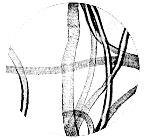
Fig. 4.—Photo-micrograph
of wool fibres, × 250.
(R. J. M. Buchanan.)
Fig. 4.—Microscopic photo of wool fibers, × 250.
(R. J. M. Buchanan.)
2. Make accurate notes of the position and shape of the stains on the material examined.
2. Take detailed notes on the location and shape of the stains on the material being examined.
3. Take one stain, if there are several, or part if single, and note the solubility of it in water, or in a mixture of water and some other substance. The solubility of the colouring matter is greater if the stain be recent than if it be old. The older the stain the less soluble it becomes, as the hæmoglobin is gradually changed in course of time to insoluble hæmatin.
3. If there are multiple stains, choose one, or if there's only one, take a part of it, and observe how well it dissolves in water or in a combination of water and another substance. The coloring matter dissolves better if the stain is fresh rather than old. The older the stain, the less it dissolves, as the hemoglobin gradually converts over time into insoluble hematin.
An endeavour should be made to obtain a solution for microscopical, chemical, and spectroscopical examination. The solvent, in order to obtain the blood corpuscles in as natural a form as possible, should approach in its specific gravity the liquor sanguinis.
An effort should be made to find a solution for microscopic, chemical, and spectroscopic examination. The solvent, to preserve the blood cells in as natural a form as possible, should have a specific gravity similar to the liquor sanguinis.
The following solvents fulfil this purpose:
The following solvents serve this purpose:
(a) Glycerine and water, 1 to 7 (sp. gr. 1030).
(a) Glycerin and water, 1 to 7 (specific gravity 1.030).
(b) Pacini‘s solution of chloral hydrate in water (1 in 10).
(b) Pacini’s solution of chloral hydrate in water (1 in 10).
(c) Normal saline solution.
Normal saline solution.
(d) Roussin‘s solution of glycerine 3 parts, sulphuric acid 1 part (by weight), and water so that the mixture shall have a sp. gr. of 1028.
(d) Roussin's solution consists of 3 parts glycerin, 1 part sulfuric acid (by weight), and water, adjusted so that the mixture has a specific gravity of 1028.
(e) Saturated solution of borax in distilled water.
(e) Saturated solution of borax in distilled water.

Fig. 5.—Photo-micrograph
of flax fibres, × 250.
(R. J. M. Buchanan.)
Fig. 5.—Photo-micrograph of flax fibers, × 250.
(R. J. M. Buchanan.)
The technique of examination has to be varied in certain details, according to the material upon which the stain is. Stains may have to be examined upon cloth-fabrics, wood, plaster, metal, or leather. These will be taken separately, and the methods of examination described which will prove most reliable in each case.
The method of examination needs to change in specific ways depending on the material that the stain is on. Stains might need to be examined on fabric, wood, plaster, metal, or leather. Each of these will be addressed individually, and the examination methods that will be most reliable for each case will be described.
1. Cloth-fabrics.—Cut out a stain, or part of one, and macerate it in a quantity of one of the solvents mentioned above, sufficient for the purpose. If the stain be very small, squeeze with fine forceps one or more drops upon microscope slides for microscopic, and keep the remainder of the solution for spectroscopic, examination. In dyed fabrics, which have been mordanted, the mordant may fix the blood-stain so as to prevent solution, and especially so when the attempts have been made to wash out the stain with soap and water.
1. Cloth fabrics.—Cut out a stain, or part of it, and soak it in one of the solvents mentioned earlier, using enough for the task. If the stain is very small, use fine forceps to squeeze one or more drops onto microscope slides for microscopic examination, and keep the rest of the solution for spectroscopic analysis. In dyed fabrics that have been treated with a mordant, the mordant may fix the blood stain so that it can't be removed, especially if attempts have been made to wash it out with soap and water.
To make a solution of the stain in such cases it is best to use distilled water to which a small quantity of ammonia or citric acid has been added; in one or other of these the colouring matter will dissolve.
To make a solution for the stain in these cases, it's best to use distilled water with a small amount of ammonia or citric acid added; the color will dissolve in either one of these.
2. Wood.—Note the kind of wood, cut off a thin shaving and treat with one of the solvents mentioned above. If on wood containing [Pg 92] tannic acid, such as oak or elm, the best solvent is a two per cent. solution of hydrochloric acid.
2. Wood.—Identify the type of wood, shave off a thin piece, and test it with one of the solvents mentioned earlier. If the wood has tannic acid, like oak or elm, the best solvent is a 2% solution of hydrochloric acid.
3. Plaster.—Scrape off some of the stained plaster, and treat as for cloth or wood.
3. Plaster.—Remove some of the stained plaster, and treat it like you would cloth or wood.
4. Metal.—If the stain be upon a clean and unrusted metal, e.g. the clean blade of a knife, then gently heat the metal on the side opposite to the stain, when the latter, if recent, will peel off or can be easily detached. This requires some care and dexterity. It is easy, however, to scrape the stain off into a watch-glass, and this procedure is necessary when the metal is rusted and the stain mixed with the rust, or when the stain is thin.
4. Metal.—If the stain is on a clean, unrusted piece of metal, e.g. the clean blade of a knife, gently heat the metal on the side opposite the stain. If the stain is recent, it will either peel off or can be easily removed. This requires some care and skill. However, it's easy to scrape the stain into a watch glass, and this method is necessary when the metal is rusted, and the stain is mixed with rust, or when the stain is thin.
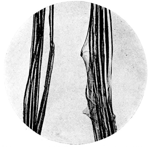
Fig. 6.—Photo-micrograph
of silk fibres, × 250.
(R. J. M. Buchanan.)
Fig. 6.—Photomicrograph of silk fibers, × 250.
(R. J. M. Buchanan.)
If on iron and mixed with rust the borax solution may be used, with a drop or two of solution of ammonia; use a fine camel-hair pencil dipped in the solution, and brush the stain off into a watch-glass. Becker advises that stains mixed with rust should be digested with a weak solution of ammonia and common salt for a few hours; decant the solution and evaporate it upon a microscope slide to dryness, then test the residue by the “hæmin test.”
If you're dealing with iron that's mixed with rust, you can use a borax solution with a drop or two of ammonia solution. Take a fine camel-hair brush, dip it in the solution, and gently scrub the stain into a watch glass. Becker suggests that stains combined with rust should be treated with a weak solution of ammonia and table salt for a few hours; then pour off the solution and let it dry on a microscope slide. After it's dried, test the residue using the "hæmin test."
Ganttner‘s test should be used to a portion of a stain upon metal when thin or mixed with rust. It may be carried out upon the metal itself or upon a scraping of the stain in a watch-glass resting upon a black surface. Moisten the scraping in a watch-glass with a drop or two of distilled water rendered feebly alkaline, then add a minute drop of hydrogen peroxide. Wherever blood is present bubbles of gas develop, which give the material a white beaded appearance. The froth develops from the outside of the drop towards the centre when the stain is mainly composed of blood. In a scraping consisting of mixed particles [Pg 93] of rust and blood, the reaction only appears upon the particles of blood, and rust to which blood adheres; it does not take place on those particles of rust free from blood. Before adding the peroxide of hydrogen it may be necessary to dissipate any air-bubbles which may cling to the scraping in the alkaline water by gentle agitation with the point of a fine glass rod. Should the above reaction with peroxide of hydrogen not take place, then one can rest assured that no blood is present. The test, however, is a negative one; it is not a positive test for blood only; other fluids and exudations from the body, such as saliva and pus, give the reaction. The reaction will take place with blood-stains of any age.
Ganttner's test should be used on a portion of a stain on metal when it's thin or mixed with rust. It can be performed on the metal itself or on a scraping of the stain placed in a watch glass sitting on a black surface. Moisten the scraping in the watch glass with a drop or two of slightly alkaline distilled water, then add a tiny drop of hydrogen peroxide. If blood is present, bubbles of gas will form, giving the material a white beaded appearance. The froth develops from the edge of the drop toward the center when the stain mainly consists of blood. In a scraping made up of mixed particles of rust and blood, the reaction only appears on the particles of blood and the rust that blood clings to; it won’t occur on rust particles that are free of blood. Before adding the hydrogen peroxide, it might be necessary to remove any air bubbles that may adhere to the scraping in the alkaline water by gently stirring with the tip of a fine glass rod. If the reaction with hydrogen peroxide doesn’t happen, you can be sure there’s no blood present. However, this test is negative; it does not confirm blood exclusively, as other body fluids and secretions, such as saliva and pus, can produce the same reaction. The reaction will occur with blood stains of any age. [Pg 93]
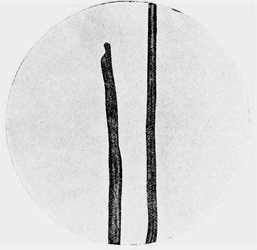
Fig. 7.—Photo-micrograph
of cotton fibres, × 250.
(R. J. M. Buchanan.)
Fig. 7.—Photo-micrograph of cotton fibers, × 250.
(R. J. M. Buchanan.)
In examining a clasp-knife or any hinged weapon for blood-stains, the instrument should be taken to pieces and all the hinges and recesses carefully examined, for in these places blood may be found, although the weapon had previously been wiped clean, and appear free from stains.
In checking a clasp knife or any hinged weapon for blood stains, the tool should be disassembled, and all the hinges and crevices thoroughly inspected, since blood can be present in these areas even if the weapon has been wiped clean and looks free of stains.
5. On Leather.—The tannic acid in leather forms a compound with blood which is insoluble in the solvents generally used. A thin shaving of the stained portion should be taken and folded, with the stained surface outwards, in the form of a loop. If the outer surface of the loop with the stain be made to touch the surface of the glycerine and water solution, at the same time taking care that the leather itself be not moistened, a recent stain may yield a sufficient quantity of colouring matter for the purposes of examination. Failing this, the shaving should be digested in a small quantity of a two per cent. solution of hydrochloric acid in distilled water (Sorby). [Pg 94]
5. On Leather.—The tannic acid in leather forms a compound with blood that doesn't dissolve in the usual solvents. You should take a thin slice of the stained area and fold it, with the stained side facing out, into a loop. If you make sure that the outer part of the loop with the stain touches the surface of the glycerine and water solution, while keeping the leather itself dry, a fresh stain might release enough color for examination. If that doesn't work, you should soak the slice in a small amount of a two percent solution of hydrochloric acid in distilled water (Sorby). [Pg 94]
Microscopical Examination
The microscopical examination of blood-stains, for the purpose of identifying the presence of the red blood corpuscles, is especially applicable to recent stains. In these the corpuscles may retain, to a great extent, their normal characters; but their condition varies with the age of the stain; they become altered in appearance and irregular in shape with increasing age, until a stage is reached when they become completely disintegrated and unrecognisable. Having obtained a solution of a stain by one of the methods recommended, a few drops should be placed upon a clean microscope slide and covered with a No. 1 cover-glass. In a recent stain, where minute coagula are present, one may be placed on a microscope slide and moistened by breathing upon it several times, and then covering it with a No. 1 slip, or a drop of the glycerine solution may be allowed to act upon it on the slide until it be sufficiently moistened, when it should be covered in the same way. The preparation should be examined through the microscope with a good lens (preferably a ¹/₁₂th oil immersion), magnifying 300 to 400 diameters, and if any corpuscles be found, their characters should be carefully observed and noted.
The microscopic examination of bloodstains, to identify the presence of red blood cells, is especially useful for recent stains. In these, the cells often maintain their normal appearance, but their condition changes as the stain gets older; they become altered and irregular in shape over time until they are completely disintegrated and unrecognizable. After obtaining a solution from a stain using one of the recommended methods, place a few drops on a clean microscope slide and cover it with a No. 1 cover glass. For a recent stain that has tiny clots, you can put one on a microscope slide and moisten it by breathing on it several times, then cover it with a No. 1 slip, or let a drop of glycerin solution act on it on the slide until it is sufficiently moistened, then cover it in the same way. The preparation should be examined under the microscope with a good lens (preferably a ¹/₁₂th oil immersion) magnifying 300 to 400 times, and if any cells are found, their characteristics should be carefully noted.
All such specimens should be carefully preserved and labelled with a description of the method of preparation, the case to which they belong, the date of preparation, and the signature of the individual who has made and examined them. It is essential that the preparation and examination of the specimen should be made by the same individual. In certain cases the conditions may be sufficiently favourable to allow of the production of stained specimens, which can be mounted so as to retain their original characters permanently. In every case it is advisable to pursue the investigation with this object in view.
All samples should be carefully preserved and labeled with a description of the preparation method, the case they belong to, the preparation date, and the signature of the person who prepared and examined them. It's important that the same person handles both the preparation and examination of the specimen. In some instances, conditions may be good enough to create stained specimens, which can be mounted to keep their original characteristics permanently. In every case, it’s advisable to pursue the investigation with this goal in mind.
This process is especially applicable to recent blood-stains, in which, from preliminary examination, the presence of blood corpuscles has been determined; where complete disintegration of the blood corpuscles has taken place it would not be of any value.
This process is particularly useful for fresh blood stains, where, based on an initial examination, blood cells have been detected; if the blood cells have completely broken down, it wouldn't be useful.
It may so happen that by means of stained specimens the identity of blood corpuscles may be more easily established, when the result of examination is uncertain in a specimen not so prepared.
It might turn out that using stained specimens makes it easier to identify blood cells when the examination results are unclear in an unprepared sample.
By the action of certain dyes upon the corpuscles their special features are rendered easier of recognition. Any of the approved methods of preparing blood films for general clinical purposes, which will suit the circumstances, may be employed. An easy and reliable method is as follows. A drop of the solution of the blood-stain properly prepared as previously recommended, or if obtainable a small coagulum moistened with normal saline solution, is placed on a clean coverslip and spread evenly over its surface with the aid of a fine glass rod. The film is allowed to dry in the air, covered with a watch-glass for protection against dust. When dry it is passed three times through the flame of a Bunsen burner, or placed in a mixture of [Pg 95] equal parts of absolute alcohol and ether, to fix it. After fixation it should be placed for a minute or more in an aqueous solution of eosin.
By using certain dyes on the blood cells, their unique characteristics become easier to identify. Any of the standard methods for preparing blood films for general clinical use that fit the situation can be used. A simple and reliable method is outlined as follows. A drop of the prepared blood-stain solution, or if available, a small clump of blood moistened with normal saline solution, is placed on a clean coverslip and evenly spread across its surface with a fine glass rod. The film is left to air dry, covered with a watch glass to protect it from dust. Once dry, it is passed through the flame of a Bunsen burner three times, or placed in a mixture of equal parts absolute alcohol and ether to fix it. After fixation, it should be soaked for a minute or more in an aqueous solution of eosin.
Any excess of stain should be removed by washing in distilled water, and the specimen allowed to drain by standing it on edge upon a piece of filter paper; it should then be allowed to dry, and then counterstained with a freshly prepared aqueous solution of methylene blue, hæmatoxylin solution, or other nuclear dye. Wash again in distilled water, allow to dry, and mount in Canada balsam. By this method the corpuscle will be stained pink, and if nucleated, the nucleus will be stained by the methylene blue, hæmatoxylin, or other nuclear stain which may have been used.
Any excess stain should be removed by washing with distilled water, and the specimen should be allowed to drain by standing it on edge on a piece of filter paper. It should then be left to dry and counterstained with a freshly prepared aqueous solution of methylene blue, hematoxylin solution, or other nuclear dye. Wash again with distilled water, allow it to dry, and mount it in Canada balsam. Using this method, the corpuscle will be stained pink, and if it has a nucleus, the nucleus will be stained with the methylene blue, hematoxylin, or other nuclear stain that was used.
Leishman‘s stain may be used. This stain being a methyl-alcohol solution is used for fixing and staining at the same time. A few drops of the stain is placed on the dried film; after standing until evaporation is almost complete, distilled water is dropped on to the slide, and left to stand for two or three minutes, it is then drained off, and a few more drops of distilled water added until the film is pink in colour, then dried with filter paper. Red corpuscles are tinted red with the eosin, and nuclei of leucocytes or nucleated red cells violet or deep blue. The specimen may be examined direct with the oil immersion or mounted in Canada balsam.
Leishman’s stain can be used. This stain, which is a methyl-alcohol solution, is used for both fixing and staining at the same time. A few drops of the stain are placed on the dried film; after sitting until evaporation is nearly complete, distilled water is added to the slide and left for two or three minutes. It is then drained off, and a few more drops of distilled water are added until the film turns pink, then dried with filter paper. Red blood cells are tinted red with the eosin, while the nuclei of white blood cells or nucleated red cells appear violet or deep blue. The specimen can be examined directly with oil immersion or mounted in Canada balsam.
When examining specimens prepared from blood-stains, it is necessary to search carefully for other cellular structures such as epithelial cells, spermatozoa, or fragments of hair.
When looking at samples taken from bloodstains, it's important to carefully check for other types of cells, like epithelial cells, sperm cells, or bits of hair.
It may be advisable, in certain cases where the amount of material submitted for examination is small, to centrifugalise some of the solution in a fine glass tube, in order to determine any cellular elements present to one spot. By making use of this concentrated portion containing the cellular elements for the preparation of a microscopic specimen, one not only facilitates the microscopical examination, but is able to place more reliance upon the results obtained.
It might be a good idea, in some situations where the amount of material submitted for examination is small, to spin some of the solution in a fine glass tube to gather any cellular elements in one spot. By using this concentrated portion with the cellular elements to prepare a microscopic sample, you not only make the microscopic examination easier but also can rely more on the results obtained.
The Results of Microscopical Examination of
Blood-Stains in Their Medico-Legal Relations.
The Results of Microscopic Examination of
Bloodstains in Their Legal Context.
As previously stated, the examination of alleged blood-stains from a medico-legal standpoint is pursued essentially for the purpose of testifying as to whether they have been produced by blood or not. Where the examination yields a negative result, further procedure is necessary with a view of identifying the true nature of the stain. Should, however, the result be positive, the question arises as to the possibility of distinguishing between human blood and the blood of other animals, and determining the exact animal from which the blood has been derived. Such an examination should be pursued in full recognition of its importance as a factor towards the establishment of truth essential to the administration of justice.
As mentioned before, examining suspected blood stains from a legal perspective is mainly done to determine whether they are actually blood or not. If the examination shows a negative result, additional steps are needed to identify what the stain truly is. However, if the result is positive, the next question is whether it’s possible to tell if the blood is human or from another animal, and to identify exactly which animal it came from. This examination should be taken seriously because it plays a crucial role in establishing the truth necessary for delivering justice.
To fulfil this obligation the methods employed should be so selected as to produce results bearing testimony free from any possibility of doubt.
To meet this obligation, the chosen methods should be selected to produce results that are undeniably reliable.
Certain differences exist, and may be detected by microscopical [Pg 96] examination, between the red corpuscles of human blood and those of some other animals sufficiently well marked to render differentiation possible. The differences are those of form and structure.
Certain differences exist that can be seen through microscopic examination between the red blood cells of humans and those of some other animals, which are distinct enough to allow for differentiation. The differences are in their shape and structure.
(1) In man the red corpuscles appear as circular biconcave discs, averaging ¹/₃₅₀₀ of an inch in diameter, and are non-nucleated.
(1) In man, red blood cells look like circular biconcave discs, averaging about ¹/₃₅₀₀ of an inch in diameter, and they don't have a nucleus.
The red corpuscles of mammals present the same features, with the exception of the
The red blood cells of mammals have the same characteristics, except for the
(2) Camel tribe, in which the corpuscles are oval in form, but non-nucleated.
(2) Camel tribe, where the cells are oval in shape, but lack a nucleus.
(3) In birds, fishes, reptiles, and amphibians the red corpuscles are oval in shape, and possess a nucleus.
(3) In birds, fishes, reptiles, and amphibians, the red blood cells are oval-shaped and have a nucleus.
Guided by the above facts, one is able to testify whether or not the corpuscles exhibit the characters of mammalian blood.
Based on the information provided, one can determine whether the corpuscles show the characteristics of mammalian blood.
Many attempts have been made with a view to establishing a reliable means of differentiation between the red blood corpuscles of man and other mammals (the camel excepted), and with a certain degree of success, such as might be expected, under select conditions favourable to histological research, but which do not obtain in medico-legal practice. Differences in size of the red corpuscles, as revealed by micrometric measurement, have been suggested as a possible means of distinguishing between the blood of different mammals. Of the common animals, the red blood corpuscles of the sheep present the most marked difference in size compared with those of man. The following table of the dimensions of red blood corpuscles is derived from measurements made by Treadwell, and quoted by White (The Medico-Legal Journal, New York, 1895):
Many efforts have been made to find a reliable way to differentiate between human red blood cells and those of other mammals (except for camels), achieving some success under specific conditions that are favorable for histological research, but these conditions are not present in legal medicine. Differences in the sizes of red blood cells, measured with micrometers, have been proposed as a potential method to distinguish the blood of different mammals. Among common animals, sheep's red blood cells show the most significant size difference compared to humans. The following table of red blood cell dimensions comes from measurements made by Treadwell and cited by White (The Medico-Legal Journal, New York, 1895):
| μ. | |
| Human | 7.940 |
| Dog | 6.918 |
| Rabbit | 6.365 |
| Ass | 6.293 |
| Pig | 6.101 |
| Horse | 5.503 |
| Cat | 5.463 |
| Ox | 5.436 |
| Sheep | 4.745 |
Menstrual blood contains no fibrin, has an acid reaction due to the vaginal mucus which keeps it fluid, and contains squamous epithelial cells. None of these characters can be differentiated on fabrics, especially when contaminated with urinary stains in addition. Hence, in cases of alleged rape, no distinction can be drawn between blood-stains on the underclothing of the female, which may have arisen from hæmorrhage the result of violence to the sexual parts, and those which might have arisen from the ordinary menstrual flow or metrorrhagia. The detection of spermatozoa, however, would add considerable value to the observation. [Pg 97]
Menstrual blood has no fibrin, is acidic because of the vaginal mucus that keeps it liquid, and includes squamous epithelial cells. These characteristics can't be distinguished on fabric, especially when mixed with urine stains. Therefore, in cases of alleged rape, there's no way to tell the difference between blood stains on a woman's underwear, which might have come from bleeding due to trauma to the genital area, and those that could have resulted from normal menstrual flow or metrorrhagia. However, the presence of spermatozoa would significantly enhance the findings. [Pg 97]

Fig. 8.—Measurement of Blood Corpuscles.
Fig. 8.—Measurement of Blood Cells.
Photo-micrograph of human red blood corpuscles, × 800. Each corpuscle in diameter covers two divisions of the scale. Compare with sheep‘s blood, Fig. 9.
Photo-micrograph of human red blood cells, × 800. Each cell in diameter covers two divisions of the scale. Compare with sheep's blood, Fig. 9.
(R. J. M. Buchanan.)
(R. J. M. Buchanan.)

Fig. 9.—Measurement of Blood Corpuscles.
Fig. 9.—Measuring Blood Cells.
Photo-micrograph of red blood corpuscles from the sheep, × 800. The diameter of the corpuscle covers one division of the scale. Compare with human blood, Fig. 8.
Photo-micrograph of red blood cells from the sheep, × 800. The diameter of the cell covers one division of the scale. Compare with human blood, Fig. 8.
(R. J. M. Buchanan.)
(R. J. M. Buchanan.)
[Pg 98] Blood Crystals.—Professor Preyer of Jena pointed out many years ago that the hæmoglobin crystals from the blood of some animals differed in shape from those of man, and this fact has given rise to many attempts to trace the identity of the blood to the animal from which it has been derived. The results have not been of sufficient value to establish it as trustworthy for medico-legal purposes. Dr. Monckton Copeman (B. M. J., vol. ii. p. 190, 1889) has carefully investigated the subject, and his researches, partly confirmed by Professor Glaister of Glasgow, show that from the guinea-pig, rat, and squirrel, crystals of hæmoglobin may be easily obtained, but the solubility of human hæmoglobin renders it much more difficult to crystallise. Crystals may, however, be obtained in the following ways:
[Pg 98] Blood Crystals.—Many years ago, Professor Preyer from Jena noted that the hemoglobin crystals in the blood of some animals have different shapes than those of humans. This observation has led to numerous attempts to identify the blood's source animal. However, the findings haven't been reliable enough for forensic purposes. Dr. Monckton Copeman (B. M. J., vol. ii. p. 190, 1889) conducted a thorough investigation on the topic, and his research, which was partly validated by Professor Glaister from Glasgow, reveals that hemoglobin crystals can be easily obtained from guinea pigs, rats, and squirrels, while the solubility of human hemoglobin makes it much harder to crystallize. Nevertheless, crystals can still be produced in the following ways:
(a) By feeding leeches on human blood, crystals may be found, after some weeks, in the gastric dilatation of the alimentary canal.
(a) By feeding leeches human blood, crystals may be found, after a few weeks, in the swollen stomach of the digestive tract.
(b) By diluting human blood with the fluid from hydrocele, ascites, or pleurisy when they have undergone decomposition.
(b) By mixing human blood with the fluid from hydrocele, ascites, or pleurisy after they have decomposed.
(c) By adding crystals of glycocholate or taurocholate of soda to human blood.
(c) By adding crystals of glycocholate or taurocholate of sodium to human blood.
(d) By adding a drop of cat‘s bile to human blood on a microscope slide, but the crystals are those of reduced hæmoglobin.
(d) By adding a drop of cat's bile to human blood on a microscope slide, the crystals observed are those of reduced hemoglobin.
Crystals of human hæmoglobin appear in the form of rectangular plates, with a greenish or pale claret colour. On spectroscopic examination they exhibit the characters of reduced hæmoglobin, in contradistinction to the crystals derived from the lower animals, which produce the spectrum of oxyhæmoglobin.
Crystals of human hemoglobin appear as rectangular plates, with a greenish or light claret color. When examined spectroscopically, they show the characteristics of reduced hemoglobin, unlike the crystals from lower animals, which exhibit the spectrum of oxyhemoglobin.
The blood of the bullock, sheep, and pig is very difficult to crystallise. By the method adopted by Gamgee of adding to defibrinated blood one-sixteenth its volume of ether, shaking until the mixture becomes transparent, and allowing to stand in an ordinary temperature for 48 hours, crystals may be obtained from the blood of the following animals:
The blood of the bull, sheep, and pig is really hard to crystallize. Using the method proposed by Gamgee, which involves adding one-sixteenth of its volume of ether to defibrinated blood, shaking it until the mixture is clear, and letting it sit at room temperature for 48 hours, crystals can be obtained from the blood of these animals:
| 1. | Horse. |
| 2. | Bullock. |
| 3. | Sheep. |
| 4. | Pig. |
| 5. | Dog. |
| 6. | Cat. |
| 7. | Rabbit. |
| 8. | Squirrel. |
| 9. | Guinea-pig. |
| 10. | Rat. |
| 11. | Mouse. |
| 12. | Chicken. |
| 13. | Pigeon. |
Crystals from human blood are not easily obtainable by this process, but when they are, they always give the spectrum of reduced hæmoglobin, whereas those from the animals mentioned above give the spectrum of oxyhæmoglobin.
Crystals from human blood are not easy to get using this process, but when they are obtained, they always show the spectrum of reduced hemoglobin, while those from the animals mentioned earlier show the spectrum of oxyhemoglobin.
Chemical Examination
Having obtained a coloured solution from a supposed blood-stain, if sufficient in quantity, apply the following chemical tests to separate portions:
Having obtained a colored solution from a suspected bloodstain, if there's enough of it, apply the following chemical tests to separate portions:
1. Add a few drops of a weak solution of ammonia in distilled water. The colour may remain unchanged, or, at the most, a slight heightening may take place, if it be due to blood. If the solution of ammonia be too strong, a brown colour may be produced if blood be present.
1. Add a few drops of a weak ammonia solution in distilled water. The color may stay the same, or at most, there may be a slight increase if blood is present. If the ammonia solution is too strong, it may turn brown if blood is present.
2. Heat to boiling, when the following changes take place if blood be present:
2. Heat until it boils, and the following changes occur if blood is present:
(a) The colour may disappear.
The color may disappear.
(b) Coagulation follows.
Coagulation happens next.
(c) A precipitate falls, dirty grey or brown in colour, depending upon the amount of colouring matter present.
(c) A precipitate falls, dirty gray or brown in color, depending on the amount of coloring matter present.
On adding caustic potash to the precipitate it will dissolve, and the solution formed will appear greenish by transmitted and red by reflected light. This phenomenon is called the dichroism of blood. Authorities differ in opinion as to whether the colour is green by transmitted and red by reflected light, or vice versa. “As a matter of fact, the phenomenon is chameleon-like as regards colour, so that both sets of observers may be considered right or wrong” (Glaister).
When you add caustic potash to the precipitate, it dissolves, and the resulting solution looks greenish when viewed through and red when seen by reflection. This effect is known as the dichroism of blood. Experts disagree on whether the color is green when transmitted and red when reflected, or vice versa. “In reality, the phenomenon changes colors like a chameleon, so both groups of observers can be seen as either right or wrong” (Glaister).

Fig. 10.—Photo-micrograph of red blood
corpuscles from domestic fowl, × 250.
(R. J. M. Buchanan.)
Fig. 10.—Microscopic image of red blood cells from domestic chickens, × 250.
(R. J. M. Buchanan.)

Fig. 11.—Photo-micrograph of blood
corpuscles of fish, × 250.
(R. J. M. Buchanan.)
Fig. 11.—Photo-micrograph of fish blood cells, × 250.
(R. J. M. Buchanan.)

Fig. 12.—Photo-micrograph of blood
corpuscles from a dried stain of the blood of a codfish, × 250.
(R. J. M. Buchanan.)
Fig. 12.—Photo-micrograph of blood cells from a dried stain of codfish blood, × 250.
(R. J. M. Buchanan.)
3. Add tincture of guaiacum, freshly prepared: an opaque, cream-coloured precipitate of the guaiac resin will form in the aqueous solution. On the addition of ozonic ether, turpentine, or peroxide of hydrogen, a blue colour will be produced at the junction of the fluids: proportionate to the amount of blood-colouring matter present, the blue colour will vary in intensity.
3. Add freshly prepared tincture of guaiacum: an opaque, cream-colored precipitate of guaiac resin will form in the watery solution. When you add ozonic ether, turpentine, or hydrogen peroxide, a blue color will appear where the fluids meet: the intensity of the blue will vary depending on the amount of blood-colored matter present.
This test, known as Day‘s or Schönbein‘s, is extremely delicate, and reacts to no coloured substance except blood.
This test, known as Day's or Schönbein's, is very sensitive and only reacts to blood.
In cases where the blood-stain is small, the test may be applied as follows. Moisten a pure white filter paper with a drop of distilled water, or one of the solutions recommended in the section on physical examination, and touch the stain with the moistened portion. On adding a drop of tincture of guaiacum followed by a drop of ozonic ether to the wet filter paper the blue colour will be produced and easily recognised on the white surface.
In cases where the bloodstain is small, the test can be done like this. Dampen a pure white filter paper with a drop of distilled water or one of the solutions mentioned in the section on physical examination, then touch the stain with the damp part. When you add a drop of guaiacum tincture followed by a drop of ozonic ether to the wet filter paper, a blue color will appear and will be easily visible on the white surface.
The guaiacum test, although extremely delicate, can only be accepted as providing negative evidence. The absence of reaction proves the absence of blood, except in very old blood-stains, which may not respond [Pg 101] to the test. The blue colour produced indicates that the substance may be blood, but it cannot be accepted without corroboration. Gluten, raw potato, milk, bile, sweat (Ogston), and other oxidising substances give a blue colour with guaiacum and ozonic ether; some substances give the blue colour with guaiacum alone.
The guaiacum test, while very sensitive, can only be considered as indicating negative results. The lack of a reaction confirms there is no blood, except in very old bloodstains, which might not react to the test. The blue color produced suggests that the substance could be blood, but it requires further confirmation. Gluten, raw potato, milk, bile, sweat (Ogston), and other oxidizing substances can produce a blue color with guaiacum and ozonic ether; some substances create the blue color with guaiacum alone.
With blood, however, the test is sufficiently delicate to detect one drop in six ounces of water.
With blood, though, the test is sensitive enough to detect one drop in six ounces of water.
4. Nitric acid added to a portion of the solution of blood in distilled water produces a whitish-grey precipitate.
4. Nitric acid added to a part of the blood solution in distilled water creates a whitish-grey precipitate.

Fig. 13.—Photo-micrograph of
frog‘s blood showing oval nucleated red corpuscles, × 250.
(R. J. M. Buchanan.)
Fig. 13.—Photo-micrograph of frog's blood showing oval nucleated red blood cells, × 250.
(R. J. M. Buchanan.)
5. Hæmin Crystals.—Concentrate a portion of the solution upon a microscope slide, add to it a minute crystal of chloride of sodium and a few drops of glacial acetic acid. Heat gently to dryness or to a lesser degree under a coverslip; examine with the microscope; if blood be present, crystals of hæmin, or the hydrochloride of hæmatin, will be found. They are of a yellowish-red to brownish-black colour with a metallic lustre. They occur in rhomboidal prisms, or six-sided in shape, or in the form of “whetstones,” often in clusters; many of the crystals exhibit a lipped projection on one side. They are known as Teichmann‘s crystals. It is well to verify their origin from blood by placing upon them a drop of hydrogen peroxide, when they will give off bubbles of oxygen gas.
5. Hæmin Crystals.—Concentrate a portion of the solution on a microscope slide, add a tiny crystal of sodium chloride and a few drops of glacial acetic acid. Heat gently until dry or to a lesser extent under a coverslip; examine under the microscope; if blood is present, crystals of hæmin, or the hydrochloride of hæmatin, will be visible. They appear in yellowish-red to brownish-black color with a metallic sheen. They can be found in rhomboidal prisms, or six-sided shapes, or in the form of “whetstones,” often in clusters; many of the crystals show a lipped projection on one side. They are known as Teichmann's crystals. It is advisable to confirm their origin from blood by placing a drop of hydrogen peroxide on them, as they will release oxygen gas bubbles.
They are insoluble in water, alcohol, and dilute acetic and hydrochloric acids. They dissolve in boiling acetic or hydrochloric acids, and the caustic alkalies. They respond to the guaiacum test, and the ash produced by incineration shows the presence of iron by the red [Pg 102] colour produced on the addition of a drop of hydrochloric acid and a solution of potassium sulphocyanide.
They don't dissolve in water, alcohol, or dilute acetic and hydrochloric acids. They do dissolve in boiling acetic or hydrochloric acids, as well as in caustic alkalis. They give a positive result on the guaiacum test, and the ash generated by burning shows the presence of iron by the red color that appears when you add a drop of hydrochloric acid and a solution of potassium thiocyanate. [Pg 102]
The production, by the methods described, of such crystals affords conclusive proof of the presence of blood.
The production of these crystals using the described methods provides clear evidence of the presence of blood.
Spectroscopic Examination
To a portion of the coloured solution, filtered if necessary, the spectroscopic tests should be applied. The following points must be remembered in carrying out a spectroscopic examination:
To a portion of the colored solution, filtered if needed, the spectroscopic tests should be performed. The following points must be kept in mind when doing a spectroscopic analysis:
(a) The colouring matter of fresh blood is hæmoglobin, and it may exist in two states, according to the degree of its combination with oxygen.
(a) The color of fresh blood is hemoglobin, and it can exist in two forms, depending on how much oxygen it is combined with.
In arterial blood it is present as oxidised hæmoglobin, and the same obtains in blood which has been exposed to the air under certain conditions and for a varying period of time.
In arterial blood, it's found as oxidized hemoglobin, and the same is true for blood that has been exposed to air under certain conditions and for varying lengths of time.

Fig. 14.—Photo-micrograph of
crystals of hæmin, × 250.
(R. J. M. Buchanan.)
Fig. 14.—Photo-micrograph of crystals of hemin, × 250.
(R. J. M. Buchanan.)
In venous blood, especially when obtained under conditions preventing oxidation, as from the heart cavity of an animal newly asphyxiated, it is present as deoxidised hæmoglobin.
In venous blood, especially when taken under conditions that prevent oxidation, such as from the heart cavity of a recently asphyxiated animal, it appears as deoxygenated hemoglobin.
(b) In dry stains, especially if they have been subjected to the action of impure air containing the products of coal-combustion, the colouring matter becomes changed into methæmoglobin, or hæmoglobin in which its combination with oxygen has been altered in such a way that a current of a neutral gas, such as hydrogen or nitrogen, will not dissociate it, as it does with oxyhæmoglobin. Such stains have a brownish colour, and may give an acid reaction.
(b) In dry stains, especially those exposed to dirty air with coal combustion byproducts, the coloring material turns into methæmoglobin, or hemoglobin that has changed in a way that neutral gases like hydrogen or nitrogen can't break it apart, unlike oxyhemoglobin. These stains appear brownish and may show an acidic reaction.
On examining the solution of the colouring matter from a blood-stain with the spectroscope, the spectrum will vary according to its condition and the nature of the solvent used.
On examining the solution of the coloring matter from a bloodstain with the spectroscope, the spectrum will change depending on its condition and the type of solvent used.
The spectra of hæmoglobin and its derivatives are characteristic, and afford conclusive evidence of the presence of blood. The spectra must be recognised, however, in more than one condition. Other substances may yield spectra very similar to that of oxyhæmoglobin, but when subjected to certain tests they do not alter in the same way. They cannot be made to give the spectra of reduced hæmoglobin and reduced hæmatin, and any colouring matter which may be made to yield the spectra of reduced hæmoglobin and reduced hæmatin is derived from blood.
The spectra of hemoglobin and its derivatives are distinctive and provide clear proof of the presence of blood. However, these spectra need to be identified under multiple conditions. Other substances can produce spectra that are very similar to oxyhemoglobin, but they don't change in the same way when tested. They can't be made to show the spectra of reduced hemoglobin and reduced hematin, and any coloring material that can produce the spectra of reduced hemoglobin and reduced hematin comes from blood.
Blood Spectra
1. Oxidised hæmoglobin (O₂Hb) is characterised by the presence in its solar spectrum of two absorption bands between the D and E lines. The first band commences at the D line and extends a short distance towards the E. The second commences at a little distance from it, and terminates at the E line; it is about twice the breadth of the first. The band at D is more defined than the other (Fig. 15, 1).
1. Oxidized hemoglobin (O₂Hb) is characterized by two absorption bands in its solar spectrum located between the D and E lines. The first band starts at the D line and stretches a short distance toward the E. The second band begins a bit away from it and ends at the E line; it is about twice as wide as the first. The band at D is more distinct than the other. (Fig. 15, 1)
2. Deoxidised or reduced hæmoglobin presents one broad band occupying almost the whole of the space between D and E slightly to the left of these lines (Fig. 15, 2).
2. Deoxygenated or reduced hemoglobin shows one wide band covering almost the entire area between D and E, just to the left of these lines (Fig. 15, 2).
3. Methæmoglobin presents two bands between D and E, in the same position as those of O₂Hb, but in addition a third band between C and D and near to the former (Fig. 15, 3).
3. Methæmoglobin shows two bands between D and E, just like O₂Hb, but it also has a third band between C and D, close to the first one (Fig. 15, 3).
A solution of oxyhæmoglobin or methæmoglobin may be reduced by the addition of a reducing agent, such as Stokes‘ reagent, consisting of ferrous sulphate with a small quantity of tartaric acid dissolved in water and rendered alkaline at the time of using with ammonia, or, better still, by the addition of ammonium sulphide. The spectrum will change to that of reduced hæmoglobin.
A solution of oxyhemoglobin or methemoglobin can be reduced by adding a reducing agent, like Stokes’ reagent, which is ferrous sulfate mixed with a little bit of tartaric acid dissolved in water and made alkaline with ammonia at the time of use, or even better, by adding ammonium sulfide. The spectrum will then change to that of reduced hemoglobin.
4. Acid hæmatin presents a spectrum with a band between D and E, commencing at a little distance from D and ending at E, also a narrower band between C and D and commencing at C. It is a difficult spectrum to obtain.
4. Acid hematin shows a spectrum with a band between D and E, starting a bit away from D and finishing at E, as well as a narrower band between C and D that begins at C. It’s challenging to get this spectrum.
5. Alkaline hæmatin presents a spectrum with a single band between C and D near to the D line. It is more difficult to obtain than the spectrum of acid hæmatin.
5. Alkaline hematin shows a spectrum with a single band between C and D close to the D line. It's harder to obtain than the spectrum of acid hematin.
It is not necessary, however, to obtain these spectra, viz. 5 and 6, but it is necessary to reduce solutions of either acid or alkaline hæmatin in order to obtain the spectrum of reduced hæmatin. To do so proceed as follows. To some of the solution of colouring matter obtained from the stain add a small quantity of a 20 per cent. solution of sodium hydrate; the solution will alter in colour, and the spectrum of O₂Hb or MetHb will disappear. On adding to this solution of alkaline hæmatin a few drops of ammonium sulphide, or Stokes‘ fluid, it becomes claret-coloured, and on examination with the spectroscope the spectrum [Pg 104] of reduced hæmatin will be seen. This is the most pronounced of all blood spectra. Its production can be hastened by gently warming the solution.
It's not necessary to obtain these spectra, specifically 5 and 6, but you do need to reduce solutions of either acid or alkaline hematin to get the spectrum of reduced hematin. Here's how to do it. Take some of the coloring solution from the stain and add a small amount of a 20 percent sodium hydroxide solution; the solution will change color, and the spectrum of O₂Hb or MetHb will disappear. When you add a few drops of ammonium sulfide or Stokes' fluid to this alkaline hematin solution, it will turn claret-colored, and when you look at it with a spectroscope, you'll see the spectrum of reduced hematin. This is the most distinct of all blood spectra. You can speed up its formation by gently warming the solution.
If the stain be old and already changed into hæmatin, its solution will yield the spectrum of acid hæmatin, and will give the spectrum of reduced hæmatin on the addition of ammonium sulphide or Stokes‘ fluid without previous alkalisation.
If the stain is old and has already turned into hematin, its solution will show the spectrum of acid hematin and will produce the spectrum of reduced hematin when ammonium sulfide or Stokes' fluid is added without prior alkalization.
6. Reduced hæmatin presents a spectrum with a dark band about midway between D and E, and a broad but paler band commences near the E line and extends to the b line (Fig. 15, 4).
6. Reduced hematin shows a range with a dark band about halfway between D and E, and a wide but lighter band starts close to the E line and stretches to the b line (Fig. 15, 4).

Fig. 15.—Blood Spectra.
Fig. 15.—Blood Spectra.
| 1. Oxyhæmoglobin. | 3. Methæmoglobin. | 5. CO hæmoglobin. |
| 2. Reduced hæmoglobin. | 4. Reduced hæmatin. | 6. CO hæmoglobin and |
| reduced hæmoglobin. |
In cases of death by asphyxia, in which the hæmoglobin is in combination with CO₂, the blood, if removed and examined immediately after death, gives a spectrum of reduced hæmoglobin, but on exposure to the air it rapidly changes to oxyhæmoglobin. The period after death at which blood is usually submitted for medico-legal examination is sufficiently late to allow of this change, and so prevents the possibility of determining death by asphyxia by spectroscopic examination of the blood. Where death has been caused by the action of carbon monoxide, the blood is of a cherry-red colour, and it will retain this colour unchanged for a long time, in fact for years, due to a stable combination of the CO and hæmoglobin, called carboxyhæmoglobin. Such blood yields a very characteristic spectrum, with two bands similar to those of O₂Hb, but nearer to the violet end (Fig. 15, 6). Their position should be assured by accurate measurement and comparison with a spectrum of O₂Hb. The CO hæmoglobin, however, cannot be reduced; on the addition of ammonium sulphide or Stokes‘ fluid the bands remain unaltered. [Pg 105]
In cases of death by asphyxiation, where hemoglobin is combined with CO₂, the blood, if removed and examined right after death, shows a spectrum of reduced hemoglobin. However, when exposed to air, it quickly changes to oxyhemoglobin. The time after death when blood is typically submitted for forensic examination is usually late enough for this change to occur, which prevents the possibility of determining death by asphyxiation through a spectroscopic examination of the blood. If death was caused by carbon monoxide exposure, the blood appears cherry-red, and it maintains this color for a long time, even for years, due to a stable combination of CO and hemoglobin known as carboxyhemoglobin. This blood produces a very distinctive spectrum, with two bands similar to those of O₂Hb, but closer to the violet end (Fig. 15, 6). Their position should be confirmed by precise measurement and comparison with a spectrum of O₂Hb. However, the CO hemoglobin cannot be reduced; when ammonium sulfide or Stokes’ fluid is added, the bands remain unchanged. [Pg 105]
In cases where the amount of fluid obtainable for examination is very small, recourse must be had to the micro-spectroscope, using a Sorby‘s cell to hold the fluid, and substituting for the eye-piece of the microscope a specially constructed spectroscope arranged so as to throw the spectrum of a known solution of blood-colouring matter alongside of that yielded by the solution under examination. Artificial light should be used, and the D line located by placing in the flame a platinum wire carrying a salt of sodium.
In situations where the amount of fluid available for testing is very small, a micro-spectroscope needs to be used, utilizing a Sorby’s cell to hold the fluid. The eye-piece of the microscope is replaced with a specially designed spectroscope that allows for the comparison of the spectrum of a known blood-colored solution with the one being analyzed. Artificial light should be used, and the D line should be identified by placing a platinum wire containing sodium salt in the flame.
Biological Tests for Blood
The results of the experimental investigations of Friedenthal,[3] Deutsch,[4] Uhlenhuth,[5] Wasserman and Schutze,[6] Nuttall,[7] Tarchetti,[8] Grünbaum,[9] Metchnikoff,[10] and M‘Weeney[11] into “blood relationships” have led to the suggestion of a new method—the “biological test,” by which different kinds of mammalian blood may be distinguished one from the other. Their experiments show in a general way that the “serum from an animal which has been injected intraperitoneally with any given organic fluid will, if mixed in small quantity with a dilute solution of the fluid used for the injection, produce a more or less definite precipitate.” If human defibrinated blood be injected into the peritoneal cavity of a rabbit, the serum obtained from the rabbit‘s blood, when mixed with a clear solution of the blood of man or ape, will produce a precipitate, and agglutinate the red blood corpuscles if present: the same reaction will not follow if such serum be added to a solution of the blood of any other animal.
The results of the experimental investigations by Friedenthal, Deutsch, Uhlenhuth, Wasserman and Schutze, Nuttall, Tarchetti, Grünbaum, Metchnikoff, and M‘Weeney into “blood relationships” have led to the suggestion of a new method—the “biological test,” which allows different types of mammalian blood to be distinguished from one another. Their experiments generally indicate that the “serum from an animal that has been injected intraperitoneally with any given organic fluid will, if mixed in small amounts with a diluted solution of the fluid used for the injection, produce a more or less definite precipitate.” If human defibrinated blood is injected into the peritoneal cavity of a rabbit, the serum obtained from the rabbit's blood, when mixed with a clear solution of human or ape blood, will produce a precipitate and agglutinate the red blood cells if they are present; the same reaction won’t occur if that serum is added to a solution of blood from any other animal.
The introduction of substances of albuminous or proteid nature into the body of an animal, and which can be taken up by its cells as a food, produces in the body of the animal a series of substances called antibodies, of which one is designated precipitin from its power of producing a precipitate with the substance introduced. The organic substance introduced capable of producing antibodies is called the antigen. The precipitin is formed and present in the blood serum of the inoculated animal.
The introduction of substances that are protein-based into an animal's body, which can be absorbed by its cells as food, leads to the production of a series of substances known as antibodies. One of these is called precipitin because it can create a precipitate with the introduced substance. The organic substance that triggers the production of antibodies is referred to as the antigen. Precipitin is formed and found in the blood serum of the inoculated animal.
Metallic poisons, alkaloids and carbohydrates, do not produce antibodies; some vegetable proteids do.
Metallic poisons, alkaloids, and carbohydrates don't create antibodies; some plant proteins do.
The serum containing the precipitin is called the antiserum.
The serum that has the precipitin is called the antiserum.
The precipitin in the antiserum is specific, and produces the effect only with its own antigen, not with that derived from another species of animal, but will act with one derived from a closely allied species, e.g. man and the higher apes, sheep, and goat. [Pg 106]
The precipitin in the antiserum is specific and only reacts with its own antigen, not with those from different animal species, but it will react with those from closely related species, like humans and higher primates, sheep, and goats. [a id="Page_106"] [Pg 106]
In order to eliminate this the antigen should be diluted to 1:1000.
To get rid of this, the antigen should be diluted to 1:1000.
The blood used as antigen may be dried and sterilised, thus it may be kept for a long time, and when required for use dissolved in normal saline.
The blood used as an antigen can be dried and sterilized, so it can be stored for a long time, and when needed, it can be dissolved in normal saline.
The most convenient human antigen for general use is ascitic or pleuritic fluid, and may be kept in good condition when mixed with a small quantity of chloroform.
The most convenient human antigen for general use is ascitic or pleuritic fluid, and it can be stored properly when mixed with a small amount of chloroform.
To produce the antiserum 3 to 10 c.c. of the antigen serum is injected into a vein or the peritoneal cavity of a rabbit, and repeated at an interval of four or five days until 25 c.c. have been injected. The serum of the rabbit is then tested with the antigen diluted 1:100. The injections are then continued until 70 to 80 c.c. have been administered. The animal is then killed, the blood collected, and stood in a refrigerator for the serum to separate. This is then drawn into sterile pipettes, sealed, and kept in the cold. For purposes of preservation one-tenth its volume of 5 per cent. phenol may be added before pipetting. The antiserum may be dried on slips of black paper for future use.
To produce the antiserum, 3 to 10 c.c. of the antigen serum is injected into a vein or the peritoneal cavity of a rabbit, with follow-up injections every four to five days until a total of 25 c.c. has been administered. The rabbit's serum is then tested with the antigen diluted 1:100. Injections are continued until 70 to 80 c.c. have been given. The rabbit is then euthanized, the blood collected, and left in a refrigerator for the serum to separate. This serum is then drawn into sterile pipettes, sealed, and stored in the cold. To help with preservation, one-tenth of its volume of 5 percent phenol may be added before pipetting. The antiserum can be dried on black paper slips for future use.
For medico-legal purposes, the antiserum should always be tested with its antigen to be sure of its efficacy.
For legal medical purposes, the antiserum should always be tested with its antigen to ensure its effectiveness.
The antigen to be tested should be diluted 1:1000 at least, as potent antisera may give reactions with strong solutions of antigens derived from other species of animals. Various antisera may be made by inoculating rabbits with the serum of different animals.
The antigen being tested should be diluted at least 1:1000, as strong antisera can react with concentrated antigen solutions from different animal species. Different antisera can be produced by injecting rabbits with the serum from various animals.
Metchnikoff has shown that intraperitoneal injection is not absolutely necessary in order to produce an antiserum, feeding a rabbit on the blood will act in the same way.
Metchnikoff has demonstrated that intraperitoneal injection isn't strictly necessary to produce an antiserum; feeding a rabbit with blood can have the same effect.
The procedure for testing a stain is as follows. First of all the stain must be proved to be blood by the usual methods; this because the antiserum will give reactions with other albuminous substances, e.g. mucus, pus, semen, milk, or albuminous urine derived from the animal providing the antigen.
The process for testing a stain is as follows. First, the stain must be confirmed as blood using standard methods. This is important because the antiserum can react with other protein-based substances, e.g. mucus, pus, semen, milk, or protein-rich urine from the animal that provided the antigen.
Having proved the stain to be blood, a solution of it should be made in normal saline, sufficiently strong to give the HNO₃ reaction for albumin, or to foam when shaken up. If the amount of solution be small, the tests can be carried out in capillary tubes or pipettes. The solution of the stain must be cleared by filtration or the centrifuge.
Having confirmed that the stain is blood, it should be dissolved in normal saline at a strength that will trigger the HNO₃ reaction for albumin or create foam when shaken. If the solution is
The tests must be controlled by comparison with known human blood, and blood from several domestic animals. For this purpose the various antisera should be kept in stock.
The tests must be controlled by comparing them with known human blood and blood from several domestic animals. For this purpose, the different antisera should be kept in stock.
Two sets, A and B, of six small tubes are used, into each of A is placed 0.05 c.c. of human antiserum from a sensitised rabbit. Into each tube of Set A is then added double the amount of diluted antigen, as follows:
Two sets, A and B, each containing six small tubes, are used. Into each tube of Set A, 0.05 c.c. of human antiserum from a sensitized rabbit is added. Then, double the amount of diluted antigen is added to each tube of Set A, as follows:
| Set A — | 1. | Receives | extract | of known | human | blood | -stain. | |
| 2. | “ | “ | “ | ox | “ | “ | ||
| 3. | “ | “ | “ | horse | “ | “ | ||
| 4. | “ | “ | “ | sheep | “ | “ | ||
| 5. | “ | “ | “ | pig | “ | “ | ||
| 6. | “ | “ | “ | stain | under observation. | |||
| Set B — | 1. | Receives | normal | rabbit serum | 0.05 | c.c. |
| 2. | “ | human | antiserum | 0.05 | “ | |
| 3. | “ | ox | “ | 0.05 | “ | |
| 4. | “ | horse | “ | 0.05 | “ | |
| 5. | “ | sheep | “ | 0.05 | “ | |
| 6. | “ | pig | “ | 0.05 | “ |
To each tube in Set B is now added about 0.1 c.c. of the extract of the suspected stain.
To each tube in Set B, add about 0.1 c.c. of the extract from the suspected stain.
It will thus be seen that in Set A known antigen is added to known precipitin, with the exception of tube 6, which receives suspected antigen, while in Set B the suspected antigen is added to known antisera, with the exception of No. 1, which contains normal rabbit serum.
It can be seen that in Set A, a known antigen is added to a known precipitin, except for tube 6, which gets the suspected antigen. In Set B, the suspected antigen is added to known antisera, except for No. 1, which has normal rabbit serum.
The tubes are observed in an hour and the results noted. Should the stain be human blood, positive reactions will be present in tubes 1 and 6 of stand A, and tube 2 of B.
The tubes are checked after an hour and the results are recorded. If the stain is human blood, positive reactions will show in tubes 1 and 6 of stand A, and tube 2 of stand B.
The negative reactions in the other tubes in stand A prove that the antiserum used is specific, and will only react with its own antigen; in stand B that the stain is composed of specific human antigen, reacting only with its own antiserum.
The negative reactions in the other tubes in stand A show that the antiserum used is specific and will only react with its own antigen; in stand B, that the stain is made up of specific human antigen, reacting only with its own antiserum.
Should the stain suspected be other than human, and derived from one of the other animals used for antiserum in stand B, then the positive reaction will occur in the particular tube.[12] The test is now recognised as very sensitive and reliable, and is also applicable to the detection of the flesh or fluids of animals, and has thus been used for the detection of meat stuffs.
If the stain in question is something other than human and comes from one of the other animals used for antiserum in stand B, then the positive reaction will happen in that specific tube.[12] The test is now known to be very sensitive and reliable, and it can also be used to detect the flesh or fluids of animals, making it useful for identifying meat products.
Tarchetti advises the following procedure in the examination of blood-stains: Dissolve the stain in a few drops of a 0.9 per cent. aqueous solution of sodium chloride, filter, and divide the filtrate into two portions; to one (a) add 0.5 c.c. of the prepared rabbit serum (the so-called antiserum); to the other (b) serum from a rabbit which has not been injected with human blood. Both are to be placed in an incubator at 37° C. for an hour. By this time, if the solution of the stain be of human or anthropoid origin, the contents of the tube (a) will have become turbid, the contents of tube (b) will remain clear. From a series of experiments with blood-stains of man and other animals on a variety of materials, Tarchetti states that this method is reliable. Prepared rabbit “human antiserum” has been shown to have no such reaction with the blood of the pig, ox, calf, mouse, or rat.
Tarchetti recommends the following method for examining bloodstains: Dissolve the stain in a few drops of a 0.9 percent aqueous solution of sodium chloride, filter it, and split the filtrate into two parts; to one (a), add 0.5 c.c. of the prepared rabbit serum (known as antiserum); to the other (b), use serum from a rabbit that hasn't been injected with human blood. Both should be placed in an incubator at 37° C. for an hour. By this time, if the stain comes from a human or anthropoid source, the contents of tube (a) will become cloudy, while the contents of tube (b) will stay clear. Based on a series of tests with bloodstains from humans and other animals across various materials, Tarchetti confirms that this method is trustworthy. Prepared rabbit “human antiserum” has been shown to not react with the blood of pigs, cows, calves, mice, or rats.
From the result of his investigations Grünbaum points out that these reactions must be looked upon rather as “special” than “specific,” in view of the fact that his “chimpanzee antiserum” gave a slight but distinct turbidity after a few hours with horse blood. He also suggests a method for the microscopical application of the “biological test,” by using a 1 per cent. blood solution with a drop of [Pg 108] “antiserum.” This method has enabled him to distinguish between human and anthropoid blood, the reaction occurring earlier and being more complete when the “antiserum” is used on its own blood.
From his investigations, Grünbaum points out that these reactions should be considered more “special” than “specific,” since his “chimpanzee antiserum” caused a slight but noticeable turbidity after a few hours with horse blood. He also proposes a method for microscopically applying the “biological test” by using a 1 percent blood solution with a drop of [Pg 108] “antiserum.” This method has allowed him to tell the difference between human and anthropoid blood, with the reaction happening sooner and being more complete when the “antiserum” is applied to its own blood.
Vegetable and other Stains
which resemble Blood
Certain vegetable colouring matters give spectra which may be mistaken for blood, from their close similarity. Of these cochineal dissolved in a solution of alum gives two bands similar to O₂Hb. On the addition of boric acid the bands move to the violet end of the spectrum, but they are unaffected if the colouring matter be blood. Lac-dye, alkanet root, madder, and others also give spectra resembling O₂Hb, but they are changed or disappear on adding ammonia or sulphite of potassium, while the spectrum of blood remains unaltered.
Certain vegetable colorants produce spectra that can be confused with blood due to their close resemblance. For example, cochineal dissolved in an alum solution creates two bands similar to O₂Hb. When boric acid is added, the bands shift toward the violet end of the spectrum, but they remain unchanged if the colorant is blood. Lac-dye, alkanet root, madder, and others also generate spectra resembling O₂Hb, but these change or disappear when ammonia or potassium sulfite is added, whereas the blood spectrum remains unchanged.
As stated previously, spectra of colouring matters other than blood are not capable of being altered by reducing agents, so that, however similar they may be to O₂Hb, they cannot be accepted as derived from blood unless the spectra of reduced Hb and reduced hæmatin can be obtained in the way described.
As mentioned earlier, the spectra of coloring substances other than blood cannot be changed by reducing agents. So, no matter how similar they might look to O₂Hb, they can't be considered as coming from blood unless the spectra of reduced Hb and reduced hematin can be obtained as described.
Cochineal, colours of certain roots and wood, turn crimson on the addition of ammonia, logwood bluish-black.
Cochineal, colors from certain roots and woods, turn bright red when ammonia is added, while logwood becomes bluish-black.
The colour of the rose and certain flowers turn green on adding ammonia.
The color of the rose and some flowers turns green when ammonia is added.
Fruit-stains from mulberry, currants, gooseberries, &c., turn bluish-green with ammonia.
Fruit stains from mulberries, currants, gooseberries, etc., turn bluish-green with ammonia.
Vegetable stains have their colour heightened by the action of dilute acids.
Vegetable stains become more vibrant when exposed to diluted acids.
Chlorine bleaches fruit-stains, but turns the colour of blood-stains to an olive-green.
Chlorine removes fruit stains but changes the color of blood stains to olive green.
Red dyes fixed by a mordant are not influenced by ammonia.
Red dyes set with a mordant aren’t affected by ammonia.
Iron stains are usually blackened by ammonium sulphide.
Iron stains are typically darkened by ammonium sulfide.
Red paint may contain red oxide of iron; digest with hydrochloric acid and test for iron, by adding ferrocyanide of potassium to obtain the Prussian blue. Iron stains may be of a reddish-brown or orange colour, and insoluble in water, so that HCl is used to dissolve them.
Red paint may include red oxide of iron; mix it with hydrochloric acid and test for iron by adding potassium ferrocyanide to produce Prussian blue. Iron stains can appear reddish-brown or orange, and they are insoluble in water, so HCl is used to dissolve them.
Citrate and malate of iron stains are soluble in water; the addition of ammonia to an aqueous solution produces no change; guaiacum will give a blue reaction if a persalt of iron be present. The addition of hydrochloric acid and ferrocyanide of potassium will give the Prussian blue reaction. A drop of nitric acid added to the solution will oxidise the iron to the ferric state, and on the addition of a few drops of fresh-made aqueous solution of sulphocyanide of potassium the port-wine colour of sulphocyanide of iron will be produced.
Citrate and malate of iron stains dissolve in water; adding ammonia to an aqueous solution doesn’t change anything; guaiacum will turn blue if there's a persalt of iron present. Adding hydrochloric acid and potassium ferrocyanide will result in the Prussian blue reaction. A drop of nitric acid added to the solution will oxidize the iron to the ferric state, and when a few drops of freshly made aqueous potassium thiocyanate solution are added, the port-wine color of iron thiocyanate will be produced.
A control test must be made with distilled water to prove the purity of the reagents, and the two results compared with each other.
A control test should be done with distilled water to confirm the purity of the reagents, and the two results should be compared.
Aniline stains resembling blood are changed to greenish-yellow or yellow on the addition of dilute nitric acid. Eosin stains produce a [Pg 109] fluorescent solution when dissolved in water. Grease, tar, pitch, snuff, and paint may be mistaken for blood, especially on dark fabrics. They may be detected by two methods:
Aniline stains that look like blood turn greenish-yellow or yellow when you add dilute nitric acid. Eosin stains create a [Pg 109] fluorescent solution when they're dissolved in water. Grease, tar, pitch, snuff, and paint can be confused with blood, especially on dark fabrics. They can be identified by two methods:
(a) The Wet Method.—Having failed to obtain a solution by the aid of the ordinary solvents for blood, other solvents must be used; ether or benzene for grease, paint, or tar. The solution obtained must be examined with the spectroscope.
(a) The Wet Method.—After not being able to get a solution using regular solvents for blood, alternative solvents need to be used; ether or benzene for grease, paint, or tar. The resulting solution must be examined with a spectroscope.
(b) The Dry Method.—Place the cloth or other fabric stain down upon a clean white filter paper; then on pressing the cloth with a hot laundry iron, grease, tar, or pitch will stain the paper, paint or snuff will not.
(b) The Dry Method.—Lay the stained cloth or fabric flat on a clean white filter paper; then, by pressing the cloth with a hot iron, grease, tar, or pitch will leave a mark on the paper, but paint or snuff won't.
BURNS AND SCALDS
Burns and Scalds are lesions characterised by a more or less marked destruction of the tissues of the body, caused by the action upon its internal or external surfaces of a temperature higher than that of the body itself, or by the action of corrosive chemical substances.
Burns and scalds are injuries defined by varying degrees of tissue damage in the body, resulting from exposure to temperatures higher than the body’s own or from corrosive chemical substances.
Burns are produced in the following ways:
Burns occur in these ways:
By exposure to radiant heat.
By exposure to heat.
By the direct application of flame.
By applying flame directly.
By contact with heated solids.
By contact with hot solids.
By contact with solid bodies which have become liquefied by heat, such as metals in a state of fusion.
By coming into contact with solid materials that have melted due to heat, like metals in a liquid state.
By friction.
By rubbing.
By lightning, electricity, and X-rays.
By lightning, electricity, and X-rays.
By contact with corrosive chemical substances, solid or liquid.
By coming into contact with corrosive chemical substances, whether solid or liquid.
Scalds are produced by the application of heated liquids, at or near their boiling-points, or in a gaseous form—as steam.
Scalds happen when hot liquids, at or near their boiling points, or in a gaseous form—like steam, come into contact with the skin.
The injuries produced will depend upon the degree of temperature, the period of exposure to its action, and the extent of surface involved.
The injuries caused will depend on the temperature level, the length of time of exposure, and the size of the affected area.
The danger to life depends more on the extent of surface injured than the intensity of the burn or scald upon a limited area, unless the position of the burn render it peculiarly dangerous. Even though the injuries be comparatively superficial, if they involve one-third or one half of the surface of the body they must be regarded as fatal. They may prove fatal by shock, by asphyxia, by constant and profuse discharge from the injured surface, from absorption of septic matter, from secondary inflammations of internal organs and serous membranes—pleurisy, peritonitis, meningitis, perforating ulcer of the duodenum. Children succumb more quickly than adults to burns and scalds—the simplest, in their case, often proving fatal.
The risk to life is more about how much skin is damaged than the severity of a burn or scald in a small area, unless the location of the burn makes it particularly dangerous. Even if the injuries are relatively minor, if they affect one-third or one-half of the body's surface, they should be considered fatal. They can be lethal due to shock, asphyxia, constant and heavy discharge from the damaged area, absorption of harmful substances, or secondary inflammation of internal organs and membranes—like pleurisy, peritonitis, meningitis, or perforating ulcers in the duodenum. Children are more likely to die from burns and scalds than adults—the simplest cases can often be fatal for them.
The cause of early death from burns and scalds is looked upon as a disorder of the blood following injury to the red corpuscles by the heat, and that this is more easily brought about in children, because of the thinness of the skin, and the red corpuscles being less capable of resistance.
The reason for early death from burns and scalds is viewed as an issue with the blood after the heat damages the red blood cells. This is more likely to happen in children because their skin is thinner, and their red blood cells are less able to withstand damage.
The following table gives the different degrees of burns:
The following table shows the various degrees of burns:
1. Superficial inflammation, characterised by redness without blistering.
1. Superficial inflammation, marked by redness without blisters.
3. Destruction of the superficial layers of the true skin.
3. Damage to the outer layers of the dermis.
4. Destruction of the true skin and subcutaneous cellular tissues.
4. Damage to the real skin and underlying cellular tissues.
5. The superficial and deep parts converted into a charred mass.
5. The top layers and the deeper parts turned into a burnt mass.
6. Entire carbonisation of the parts.
6. Complete carbonization of the components.
Post-mortem Appearances
These will vary according to the extent of the injuries, the length of time the individual lived after receiving them, and the causative agent.
These will vary based on the severity of the injuries, how long the person lived after getting them, and the cause.
External Appearances.—1. Burns.—Radiant heat whitens the skin, flames blacken it, from deposit of carbonaceous material. The hair and clothing of the body are singed. Blisters may be present on various parts, and roasted patches of the skin or deeper parts may be present. The flame of an explosive, such as a mixture of coal gas and air, scorches and mummifies the skin. The skin is blackened by the explosion of gunpowder, and particles of the powder may be driven into it; similar results follow explosions in coal mines, but to a greater degree. Burns caused by red-hot solids or molten metals vary in appearance according to the length of time they have remained in contact with the surface: if short, there may be injury to the skin only, with blistering; if for a longer period, there will be roasting or charring of the part, and blisters may not be present.
External Appearances.—1. Burns.—Radiant heat causes the skin to turn white, while flames blacken it due to the deposit of carbon. The hair and clothing can get singed. Blisters may appear on different areas, and there can be burned patches on the skin or deeper tissues. The flame from an explosive, like a mix of coal gas and air, can scorch and dry out the skin. Gunpowder explosions blacken the skin, and bits of the powder may embed in it; similar damage occurs with coal mine explosions, but to a greater extent. Burns from red-hot solids or molten metals look different based on how long they come into contact with the skin: if briefly, there might be skin damage only, with blistering; if longer, there may be severe burning or charring, and blisters might not form.
All stages of burns may be present.
All stages of burns might be present.
The uncovered parts of the body, as a rule, are more affected than the clothed, unless the clothes become ignited, when the converse would hold good. In cases where the clothes have been saturated with an inflammable oil like petroleum the burns are much more severe.
The exposed areas of the body typically suffer more than the covered ones, unless the clothing catches fire, in which case the opposite is true. When clothes are soaked in a flammable oil like petroleum, the burns are much worse.
2. Scalds.—The appearances produced and the severity of the result will vary directly with the boiling-point of the liquid. Boiling water and steam produce vesication; the hairs are not affected. If the steam be superheated, blistering may be absent, and the skin appear sodden and devoid of elasticity. If the person survive the injuries for some days, the skin will present appearances of reaction. After exposure to great heat the bodies of the victims are usually contorted, with the limbs flexed and the arms fixed in a defensive attitude—the “pugilistic attitude.” This condition is due to heat rigidity.
2. Scalds.—The signs and severity of the damage will depend directly on the boiling point of the liquid. Boiling water and steam cause blisters; the hair remains unaffected. If the steam is superheated, blisters may not form, and the skin might look soggy and lack elasticity. If the person survives the injuries for several days, the skin will show signs of healing. After being exposed to extreme heat, the bodies of the victims are typically twisted, with the limbs bent and the arms held in a protective position—the “pugilistic attitude.” This condition is a result of heat rigidity.
Internal Appearances.—The brain is shrunk, usually without any alteration in form, the lungs also shrunk, and the larynx, trachea, and bronchi may contain carbonaceous material; their membranes may be injected and covered with frothy mucus. The kidneys may present reddish-brown markings from altered blood, and degeneration of the epithelium of the tubules and Malpighian bodies.
Internal Appearances.—The brain is smaller, generally without any change in shape, the lungs are also smaller, and the larynx, trachea, and bronchi might have carbon deposits; their membranes might be inflamed and coated with foamy mucus. The kidneys could show reddish-brown marks from changed blood, along with deterioration of the epithelial cells in the tubules and Malpighian bodies.
The uterus and testicles resist the action of fire in a marked degree, and may be changed but slightly, although the rest of the body has been almost consumed.
The uterus and testicles are significantly resistant to fire and may only be slightly affected, even though the rest of the body has been nearly destroyed.
The blood of persons who have been exposed to the action of CO during a fire will present the usual cherry-red colour and the spectrum of COHb. A similar cherry-red colour of the blood is found in bodies of persons burnt to death which is not due to the action of CO. The cause is a physical one, the alteration in colour being due to the coagulation of the albumin in microscopical particles by the heat. In this condition the spectrum is that of O₂Hb, and can be reduced in the usual way. The same peculiar condition of the blood may be produced in corpses by exposure to a sufficiently high temperature.
The blood of people who have been exposed to CO during a fire will show the typical cherry-red color and the spectrum of COHb. A similar cherry-red color can be seen in the blood of people who have died in a fire, which isn't caused by CO. Instead, it's a physical change; the color change results from the heat causing the albumin in microscopic particles to coagulate. In this state, the spectrum is that of O₂Hb, and can be reduced in the usual way. This same unique condition of the blood can also occur in corpses that have been exposed to high temperatures.
If on the examination of the blood COHb is detected, it indicates that the person in whose body it is found was alive during the progress of the fire.
If blood tests show COHb, it means that the person in whose body it was found was alive while the fire was happening.
Corrosives.—The appearances produced by the application of corrosive chemical substances are peculiar to them, and depend upon their special actions upon the tissues. Sulphuric acid acts by rapidly extracting water from the tissues and producing local rise of temperature; nitric acid combines with the tissues to form picric acid; nitrate of silver acts upon the tissue by hyperoxidation, and combines to form albuminate of silver, nitric acid being liberated. A solution of phosphorus in carbon disulphide, known as Greek fire, by the rapid oxidation and burning of the phosphorus produces combustion of the tissues.
Corrosives.—The effects caused by corrosive chemicals are unique to each one and depend on how they specifically interact with the tissues. Sulphuric acid quickly pulls water from the tissues, causing a local increase in temperature; nitric acid reacts with the tissues to create picric acid; silver nitrate affects the tissue through hyperoxidation and combines to form silver albuminate, with nitric acid being released. A solution of phosphorus in carbon disulfide, called Greek fire, causes combustion of the tissues through the rapid oxidation and burning of the phosphorus.
The diagnosis of lesions produced by corrosives from those by fire or heated fluid or steam rests upon the absence of vesication, the presence of the stains on the skin or clothing which they produce, and the chemical analysis of the stains. Sulphuric acid produces a grey or brownish-black eschar on the body; hydrochloric acid may leave a whitish-grey stain; nitric acid produces a yellow stain on the skin, and may produce sloughing.
The diagnosis of lesions caused by corrosive substances, as opposed to those from fire, heated liquids, or steam, relies on the absence of blisters, the presence of stains on the skin or clothing, and a chemical analysis of these stains. Sulfuric acid creates a gray or brownish-black scab on the skin; hydrochloric acid may leave a whitish-gray stain; nitric acid results in a yellow stain on the skin and can cause tissue breakdown.
Was the burn inflicted before or after death?—The answer to this question depends upon careful consideration of all the evidences afforded by the external and internal appearances, and upon the presence or absence of vital reaction in the lesions found.
Was the burn caused before or after death?—The answer to this question depends on a careful examination of all the evidence provided by the external and internal appearances, as well as the presence or absence of vital reaction in the injuries observed.
Two characteristic appearances—redness and vesication—are present in burns inflicted during life when the surface of the body is not charred and the tissues destroyed. The redness affects the surface and entire substance of the true skin, which is dotted by the deep red openings of the sudoriferous and sebaceous ducts. This appearance cannot be produced after death. Blisters are formed by a temperature somewhat less than that of boiling water. Vesication, according to Orfila, is characteristic of a burn inflicted during life, and the late Sir Robert Christison found that in burns caused before and after death the vesicles in the former contained serum, the latter air. In anasarcous subjects, however, serous blisters may be formed, especially if the [Pg 113] heat employed be not too severe. A case is recorded by Taylor in which vesicles containing bloody serum were formed on the body of a man who had just been drowned and who had been put into a hot bath.
Two typical signs—redness and blisters—are seen in burns that happen while a person is alive, when the skin isn't charred and the tissues aren't destroyed. The redness affects the surface and the entire layer of the skin, which is marked by deep red openings of sweat and oil glands. This appearance cannot happen after death. Blisters form at a temperature that's slightly lower than boiling water. According to Orfila, blistering is a sign of a burn that occurs while a person is alive, and the late Sir Robert Christison discovered that burns occurring before death have vesicles filled with serum, while those after death have vesicles containing air. In people with swelling throughout the body, however, fluid-filled blisters can form, especially if the heat is not too intense. Taylor records a case where blisters filled with bloody serum appeared on a man who had just drowned and had been placed in a hot bath.
Ante-mortem vesicles in which vital reaction has taken place present the following characteristics:
Ante-mortem vesicles where vital reactions have occurred show the following characteristics:
(a) They contain serous fluid in which albumin and chlorides can be detected.
(a) They contain a clear fluid where albumin and chlorides can be found.
(b) An inflammatory red bounding line round the circumference.
(b) A red, inflamed border around the edge.
(c) Inflammatory redness of the base, and the papillæ of the skin.
(c) Inflamed redness at the base and the skin's papillae.
(d) The presence of pus, which would indicate that the person had lived at least thirty-six hours after the burn had taken place.
(d) The presence of pus, which would indicate that the person had lived for at least thirty-six hours after the burn occurred.
In burns produced after death, the surface and substance of the skin is of a dull white colour, dotted with grey openings of the sudoriferous and sebaceous ducts, and the subcutaneous tissues are uninjected. Vesicles produced by burns may have to be distinguished from the phlyctænæ, the result of advanced putrefaction. The latter possesses none of the characteristics of the former.
In burns that occur after death, the skin’s surface and inner layers appear dull white, with gray spots where the sweat and oil glands are located, and the tissues beneath are not swollen. Blisters from burns need to be distinguished from phlyctenæ, which result from advanced decay. The latter lacks any of the signs associated with the former.
If a vesicle present the following characteristics it may be accepted as a post-mortem origin without doubt: if it be small and its contents scanty, if the fluid it contain be free from albumin and chlorides, if it contain air, and if there be no signs of inflammatory reaction.
If a vesicle shows the following characteristics, it can definitely be accepted as having a post-mortem origin: if it is small and its contents are minimal, if the fluid inside is free of albumin and chlorides, if it contains air, and if there are no signs of inflammatory reaction.
Was the burning homicidal, suicidal, or accidental?—No general rules for guidance can be here laid down. In most cases the conditions under which the body is found will point less to suicide than to homicide or accident. In cases of murder, the body is often burnt in the attempt to destroy all traces of the crime. It must, however, be borne in mind that intense heat applied to the body may give rise to a wound on the surface like that caused by a cutting instrument. Casper mentions such a case, in which a wound was found over the liver, due to the application of intense heat to the body. The conjunction of robbery will greatly assist in helping to solve the difficulty. It may be very confidently stated that to dispose of a body by burning is no easy matter.
Was the burning intentional, a suicide, or an accident?—There aren't any general rules to follow in this situation. In most cases, the conditions surrounding how the body is found suggest homicide or accident rather than suicide. In murder cases, the body is often burned to eliminate any evidence of the crime. However, it should be noted that extreme heat applied to the body can create wounds on the surface that resemble those caused by cutting instruments. Casper mentions a case where a wound was found over the liver, resulting from intense heat applied to the body. The presence of robbery will greatly help in solving this issue. It can be confidently stated that getting rid of a body by burning isn't easy.
Preternatural Combustibility
The possibility of “spontaneous combustion” occurring in bodies during life has been mentioned in the earlier writings on medical jurisprudence, and cases have been recorded in which it has been alleged to have taken place. Up to the present time no undoubted case of “spontaneous combustion” during life has been seen. On the other hand, the possibility of its occurrence is contra-indicated by the following facts: that the human body must consist of 75 per cent. of its weight of water, to be compatible with life, and that a dead body steeped in methylated spirit for many months or even years will never [Pg 114] be consumed, if set on fire, in the rapid and complete manner alleged as occurring in cases of so-called “spontaneous combustion.”
The idea of "spontaneous combustion" happening in living bodies has been mentioned in earlier medical writings, and there have been cases claiming it occurred. So far, there hasn’t been a confirmed instance of "spontaneous combustion" in a living person. However, the likelihood of it happening is challenged by these facts: the human body is made up of about 75 percent water by weight, which is necessary for life, and a dead body soaked in methylated spirit for many months or even years will not ignite and be consumed in the quick and complete way that's claimed to happen in alleged cases of "spontaneous combustion." [Pg 114]
A case is recorded by Beatson (B. M. J., vol. i., 1886, p. 295) of a man, subject to foul eructations from the stomach, who got out of bed during the night and struck a match to see the time; while blowing out the light his breath took fire, producing an explosive noise sufficiently loud to awaken his wife. Such cases are very rare.
A case is documented by Beatson (B. M. J., vol. i., 1886, p. 295) about a man who suffered from bad burping. One night, he got out of bed to check the time and struck a match. When he blew out the light, his breath caught fire, creating an explosive noise loud enough to wake his wife. Such cases are extremely unusual.
It is a fact that by the action of certain micro-organisms upon organic matter inflammable gases are produced. That such an occurrence is possible, in the dead human body, is supported by cases recorded by Gull (Med. Times and Gazette, 1885) and Reynolds (Med. Chron., 1891). In Gull‘s case inflammable gases escaped through punctures made into the abdomen, and they burned spontaneously on contact with the air. In Reynold‘s case no flames were seen, but extensive and deep marks of burning were present, especially on the trunk and thighs. It has been suggested that the habitual use of alcohol in excess during life renders the tissues of the body more inflammable, but the matter is not yet decided. Tissues steeped in alcohol are not rendered more inflammable.
It is a fact that certain microorganisms interacting with organic matter produce flammable gases. Evidence supporting the possibility of this occurring in a dead human body comes from cases documented by Gull (Med. Times and Gazette, 1885) and Reynolds (Med. Chron., 1891). In Gull's case, flammable gases escaped through punctures made in the abdomen and ignited spontaneously upon contact with air. In Reynolds' case, no flames were observed, but there were extensive and deep burn marks, particularly on the trunk and thighs. It's been suggested that regular excessive alcohol consumption during life makes body tissues more flammable, though this hasn't been conclusively proven. Tissues soaked in alcohol are not more flammable.
Dr. Ogston, who cautiously avoids committing himself to the belief in “spontaneous combustion,” yet thinks that the subject of preternatural combustibility in certain conditions of the body may perhaps, to say the least of it, be set down as one still sub judice. “There is no evidence to justify the use of the word ‘spontaneous,’ but there can be no doubt that an extraordinarily high degree of combustibility occurs in rare instances, to which the term preternatural combustibility would more correctly apply” (J. Dixon Mann).
Dr. Ogston, who is careful not to jump to conclusions about the idea of “spontaneous combustion,” believes that the topic of preternatural combustibility under certain bodily conditions might still be considered sub judice. “There isn't enough evidence to support using the word ‘spontaneous,’ but it’s clear that an exceptionally high level of combustibility happens in rare cases, which is better described as preternatural combustibility” (J. Dixon Mann).
Burns due to X-Rays
Over-exposure to the action of X-rays produces burns of a peculiar character, and in cases of unprotected skin exposed to the action of the rays in those who are constantly working with them, forms of dermatitis have arisen which are intractable and tend to become epitheliomatous in character.
Overexposure to X-rays can cause burns that are quite unique. In people who work with them constantly and have unprotected skin, forms of dermatitis may develop that are difficult to treat and can become cancerous.
The results of slight over-exposure, or repeated short exposures vary from a simple redness of the skin to severe dermatitis, the hair of the part being shed. When the over-exposure or dosage has been severe, the skin may vesicate and ulcerate. I have seen this in cases where the rays have been used for reduction of the spleen in leukæmia and Banti‘s disease. The burns heal with cicatrisation of radiate shape, the skin around the scar being permeated with numerous capillary vessels which produce the appearance of capillary nævi, and in other instances large stellate superficial vessels are formed. In addition, marked pigmentation of the skin may follow the burn. X-ray burns which have produced vesication and ulceration or sloughing take a long time to heal in comparison with ordinary burns.
The effects of slight over-exposure or repeated short exposures range from mild redness of the skin to severe dermatitis, where hair in the affected area is lost. In cases of severe over-exposure or dosage, the skin can blister and develop ulcers. I've witnessed this in situations where the rays were used to reduce the spleen in leukemia and Banti's disease. The burns heal with a radiate-shaped scar, surrounded by many tiny blood vessels that create a capillary naevus appearance, and in some cases, large star-shaped superficial blood vessels form. Additionally, significant skin pigmentation may follow the burn. X-ray burns that result in blistering, ulceration, or tissue death take much longer to heal than regular burns.
In repeated exposure to the rays, even when little redness has been noticed at the time, the formation of capillary vessels may become evident some time after the exposures have been stopped. This may cause [Pg 115] disfigurement, by the formation of telangiectasis, especially if they appear on the face or neck. The falling out of the hair is produced by X-rays as a therapeutic measure in cases of ringworm, and due precaution must be taken to limit the effect and area of exposure by proper screening. The same precaution should be used to protect parts of the body other than the part which requires treatment or examination in other conditions.
With repeated exposure to the rays, even if there isn’t much redness at the time, the growth of small blood vessels may become noticeable long after the exposures have stopped. This can lead to [Pg 115] disfigurement from the formation of telangiectasis, especially if it occurs on the face or neck. Hair loss can occur from X-rays as a treatment for ringworm, so it’s important to take proper precautions to limit the effect and area of exposure with appropriate shielding. The same precautions should be taken to protect other parts of the body that don’t need treatment or examination in different situations.
With increasing knowledge of the effects of X-ray exposures, special precautions have been devised to prevent untoward effects, so that burns from over-exposure, and dermatitis amongst X-ray workers are prevented.
With growing awareness of the effects of X-ray exposure, special precautions have been developed to prevent negative impacts, reducing cases of burns from overexposure and dermatitis among X-ray workers.
CONTUSIONS AND BRUISES
In the living these injuries are accompanied with swelling, pain, and more or less discoloration of the part affected. Among malingerers it is not an uncommon practice to bruise the body to imitate the spots of purpura and scurvy. In scurvy, the condition of the gums common to that disease, and the state of the general health, will point to the true nature of the spots. The diagnosis of purpura will be assisted by noting the diffusion of the spots over the body. In old people purpuric spots frequently extend round the limbs, chiefly on one of the lower extremities. Many blood diseases are associated with ecchymoses and purpuric eruptions. Some persons are very easily bruised, and a pinch, by no means severe, will cause on their arms a severe bruise. Discoloration—ecchymosis—may take place in the skin, cellular tissue, muscles, or internal organs as a result of external injury, or it may be due to sudden and powerful contraction of a muscle or group of muscles. Not infrequently the discoloration does not appear over the seat of injury, but at some distance from it; and when the effusion is deep-seated, days may elapse before any discoloration of the skin takes place, and then it is not blue, as in superficial parts, but of a violet, greenish, or yellowish hue. A deep-seated ecchymosis may give no external sign of its presence; hence in all cases deep incisions should be made before an opinion is ventured as to the entire absence of this occurrence. This is very noticeable among the deep-seated muscles of a limb. In these cases, forty or fifty days may elapse before the deep-seated bruise shows its existence on the surface, and then only as irregular, yellowish, green, or bluish spots over the part. A very slight contusion, as a sprain of the ankle, may give rise to extended discoloration of the leg. An ecchymosis is not necessarily situated directly under the seat of injury. A blow given during life may not appear as an ecchymosis till after death. The change of colour in bruises begins at the circumference, and travels inwards. During the first three days the colour of the bruise is blue, bluish-black, or black; greenish on the fifth or sixth day; and yellow from the seventh to the twelfth. The extent of an ecchymosis depends greatly on the looseness of the cellular tissue. A slight contusion [Pg 116] causes a slight redness and swelling, and may leave no mark on the dead body, unless death has taken place within thirty-six hours. Injuries of this kind sometimes leave a parchment-like hardness and discoloration of the skin. The part looks slightly depressed, due probably to the epidermis having been partly rubbed off, and the skin then drying. Similar marks are sometimes made by blisters. These marks may be produced on the dead body by friction and exposure to the air.
In living individuals, these injuries usually come with swelling, pain, and some discoloration of the affected area. Among those faking illness, it's not uncommon to bruise the body to replicate the spots seen in purpura and scurvy. In scurvy, the condition of the gums typical for that disease, along with the overall health status, will reveal the true nature of the spots. Diagnosing purpura is easier when observing the spread of the spots across the body. In older adults, purpuric spots often appear around the limbs, especially on one of the lower legs. Many blood-related diseases are linked to ecchymoses and purpuric eruptions. Some people bruise very easily; even a mild pinch can leave severe bruises on their arms. Discoloration—ecchymosis—can occur in the skin, connective tissue, muscles, or internal organs due to external injury, or it may happen from a sudden and intense contraction of a muscle or group of muscles. Often, the discoloration doesn't show up right over the injury but instead at some distance from it. When the bleeding is deep, it can take days for any skin discoloration to show up, which may not be blue like in more superficial injuries, but may appear violet, greenish, or yellowish instead. A deep ecchymosis might not show any external signs; therefore, it's essential to make deep incisions before concluding that this occurrence is absent. This is particularly noticeable in the deep muscles of a limb. In such cases, it could take forty to fifty days before the deep bruise is visible on the surface, which will then appear as irregular yellowish, green, or bluish spots. Even a minor injury, like an ankle sprain, can cause significant discoloration of the leg. An ecchymosis isn’t always directly under the injury site. A blow sustained during life might not show as an ecchymosis until after death. The color change in bruises starts at the edges and progresses inward. In the first three days, the bruise appears blue, bluish-black, or black; it becomes greenish by the fifth or sixth day; and yellow from the seventh to the twelfth day. The size of an ecchymosis largely depends on how loose the connective tissue is. A minor bruise [Pg 116] may result in slight redness and swelling and might leave no marks on the deceased unless death occurred within thirty-six hours. Such injuries can sometimes leave behind a parchment-like hardness and discoloration of the skin. The area may look a bit sunken, likely because the outer layer of skin has been partially rubbed off and then dried out. Similar markings can also come from blisters. These marks might be created on the deceased through friction and exposure to air.
The diagnosis of ecchymosis from hypostasis has been given (see p. 46). A tolerably severe blow after death would be necessary to produce appearances similar to those produced by a slight one during life. In scourging, there are parallel ecchymosed lines, of small spots resembling petechiæ. An internal organ may be ruptured, and yet there may be no appearance of injury externally. The liver is the organ most commonly ruptured. The rupture is almost always longitudinal, and in some cases a portion of the gland is more or less detached. The spleen is also not infrequently ruptured; and this occurs most frequently in countries where ague prevails. Rupture of the lungs and brain is rare. When the pelvis is fractured, the bladder is frequently found ruptured.
The diagnosis of bruising from postmortem lividity has been established (see p. 46). A fairly significant impact after death would be needed to create marks similar to those caused by a minor one while alive. In cases of whipping, there are parallel bruised lines with small spots resembling petechiae. An internal organ may be torn, yet there might be no visible signs of injury on the outside. The liver is the organ that is most often torn. The tear is usually lengthwise, and sometimes a part of the gland is partially separated. The spleen is also frequently torn, especially in regions where malaria is common. Tears in the lungs and brain are rare. When the pelvis is fractured, the bladder is often found to be ruptured.
Death in most cases is due to internal hæmorrhage or shock, when any of the internal organs are ruptured.
Death in most cases is caused by internal bleeding or shock when any of the internal organs are torn.
Can the appearance of a bruise be produced after death?—It is possible that the appearance of a bruise inflicted during life may be produced within two hours after death, and in some rare cases even after the lapse of three hours and a quarter (Christison); but these ecchymoses are limited in extent, and when large are due to a rupture in a vein which can be readily ascertained. The experiments of the late Sir Robert Christison, relating to this question, are detailed in the Edinburgh Medical and Surgical Journal, vol. xxxi. The amount of violence required after death to produce appearances like those made before death is such as would seldom, if ever, be inflicted on a corpse, and, therefore, where we find a well-marked bruise we ought to infer that it was made before death.
Can a bruise appear after death?—It's possible for a bruise that happened while someone was alive to form within two hours after death, and in some rare cases, even after three hours and a quarter (Christison); however, these bruises are usually small, and if they're large, it's often due to a vein rupturing, which can be easily identified. The late Sir Robert Christison's experiments on this topic are discussed in the Edinburgh Medical and Surgical Journal, vol. xxxi. The level of violence needed after death to create marks like those made while alive would rarely, if ever, be applied to a corpse. Therefore, if we see a clear bruise, we should conclude that it occurred before death.
The following table, compiled from the experiments of Christison, may assist the diagnosis:
The table below, created from Christison's experiments, might help with the diagnosis:
| During Life. | After Death. |
| 1. Swelling of the part. | 1. No swelling. |
| 2. Coagulation of the blood | 2. No such appearance, unless |
| effused into the adjacent | there is a rupture of a large |
| cellular tissue, with or | vessel in the neighbourhood |
| without tumefaction. | of loose cellular tissue. |
| 3. Incorporation of blood | 3. No such appearance produced |
| with the whole thickness | by a blow after death. |
| of the true skin, rendering | |
| it black instead of white. |
The Size and Form of a Bruise should be noted.
The size and shape of a bruise should be noted.
Why?
Why?
1. In Hanging and Strangulation.—The mark due to pressure of the cord on the neck in hanging runs obliquely round the neck; in strangulation, the mark encircles the neck. The mark is frequently interrupted, and may present very varied appearances in different parts of the neck. The mark of the knot may be found under the chin.
1. In Hanging and Strangulation.—The mark from the pressure of the cord on the neck in hanging runs diagonally around the neck; in strangulation, the mark goes all the way around the neck. The mark is often broken and can look quite different in various areas of the neck. The mark from the knot may be located under the chin.
2. In Throttling.—The pressure exerted on the throat of the deceased by the fingers of his assailant may leave marks which may point to the means used to cause death.
2. In Throttling.—The pressure applied to the throat of the deceased by the fingers of their attacker may leave marks that could indicate the method used to cause death.
3. In other Cases of Death by Violence.—The impression made by the weapon used may lead to the identification of the murderer. The marks left by the wards of a large door-key once led to the identification of the assailant.
3. In other Cases of Death by Violence.—The type of weapon used can help identify the killer. The marks left by the key of a large door once helped identify the attacker.
SUFFOCATION
Death from suffocation is said to result from any impediment to the respiration which does not act by compressing the larynx or trachea.
Death from suffocation is said to happen due to any blockage to breathing that doesn’t involve squeezing the larynx or trachea.
Suffocation may therefore be caused by pressure on the chest, as in persons crushed in a crowd. It may also be due to the respiration of certain gases, or to the presence of pulverulent substances in the air, which act by choking up the air-passages. Imprisonment in any confined space may cause death from suffocation, and abscesses bursting into the trachea, or vomiting matters in drunken persons lodging in the windpipe, may be attended with a like result. Pressure on the umbilical cord whilst the child is in the maternal passages causes death from suffocation.
Suffocation can be caused by pressure on the chest, like when someone gets crushed in a crowd. It can also happen from breathing in certain gases or from dust particles in the air that block the airways. Being trapped in a confined space can lead to death from suffocation, and if an abscess bursts into the trachea or if a drunk person vomits and it ends up in the windpipe, it can have similar consequences. Pressure on the umbilical cord while the baby is in the mother's birth canal can also result in suffocation.
Signs of Death by Suffocation.—The first effect of arrest to the passage of air into the lungs is the stagnation of blood in the capillaries of the lungs. Non-arterial blood then goes to the brain and consciousness is soon lost. The respiratory sensation is then arrested by the circulation of venous blood. The left side of the heart becomes emptied, and then weak; the right side full and engorged. The great venous trunks are also more or less full, and the abdominal viscera, liver, spleen, and kidneys congested. The arrest of the heart‘s action is a secondary effect; the right side is paralysed by being too full, the left by being empty. These signs may be said to be typical, or, rather, are to be expected in death due to suffocation, but it must be distinctly stated that they are not always present. The right side of the heart is not in all cases engorged with blood; and Christison warns medical men against expecting “strongly marked appearances in every case of death from suffocation.” The heart, moreover, continues to contract after the lungs have ceased to perform their duty. Death is thus due to apnœa—that is, death beginning at the lungs—and not to syncope. Death in some cases is from neuro-paralysis or nervous apoplexy. In death by shock, which in most cases is instantaneous, both sides of the heart are equally filled. Death, the result of disease, may present all the signs of death from suffocation, and no suspicion may be aroused as to the cause of death from the post-mortem appearances, especially if putrefaction have set in. [Pg 119]
Signs of Death by Suffocation.—The first effect of stopping air from entering the lungs is that blood stagnates in the lung capillaries. Deoxygenated blood then goes to the brain, and consciousness is quickly lost. The feeling of needing to breathe stops because of the flow of venous blood. The left side of the heart becomes empty and weak, while the right side becomes full and congested. The major veins are also more or less filled, and the abdominal organs, including the liver, spleen, and kidneys, become congested. The heart's action stopping is a secondary effect; the right side is paralyzed from being too full, while the left side is paralyzed from being empty. These signs can be considered typical or expected in cases of death due to suffocation, but it's important to note that they are not always present. The right side of the heart is not engorged with blood in every case; Christison warns medical professionals not to expect “strongly marked appearances in every case of death from suffocation.” Additionally, the heart continues to beat even after the lungs have stopped working. Therefore, death occurs due to apnœa—that is, death beginning at the lungs—and not due to syncope. In some cases, death results from neuro-paralysis or nervous apoplexy. In instances of death by shock, which is usually instantaneous, both sides of the heart are equally filled. Death caused by disease can show all the signs of suffocation death, and the post-mortem appearances might not raise any suspicion about the cause of death, especially if decomposition has begun. [Pg 119]
The following table is given as an aid to diagnosis in this form of death:
The table below is provided as a tool to help diagnose this type of death:
Points to be noticed in forming a
Diagnosis of Death by Suffocation
Things to consider when establishing a
Diagnosis of Death by Suffocation
1. The Blood.—There is unusual fluidity of the blood found in death by suffocation, however produced. This condition is sometimes present in deaths due to certain diseases, fevers, &c., and in cases of narcotic poisoning. Even with the blood in this condition, the presence of coagula in the cavities of the heart is not infrequent. The colour of the blood is changed to a dark purple, but in suffocation by carbon monoxide it is red.
1. The Blood.—There is unusual fluidity of the blood found in deaths caused by suffocation, no matter the reason. This condition can also be observed in deaths related to certain diseases, fevers, etc., and in cases of narcotic poisoning. Even when the blood is in this state, it's not uncommon to find clots in the heart's cavities. The color of the blood shifts to a dark purple, but in cases of suffocation by carbon monoxide, it remains red.
2. Animal Heat.—In persons who have died from suffocation the animal heat is long retained.
2. Animal Heat.—In people who have died from suffocation, body heat is retained for a long time.
3. Cadaveric Rigidity.—Other things being equal, the rigor mortis is as well marked in this kind as in other forms of death.
3. Cadaveric Rigidity.—When everything else is equal, the rigor mortis is as pronounced in this type as in other forms of death.
4. The Lungs.—Hyperæmia of the lungs is rarely absent. In most cases both lungs are engorged in about equal proportions. Hypostasis—post-mortem stains—must not be mistaken for capillary engorgement.
4. The Lungs.—Hyperemia of the lungs is almost always present. In most instances, both lungs are filled with blood to about the same degree. Hypostasis—post-mortem stains—should not be confused with capillary engorgement.
5. The Heart.—Engorgement of the right side of the heart, the left being empty, or nearly so. It is advisable always to examine the heart first, and then the lungs. The pulmonary artery is also much congested.
5. The Heart.—Swelling of the right side of the heart, while the left side is empty or almost empty. It's always a good idea to check the heart first, and then the lungs. The pulmonary artery is also very congested.
6. Capillary Ecchymoses.—These appear as purplish-red spots on the pulmonary pleuræ, on the surface of the heart, aorta, in the thymus, and on the diaphragm. They may appear on the above-mentioned parts in a fœtus suffocated in utero by pressure on the cord. These ecchymoses are rarely seen on adults, most frequently on infants, due probably to the thinness of the coats of the capillaries, which are ruptured in the efforts made to breathe. They are not a positive sign of death from suffocation, as they have been seen in death due to cholera, typhus, and other diseases. They are present also where death is due to hanging, drowning, &c.
6. Capillary Ecchymoses.—These show up as purplish-red spots on the lungs' lining, on the heart's surface, the aorta, in the thymus, and on the diaphragm. They can appear in a fetus that has been suffocated in utero due to pressure on the umbilical cord. These ecchymoses are rarely found in adults, but more commonly in infants, likely because the walls of their capillaries are thinner and rupture during attempts to breathe. They are not a definitive sign of death by suffocation, as they have also been observed in deaths caused by cholera, typhus, and other illnesses. They are also present in cases where death is due to hanging, drowning, etc.
7. Condition and Appearance of the Trachea.—The mucous membrane of the trachea is injected, and appears of a cinnabar-red colour. This is present in every case of death by suffocation, and must not be confounded with the dirty cherry-red or brownish-red coloration due to putrefaction. Remember also that the trachea putrefies early. If suffocation be slowly produced, a quantity of frothy mucus may be found in the windpipe, and also in the smaller tubes of the lungs. Always examine, especially in cases of supposed infanticide, the trachea for foreign bodies, the presence of soot, &c. The presence of sand, ashes, &c., in the œsophagus and stomach in persons buried in these materials, is presumptive of the person having been placed in them prior to death.
7. Condition and Appearance of the Trachea.—The mucous membrane of the trachea is swollen and looks a bright red color. This is found in every case of death by suffocation and should not be confused with the dark red or brownish-red color caused by decay. Keep in mind that the trachea starts to decompose quickly. If suffocation happens gradually, you may find a lot of frothy mucus in the windpipe and the smaller tubes of the lungs. Always check the trachea for foreign objects, soot, etc., especially in suspected infanticide cases. The presence of sand, ashes, etc., in the esophagus and stomach of individuals buried in these materials suggests that they were placed there before death.
8. Kidneys, Vena Cava, &c.—The quantity of blood in the kidneys is always considerable. The abdominal veins are all more or less congested, and the external surface of the intestines presents numerous traces of venous congestion.
8. Kidneys, Vena Cava, &c.—The amount of blood in the kidneys is always significant. The abdominal veins are all somewhat congested, and the outer surface of the intestines shows several signs of venous congestion.
9. The Brain.—Apoplexy of the brain, as secondary to the pulmonary apoplexy, may be more or less present, attended by its well-known appearances.
9. The Brain.—Brain hemorrhage, as a secondary effect of pulmonary hemorrhage, may be present to varying degrees, accompanied by its typical symptoms.
10. Face, Tongue, and Mouth.—The expression of the face is not characteristic of death by suffocation, and differs in no particular from that common to other forms of death, being more frequently pale than turgid; and the starting of the eyes, popularly ascribed to this form of death, is not often seen. The tongue may or may not be protruded beyond the teeth. The presence of froth about the mouth is not constant, and is of common occurrence in those dying from natural causes. The tympanum may be ruptured. [Pg 120]
10. Face, Tongue, and Mouth.—The expression on the face doesn’t really suggest death by suffocation and is similar to what you see in other types of death, often looking more pale than swollen; the bulging of the eyes, which people often link to this kind of death, isn’t usually observed. The tongue might stick out from between the teeth or it might not. The presence of froth around the mouth isn’t consistent and often appears in people who die from natural causes. The eardrum may be ruptured. [Pg 120]
Was the suffocation homicidal, suicidal, or accidental?—Suffocation may occur accidentally during the act of swallowing, and by foreign bodies placed carelessly in the mouth and then drawn suddenly into the windpipe, or by blocking the pharynx or œsophagus, also from being smothered by sinking into sand, grain, mud, and such-like, or by the bed-clothes in cases of epilepsy during a fit. Examine the lips for the presence of ecchymosis and other marks of violence. A man, some years ago, was accused of having caused the death of his wife by strangulation, for which he was indicted, and tried before the High Court of Justiciary in Scotland. The post-mortem examination revealed the cause of death as due to suffocation, and the following injuries were found on dividing the windpipe, which contained a quantity of frothy mucus: in the interior of the larynx there was a considerable extravasation of blood lying beneath the investing membrane, and passing up on both sides and behind, as far as the chink of the glottis, and above that opening into the ventricles of the larynx. There was here, also, a fracture of the right wing of the thyroid cartilage, by which its lowest horn was wholly detached, and the cricoid cartilage was broken in two places at opposite sides of its ring. The defence was that she had fallen accidentally while in a state of drunkenness, and had thus produced the fatal injuries.
Was the suffocation caused by homicide, suicide, or was it accidental?—Suffocation can happen accidentally during swallowing, by foreign objects carelessly placed in the mouth and suddenly pulled into the windpipe, or by blocking the throat or esophagus. It can also occur from being smothered in sand, grain, mud, or similar substances, or by bedclothes during an epileptic seizure. Check the lips for signs of bruising and other marks of violence. A man was accused years ago of killing his wife by strangulation, which led to an indictment and trial before the High Court of Justiciary in Scotland. The post-mortem examination showed that the cause of death was suffocation, along with the following injuries found upon cutting open the windpipe, which contained a quantity of foamy mucus: inside the larynx, there was significant bleeding under the membrane, extending on both sides and behind, reaching as far as the opening of the glottis and above into the laryngeal ventricles. There was also a fracture of the right wing of the thyroid cartilage, completely detaching its lowest horn, and two breaks in the cricoid cartilage located at opposite sides of its ring. The defense claimed she had fallen accidentally while drunk, resulting in the fatal injuries.
The man was acquitted, the legal opinion in favour outweighing the medical opinion against the theory of accident. The above case created some discussion at the time, and induced Dr. Keiller to make several experiments as to the possibility of fracturing the cartilages of the larynx. The following are his conclusions:
The man was found not guilty, as the legal opinion in favor was stronger than the medical opinion against the accident theory. This case sparked some debate at the time and prompted Dr. Keiller to conduct several experiments on the possibility of fracturing the cartilages of the larynx. Here are his conclusions:
1. That ordinary falls on the human larynx are apparently not capable of producing fractures of its cartilages, and even falls from a height with superadded force appear to be unlikely to do so.
1. Those ordinary falls onto the human larynx apparently can't cause fractures of its cartilages, and even falls from a height with extra force seem unlikely to cause that.
2. That severe pressure applied from before backwards, so as strongly to compress the larynx against the vertebral column, or violent blows inflicted over the larynx by means of a heavy body, are sufficient to cause fractures of the larynx. Fractures so produced, however, will be most discernible on the internal surface, and generally in or near the mesial line.
2. That intense pressure applied from the front backwards, strong enough to compress the larynx against the spine, or heavy impacts to the larynx from a solid object, can lead to fractures of the larynx. However, these fractures will mostly be noticeable on the inner surface, typically in or near the middle line.
3. Violent compression applied to the sides of the larynx (as in ordinary manual throttling or strangulation by grasping), is, of all applied forces, the most likely to produce fractures of the alæ of the thyroid cartilage, or even of the cricoid cartilage, and fractures so produced are most perceptible, as well as most extensive, on the external surface of the larynx. By this lateral mode of applying force, the hyoid bone is almost most readily broken.
3. Violent compression applied to the sides of the larynx (like in regular manual throttling or strangulation by grabbing) is, among all applied forces, the most likely to cause fractures of the alæ of the thyroid cartilage, or even the cricoid cartilage. These fractures are the most noticeable and extensive on the external surface of the larynx. This lateral way of applying force can also easily break the hyoid bone.
4. That the condition of the larynx in regard to the absence or presence of ossific deposit materially influences its liability to fracture from external violence. If altogether cartilaginous, partial slits or splittings may be produced. If partly ossified, fractures may be produced by a comparatively moderate degree of applied violence, and if extensively or entirely ossified, extreme violence will generally be required to produce laryngeal fracture (Edinburgh Medical Journal, 1855-56).
4. The state of the larynx, whether it has ossification or not, significantly affects its risk of breaking from external force. If it's completely cartilaginous, there may be partial tears or splits. If it's partly ossified, fractures can happen with a relatively moderate amount of force, and if it's mostly or completely ossified, it usually takes a lot of force to cause a laryngeal fracture (Edinburgh Medical Journal, 1855-56).
Homicidal suffocation may be due to forcibly introducing foreign bodies into the air-passages, especially in children; adults suffocated thus are generally under the influence of alcohol or drugs, or enfeebled from disease. Suicidal suffocation by these means is generally limited to lunatics.
Homicidal suffocation can happen when foreign objects are forcefully inserted into the airways, especially in children; adults who experience this type of suffocation are usually under the influence of alcohol or drugs, or weakened by illness. Suicidal suffocation in this way is typically confined to those with mental illnesses.
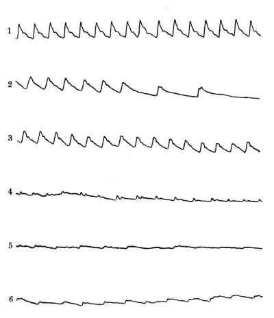
Fig. 16.—The pulse in this case became slower than normal. Five minutes after the drop the type reached that of bradycardia, then recovered itself, and even 14½ minutes after the drop was beating with normal frequency, but in the meantime had become very feeble.
Fig. 16.—In this case, the pulse slowed down below normal. Five minutes after the drop, it reached the level of bradycardia, then got back on track, and even 14½ minutes after the drop, it was beating at a normal rate, although in the meantime it had become very weak.
- 1. The day before execution, 102 per minute.
- 2. 5 minutes after the drop, 54 per minute.
- 3. 7 minutes, 102 per minute.
- 4. 10 minutes, 96 per minute.
- 5. 13 minutes, 66 per minute.
- 6. 14½ minutes, 72 per minute.

Fig. 17.—The pulse in this case became accelerated and then fell again, but was feeble throughout.
Fig. 17.—In this case, the pulse quickened and then slowed down again, but it remained weak the whole time.
- 1. The day before execution, 84 per minute.
- 2. 3½ minutes after the drop, 162 per minute.
- 3. 4 minutes, 156 per minute.
- 4. 5½ minutes, 132 per minute.
- 5. 6½ minutes, 102 per minute.
- 6. 9 minutes, 84 per minute.
- 7. 11½ minutes, 78 per minute.
- 8. 12½ minutes, 78 per minute.

Fig. 18.—In this case the pulse rate increased, and then fell to 72 per minute towards the end.
Fig. 18.—In this case, the pulse rate went up, then dropped to 72 beats per minute by the end.
- 1. 1 week before execution, 60 per minute.
- 2. 2½ minutes after the drop, 162 per minute.
- 3. 3 minutes, 138 per minute.
- 4. 3½ minutes, 132 per minute.
- 5. 4½ minutes.
- 6. 5¼ minutes, 168? per minute.
- 7. 5¾ minutes, 108 per minute.
- 8. 7 minutes.
- 9. 8½ minutes.
- 10. 10 minutes, 72 per minute.
- 11. 12 minutes.
[Pg 121] Homicidal suffocation by compression of the chest has been effected in infants; in adults it is combined with the covering of the nose and mouth. The victims are generally old or enfeebled. Suffocation of infants may be homicidal, or accidental, due to “overlaying”; in the latter the greatest mortality is amongst the youngest infants. A child ten months old may struggle and free itself or awaken the mother.
[Pg 121] Homicidal suffocation by chest compression has occurred in infants; in adults, it usually involves covering the nose and mouth. The victims are typically elderly or weakened individuals. Infant suffocation can be either intentional or accidental, often from "overlaying"; in accidental cases, the highest risk is among the youngest infants. A ten-month-old child may fight back and free itself or wake the mother.
In France a favourite mode of committing suicide by suffocation is the use of irrespirable gases—carbonic acid, carbon monoxide, and the like. Collateral circumstances must be taken into consideration, and will more or less help to point to the true cause of death.
In France, a common method of committing suicide by suffocation is using non-breathable gases—like carbon dioxide and carbon monoxide. Other factors need to be considered, which will help indicate the actual cause of death.
The cause and nature of the death in all of its forms just mentioned are in general the same. Pressure on the trachea—thus arresting respiration—and also on the important vessels and nerves of the neck, results in death, which may be brought about in four different ways:
The cause and nature of death in all its forms mentioned earlier are basically the same. Pressure on the trachea—thereby stopping respiration—and also on the key vessels and nerves in the neck leads to death, which can happen in four different ways:
- 1. Cerebral congestion, or apoplexy.
- 2. Congestion of the lungs and heart—apnœa or asphyxia.
- 3. Combination of above—apoplexy and asphyxia or apnœa.
- 4. Neuro-paralysis—nervous apoplexy, or syncope.
The following table will show the relative frequency of each form of death:
The table below shows how often each type of death occurs:
| Remer. | Casper. | ||
| Apoplexy | 9 | 9 | |
| Asphyxia | 6 | 14 | |
| Mixed | 68 | 62 | |
| 83 | 85 |
Traumatic asphyxia occurs when a heavy weight such as a fall of earth or masonry compresses the chest, and thoracic respiration is impossible. The head and neck appear ecchymosed, the purplish-blue lividity generally ending abruptly at the lower part of the neck or upper part of the thorax, about the level of the clavicles. The conjunctivæ are ecchymosed, and there may be epistaxis. This discoloration does not disappear on pressure by the finger, indicating its petechial character. If the person survive long enough, the discoloration gradually disappears, passing through the colour changes of an ordinary ecchymosis.
Traumatic asphyxia happens when a heavy object, like a collapse of earth or bricks, crushes the chest, making it impossible to breathe normally. The head and neck show bruising, and the purplish-blue discoloration usually stops sharply at the lower part of the neck or the upper part of the chest, around the collarbone area. The eyes may also be bruised, and there could be nosebleeds. This discoloration doesn't fade when pressed with a finger, which shows it's petechial. If the person lives long enough, the discoloration will slowly fade, going through the color changes of a normal bruise.
The effects on the eyes may be such as to be followed by changes in the retina, and optic atrophy leading to loss of vision.
The effects on the eyes can result in changes to the retina and optic nerve damage, leading to vision loss.
Parts of the neck pressed upon by clothing may escape ecchymosis, and present a white mark of almost normal skin.
Parts of the neck covered by clothing might not show bruising and could have a white mark that looks almost like normal skin.
HANGING
Death by hanging is caused by the more or less perfect suspension of the body by a cord applied around the neck, the weight of the body acting as the constricting force.
Death by hanging occurs when the body is more or less perfectly suspended by a cord tied around the neck, with the weight of the body acting as the tightening force.
The body need not hang completely, partial suspension is sufficient to [Pg 122] cause death. The ligature surrounds the neck above the thyroid cartilage, the epiglottis is pushed against the back of the pharynx, the base of the tongue and the soft parts are pressed into the cavity of the pharynx, so that the obliteration of the naso-pharyngeal and the laryngeal passages is complete. Death is due to asphyxia and pressure on the cervical blood-vessels, even if the air-passages be not completely occluded; stoppage of the cerebral circulation being sufficient to cause death; in such a case the signs of asphyxia are absent.
The body doesn't need to be fully suspended; partial suspension is enough to cause death. The ligature goes around the neck above the thyroid cartilage, pushing the epiglottis against the back of the throat. The base of the tongue and the soft tissues are pushed into the throat, completely blocking the nasal and airway passages. Death results from asphyxia and pressure on the neck's blood vessels, even if the airways aren't entirely blocked; halting blood flow to the brain is enough to lead to death, and in such cases, signs of asphyxia may not be present. [Pg 122]
Consciousness is quickly lost, due to pressure on the vessels, and, for this reason, in accidental or suicidal hanging the person is prevented from making any effort to save himself.
Consciousness is quickly lost because of pressure on the blood vessels, and for this reason, in accidental or suicidal hanging, the person is unable to make any effort to save themselves.
Post-mortem Appearances.—The external appearances are more or less those described under “Death from suffocation.” In the greater number of cases the face bears a quiet, placid expression, no turgidity or lividity being noticeable. The eyes are usually half open, but not protruded, and the condition of the pupils variable. The tongue may be protruded, but just as often not. The face may be pale or livid, and a bluish colour present on the free border of the lips. Cyanosis and swelling of the face are only present when the death agony has been long; they soon disappear.
Post-mortem Appearances.—The external appearances are generally similar to those described under “Death from suffocation.” In most cases, the face has a calm, peaceful expression, with no swelling or discoloration noticeable. The eyes are typically half open, but not bulging, and the state of the pupils varies. The tongue may be sticking out, but just as often it isn’t. The face can be pale or discolored, and there may be a bluish tint on the edges of the lips. Cyanosis and swelling of the face only occur when the dying process has been prolonged; they quickly fade away.
Ecchymosis of the conjunctiva, and on the outer surfaces of the lids may be present. The escape of urine, fæces, and semen may take place, but are not characteristic, and are extremely rare.
Ecchymosis of the conjunctiva and on the outer surfaces of the eyelids may occur. The leakage of urine, feces, and semen can happen, but these are not typical and are very rare.
Casper states that in not one of the many cases he had examined of persons hanged has he ever “found an erection of the male organ,” and he also asserts that the emission of semen is extremely rare. Seminal emissions take place more frequently in persons who have been shot, and also in those who have been poisoned by irrespirable gases or by hydrocyanic acid. As a test of strangulation, it is therefore worthless. Tardieu, however, only noticed the escape of urine and fæces in two out of forty-one cases; it is by no means a test of hanging, as it may occur after death if the body is shaken in a cart, or roughly used when first found. A fat person dying of apoplexy may have a mark round the neck as if strangled. Injury to the spinal cord due to fracture or dislocation of the cervical vertebræ is rare in suicidal hanging. Fracture of the spinal ligaments and of the hyoid bone is also rare. Rupture of the internal and middle coats of the carotid arteries sometimes occurs. But it appears that considerable damage is done to the soft parts of the neck by the present judicial mode of hanging with the “long drop.”
Casper mentions that in all the cases he has looked at of people who were hanged, he has never “found an erection of the male organ,” and he also claims that semen release is very rare. Seminal emissions happen more often in people who have been shot, as well as in those who have been poisoned by harmful gases or hydrocyanic acid. Therefore, it’s not a reliable test for strangulation. Tardieu, however, only observed the release of urine and feces in two out of forty-one cases; it’s definitely not a reliable indicator of hanging, as it can happen after death if the body is moved in a cart, or treated roughly when first discovered. A heavy person dying from a stroke might have a mark around the neck that looks like strangulation. Damage to the spinal cord from fractures or dislocation of the cervical vertebrae is uncommon in suicidal hangings. Fractures of the spinal ligaments and the hyoid bone are also rare. Sometimes, there can be a rupture of the inner and middle layers of the carotid arteries. However, it appears that significant damage is inflicted on the soft tissues of the neck by the current judicial method of hanging with the “long drop.”
Dr. Dyer has recorded (New York Medical Journal, vol. iii., 1866) some experiments he made on the eyes of a man and some dogs killed by hanging. He found certain transverse fissures across the lens, which he is inclined to think are characteristic of this mode of death. Dr. R. F. Hutchinson states that an invariable sign of death from hanging is the flow of saliva out of the mouth, down the chin, and straight down the chest. The appearance is unmistakable and invariable, and could not occur in a body hung up after death, the secretion of saliva being a living act (Chevers). Death from hanging may take place although the toes or other parts of the body [Pg 123] rest on the ground. Death is complete in four or five minutes.
Dr. Dyer recorded some experiments he conducted on the eyes of a man and some dogs that were killed by hanging in the New York Medical Journal, vol. iii., 1866. He discovered certain transverse lines across the lens, which he believes are characteristic of this method of death. Dr. R. F. Hutchinson notes that a consistent sign of death from hanging is the flow of saliva from the mouth, down the chin, and straight down the chest. This appearance is clear and consistent, and could not happen in a body hung after death, as saliva secretion is a living process (Chevers). Death from hanging can occur even if the toes or other parts of the body [Pg 123] are resting on the ground. Death is usually complete within four or five minutes.
Marks of the Cord, &c.—The mark of the cord is nearly always present. It varies with the breadth and hardness of the ligature, but is often interrupted. Its irregularities are reproduced on the skin. It is sometimes only seen on one side. In strangling, the mark is low down, most frequently encircling the neck; in hanging, the mark is generally above or on the thyroid cartilage, and carried obliquely upwards. The mark of the cord may be of a dirty yellowish-brown colour, and, when cut into, feels more or less hard and leathery. In general appearance it is not unlike the mark left by mustard-plasters or blisters applied within a short time of death. This effect is probably produced by the rubbing off of the epidermis, and subsequent drying up of the cutis on exposure to the air. At other times the mark may be of a dirty reddish or bright blue colour; or, lastly, there may be little or no mark present, or the edges may assume a livid red coloration, being nothing more or less than a post-mortem stain.
Marks of the Cord, &c.—The mark of the cord is almost always present. It changes depending on the width and tightness of the ligature but is often broken. Its irregularities show up on the skin. Sometimes, it's only visible on one side. In cases of strangling, the mark is usually low down, often wrapping around the neck; in hanging, the mark is typically above or on the thyroid cartilage and runs at an angle upwards. The mark of the cord can appear as a dirty yellowish-brown color, and when cut into, it feels somewhat hard and leathery. In general, it resembles the mark left by mustard plasters or blisters applied shortly before death. This effect is likely caused by the rubbing off of the outer skin and the drying of the skin underneath when exposed to the air. At other times, the mark may be a dirty reddish or bright blue color; or, in some cases, there may be little to no mark at all, or the edges may take on a livid red color, which is nothing more than a post-mortem stain.
May the mark of the cord be produced after death?—On this point Casper says: “That any ligature with which any body may be suspended or strangled, not only within a few hours, but even days after death, especially if the body be forcibly pulled downwards, may produce a mark precisely similar to that which is observed in most of those hanged while alive.” And the same authority also adds that “the mark of the cord is a purely cadaveric phenomenon.”
Can a ligature mark appear after death?—On this topic, Casper states: “Any ligature used to suspend or strangle a body can create a mark, not just within a few hours, but even days after death, particularly if the body is forcibly pulled downwards, which can result in a mark similar to what’s seen on most people who are hanged while alive.” He also notes that “the ligature mark is purely a post-mortem phenomenon.”
Accidental hanging is rare, and generally happens with children while playing at hanging, or by accidentally becoming entangled in a window-blind cord or swing rope, or by the neck-band of an article of clothing by which they may become accidentally suspended from the spike of a railing.
Accidental hanging is uncommon and usually occurs with children who are playing at hanging or by unintentionally getting caught in a window-blind cord or swing rope, or by the neckband of a piece of clothing that causes them to be unintentionally suspended from a railing spike.
Homicidal hanging is rare, but the body may be suspended after death from violence, to simulate suicide; and it may have to be decided whether the hanging took place during life or after death.
Homicidal hanging is uncommon, but the body may be hung after death from violence to make it look like suicide; it may need to be determined whether the hanging happened while the person was alive or after they died.
The mark of the cord is of no assistance, rents in the carotids with extravasation into the coats of the vessels indicate ante-mortem suspension. The flow of saliva down the chin to the body indicates suspension before death. It is important to examine the body for injuries which could not be self-inflicted, and to remember the possibility of poison having been administered with suspension after death.
The mark from the cord doesn't help, and tears in the carotid arteries with bleeding into the vessel walls suggest ante-mortem suspension. Saliva pooling down the chin to the body indicates suspension before death. It's crucial to check the body for injuries that couldn't have been self-inflicted and to consider the possibility that poison was given after death along with the suspension.
Suicidal hanging is the most common, as it is a favourite mode of death with suicides. The absence of marks of injury on the body found suspended, and the want of evidence as to a previous struggle having taken place, all point to suicide. The fact that the feet are found in contact with the ground does not militate against the probability of suicidal hanging; and it appears that in India the natives seldom hang themselves from any height, and are most frequently found with their feet on the ground. A person may take poison first, and hang himself before the poison has had time to prove fatal. [Pg 124]
Suicidal hanging is the most common method, as it's a preferred way for people to end their lives. The absence of injury marks on the body found suspended, along with a lack of evidence indicating a struggle, all suggest suicide. The fact that the feet are touching the ground doesn't reduce the likelihood of suicidal hanging; in India, for instance, it's common for individuals to hang themselves from low heights, often with their feet still on the ground. A person might also take poison first and hang themselves before the poison has a chance to become fatal. [Pg 124]
STRANGLING
Death is due to pressure made on the neck by any form of ligature carried circularly round the neck, without suspension. The cord in hanging is, as a rule, placed more obliquely than in strangling.
Death is caused by pressure on the neck from any type of ligature wrapped around it, without being suspended. The cord in a hanging usually positions itself at a more diagonal angle than in strangulation.
The mode of death is the same as in hanging. The post-mortem appearances are similar to those of hanging, practically those of asphyxia. The mark on the neck is the principal feature. In position it is generally horizontal and situated below or on a level with the thyroid cartilage. It more completely encircles the neck. It may be interrupted in places if an irregular ligature has been used, causing irregular pressure. Its character depends largely upon the nature of the ligature. If the constriction has been uniform a continuous depression is produced which may be marked by ecchymosis. If the skin has been abraded, the line dries, and has a brownish, parchment-like appearance and feeling. If the ligature has been removed before life is quite extinct, the depressed line may disappear or be but slightly evident.
The way someone dies is the same as in hanging. The post-mortem signs are similar to those of hanging, mainly like asphyxia. The mark on the neck is the main feature. It's usually horizontal and positioned below or at the level of the thyroid cartilage. It more completely wraps around the neck. It might be interrupted in some places if an uneven ligature has been used, causing uneven pressure. Its appearance largely depends on the type of ligature. If the constriction has been even, a continuous depression is created, which may be marked by bruising. If the skin has been scraped, the line dries out and takes on a brownish, parchment-like look and feel. If the ligature has been taken off before death is fully achieved, the depressed line may fade or be only slightly visible.
A soft, broad ligature may leave no mark on the neck, if not applied too tightly, or for too long a time. Should the victim have been strangled in the recumbent posture, and dragged upwards and backwards by the ligature, the mark will be on a slant as in hanging. According to the amount of violence used injuries may be caused to the deeper structures of the neck, such as effusion of blood into muscles, fractures of the thyroid or cricoid cartilage; rupture of the tympanum and epistaxis may take place.
A soft, wide ligature might not leave any marks on the neck if it isn’t pulled too tightly or for too long. If the victim was strangled while lying down and then pulled up and back with the ligature, the mark will be slanted, similar to those from hanging. Depending on the level of force used, there can be injuries to the deeper parts of the neck, like bleeding in the muscles, fractures to the thyroid or cricoid cartilage; there may also be a rupture of the eardrum and nosebleeds.
Accidental strangulation may occur when a cord suspending a weight on the back and passing across the chest slips and encircles or compresses the neck.
Accidental strangulation can happen when a cord holding a weight on the back and going across the chest slips and wraps around or tightens on the neck.
Falling out of bed, with entanglement in the clothes or nightdress, may cause strangling by tightening of the neck-band round the neck.
Falling out of bed while getting tangled in clothes or a nightgown can lead to strangulation if the neckband tightens around the neck.
Homicidal strangling is as common as homicidal hanging is rare. It is difficult to hang a man, but easy to strangle him, because consciousness is rapidly lost, and the victim is unable to offer any resistance once the cord is tightened round the neck. In homicidal strangling the murderer generally uses a great deal more violence than is necessary, and so there is found after death much more local injury in such cases.
Homicidal strangling is as common as homicidal hanging is rare. It’s hard to hang someone, but easy to strangle them, because they quickly lose consciousness and can’t resist once the cord is tightened around their neck. In cases of homicidal strangling, the killer usually uses a lot more violence than needed, which results in significantly more local injury after death.
Severe local injuries, such as fracture of the laryngeal cartilages or hyoid bone, denote homicide, as they are rarely noticed in suicidal strangulation.
Severe local injuries, like fractures of the laryngeal cartilages or hyoid bone, indicate homicide, as they are rarely seen in cases of suicidal strangulation.
There may also be signs of general violence about the face, neck, chest, or other parts of the body. The position of the knot affords no material help, as the murderer may tie it in any position, but more than one knot, especially if in different positions, points to homicide. It must be remembered that finger-nails or other marks in the vicinity of the ligature may be present in cases of suicides, from the slipping of the cord or the determined attempts of the suicide to carry [Pg 125] out the act, or plucking at the cord involuntarily. When a person is first strangled, then hung, there would be two marks—one probably horizontal, the other slanting.
There may also be signs of general violence on the face, neck, chest, or other parts of the body. The position of the knot isn’t very helpful since the murderer can tie it in any way. However, if there’s more than one knot, especially if they’re in different positions, it suggests homicide. It's important to remember that nail marks or other signs around the ligature can also be present in suicide cases due to the slipping of the cord or the person's determined attempts to go through with it, or even involuntary tugging at the cord. When someone is first strangled and then hung, you would see two marks—one likely horizontal and the other slanting.
Suicidal strangulation is rare. The knot is generally adjusted at the front or side of the neck, the cord may encircle the neck several times. Injuries to the deep structures of the neck are absent. Signs of general violence are not necessarily present.
Suicidal strangulation is uncommon. The knot is usually positioned at the front or side of the neck, and the cord might wrap around the neck multiple times. There are no injuries to the deep structures of the neck. Signs of overall violence are not always evident.
If there be two marks upon the neck, one due to an attempt at strangulation, the other to suspension, in a suicide, the first would be the less marked, the latter more pronounced, whereas in homicide the strangulation mark would be most distinct.
If there are two marks on the neck, one from an attempt to strangle and the other from hanging in a suicide, the strangulation mark would be less noticeable, while the hanging mark would be more prominent. In contrast, in a homicide, the strangulation mark would be the most distinct.
THROTTLING
Throttling is strangling by means of the hand or hands alone, and is due to constant pressure of the fingers upon the throat. Very little pressure is required to occlude the glottis; it can be done with slight pressure of the thumb and forefinger on the side of the thyroid cartilage.
Throttling is when someone chokes another person using only their hands, applying constant pressure with their fingers on the throat. It takes very little pressure to block the airway; it can be done with just a light pinch of the thumb and index finger on the side of the thyroid cartilage.
The impression of the fingers and thumb upon the throat have characteristic marks. They are usually to be seen on both sides of the throat. The thumb mark is on one side, and the marks of the fingers, separated from one another or clustered together, are on the other, the thumb mark being the highest. When grasped from the front by the right hand, the thumb mark will be on the right side of the throat. If the victim be throttled by the two hands at the same time, as when on the ground, the thumb marks are on the same side. If the assailant be left-handed, and has used this hand to grip the throat, the thumb mark will be on the left side of the victim. The finger marks are one above the other. The marks may appear as ecchymoses if the examination be made soon after death; if later, they may appear and feel like parchment, and of a brownish colour. Crescentic marks of finger-nails may be present, also other scratches in the vicinity. Other marks of general violence may be present, and should the victim have fallen to the ground, the head may be injured. Much blood is effused in the tissues of the neck and the laryngeal cartilages, and the hyoid may be found fractured; the carotids may escape injury, but not always.
The impressions of fingers and thumbs on the throat have distinct marks. They are generally visible on both sides of the throat. The thumb mark is on one side, while the marks of the fingers, either separate or grouped together, are on the other side, with the thumb mark being the highest. When someone is grabbed from the front with the right hand, the thumb mark will be on the right side of the throat. If the victim is choked with both hands simultaneously, like when they are on the ground, the thumb marks will appear on the same side. If the attacker is left-handed and used that hand to grab the throat, the thumb mark will be on the left side of the victim. The finger marks will be positioned one above the other. These marks may look like bruises if examined shortly after death; if checked later, they may appear and feel like parchment and take on a brownish color. Crescent-shaped marks from fingernails may also be visible, along with other scratches nearby. Additional signs of general violence could be present, and if the victim fell to the ground, their head might be injured. A lot of blood accumulates in the tissues of the neck and around the laryngeal cartilages, and the hyoid bone could be fractured; while the carotid arteries might not be injured, that isn’t always the case.
The mucous membrane of the cheeks may be found lacerated. Where the victim is thrown to the ground and knelt upon, fracture of the ribs and ecchymosis of the chest wall may occur.
The inside of the cheeks may be torn. If the victim is pushed to the ground and knelt on, they might suffer broken ribs and bruising on the chest.
In a case examined by me of combined strangling and throttling, marks of the ligature and fingers were both present, and on the clavicles separate marks produced by the knuckles while tying the cord. These knuckle marks did not show up till some time after death. Effusions of blood were present in the tissues of the neck on each side of the larynx, and amongst the muscles on both sides, and in the sheath of the left carotid artery.
In a case I looked at involving both strangling and throttling, there were clear marks from the ligature and fingers, along with distinct marks from the knuckles where the cord was tied on the collarbones. These knuckle marks only became visible some time after death. There were also blood effusions in the neck tissues on either side of the larynx, as well as among the muscles on both sides and in the sheath of the left carotid artery.
The hyoid bone was fractured in two places in its left half. The thyroid cartilage was fractured vertically on each side of the middle [Pg 126] line into three pieces, the central portion having fallen behind the other two into the cavity of the larynx. The right wing of the thyroid was comminuted. The cornua were fractured. The cricoid cartilage was also fractured posteriorly, and into three pieces in front. Effusions of blood in the fractured areas had formed beneath the mucous membrane. Effusion of blood was present on the front wall of the pharynx and the upper part of the œsophagus. The right carotid artery was atheromatous, and linear and star-shaped fissures were present, the latter surrounded by a ring of sub-intimal effusion of blood.
The hyoid bone was broken in two places on its left side. The thyroid cartilage was cracked vertically on both sides of the middle line into three pieces, with the central piece having fallen behind the other two into the larynx cavity. The right wing of the thyroid was shattered. The cornua were fractured. The cricoid cartilage was also broken at the back and into three pieces at the front. Blood had pooled in the fractured areas beneath the mucous membrane. Blood was present on the front wall of the pharynx and the upper part of the esophagus. The right carotid artery showed signs of atherosclerosis, with linear and star-shaped cracks present, the latter surrounded by a ring of sub-intimal blood pooling. [Pg 126]
On the right side of the thorax the fifth rib was fractured at the junction with its cartilage, and on the left side the second, third, fourth, fifth, and sixth ribs were fractured close to their cartilages, and again from three to five inches further back. Blood effusions in the tissues of the chest wall and under the pleuræ were present in the region of the fractures.
On the right side of the chest, the fifth rib was broken at the spot where it meets its cartilage, and on the left side, the second, third, fourth, fifth, and sixth ribs were broken near their cartilages, as well as three to five inches further back. There was bleeding in the tissues of the chest wall and under the pleurae around the fractures.
A handkerchief was found tight on the neck and tied with two knots both fastened securely. It was probable that the handkerchief had been tied on after the throttling, as the knot ends were too short to allow a sufficient pull on them to cause the injuries. Throttling may be regarded as a homicidal act; although one or two instances of suicidal throttling have been recorded in the insane.
A handkerchief was found tightly wrapped around the neck, secured with two knots that were fastened firmly. It was likely that the handkerchief was tied on after the choking, since the knot ends were too short to create enough tension to cause the injuries. Choking can be viewed as an act of murder, although there have been one or two cases of suicidal choking documented among the mentally ill.
Judicial Hanging.—In judicial hanging the prisoner is suspended by a rope with a running noose around the neck, after a sudden drop of from six to eight feet according to the weight of the body. The noose may be arranged with the knot or slip-ring fixed at the side below the ear, or in front so as to jerk the head backwards. The sudden and severe strain upon the neck produces fracture or dislocation of the spinal column at the second or third cervical vertebra, with rupture of the spinal cord. Other local injuries occur, such as rupture of cervical muscles, fracture of the larynx, and even lacerated wounds of the neck. The head has even been severed completely from the body, and the deep structures of the neck have even been so lacerated that the body has hung by skin only, stretched to the thickness of two or three fingers. Death is said to take place from shock, pressure on the vagi, and asphyxia, probably also cerebral apoplexy. When death is instantaneous, the body hangs motionless, the head fallen over the side opposite the knot, and the neck stretched.
Judicial Hanging.—In judicial hanging, the prisoner is suspended by a rope with a running noose around their neck, after a sudden drop of six to eight feet depending on their weight. The noose can be positioned with the knot or slip-ring fixed to the side below the ear, or in front to jerk the head backward. The sudden and intense strain on the neck causes a fracture or dislocation of the spinal column at the second or third cervical vertebra, resulting in rupture of the spinal cord. Other local injuries can occur, such as tearing of cervical muscles, fracture of the larynx, and even deep lacerations of the neck. In some cases, the head has been completely severed from the body, and the deeper structures of the neck can be so damaged that the body hangs by skin alone, stretched to the thickness of two or three fingers. Death is said to occur from shock, pressure on the vagus nerves, and asphyxia, and possibly also from cerebral apoplexy. When death is instantaneous, the body hangs motionless, the head tilted to the side opposite the knot, and the neck elongated.
The heart may, however, continue to beat for a varying period after apparent death—in some cases even as long as 14½ minutes. The following interesting series of pulse tracings were taken by Dr. Llewellyn Morgan, of Liverpool, and kindly placed at my disposal. In No. 1 the heart beats could be recorded at the wrist for 14½ minutes after the drop; in No. 2 for 12½ minutes; and in No. 3 for 12 minutes. The frequency and character of the beat is variable, but in each case shows a practically normal rate towards the end. (See Figs. 16, 17, 18.)
The heart can keep beating for a while after someone appears to be dead—in some cases, for as long as 14½ minutes. Dr. Llewellyn Morgan from Liverpool recorded the following interesting series of pulse tracings and generously shared them with me. In No. 1, heartbeats could be detected at the wrist for 14½ minutes after the drop; in No. 2 for 12½ minutes; and in No. 3 for 12 minutes. The frequency and quality of the heartbeat vary, but in each case, it shows a nearly normal rate toward the end. (See Figs. 16, 17, 18.)
Apart from the local injuries to the neck, the external appearances in judicial hanging are similar to those in other forms.
Apart from the local neck injuries, the outside signs of judicial hanging look similar to those in other methods.
Death by drowning occurs when the breathing is arrested by watery or semi-fluid substances, blood, urine, or the muddy semi-fluid matter found in cesspools and marshes. It is not necessary for the whole body to be submerged. Death may result if the face alone be immersed, as in the case of a man in a fit of drunkenness being drowned in the water contained in the imprint of a horse‘s hoof left in the mud.
Death by drowning happens when breathing stops because of water or semi-fluid substances like blood, urine, or the muddy stuff in cesspools and marshes. The entire body doesn’t have to be fully submerged. Even if just the face is under, it can lead to death, like when a man, in a drunken stupor, drowns in the water collected in a horseshoe print left in the mud.
In addition to the changes in the internal organs, identical with those present in persons who have died from suffocation or hanging, water is found in the lungs or stomach.
In addition to the changes in the internal organs, the same as those seen in people who have died from suffocation or hanging, water is found in the lungs or stomach.
Death may be due to—
Death may be caused by—
- (a) Apoplexy.
- (b) Asphyxia.
- (c) A combination of the two.
- (d) Neuro-paralysis.
Death from pure apoplexy is rare; and it may be affirmed that death from syncope never occurs in the drowned without leaving some signs of asphyxia.
Death from pure apoplexy is rare; and it can be said that death from fainting never happens in drowning victims without leaving some signs of asphyxia.
It is more difficult to restore the drowned than those dying from mere stoppage of air from entering the lungs. Few if any persons recover who have been submerged four minutes, and even in cases where this time has been exceeded, followed by recovery, this result is probably due to the person fainting before entering the water.
It’s harder to revive someone who has drowned than someone who has simply stopped breathing. Very few people, if any, survive after being underwater for four minutes, and even in cases where recovery does happen after that time, it’s likely because the person fainted before going into the water.
In death from drowning, the lungs are distended and overlap the heart, and have a peculiar spongy feeling. They also contain a quantity of frothy fluid, which cannot be produced in the dead body, as it is the result of the violent efforts made by the individual to breathe in the act of dying. This frothy condition of the fluid in the lungs is an important sign of death by drowning, especially if the fluid corresponds with that in which the individual is said to have perished. It is just possible, however, that the person may have been first suffocated, and then thrown into the water, froth in the trachea being found in those suffocated; but in this case the froth is small in quantity, and not watery. The froth in the drowned is like that made with soap and water, and is not viscid, thus differing from bronchitic exudation. Water in the stomach is an important indication of death from drowning, especially if the water contained in the stomach can be shown to possess the same characters as that in which the body was found. Water in the intestines is still more important. In a great number of cases this, however, must be next to impossible; when it can [Pg 128] be identified, the value of this sign is enhanced by the fact that water does not enter the stomach in those submerged after death, unless putrefaction be far advanced, or the body has lain in very deep water. Casper concluded that a person had been drowned, by finding a small quantity of mud in the stomach after putrefaction had set in. Water, however, may be absent from the stomach if the person fall into the water in a state of syncope, and it may be present if the person has taken a draught of water before submersion.
In drowning, the lungs are swollen and press against the heart, feeling spongy. They also contain a lot of frothy fluid, which can't form in a deceased body, as it results from the person's desperate attempts to breathe while dying. This frothy fluid in the lungs is a key indicator of death by drowning, especially if the fluid matches the water where the person drowned. However, it’s possible that the person may have been suffocated first and then thrown into the water; in cases of suffocation, froth can be found in the trachea, but it tends to be small in quantity and not watery. The froth in drowning victims resembles that made with soap and water and is not sticky, unlike bronchitic fluid. Water in the stomach is a significant indication of drowning, particularly if the water matches the type found at the scene of the incident. Water in the intestines is even more telling. However, in many instances, this may be nearly impossible to determine; when it can be identified, it adds to the evidence since water doesn't enter the stomach of those who are submerged after death unless decomposition is advanced or the body has been in very deep water. Casper concluded someone had drowned after finding a small amount of mud in the stomach post-decomposition. However, water might be absent from the stomach if the person fell into the water while fainting, and it might be present if the person drank water before submersion.
The effect of season on putrefaction in water is shown in the following table:
The impact of the season on decay in water is displayed in the table below:
| Summer. | Winter. | ||||
| 5 to 8 | hours | produce | as much change | as 3 to 5 | days. |
| 24 | “ | “ | “ | 4 to 8 | “ |
| 4 | days | “ | “ | 15 | “ |
| 10 to 12 | “ | “ | “ | 28 to 42 | “ |
| (Devergie.) | |||||
Of the external signs, the presence of sand, gravel, or mud under the nails may or may not be an important sign, for sand or mud may collect under the nails during the efforts to drag the body from the water; but weeds, &c., grasped in the hands show that there has been a struggle, and point to death from drowning. The cutis anserina—goose skin—present generally on the anterior surface of the body, and not, however, peculiar to death from drowning, is important as a sign of recent vitality. The face of those who have been drowned, and then quickly removed from the water, is pale, and in most cases not swollen; the eyes may or may not be closed; and not infrequently round the mouth there is more or less froth, especially when death is due to apnœa. In summer, however, after two or three days, and longer in winter, the face assumes a reddish or bluish-red coloration, putrefaction taking place about the head and upper extremities earlier than in other forms of death. The contraction or retraction of the penis is a well-marked sign of death by drowning, and Casper asserts that he has “not observed anything similar so constantly after any other kind of death.” Ogston states that he has met with two cases of erection of the penis in the drowned.
Of the external signs, finding sand, gravel, or mud under the nails might not be significant since those materials can get trapped while pulling the body from the water. However, weeds and similar debris held in the hands indicate a struggle and suggest death by drowning. The cutis anserina—goosebumps—commonly seen on the front of the body, while not unique to drowning, is an important indicator of recent vitality. The faces of drowning victims who are quickly pulled from the water are usually pale and not typically swollen; the eyes may be closed or open, and there often is some froth around the mouth, particularly if death resulted from asphyxiation. In summer, after two or three days, and even longer in winter, their faces may take on a reddish or bluish-red tint, with decomposition occurring around the head and upper limbs more quickly than in other types of death. The contraction or retraction of the penis is a clear sign of death by drowning, and Casper claims he has “not seen anything similar so consistently after any other type of death.” Ogston notes he has encountered two cases of penile erection in drowning victims.
The question as to how long a body may remain in the water before it floats has given rise to considerable discussion, without, however, arriving at any very definite conclusion. It may be stated in general terms that, as floating depends to some extent on the rapidity in which putrefaction supervenes on submersion, bodies float earlier in summer than in winter, in salt than fresh water, clothed than naked. In India bodies have floated in twenty-four hours after immersion. Females and children float more readily than males. A body from various causes may float within a few hours of its submersion, especially if the body be that of a female, fat and clothed. The old idea that the body of a person thrown into water during life sinks, but that a dead body under like conditions floats, is a fiction now exploded.
The question of how long a body can stay in water before it floats has sparked a lot of debate, but there hasn't been a clear answer. Generally speaking, floating happens sooner when decomposition sets in quickly after submersion. Bodies tend to float faster in summer compared to winter, in saltwater versus freshwater, and when clothed versus when naked. In India, bodies have been known to float just twenty-four hours after being immersed. Women and children float more easily than men. A body can float within a few hours of submersion for various reasons, especially if it’s a female body that is fat and clothed. The old belief that a living person sinks when thrown into water, but a dead body floats under the same circumstances, has been disproven.
The signs to be sought for in the drowned are—(1) Absence of any injury. (2) Cutis anserina and retracted penis. (3) Water and mud in the stomach. (4) Froth in the air-passages. (5) Distended lungs. (6) General signs of death by asphyxia.
The signs to look for in drowning victims are—(1) No obvious injuries. (2) Goosebumps and a retracted penis. (3) Water and mud in the stomach. (4) Froth in the airways. (5) Swollen lungs. (6) General signs of death by suffocation.
It should be remembered that the fact of the hands being tied together, or to the feet, does not militate against suicide by drowning.
It should be remembered that having the hands tied together, or to the feet, does not rule out the possibility of suicide by drowning.
If wounds and other injuries be found on the body, the question arises as to whether the injuries were sufficient in themselves to cause death, and then as to whether they were caused during life. A person jumping from a height into the water may sustain severe injuries—dislocation of both arms, fracture of the skull and of the vertebræ, or even lacerated wounds of more or less severity. The absence of the signs proper to death by drowning, coupled with the presence of external injuries, would point to death by violence prior to immersion. The presence of signs of drowning, and injuries, sufficient to have caused death in themselves, would indicate that they had occurred after death.
If there are wounds and other injuries on the body, the question comes up about whether these injuries were enough on their own to cause death, and then whether they happened while the person was alive. A person jumping from a height into the water could suffer serious injuries—like dislocated arms, a fractured skull, and broken vertebrae, or even severe lacerations. If there are no signs typical of drowning, along with the presence of external injuries, it suggests death by violence occurred before entering the water. If signs of drowning are present, along with injuries that could have independently caused death, it would suggest those injuries happened after death.
The following considerations may assist in forming an opinion:
The following points may help in forming an opinion:
- 1. Previous history of person found in the water.
- (a) Any history of suicidal tendency.
- (b) Any motive that would render suicide probable.
- 2. Height from which the person fell.
- 3. Absence or presence of signs of death by drowning.
- 4. Absence of stakes or other objects in the water that might
- have caused injuries to anyone falling against them.
The time required to cause death by drowning is so short that persons seldom recover after submersion for three or four minutes; but the cessation of respiration is no guide to the extinction of life, and an attempt at resuscitation should always be made, for if the respiration be fairly restored the heart will soon act. Nay more, as pointed out before, in cases of so-called asphyxia, the heart may continue to act for several minutes after the entrance of air to the lungs has been arrested, and in judicial hanging it frequently happens that the pulse at the wrist can be felt for ten or twelve minutes after suspension. (See Figs. 16, 17, 18.)
The time it takes to drown is so brief that people rarely recover after being submerged for three or four minutes. However, the stopping of breathing doesn’t necessarily mean that life has ended, and an attempt at resuscitation should always be made. If breathing is successfully restored, the heart will likely start to function again soon. Furthermore, as noted earlier, in cases of so-called asphyxia, the heart can keep beating for several minutes even after air stops reaching the lungs. In cases of judicial hanging, it often happens that a pulse can still be felt at the wrist for ten to twelve minutes after suspension. (See Figs. 16, 17, 18.)
Recapitulation of the Post-mortem
Appearances in the Drowned
I. External
1. In the Skin.—Rose-coloured patches may be present on the face and neck. The condition of goose skin—cutis anserina—is hardly ever absent even in summer. The cutis anserina is not, however, characteristic of drowning, as it may be present in other forms of violent death, and also in some persons during life. It is a vital act, the result of nervous shock, and does not depend upon the temperature of the water for its production; still, it points to recent vitality.
1. In the Skin.—You might notice pink patches on the face and neck. The condition known as goose skin—cutis anserina—is almost always present, even in summer. However, cutis anserina isn't exclusive to drowning, as it can occur in other forms of violent death and also in some people while they are alive. It's a vital response caused by nervous shock and isn't affected by the water temperature; nonetheless, it indicates recent life.
2. The Tongue.—The tongue is just as often found behind the jaws as between them (Casper).
2. The Tongue.—The tongue is just as often found behind the jaws as it is between them (Casper).
3. The Hands and Feet.—The hands and feet acquire a greyish-blue colour when the body has lain in the water from twelve to twenty-four hours. The skin also becomes corrugated in longitudinal [Pg 130] folds. The greyish-blue condition of the hand is known as the “cholera hand.” The nails may contain particles of sand and weeds. “No corrugation or discoloration of the skin of the hands or feet is ever observed on the body of anyone drowned who has been taken out of the water within half an hour, within two, six, or even eight hours” (Casper). The same authority states he has produced these effects by laying the hands after death in water, or wrapping them in cloths kept constantly wet for a few days.
3. The Hands and Feet.—The hands and feet turn a greyish-blue color when the body has been in the water for twelve to twenty-four hours. The skin also becomes wrinkled in long folds. The greyish-blue appearance of the hand is referred to as the “cholera hand.” The nails may have bits of sand and seaweed. “No wrinkling or discoloration of the skin on the hands or feet is ever seen on the body of someone who has drowned and been taken out of the water within half an hour, two hours, six hours, or even eight hours” (Casper). The same expert claims he has produced these effects by placing the hands in water after death or wrapping them in cloths that remain wet for a few days.
4. The Genitals.—Contraction of the penis and dartos is an almost constant symptom, and Casper has “not observed anything similar so constantly after any other kind of death.” It is due, probably, to the same cause as the cutis anserina, which Brettner attributes to “bundles of unstriped muscular fibres, lying in the upper stratum of the true skin, surrounding the sebaceous glands, and forcing them forwards by their contraction, thus making the cutis anserina. Precisely similar unstriped muscles are found in the subcutaneous cellular tissue of the penis; they run principally parallel to the long axis of the member, but very often large bundles run across it.” The action of cold and fright is to induce contraction of these cutaneous muscles, with a resulting contraction of the penis.
4. The Genitals.—Contraction of the penis and dartos is almost always a symptom, and Casper has “not seen anything similar so consistently after any other kind of death.” This is probably due to the same reason as the cutis anserina, which Brettner attributes to “bundles of unstriped muscle fibers that lie in the upper layer of true skin, surrounding the sebaceous glands and pushing them forward through their contraction, thus creating the cutis anserina. Identical unstriped muscles are found in the subcutaneous tissue of the penis; they mainly run parallel to the length of the penis, but often large bundles run across it.” The effects of cold and fear induce the contraction of these skin muscles, resulting in the contraction of the penis.
II. Internal
1. The Brain.—Cerebral hyperæmia is most rare in the drowned, but cerebral hypostasis is not infrequently mistaken for it.
1. The Brain.—Cerebral hyperemia is very rare in drowning victims, but cerebral hypostasis is often incorrectly identified as it.
2. The Trachea.—The mucous membrane of the trachea and larynx is always more or less injected, and is of a cinnabar-red, which must not be mistaken for the dirty, brownish-red colour, the result of putrefaction. A white froth, but seldom bloody, is also found in varying quantity in the trachea, and is a most important sign of vital reaction, but its diagnostic value is destroyed by putrefaction. Sometimes a portion of the contents of the stomach may be found in the trachea. When this occurs it is due to the act of coughing, induced by the admission of water into the lungs. The contents of the stomach are forced into the mouth, and then drawn into the lungs during the next attempt at inspiration. This indicates that the person entered the water during life. In cases where death has taken place from syncope little or no froth may be found in the trachea.
2. The Trachea.—The mucous membrane of the trachea and larynx is usually somewhat swollen and has a bright red color that shouldn’t be confused with the dirty, brownish-red shade that results from decay. A white froth, though rarely bloody, can also be present in varying amounts in the trachea and is a significant sign of vital reaction, but its diagnostic value is compromised by decay. Occasionally, some of the stomach contents might be found in the trachea. This happens when coughing is triggered by water entering the lungs. The stomach contents are pushed into the mouth and then inhaled into the lungs during the next breath. This suggests that the person was alive when they entered the water. In cases where death resulted from fainting, there may be little to no froth in the trachea.
3. The Lungs.—The lungs are completely distended, almost entirely overlapping the heart, and pressing close to the ribs. They are spongy to the feel, and when cut into, a considerable quantity of bloody froth escapes. The froth found in the lungs is the result of the powerful attempts to breathe, and cannot be produced by artificial means. It adheres not to the sides of the bronchial tubes, as does the exudation of bronchitis or pneumonia. The distension of the lungs is due partly to an actual hyperæmia, and partly to inhaled fluid. Water is present in the pleural cavities.
3. The Lungs.—The lungs are fully expanded, nearly covering the heart and pressing against the ribs. They feel spongy, and when cut, a significant amount of bloody froth comes out. The froth in the lungs is caused by intense attempts to breathe and cannot be created artificially. It doesn't stick to the walls of the bronchial tubes like the fluid from bronchitis or pneumonia. The expansion of the lungs is due to both actual excess blood flow and inhaled fluid. Water is found in the pleural cavities.
4. The Heart and Great Vessels.—As is common to other forms of asphyxia, the left side of the heart is entirely, or almost entirely, empty; the right, on the contrary, is engorged. This condition of the heart is, therefore, not a diagnostic sign of drowning, and is absent in the drowned when death takes place by neuro-paralysis; in fact, in some cases of undoubted drowning, both sides have been found empty, probably, however, the result of putrefaction (Ogston). The same may be said of the accompanying congestion of the pulmonary artery.
4. The Heart and Great Vessels.—Like other types of asphyxia, the left side of the heart is completely or almost completely empty, while the right side is swollen with blood. So, this condition of the heart isn't a definitive sign of drowning, and it's not present in drowning victims who die from neuro-paralysis. In fact, in some clear cases of drowning, both sides of the heart have been found empty, likely due to decomposition (Ogston). The same applies to the congestion of the pulmonary artery.
5. The Blood.—As is common in all forms of death where respiration has been arrested, the blood is found to be remarkably fluid, and of a cherry-juice colour. M. Faure in his monograph on asphyxia states that he has found large and firm clots in the right side of the heart in the drowned who have not remained long under water. The blood is diluted with water, most marked in the left side of the heart. The amount of dilution is greater in slow drowning. [Pg 131]
5. The Blood.—As is typical in all types of death where breathing has stopped, the blood is notably fluid and has a color similar to cherry juice. M. Faure, in his study on asphyxia, mentions that he has found large and solid clots in the right side of the heart of individuals who drowned but were not submerged for long. The blood is diluted with water, especially evident in the left side of the heart. The level of dilution is greater in cases of slow drowning. [Pg 131]
6. The Stomach.—Casper considers that the presence of fluid in the stomach, corresponding to that in which the body is found, is “an irrefragable proof of the actual occurrence of death from drowning,” and that the swallowing of it must have been a vital act of the individual dying in the water. The absence of water from the stomach does not negative death from drowning. Water is not always present. It is possible for it to reach the stomach of a submerged body after death.
6. The Stomach.—Casper believes that the presence of fluid in the stomach, matching the water in which the body is found, is “undeniable proof that drowning actually caused death,” and that swallowing it must have been a vital action of the person who died in the water. The absence of water in the stomach doesn’t rule out death by drowning. Water isn’t always there. It can enter the stomach of a submerged body after death.
Water in the intestines is a more reliable sign of death from drowning, and indicates submersion during life. It is only after very long submersion and under great pressure that water finds its way into the intestines after death.
Water in the intestines is a more reliable sign of death from drowning and indicates that the person was submerged while alive. It's only after a long time underwater and under significant pressure that water enters the intestines post-mortem.
N.B.—Putrefaction in the drowned in most cases commences in the upper part of the body, and extends downwards. The face, head, and neck are first attacked. This is the reverse of putrefaction in air.
N.B.—Decomposition in drowning victims usually starts in the upper body and moves downward. The face, head, and neck are the first areas to be affected. This is the opposite of decomposition in the open air.
DEATH FROM STARVATION
Death from starvation may be due to the total withdrawal of food, to prolonged insufficiency, defective quality, inability to swallow it, and inability to retain it.
Death from starvation can result from a complete lack of food, long-term insufficiency, poor quality food, trouble swallowing, or being unable to keep food down.
Death from starvation may occur during famines, amongst ship-wrecked sailors, and persons entombed in mines or pits, and is due to sheer privation. It may follow criminal starvation, wilful refusal to take food as a form of suicide, and it has been noted in cases of hysteria and lunacy.
Death from starvation can happen during famines, among shipwrecked sailors, and for people trapped in mines or pits, and it results from extreme deprivation. It can also happen due to intentional starvation, where someone refuses to eat as a form of suicide, and it has been observed in cases of hysteria and insanity.
It may result from mechanical hindrance to the entrance of food into the body from ankylosis of the jaws, or its passage through the alimentary tract from stricture of the œsophagus or stomach, from cancer or cicatrisation after injury from swallowing corrosive substances. Amongst other diseases, tuberculosis, malignant disease, and diabetes mellitus are the chief which produce external appearances of starvation.
It can happen due to mechanical obstacles preventing food from entering the body because of jaw joint fusion, or issues along the digestive tract like esophageal or stomach strictures, cancer, or scarring from injury after swallowing caustic substances. Other diseases, including tuberculosis, cancer, and diabetes, are the main conditions that create visible signs of starvation.
In the withdrawal or deprivation of food for criminal purposes the victims are usually old, helpless, or feeble-minded persons, or young children.
In cases where food is taken away or denied for criminal reasons, the victims are often elderly, defenseless, intellectually disabled individuals, or young children.
Symptoms.—These depend on the previous state of nutrition. Starvation may be regarded as acute when death occurs within fourteen days from the withdrawal of food, chronic when at a longer period.
Symptoms.—These depend on the previous state of nutrition. Starvation is considered acute when death occurs within fourteen days after food is stopped, and chronic when it lasts for a longer period.
The symptoms of hunger vary: they are said to pass off in forty-eight hours, but may last for several days acutely.
The symptoms of hunger vary: they are said to go away in forty-eight hours, but can last for several days intensively.
The body temperature falls below normal, and the fall may be two or three degrees before death. The pulse gradually increases in frequency day by day. The chief sign is loss of body weight. Chossat‘s experiments on pigeons showed that when they were totally deprived of food, the surplus fat of the body was lost first, then the fatty coverings of internal organs, the interstitial fat of muscles last of all; the muscles themselves also wasted. A peculiar odour like acetone emanates from those who have been starved, and towards and after death the odour is putrescent.
The body temperature drops below normal, often by two or three degrees before death. The pulse gradually speeds up day by day. The main sign is weight loss. Chossat's experiments on pigeons revealed that when they were completely deprived of food, the body burned through its stored fat first, then the fatty layers around internal organs, and finally the fat in muscles; the muscles also shrank. A distinctive smell similar to acetone comes from those who have been starved, and as death approaches, the smell becomes more decayed.
In addition to the above signs, there are anæmia, sunken, glistening eyes with dilated pupils, prominence of bony projections, pale and dry lips and tongue, parched mouth and throat, weakness of the voice, [Pg 133] sunken abdomen, wasted limbs, constipated bowels, urine scanty and turbid. There are pains in the abdomen, relieved by pressure; great thirst, a dusky, dry skin, occasionally purpuric eruptions, exhaustion, ultimately delirium ending in death.
In addition to the signs mentioned above, there are symptoms like anemia, sunken and shiny eyes with dilated pupils, noticeable bone projections, pale and dry lips and tongue, a dry mouth and throat, a weak voice, [Pg 133] a sunken abdomen, thin limbs, constipation, and scanty, cloudy urine. There are abdominal pains that feel better with pressure, intense thirst, a dark and dry skin, occasional purple spots, exhaustion, and eventually delirium that can lead to death.
Post-mortem Appearances.—There is emaciation of the whole body, dry, wrinkled skin of a brown colour; the muscles are flabby and wasted, the abdomen sunken, the eyes red and open; this appearance is not common in death from other causes. The mouth and throat are dry even to aridity. The heart, lungs, and blood-vessels are collapsed, and contain but little blood. The abdominal viscera are shrunken and without enveloping fat. The omentum is devoid of fat, and clear; the gall bladder is full of dark bile; the urinary bladder may be quite empty. The stomach and intestines are collapsed, contracted, and empty, and the walls extremely thinned.
Post-mortem Appearances.—The entire body shows signs of severe weight loss, with dry, wrinkled, brown skin; the muscles are weak and depleted, the abdomen is sunken, and the eyes are red and open. This appearance is not typical in deaths from other causes. The mouth and throat are dry to the point of being parched. The heart, lungs, and blood vessels are collapsed and contain very little blood. The internal organs in the abdomen are shriveled and lack surrounding fat. The omentum has no fat and looks clear; the gallbladder is filled with dark bile; the urinary bladder may be completely empty. The stomach and intestines are collapsed, contracted, and empty, with the walls being extremely thin.
Diagnosis.—The absence of any other cause of death—such as cancer of the stomach, stricture of the œsophagus, &c.—and the previous history of the case will assist in forming an opinion, care being taken not to confound the results of wasting disease with those due to starvation.
Diagnosis.—The lack of any other cause of death—like stomach cancer, esophagus stricture, etc.—and the patient’s previous history will help in forming a conclusion, while being careful not to mix up the effects of wasting disease with those from starvation.
Legal Relations.—The question of death from starvation may be raised in a case of infanticide by omission. Although rare as an act of homicide, it must be remembered that the law does not require the absolute deprivation of food to be proved, but only that the necessary quantity and quality of food has been withheld; but malice at the same time must be proved. In cases of infanticide by starvation, the mother and not the father is responsible for the proper feeding of the child; but in the case of an apprentice, the master and not his wife is bound to supply proper food to such apprentice.
Legal Relations.—The issue of death from starvation can come up in a case of infanticide by omission. While it's uncommon as a homicide, it's important to note that the law doesn't require proof of total lack of food, just that the necessary amount and quality of food has been withheld; however, malice must also be demonstrated. In cases of infanticide due to starvation, the mother, not the father, is responsible for ensuring the child is fed properly; in the case of an apprentice, the master, not his wife, is obligated to provide appropriate food for the apprentice.
In questions of survivorship, and in criminal cases, the medical witness may be asked how long a person may survive after complete withdrawal of food. Little is known as to the length of time required to cause death by starvation, but it is certain that life may be prolonged for some time without food, if water be allowed. Starvation is less rapid in its effects if the body be kept warm. In a case recorded in the Lancet, a man who had been shut up in a coal-mine for twenty-three days, with only a little dirty water to drink, lived three days after his liberation, and then died of exhaustion. In adults the average is from seven to ten days without water. Tidy (Legal Medicine, vol. i. p. 392) is of the opinion that the young die first, then adults, and the aged last. Taking into account the enfeebled vitality of the aged, it is more probable that the young or middle-aged adult would survive the longest. Where water is freely obtainable, life may be prolonged to the fifty-eighth day (Foderé, vol. ii. p. 276) or even more.
In cases involving survivorship and criminal matters, medical witnesses might be asked how long a person can survive after completely stopping food intake. Not much is known about how long it takes for starvation to cause death, but it's clear that life can be extended for some time without food as long as water is available. Starvation progresses more slowly if the body is kept warm. In a case documented in the Lancet, a man who had been trapped in a coal mine for twenty-three days, with only a bit of dirty water to drink, lived for three days after being rescued before dying from exhaustion. For adults, the average survival time without water is about seven to ten days. Tidy (Legal Medicine, vol. i. p. 392) believes that the young tend to die first, then adults, with the elderly surviving the longest. However, considering the weakened vitality of older individuals, it's more likely that young or middle-aged adults would survive the longest. When water is easily accessible, life can be extended to the fifty-eighth day (Foderé, vol. ii. p. 276) or even longer.
Apart from age, account must be taken of the condition of the person in reference to bodily health prior to the withdrawal of food.
Apart from age, it’s important to consider the individual's health status regarding their physical condition before stopping food intake.
Male Children
Boys
| Age. | Weight in Pounds. |
|
|---|---|---|
| At birth | 6.8 | |
| One month | 7.4 | |
| Two | months | 8.4 |
| Three | “ | 9.6 |
| Four | “ | 10.8 |
| Five | “ | 11.8 |
| Six | “ | 12.4 |
| Seven | “ | 13.4 |
| Eight | “ | 14.4 |
| Nine | “ | 15.8 |
| Ten | “ | 16.8 |
| Eleven | “ | 17.8 |
| Twelve | “ | 18.8 |
| Age. | Height in Inches. | Weight in Pounds. | |||
|---|---|---|---|---|---|
| Female. | Male. | Female. | Male. | ||
| One | year | 27.5 | 33.50 | .... | 18.8 |
| Two | years | 32.33 | 33.70 | 25.3 | 32.5 |
| Three | “ | 36.23 | 36.82 | 31.6 | 34.0 |
| Four | “ | 38.26 | 38.46 | 36.1 | 37.3 |
| Five | “ | 40.55 | 41.03 | 39.2 | 39.9 |
| Six | “ | 42.88 | 44.00 | 41.7 | 44.4 |
| Seven | “ | 44.45 | 45.97 | 47.5 | 49.7 |
| Eight | “ | 46.60 | 47.05 | 52.1 | 54.9 |
| Nine | “ | 48.73 | 49.70 | 55.5 | 60.4 |
| Ten | “ | 51.05 | 51.84 | 62.0 | 67.5 |
| Eleven | “ | 53.10 | 53.50 | 68.1 | 72.0 |
| Twelve | “ | 55.66 | 54.59 | 76.4 | 76.7 |
Table of Ages, Heights, and Weights
of Males and Females from
13 to 30-35 Years of Age
Table of Ages, Heights, and Weights
of Males and Females from
13 to 30-35 Years Old
Recapitulation of the Post-mortem Appearances
of Death by Starvation
Summary of the Post-mortem Signs
of Death by Starvation
1. In the Body generally.—Marked general emaciation of the body. The skin is dry and shrivelled, sometimes more or less covered with unhealthy-looking pimples, the muscles soft, reduced in size, and free from fat. A peculiar fœtid acrid odour is given off from the body.
1. In the Body generally.—There is significant overall weight loss. The skin is dry and wrinkled, sometimes somewhat covered with unhealthy-looking pimples. The muscles are weak, smaller, and have no fat. A distinct foul, sharp smell is emitted from the body.
2. In the Solid Viscera of the Thorax and Abdomen.—The liver is small, the gall-bladder distended with bile, and the heart and kidneys deprived of any surrounding fat. All the internal organs are shrivelled and bloodless.
2. In the Solid Viscera of the Thorax and Abdomen.—The liver is small, the gallbladder is filled with bile, and both the heart and kidneys have no fat around them. All the internal organs are shriveled and devoid of blood.
3. In the Stomach and Intestines.—The stomach in some cases is quite healthy, but more or less stained with bile; in others it is found collapsed, contracted, empty, and the mucous membrane more or less ulcerated. The intestines are thin, contracted, empty, and so shrunken that the canal is almost obliterated. According to the late Dr. Duncan, the intestines are frequently found inflamed and ulcerated.
3. In the Stomach and Intestines.—In some cases, the stomach is fairly healthy but has some bile staining; in others, it appears collapsed, shriveled, empty, and the mucous membrane is somewhat ulcerated. The intestines are thin, contracted, empty, and so shrunken that the canal is nearly blocked. According to the late Dr. Duncan, the intestines are often inflamed and ulcerated.
DEATH FROM COLD
This form of death is rare in England, but is more common in countries where the winters are severe. Anything that depresses the vital powers renders the individual more or less susceptible to cold; such, for instance, as drunkenness, previous illness, or deficiency in the amount of food. The following post-mortem appearances are given by Ogston, who holds that they point, in the absence of any other obvious cause of death, “if not with absolute certainty, yet with high probability,” to death caused by cold:
This type of death is uncommon in England but more frequent in countries with harsh winters. Anything that weakens the body's vital functions makes a person more vulnerable to cold, such as drunkenness, prior illnesses, or not getting enough food. The following post-mortem signs are noted by Ogston, who believes that they indicate, in the absence of any other clear cause of death, “if not with absolute certainty, at least with high probability,” that the cause of death was cold:
1. An arterial hue of the blood generally, except when viewed in mass within the heart; the presence of this coloration not having been noted in two instances.
1. A bright red color of the blood overall, except when seen in larger quantities inside the heart; this coloration hasn’t been observed in two cases.
2. An unusual accumulation of blood, as in Quelmalz and Cappel‘s cases, on both sides of the heart, and in the larger blood-vessels of the chest, arterial and venous.
2. An unusual buildup of blood, like in Quelmalz and Cappel's cases, on both sides of the heart and in the larger blood vessels of the chest, both arterial and venous.
3. Pallor of the general surface of the body, and anæmia of the viscera most largely supplied with blood. The only exceptions to this were moderate congestion of the brain in three cases, and of the liver in seven of them.
3. Pale skin all over the body, and anemia in the organs that get the most blood supply. The only exceptions were some mild congestion in the brain in three cases and in the liver in seven of them.
4. Irregular and diffused dusky-red patches—“frost erythems”—on limited portions of the exterior of the bodies, encountered in non-dependent parts, these patches contrasting forcibly with the pallor of the skin and general surface.
4. Irregular and spread-out dusky-red patches—“frost erythems”—on specific areas of the body that aren't dependent, these patches standing out sharply against the pale skin and overall surface.
These signs are not so well marked in children as in adults. The late Sir Benjamin Brodie considered that the effect of cold is to destroy the principle of vitality equally in every part, and that it does not exclusively disturb the functions of any particular organ. The fact of a body being found frozen is no proof that death has been brought about by cold.
These signs are less noticeable in children than in adults. The late Sir Benjamin Brodie believed that the effect of cold is to equally undermine the principle of vitality in every part, rather than only affecting the functions of a specific organ. Just because a body is found frozen doesn’t necessarily mean that death was caused by the cold.
Symptoms.—Exposure to severe cold produces loss of energy, lethargy, followed by drowsiness, with an intense desire to sleep, which, if gratified, passes on to stupor and coma. There may be delusions before the coma. The primary cause of death is attributed to the lessened dissociation of oxygen from the hæmoglobin to the tissues, and a lessened power of the latter to utilise it. [Pg 136]
Symptoms.—Being exposed to extreme cold leads to a loss of energy and extreme tiredness, followed by a strong urge to sleep. If this urge is satisfied, it can progress to stupor and coma. Delusions may occur before reaching coma. The main cause of death is linked to reduced oxygen separation from hemoglobin to the tissues and the tissues' decreased ability to utilize it. [Pg 136]
Diagnosis.—The general appearance of the deceased, and the absence of any other cause of death, together with the appearances just mentioned, will assist in forming an opinion on this difficult subject. The body lies as if in a deep and calm, sleep, without any external appearance to guide us as to the cause of death, except perhaps a swelling of the extremities, which has come on prior to death. If a body be found buried in snow, and putrefaction present, death did not in all probability take place from cold, provided that the cold has been severe and continuous. Death from cold is generally accidental, except in newly-born children, when it may be either accidental or homicidal, according to circumstances.
Diagnosis.—The overall appearance of the deceased and the absence of any other cause of death, along with the signs mentioned earlier, will help in forming an opinion on this challenging issue. The body appears to be in a deep and peaceful sleep, without any visible signs to indicate the cause of death, except possibly some swelling in the extremities that occurred before death. If a body is found buried in snow and shows signs of decay, it is unlikely that death resulted from the cold, especially if the cold was severe and consistent. Death from cold is usually accidental, except in newborns, where it can be either accidental or intentional, depending on the situation.
When freezing of the body has taken place prior to the onset of rigor mortis, the latter comes on after the body thaws. This, combined with the other post-mortem signs given above, affords evidence of the strong probability that death had resulted from exposure to cold. In view of the red colour of the skin being similar to that caused by poisoning with CO, a spectroscopic examination of the blood should be made.
When the body freezes before rigor mortis sets in, rigor mortis will occur after the body thaws. This, along with the other post-mortem signs mentioned earlier, provides strong evidence that death likely resulted from exposure to cold. Given that the red color of the skin resembles that caused by carbon monoxide poisoning, a spectroscopic examination of the blood should be conducted.
DEATH FROM EXPOSURE TO HEAT
The results from exposure to excessive heat manifest themselves in various ways. Any condition which may lessen the resistance of the body to external heat predisposes to heat-stroke; such as privation, fatigue, mental emotions, alcohol, over-eating, and especially previous attacks of the disease.
The effects of being exposed to extreme heat show up in different ways. Any situation that weakens the body’s ability to handle heat increases the risk of heat stroke, including hunger, exhaustion, stress, alcohol consumption, overeating, and especially having had previous episodes of the illness.
Sunstroke occurs in those who work under the direct rays of the sun, when the air is hot, still, and humid.
Sunstroke happens to people who work in direct sunlight when the weather is hot, calm, and humid.
Heat-stroke or thermic fever affects those working in places which are excessively hot and confined, as in glass-works, foundries, stoke-holds, boiler-houses, sugar-refineries, paper-mills, &c.
Heat-stroke or thermic fever affects people working in excessively hot and confined places, such as glass factories, foundries, stoke-holds, boiler rooms, sugar refineries, paper mills, etc.
Heat exhaustion and prostration are brought about in a similar manner, but the effects are not the same, and usually transient and less severe.
Heat exhaustion and heat prostration happen in similar ways, but their effects are different, and they are typically temporary and less severe.
Sunstroke or heat-stroke may occur in two forms, the asphyxial or apoplectic, and the hyperpyrexial; it is also classified as sthenic or asphyxial, and asthenic or syncopal.
Heatstroke or heat stroke can happen in two types, the asphyxial or apoplectic, and the hyperpyrexial; it is also categorized as sthenic or asphyxial, and asthenic or syncopal.
Asphyxial sunstroke or heat apoplexy is probably the least frequent; prodromal symptoms are headache, vertigo, disturbances of vision, dyspnœa, and dry skin. In some cases sudden unconsciousness, with or without convulsions, may occur, and death rapidly follows.
Asphyxial sunstroke or heat apoplexy is likely the least common; early symptoms include headache, dizziness, vision problems, shortness of breath, and dry skin. In some cases, sudden unconsciousness may occur, with or without convulsions, and death can happen quickly afterward.
The coma may not be profound; there may be nausea and vomiting of dark material, bounding pulse, stertorous breathing, contracted pupils, and frequent micturition. The body exhales a “mousey odour.” There may be involuntary dejection, with the same pungent odour, and very watery. Delirium is present in some cases. The temperature may be subnormal, or rise to 102° F., occasionally even to 106° F. When fatal, the coma deepens, the pulse becomes rapid and feeble, and there is Cheyne-Stokes respiration. [Pg 137]
The coma might not be severe; there could be nausea and vomiting of dark material, a rapid pulse, noisy breathing, small pupils, and frequent urination. The body has a “mousy smell.” There might be involuntary bowel movements with the same strong odor, and they're very watery. Delirium occurs in some cases. The temperature might be low or rise to 102° F., sometimes even reaching 106° F. When fatal, the coma gets deeper, the pulse becomes fast and weak, and there is Cheyne-Stokes respiration. [Pg 137]
In Hyperpyrexial cases the symptoms are similar to the asphyxial, but the temperature may reach 110-115° F., and in profound coma death takes place from asphyxia. A subconscious or automatic state, in which the person may go on working, may precede the “stroke.”
In Hyperpyrexial cases, the symptoms are similar to those of asphyxia, but the temperature can rise to 110-115° F. In severe coma, death occurs due to asphyxia. A subconscious or automatic state, where the person may continue to work, can occur before the “stroke.”
In cases which are not so rapid, pneumonia, meningitis, cardiac and respiratory paralysis may occur and prove fatal.
In slower cases, pneumonia, meningitis, heart failure, and respiratory paralysis can happen and may be deadly.
Heat exhaustion may be more gradual in its onset, or come on suddenly. Prodromal dizziness, faintness, nausea, headache, drowsiness, epigastric and lumbar pains may precede the prostration and muscular weakness. Fever supervenes, a rapid pulse, and in severe cases collapse. Consciousness is seldom completely lost, and in favourable cases is quickly regained. When there is marked prostration the heart may fail.
Heat exhaustion can develop gradually or appear suddenly. Early symptoms like dizziness, faintness, nausea, headache, drowsiness, and pain in the stomach and lower back may occur before the extreme fatigue and muscle weakness. Fever may follow along with a rapid heartbeat, and in severe cases, it can lead to collapse. Awareness is rarely completely lost, and in favorable situations, it returns quickly. When there is significant fatigue, the heart may fail.
The condition known as heat prostration is a milder form, in which there is little or no fever, and the circulation remains good.
The condition known as heat prostration is a milder form, in which there is little or no fever, and the circulation remains good.
After suffering from an attack of heat-stroke certain sequelæ have been noted. The patients are rendered sensitive to slight elevations of temperature, and, during warm weather, experience chromatopsia, headaches, irritability, irascibility, and even delirium. Epilepsy and insanity may persist during life. Fiske from his investigations concludes that the habits of those working in hot atmospheres, and the degree of temperature, are not nearly so important as the absence of thorough ventilation as causative factors in the disease.
After experiencing heat stroke, certain lasting effects have been observed. Patients become sensitive to small increases in temperature and may face issues like color vision disturbances, headaches, irritability, mood swings, and even delirium during hot weather. Conditions like epilepsy and mental illness can persist throughout their lives. Fiske, based on his research, concludes that the habits of people working in hot environments and the temperature itself are not as significant as the lack of proper ventilation in contributing to this condition.
Post-mortem Appearances.—Rigor mortis comes on early, and is pronounced and disappears quickly. Putrefaction is early in onset. The brain and cord, the lungs and spleen and splanchnic area exhibit venous engorgement.
Post-mortem Appearances.—Rigor mortis sets in quickly, is well-defined, and fades rapidly. Decomposition begins early. The brain and spinal cord, lungs, spleen, and abdominal area show signs of venous congestion.
The blood is fluid, and the red corpuscles crenated. Extravasations of blood may be present in the skin, the serous membranes, and cavities, the superior sympathetic ganglia, and the vagi and phrenics. The left ventricle is markedly contracted, the right dilated and filled with blood.
The blood is liquid, and the red blood cells are shriveled. Blood leaks might be found in the skin, the serous membranes, and cavities, the upper sympathetic ganglia, and the vagus and phrenic nerves. The left ventricle is noticeably contracted, while the right ventricle is enlarged and full of blood.
Van Gieson records acute parenchymatous degeneration of the whole of the cerebro-spinal nervous system, with chromatolysis of the cells. Parenchymatous changes may be present in the liver and kidneys.
Van Gieson notes severe parenchymatous degeneration throughout the entire cerebro-spinal nervous system, along with the breakdown of cell structure. Parenchymatous changes might also be found in the liver and kidneys.
DEATH BY LIGHTNING AND
ELECTRICITY
LIGHTNING
Death is not always immediate. Sometimes the clothes have been torn off the body with scarcely any personal injury. Metallic articles, especially steel, worn or carried about the person become magnetic and may be fused. The lesions which may be met with after lightning-stroke are varied, and may comprise wounds of almost any description; simple, compound, or comminuted fractures of bones; burns in the form of streaks, patches, lines, or arborescent markings; ecchymoses; singeing of the hair; impressions of metallic articles on [Pg 138] the skin. Apart from the lesions noted above, the following symptoms may be present: deafness, blindness, paralysis, loss of memory, delirium, and convulsions. Not infrequently those killed by lightning are found in the same position that they occupied during life. The question may arise as to whether the deceased died by lightning or violence. The presence of a storm at the time when death is stated to have occurred, and other attendant circumstances, will in most cases point to the true cause of death. Metallic articles should be examined with regard to their electric state. Dr. Honiball tenders this caution: “Be not too sure that in every body found dead after a thunderstorm, and where no marks upon it are found, that death was due to lightning-stroke, for it may happen that death was due solely to cardiac syncope owing to sudden and startling fright.”
Death isn't always instant. Sometimes, the clothes have been torn from the body with little to no personal injury. Metal items, especially steel, worn or carried can become magnetic and may melt. The injuries that can occur after a lightning strike vary widely and may include wounds of almost any kind; simple, compound, or shattered bone fractures; burns appearing as streaks, patches, lines, or tree-like patterns; bruising; singed hair; and impressions of metallic objects on the skin. Aside from the injuries mentioned, the following symptoms might also be present: hearing loss, vision loss, paralysis, memory loss, delirium, and seizures. Often, those killed by lightning are found in the same position they were in during life. There may be a question of whether the person died from lightning or violence. The presence of a storm at the time of death, along with other circumstances, will usually clarify the actual cause of death. Metallic items should be checked for their electric state. Dr. Honiball gives this warning: “Don’t assume that every person found dead after a thunderstorm, with no visible marks, died from a lightning strike; it’s possible that death was caused solely by cardiac failure due to sudden and shocking fright.”
Post-mortem Appearances.—Apart from the external injuries, when present, post-mortem rigidity comes on early. The head and neck may be purplish in colour, the eyes partially open and suffused, with variable pupils. The internal signs are not characteristic. The membranes and vessels of the brain may be hyperæmic. The blood has been said to be very fluid, but it may be coagulated. The internal organs may be torn, bones may be fractured, and blood-vessels ruptured.
Post-mortem Appearances.—Besides any external injuries, when they exist, rigor mortis sets in quickly. The head and neck might appear purplish, the eyes could be partially open and bloodshot, with pupils of different sizes. The internal indicators aren't specific. The membranes and blood vessels in the brain may show increased blood flow. While some say the blood is very liquid, it can also be clotted. The internal organs might be damaged, bones could be broken, and blood vessels may have ruptured.
Industrial Energy
With the increased use of electricity for motive power and illumination instances of injury and even death have occurred, and probably will become more frequent. One of the most important safeguards of the body against the effects of electricity is its high degree of resistance, especially if the body surface be dry. Moisture of the body surface lessens the resistance and increases the liability to injurious effects.
With the growing use of electricity for power and lighting, injuries and even fatalities have happened, and they're likely to become more common. One of the main protections our bodies have against the effects of electricity is their high resistance, especially when the skin is dry. When the skin is moist, resistance decreases, making injuries more likely.
The body is a bad conductor; it is said to be three million times less than mercury, and fifteen million times less than copper; the nerves conduct like metals. The danger of electric shocks depends upon the amount of current passing through the body, the kind of contact, and the insulation of the body at the time. Moisture of the body or clothes will increase the effects of the shock. Much depends upon the quality, duration, strength, and density of the current, and the direction in which it passes.
The body doesn't conduct electricity well; it's said to be three million times worse than mercury and fifteen million times worse than copper. However, the nerves conduct electricity like metals do. The risk of electric shocks depends on how much current flows through the body, the type of contact, and how insulated the body is at that moment. Moisture on the body or clothes will amplify the effects of the shock. A lot also relies on the quality, duration, strength, and density of the current, as well as the direction it travels.
The continuous current is less severe than the interrupted, and the alternating is the most powerful in its effects. An alternating current of 300 volts has caused death, and one of 1500 volts would certainly prove fatal, whereas for the continuous current it would take 3000 volts to prove fatal.[13] Cases are recorded when with good contact even so low as 65 volts has proved fatal. The Board of Trade forbids the introduction into dwelling-houses of currents of more than 250 volts, unless for special purposes and with special permission. [Pg 139]
The continuous current is less dangerous than the interrupted current, and the alternating current is the most powerful in its effects. An alternating current of 300 volts has caused fatalities, and one of 1500 volts would definitely be lethal, while for the continuous current it would take 3000 volts to be deadly. Cases have been reported where even a low voltage, as low as 65 volts, has been fatal with good contact. The Board of Trade prohibits the use of currents over 250 volts in homes, except for specific purposes and with special permission. [Pg 139]
As the current enters or leaves the body it may cause local injury. Tetanic muscular contractions with pain, and pain from stimulation of nerve endings in the skin with erythema may occur. Burns produced by electricity may be accompanied by much local destruction of tissue and slow healing.
As the current flows into or out of the body, it can cause localized injuries. You might experience painful muscle contractions, as well as pain from stimulation of nerve endings in the skin, which can result in redness. Burns from electricity can lead to significant local tissue damage and slow healing.
There may be ascending neuritis from injury to nerves.
There can be ascending neuritis due to nerve injury.
The shock may cause insensibility with pallor and stertorous breathing; the skin is moist, the eyes suffused, and the pupils dilated.
The shock might lead to unconsciousness, with pale skin and heavy breathing; the skin feels wet, the eyes are bloodshot, and the pupils are enlarged.
Death is due to (1) inhibition of the medulla oblongata, or (2) direct action on the heart muscle. According to the researches of Cunningham, currents which traverse the whole body transversely or longitudinally produce fibrillary contraction of the heart muscle. When the current passes through the brain, medulla, and upper cord it may cause death by respiratory paralysis. Those who have recovered from severe shock describe the sensation as peculiar rather than painful.
Death occurs because of (1) inhibition of the medulla oblongata, or (2) direct impact on the heart muscle. According to Cunningham's research, currents that move through the entire body either transversely or longitudinally can cause fibrillary contraction of the heart muscle. When the current travels through the brain, medulla, and upper spinal cord, it can lead to death by respiratory paralysis. People who have recovered from severe shock report the sensation as unusual rather than painful.
Post-mortem Appearances.—These include the local injury, if any, at the point or points of contact, some hyperæmia of the internal organs, œdema of the lungs, and fluidity of the blood. The general appearances are those of asphyxia. Kratter considers that external burns in association with subpericardial and subpleural ecchymoses, and especially subendocardial petechiæ, also the presence of congestion of the bronchi, strongly indicate death from electricity.
Post-mortem Appearances.—These include any local injuries at the points of contact, some redness of the internal organs, swelling of the lungs, and fluid blood. The overall signs suggest asphyxia. Kratter believes that external burns along with bruising beneath the pericardium and pleura, especially small spots of bleeding under the endocardium, as well as congestion in the bronchi, strongly indicate death by electrocution.
Minute hæmorrhages may occur in the meninges, and in the fourth and other ventricles. Changes have also been described in the cells of the central nervous system in experiments upon animals.
Minute hemorrhages may occur in the meninges, and in the fourth and other ventricles. Changes have also been described in the cells of the central nervous system in experiments on animals.
Treatment.—The current should be switched off at once; the patient should be removed from the conductor, the rescuer being protected with some insulating material, the attempt should not be made bare-handed.
Treatment.—The power should be turned off immediately; the patient should be disconnected from the conductor, and the rescuer should use some insulating material for protection; do not attempt to help bare-handed.
Stimulation, warmth, and artificial respiration should be resorted to. After respiration has been re-established, friction should be applied to the body. The treatment should be persevered in for several hours. Signs of life may not be seen for two hours. Venesection may be desirable.
Stimulation, warmth, and artificial breathing should be used. After breathing has been restored, friction should be applied to the body. The treatment should continue for several hours. Signs of life might not appear for two hours. Bloodletting may be necessary.
RAPE
According to the Statute 24 and 25 Vict. c. 100, sec. 48, rape in England is defined as the “carnal knowledge of a woman against her will.” In Scotland rape is held to be “the carnal knowledge of a woman forcibly, and against her will, or of a girl below twelve years of age, whether by force or not” (Hume, i. 303). An Act passed in 1885 (48 and 49 Vict. c. 69) has materially affected the law on this subject as regards the age of females. To constitute the offence of rape, there must be penetration, but proof of the actual emission of semen is not now necessary. Before the Statute 9 Geo. IV. c. 31, sec. 18, it was also necessary to prove emission, which might be proved either positively by the evidence of the woman that she felt it, or it might be presumed from circumstances; as, for instance, that the defendant, after connection with the prosecutrix, arose from her voluntarily without being interrupted in the act. The slightest penetration of the male organ within the vulva will be sufficient, and the hymen need not be ruptured (R. v. Russen, 1 East P.C. 438, 439). The resistance of the woman must be to the utmost of her power. If, however, the woman yield through fear or duress, it is still rape; but of course much will depend upon the previous character of the woman, and her conduct subsequent to the alleged outrage. The party ravished is a competent witness to prove this and every other part of the case; but the credibility of her testimony must be left to the jury. The defendant may produce evidence of the woman‘s notoriously bad character for want of chastity or common decency, or that she had before been connected with the prisoner himself; but he cannot give evidence of any other particular facts to impeach her chastity (R. v. Hodgson, R. & R. 211). She may be asked if she has had connection with other men, but she need not answer (R. v. Cockcroft, 11 Cox, 410, per Willis, J.). If she deny connection with the men named to her, they cannot be called to contradict her (R. v. Holmes, L.R. 1 C.C.R. 334).
According to Statute 24 and 25 Vict. c. 100, sec. 48, rape in England is defined as the "carnal knowledge of a woman against her will." In Scotland, rape is described as "the carnal knowledge of a woman forcibly, and against her will, or of a girl below twelve years of age, whether by force or not" (Hume, i. 303). An Act passed in 1885 (48 and 49 Vict. c. 69) has significantly impacted the law on this issue regarding the age of females. To establish the offense of rape, there must be penetration, but proof of actual emission of semen is no longer necessary. Before Statute 9 Geo. IV. c. 31, sec. 18, proving emission was also required, which could be demonstrated either by the woman stating she felt it, or it could be inferred from circumstances; for example, if the defendant, after being with the complainant, got up voluntarily without being interrupted during the act. The slightest penetration of the male organ within the vulva is sufficient, and the hymen does not need to be ruptured (R. v. Russen, 1 East P.C. 438, 439). The woman's resistance must be to the best of her ability. However, if the woman submits out of fear or under duress, it is still considered rape; but of course, much will depend on the woman's previous character and her behavior after the alleged assault. The victim is a competent witness to prove this and every other aspect of the case, but the credibility of her testimony is up to the jury. The defendant may present evidence of the woman's notoriously bad character regarding chastity or decency, or show that she had been involved with the defendant before; however, he cannot provide evidence of any other specific facts to question her chastity (R. v. Hodgson, R. & R. 211). She may be asked if she has had relations with other men, but she is not obligated to answer (R. v. Cockcroft, 11 Cox, 410, per Willis, J.). If she denies having connections with the men named, they cannot be called to contradict her (R. v. Holmes, L.R. 1 C.C.R. 334).
A rape, according to Scottish law, may be committed on a common strumpet; and in England the law goes even further, and admits the possibility of rape on the concubine of the ravisher (1 Hale, 729), “although such circumstances should certainly operate strongly with the jury as to the probability of the fact that connection was had with a woman against her will.” A husband may be guilty of rape on his wife if he hold her while another violates her, as in the case of the Earl of [Pg 141] Castlehaven, tried in 1637. Carnal knowledge of a woman by fraud, which induces her to suppose it is her husband, now constitutes a rape by the 48 and 49 Vict c. 69, which enacts that “whereas doubts have been entertained whether a man who induces a married woman to permit him to have connection with her by personating her husband, is or is not guilty of rape, it is hereby enacted and declared that every such offender shall be deemed to be guilty of rape.” It has also been decided that if a man get into bed with a woman while she is asleep, and he know she is asleep, and he have connection with her while in that state, he is guilty of rape (R. v. Mayers, 12 Cox, 311, per Lush, J.). The offence of rape is not triable at quarter sessions.
A rape, according to Scottish law, can occur with a common prostitute; and in England, the law goes further, allowing for the possibility of rape against the attacker’s mistress (1 Hale, 729), “although such circumstances would strongly influence the jury regarding the likelihood that the connection happened against the woman’s will.” A husband can be charged with rape against his wife if he restrains her while another person assaults her, as seen in the case of the Earl of Castlehaven, tried in 1637. Engaging in sexual activity with a woman through deception, making her believe he is her husband, now constitutes rape under the 48 and 49 Vict c. 69, which states that “whereas doubts have arisen whether a man who tricks a married woman into having sex with him by pretending to be her husband is guilty of rape, it is hereby declared that every such offender shall be considered guilty of rape.” It has also been ruled that if a man gets into bed with a woman while she is asleep, knowingly takes advantage of her sleeping state, and has sex with her, he is guilty of rape (R. v. Mayers, 12 Cox, 311, per Lush, J.). The crime of rape cannot be tried at quarter sessions.
Upon an indictment for rape, there must be some evidence that the act was without the consent of the woman, even when she is an idiot. In such a case, where there was no appearance of force having been used to the woman, and the only evidence of the connection was the prisoner‘s own admission, coupled with the statement that it was done with her consent, the Court held that there was no evidence for the jury (R. v. Fletcher, L.R. 1 C.C.R. 39).
Upon an indictment for rape, there must be some evidence that the act was without the woman's consent, even if she has an intellectual disability. In a case where there was no indication of force being used against the woman, and the only evidence of the encounter was the defendant's own admission, along with the claim that it was done with her consent, the Court ruled that there was no evidence for the jury (R. v. Fletcher, L.R. 1 C.C.R. 39).
In another case, where the prisoner was caught in the act by the father of an idiot girl, the learned judge told the jury that if the prisoner had connection with the prosecutrix by force, and if she was in such an idiotic state that she did not know what the prisoner was doing, and if the prisoner was aware of her being in that state, they might find him guilty of rape; but if, from animal instinct, she yielded to the prisoner without resistance, or if the prisoner, from her state and condition, had reason to believe she was consenting, they ought to acquit him. The jury found that he was guilty of an attempt at rape (R. v. Barrat, L.R. 2 C.C. 81).
In another case, where a prisoner was caught in the act by the father of a girl with a mental disability, the knowledgeable judge told the jury that if the prisoner had sex with the victim by force, and if she was in such a state that she didn’t understand what was happening, and if the prisoner knew she was in that state, they could find him guilty of rape. However, if she responded due to instinct without fighting back, or if the prisoner had a reason to believe she was consenting because of her condition, they should acquit him. The jury found him guilty of an attempted rape (R. v. Barrat, L.R. 2 C.C. 81).
Where the prosecutrix, an apparent idiot, proved that the prisoner had had connection with her, but it appeared from her examination that though she knew he was doing wrong, she made no resistance, and the prisoner, on being apprehended and charged with committing a rape upon the prosecutrix “against her will,” said “Yes, I did, and I‘m very sorry for it,” it was held that there was evidence to go to the jury of a rape (R. v. Pressy, 10 Cox, 635).
Where the victim, who seemed to have limited understanding, showed that the accused had been intimate with her, it came out during her questioning that even though she realized he was wrong, she did not resist. When the accused was arrested and charged with raping the victim “against her will,” he admitted, “Yes, I did, and I’m very sorry for it.” The court decided that there was enough evidence for the jury to consider a charge of rape (R. v. Pressy, 10 Cox, 635).
In Scotland, in the case of Hugh M‘Namara (H.C. July 24, 1848, Ark. 521), where the woman was only one degree removed from idiocy, it was laid down that “if she had shown any physical resistance, to however small an extent, the offence would be complete, in consequence of her inability to give a mental consent.”
In Scotland, in the case of Hugh M‘Namara (H.C. July 24, 1848, Ark. 521), where the woman was only one degree away from being considered mentally incompetent, it was established that “if she had shown any physical resistance, even to the slightest degree, the offense would be complete, due to her inability to give mental consent.”
In future cases the above decisions will probably be set aside in the light of the present enactment.
In future cases, the decisions mentioned above will likely be overturned based on the current law.
In the case also of a quack doctor, who, under the pretext of performing a surgical operation on a young girl of nineteen years of age, had connection with her, she at the time resisting, but believing that she was undergoing an operation, it was held, on appeal, that he was guilty of the crime of rape, and the former conviction confirmed (R. v. Hattery, C.C.).
In the case of a fake doctor who pretended to perform surgery on a nineteen-year-old girl, he had sexual contact with her while she resisted, believing she was undergoing an operation. Upon appeal, it was determined that he was guilty of rape, and the previous conviction was upheld (R. v. Hattery, C.C.).
In England, and in Ireland, and also in Scotland, unlawfully and carnally knowing a girl under thirteen years of age constitutes a felony—the attempt in the former countries constitutes a misdemeanour; [Pg 142] in Scotland, a “crime and offence.” The child may be a witness if she understands the nature of an oath or understands the duty of speaking the truth, but her evidence must be corroborated by some other material evidence in support thereof, implicating the accused. The carnal knowledge of a girl above thirteen and under sixteen, or of any female idiot or imbecile woman or girl, under circumstances which do not amount to rape, but which prove that the offender knew at the time of the commission of the offence that the woman or girl was an idiot or imbecile, constitutes a misdemeanour. Above sixteen consent does away with the crime; and it shall be a sufficient defence for the accused to show that he had reasonable cause to believe that the girl was of or above the age of sixteen years. This defence does not apply to female idiots or imbeciles.
In England, Ireland, and Scotland, having sex with a girl under thirteen years old is considered a felony; attempting it in the first two countries is a misdemeanor, while in Scotland, it’s classified as a “crime and offence.” The child can testify if she understands what it means to take an oath or the importance of telling the truth, but her testimony must be supported by other evidence linking the accused to the crime. Having sex with a girl above thirteen but under sixteen, or with any female who is mentally disabled, under circumstances that don’t amount to rape but show that the offender knew she was mentally disabled, is considered a misdemeanor. If the girl is above sixteen, her consent eliminates the crime; it’s a valid defense for the accused to prove that he reasonably believed the girl was sixteen or older. This defense doesn’t apply to mentally disabled females.
A boy under the age of fourteen was formerly in England presumed by law incapable of committing a rape (R. v. Groombridge, 7 C. & P. 582); but in Scotland there was no such provision, and a boy thirteen and a half years of age was committed for rape (Rob. Fulton, jun., Ayr, Sept. 20, 1841).
A boy under the age of fourteen was previously considered by law in England to be incapable of committing rape (R. v. Groombridge, 7 C. & P. 582); however, in Scotland, there was no such rule, and a boy who was thirteen and a half years old was charged with rape (Rob. Fulton, jun., Ayr, Sept. 20, 1841).
The recent Act before quoted provides that, instead of imprisonment, the offender, if under sixteen, may be whipped and sent to a reformatory school for not less than two or more than five years. Evidently age cannot now be pleaded as an incapability.
The recent law mentioned earlier states that instead of going to jail, a person under sixteen can be whipped and sent to a reform school for at least two years and up to five years. Clearly, age can no longer be used as an excuse.
The crime of rape appears to be most frequently perpetrated against children, probably due to the popular idea that an attack of gonorrhœa may be cured by connection being had with a virgin or healthy female.
The crime of rape seems to happen most often against children, likely because of the widespread belief that an attack of gonorrhea can be cured by having sex with a virgin or healthy female.
The following Table from Casper gives the result of his examination of one hundred and thirty-six cases of rape:
The following table from Casper shows the results of his examination of one hundred and thirty-six rape cases:
| From | 2½(!) | to | 12 | years | old | 99 |
| “ | 12 | “ | 14 | “ | 20 | |
| “ | 15 | “ | 18 | “ | 8 | |
| “ | 19 | “ | 25 | “ | 7 | |
| 47 | “ | 1 | ||||
| 68 | “ | 1 | ||||
| 136 | ||||||
In examination of a case of alleged rape, several points of interest will have to be considered, which, for the sake of convenience, will be placed in a tabular form:
In looking at a case of alleged rape, several important points need to be considered, which, for the sake of convenience, will be presented in a table:
- 1. An examination of the parts of generation.
- (a) Inflammatory redness and abrasion of the parts.
- (b) A muco-purulent secretion.
- (c) Hæmorrhage or dried blood about the genital organs.
- (d) Destruction of the hymen.
- (e) Dilatation of the vagina.
- (f) General signs of rape.
- 2. An examination of the body and limbs of the female.
- 3. Examination of the linen worn by the female and the male for
- (a) Marks of semen.
- (b) Marks of blood.
- (c) Marks of other discharges, gonorrhœa, &c.
1. An Examination of the Parts of Generation.
1. A Study of the Aspects of Reproduction.
(a) More or less inflammatory redness and abrasion of the mucous membrane lining the parts, which is never absent in children, and may last for some weeks. “In adults, virgins up to the time of the commission of the crime, this appearance is either not found at all or only faint traces of it. In those previously deflowered it is never observed.” In the case of young children the genitals may be so injured as to cause death in a few hours. The parts may therefore present all varieties of injury, from slight bruising and redness to the most fearful lacerations.
(a) More or less inflamed redness and abrasion of the mucous membrane lining the area, which is always present in children, and can last for several weeks. “In adults, especially virgins until the time of the act, this condition is either completely absent or only shows faint traces. In those who have already been deflowered, it is never seen.” In young children, the genital area may be so severely injured that it can lead to death within hours. Therefore, the area can show various types of injury, from slight bruising and redness to severe lacerations.
Caution.—Inflammatory irritation due to catarrh may occur, and be apt to mislead.
Caution.—Inflammatory irritation from catarrh can happen and may be misleading.
(b) A muco-purulent secretion, from the mucous membrane lining the vagina, of a greenish-yellow colour, more or less viscid, and soiling the linen of the girl. This secretion, in colour and consistence, cannot be distinguished from that the result of gonorrhœa. The usual period of incubation of gonorrhœa is from three to eight days; among young girls, however, this period may be shortened. The incubatory stage of soft chancre is from three to five days (Diday); that of hard chancre somewhat longer, varying from fifteen to twenty days. Enlargement of the inguinal glands and the persistence of the discharge after the use of simple treatment will tend greatly to confirm the suspicion of venereal disease. The genital organs of the male may have to be examined as to the presence of gonorrhœa or syphilis. Syringing the urethra may remove for a time the gonorrhœal discharge; care must therefore be taken in forming an opinion.
(b) A muco-purulent secretion from the mucous membrane lining the vagina, which is greenish-yellow in color, somewhat sticky, and stains the girl's clothing. This secretion, in both color and consistency, cannot be differentiated from that resulting from gonorrhea. The typical incubation period for gonorrhea is between three to eight days; however, in young girls, this period may be shorter. The incubation stage for soft chancre is around three to five days (Diday), while for hard chancre, it tends to be longer, ranging from fifteen to twenty days. Swelling of the inguinal glands and the continued discharge after basic treatment will significantly support the suspicion of a sexually transmitted disease. The male genital organs may also need to be examined for signs of gonorrhea or syphilis. Flushing the urethra may temporarily clear the gonorrheal discharge, so caution is needed when forming a diagnosis.
Caution.—Unhealthy children, and those recovering from some debilitating diseases—fever, &c.,—may suffer from purulent discharges from the vagina. Small ulcers may also be present, and may be mistaken for syphilitic ulceration Infantile leucorrhœa is not uncommon. (Percival‘s Medical Ethics.)
Caution.—Unhealthy children, and those recovering from certain debilitating illnesses—fever, etc.—may experience pus-like discharges from the vagina. Small sores may also be present and could be mistaken for syphilitic ulcers. Infantile leucorrhea is not uncommon. (Percival's Medical Ethics.)
(c) Hæmorrhage or Dried Blood about the Genital Organs.—(1) Frequently absent in young children. (2) Always found in adults, virgins at the time the rape was committed, when the vessels of the hymen are ruptured.
(c) Bleeding or Dried Blood around the Genital Organs.—(1) Often absent in young children. (2) Always present in adults, including virgins, at the time of the rape when the vessels of the hymen are broken.
(d) Destruction of the Hymen.—Most frequently, and especially in young girls, one or more lacerations of the hymen may be seen. These lacerations must be looked for within five or six days of the alleged rape, as they soon heal up, and then no certain opinion can be given as to the date of their infliction. They may also be produced by any foreign body to substantiate a charge of rape.
(d) Destruction of the Hymen.—Most often, especially in young girls, one or more tears of the hymen can be observed. These tears should be checked for within five to six days of the reported rape, as they heal quickly, and then it becomes impossible to determine when they occurred. They can also be caused by any foreign object to support a claim of rape.
(e) Dilatation of the Vagina.—This condition may be produced by the passage of hard bodies in order to substantiate a false charge of rape. Casper once examined a girl, only ten years of age, whose mother had gradually dilated her vagina with her fingers, in order to fit her for sexual intercourse with men.
(e) Dilatation of the Vagina.—This condition can occur from the insertion of hard objects to support a false accusation of rape. Casper once examined a girl, only ten years old, whose mother had slowly stretched her vagina with her fingers, preparing her for sexual activity with men.
(f) General Signs of Rape.—To the above are added certain [Pg 144] general signs, as a difficulty in walking, attended with an involuntary separation of the thighs, common to both children and adults; pain is also not infrequently present in passing water, and when the bowels are relieved. In determining the truthfulness of the statements made as to an alleged rape, the character of the woman, and the obvious inconsistencies of her statements must be taken into consideration. Moreover, if, in addition to the injuries found on the external genitals, spermatozoa be detected in the vagina, a presumption in favour of the injuries being due to sexual intercourse will be clearly made out, but the presence of spermatozoa in the vagina of a woman is no evidence of rape. Care, however, must be taken not to confound with spermatozoa an animalcule—Trichomonas vaginæ—described by M. Donné as being sometimes found in the vaginal mucus. The head of the animalcule is larger than that of a spermatozoon, and is surrounded by a row of cilia.
f General Signs of Rape.—In addition to the points mentioned above, there are some general signs, such as difficulties walking, often accompanied by an involuntary separation of the thighs, which can occur in both children and adults; pain may also frequently be present while urinating, and when having a bowel movement. When assessing the credibility of claims regarding an alleged rape, it is important to consider the character of the woman and the clear inconsistencies in her statements. Furthermore, if, alongside the injuries found on the external genitals, sperm is detected in the vagina, this supports the idea that the injuries resulted from sexual intercourse, but the presence of sperm in a woman's vagina does not necessarily prove rape. However, caution should be exercised to avoid confusing sperm with an organism—Trichomonas vaginæ—which M. Donné noted can sometimes be found in vaginal mucus. The head of this organism is larger than that of a sperm cell and is surrounded by a fringe of tiny hair-like structures.

Fig. 19.—Hymen of child
of
four years—annular type.
The illustration also shows the
prominence of the
urinary portion of
the genitals.
Fig. 19.—Hymen of a four-year-old child—annular type.
The illustration also highlights the
prominence of the
urinary part of
the genitals.
In the case of young children, the anxiety on the part of the parents of the child to push the charge, and the story of the child and that of the parent heard apart, may assist in guiding the opinion. The lesson-like way in which the child tells her story, even to the minutest details, is always suspicious. The proof of a previous defloration negatives the pretended loss of virginity at the time of the commission of the deed for which the accused is being tried. In most cases, it is best to let the patient tell her own tale, and then cross-examine. An injudicious question may put her on her guard.
In cases involving young children, parents' anxiety about supporting their child and the separate accounts of the child and parent can help shape opinions. The way the child narrates her story, even down to the smallest details, often raises suspicion. Evidence of previous sexual activity contradicts the claimed loss of virginity at the time of the incident for which the accused is being tried. In most situations, it's best to let the child share her story first and then ask follow-up questions. An inappropriate question could make her defensive.
2. Examination of the Limbs and Body of the Female for Bruises, &c.—Little value is to be placed on injuries said to be inflicted on the person of a female the result of a struggle, as these may be produced by the woman on herself in order to substantiate her story. In children, for obvious reasons, they do not occur.
2. Examination of the Limbs and Body of the Female for Bruises, &c.—Injuries claimed to be caused by a struggle involving a female should be regarded with skepticism, as these can be self-inflicted by the woman to support her narrative. In children, for obvious reasons, such injuries are not present.

Fig. 20.—Virgin hymen,
with central slit.
Fig. 20.—Virgin hymen,
with center slit.
3. Examination of the Linen.—In all cases a careful examination of the body linen of both parties should be made. With regard to the position of the stains on the chemise of the woman, M. Devergie insists that the stains on the front of the chemise are seminal, those on the back are due to blood. This distinction is too arbitrary to meet all the facts of these cases, for the position of the spots necessarily [Pg 145] depends on the respective positions of the parties at the moment of ejaculation; and, moreover, the woman is more likely to wipe the parts with the front than the back of her chemise. Mistakes may arise from—
3. Examination of the Linen.—In all cases, a careful examination of the body linen of both parties should be done. Regarding the placement of stains on the woman's chemise, M. Devergie argues that stains on the front of the chemise are seminal, while those on the back are from blood. This distinction is too simplistic to account for all the facts in these cases, as the placement of the spots depends on the positions of the parties at the moment of ejaculation; furthermore, the woman is more likely to wipe herself with the front rather than the back of her chemise. Mistakes may arise from— [Pg 145]
1. The garments being intentionally soiled with blood. This is not infrequently done in cases of false accusations.
1. The clothes are deliberately stained with blood. This is often done in cases of false accusations.
2. The menstrual discharge may be readily mistaken for that due to violence, as the two kinds of blood cannot be distinguished.
2. Menstrual bleeding can easily be confused with bleeding from violence, as there's no way to tell the two types of blood apart.
3. The red juice of fruits and grease spots have been mistaken for marks of blood and seminal stains on linen.
3. The red juice from fruits and grease stains have been confused for blood and semen marks on fabric.
The identification of blood-stains is not difficult when the stain occurs on pieces of white linen; but when, as it not infrequently happens, they have to be detected on the coarse, dirty, often stinking linen of the poor, the task becomes somewhat more difficult. The same may be said with regard to seminal spots. As a means of diagnosis in stains due to semen, the appearance and smell of the stains are of no assistance whatever. The microscope will alone give any trustworthy evidence as to the nature of the stain; and even here a caution must be added—for the fact is beyond doubt that the semen even of a healthy young man varies much, and is scarcely ever twice alike, so that the absence of spermatozoa is no proof that the spot is not seminal in its origin.
Identifying blood stains is not hard when they’re on white linen. However, when they’re found on the rough, dirty, often smelly fabric of the poor, it gets a bit trickier. The same goes for semen stains. The look and smell of these stains don’t help at all for diagnosis. Only a microscope can provide reliable evidence about the type of stain; and even then, there’s a caveat—semen from even a healthy young man varies quite a bit and is rarely the same twice, so just because sperm cells aren’t present doesn’t mean the stain isn’t from semen.
The following are the tests used for the detection of semen:
The following are the tests used to detect semen:
1. Characteristic smell when the spot is moistened.—This test is of no use, for the reasons before stated.
1. Characteristic smell when the spot is moistened.—This test is useless, for the reasons mentioned earlier.
2. Appearance when held to the light.—As uncertain as the preceding.
2. Appearance when held to the light.—Just as uncertain as before.
3. Doubtful spots upon cotton or linen—not upon wool, which usually contains sulphur—should be cut out and moistened with a few drops of oxide of lead, dissolved in liquor potassæ, and then dried at a temperature of 68° F. The stain in a few minutes becomes of a dirty yellow or sulphur-yellow colour. This change in colour proves that the mark is not a seminal stain. Semen does not contain albumen. This test only shows that the stain is not caused by albuminous compounds, which contain sulphur; but it does not follow therefore that the spot must be seminal, for marks made by gum, dextrine, and some other substances of a like nature are not changed in colour.
3. Doubtful spots on cotton or linen—not on wool, which usually contains sulfur—should be cut out and dampened with a few drops of lead oxide, dissolved in potassium hydroxide, and then dried at a temperature of 68° F. The stain will turn a dirty yellow or sulfur-yellow color in a few minutes. This color change indicates that the mark is not a semen stain. Semen doesn’t contain albumin. This test only shows that the stain isn’t caused by albuminous compounds, which contain sulfur; however, that doesn’t mean the spot must be from semen, since marks made by gum, dextrin, and some other similar substances don’t change color.
4. The Microscope.—This is by far the most reliable test, but care is required in its manipulation.
4. The Microscope.—This is definitely the most reliable test, but you need to be careful when using it.
(a) The cloth must not be rubbed between the fingers, as the spermatozoa may be damaged by the operation.
(a) The cloth shouldn't be rubbed between your fingers, as doing so could damage the sperm cells.
(b) The suspicious spot on the linen should be carefully cut out and placed in a clean watch-glass or small porcelain vessel, and then moistened with a small quantity of distilled water. The cloth may be gently moved about in the water with a glass rod, and gentle pressure made so as to thoroughly wet the cloth, which, in most cases, will be accomplished in about a quarter of an hour. A single drop should now, by gentle pressure with the fingers, be squeezed on to a clean slide, and then placed under the microscope.
(b) The suspicious stain on the linen should be carefully cut out and placed in a clean watch glass or small porcelain container, and then moistened with a small amount of distilled water. The cloth can be gently moved in the water with a glass rod, applying gentle pressure to thoroughly wet the cloth, which usually takes about fifteen minutes. A single drop should now be squeezed onto a clean slide by gently pressing with your fingers, and then placed under the microscope.
(c) Another method may be adopted. First determine the side of the cloth on which the stain is present, and cut out the stain, leaving a small strip of cloth attached to the main portion. Place the end of the strip in a little water in a watch-glass, so that the water by capillary [Pg 146] attraction may permeate the entire stain. With a thin-bladed knife gently remove the moistened stain and place it on a microscopic slide, and examine as before.
(c) Another method can be used. First, figure out which side of the cloth has the stain, and cut out the stained area, leaving a small strip of cloth still attached to the main piece. Put the end of the strip in a little water in a watch glass, so that the water can seep into the stain through capillary action. Using a thin-bladed knife, carefully remove the moistened stain and place it on a microscopic slide, then examine it as before.
Fixing and Staining.—A drop of the watery extract of the stain may be placed on a microscope slide and allowed to dry in the air, covered to protect it from dust, and then fixed by heating the slide over a spirit or Bunsen flame or by the use of absolute alcohol. When fixed, the preparation can be stained with eosin and methylene blue, methylene violet, or other nuclear dye. The film may also be fixed and stained with Leishman‘s solution in one procedure, or fixed by heat first and then stained with Leishman. The procedure is the same as for blood, viz. to cover the film with a few drops of Leishman‘s stain, let stand for two or three minutes, then add a few drops of distilled water, in two or three minutes wash in distilled water, dry with filter paper, and examine with the oil immersion. The preparation may be mounted in Canada balsam and preserved for further reference. The head of the spermatazoon takes the nuclear dyes in ordinary use. The spermatazoa are best found in the centre of the stain. If the stain be small the watery extract may be centrifuged.
Fixing and Staining.—A drop of the watery extract of the stain can be placed on a microscope slide and allowed to air dry, covered to keep it free from dust, and then fixed by heating the slide over a spirit or Bunsen flame or by using absolute alcohol. Once fixed, the preparation can be stained with eosin and methylene blue, methylene violet, or other nuclear dyes. The film can also be fixed and stained using Leishman’s solution in one step, or first fixed by heat and then stained with Leishman. The process is the same as for blood: cover the film with a few drops of Leishman’s stain, let it sit for two or three minutes, then add a few drops of distilled water. After two or three minutes, wash in distilled water, dry with filter paper, and examine with oil immersion. The preparation can be mounted in Canada balsam for future reference. The head of the spermatozoon absorbs the commonly used nuclear dyes. The spermatozoa are typically located in the center of the stain. If the stain is small, the watery extract can be centrifuged.
Florence‘s Reaction.—A useful chemical reaction has been introduced by Dr. Florence of Lyons. It depends upon the reaction between a concentrated solution of iodine in potassium iodide and human semen. A watery extract of the stain is made, and a drop placed on a microscope slide, by
Florence’s Reaction.—A valuable chemical reaction has been developed by Dr. Florence from Lyons. It relies on the reaction between a concentrated solution of iodine in potassium iodide and human semen. A watery extract of the stain is created, and a drop is placed on a microscope slide, by
| Potassium iodide | 1.65 | grms. |
| Iodine | 2.54 | “ |
| Distilled water | 30 | c.c. |
The drops are covered with a cover glass and examined; where the two drops unite, a precipitate is formed which consists of brown crystals, very similar to hæmin crystals. If there be sufficient material the precipitate may be obtained in a test tube. This test is extremely delicate, and is analogous in value to the guaiacum reaction for blood. A positive reaction does not absolutely prove that the stain is seminal, but a negative reaction proves that it is not. Dr. Florence holds the reaction is specific for human semen, as he has failed to obtain it with the seminal fluid of any other animal, or other fluids or tissues. He considers it due to the presence of an alkaloidal body in human semen, which he calls virispermin.
The drops are covered with a glass slide and examined. Where the two drops meet, a precipitate forms that consists of brown crystals, very similar to hemoglobin crystals. If there's enough material, the precipitate can be collected in a test tube. This test is very sensitive and is comparable in significance to the guaiac reaction for blood. A positive result doesn’t definitively confirm that the stain is semen, but a negative result confirms that it isn't. Dr. Florence believes the reaction is specific to human semen, as he hasn't been able to reproduce it with the seminal fluid from any other animal, or with other fluids or tissues. He attributes it to the presence of an alkaloid in human semen, which he calls virispermin.
The crystals are said by Dr. Max Richter to form on the addition of the iodine solution to decomposition products of lecithin.
Dr. Max Richter claims that the crystals form when iodine solution is added to the breakdown products of lecithin.
The author has found this reaction extremely delicate, even with minute traces. The crystals form rapidly and are easily recognisable. They, however, are evanescent and disappear on standing, so that they cannot be preserved as microscopic specimens.
The author has found this reaction to be very sensitive, even with tiny amounts. The crystals form quickly and are easy to identify. However, they are fleeting and vanish after a while, so they can’t be kept as microscopic samples.
The biological test.—This is carried out in the same way as the biological test for blood. Dr. C. G. Farnum, who proposed this test, uses semen or testicular emulsion for antigen, injecting 5 to 10 c.c. into the peritoneal cavity of a rabbit at intervals of from two to six days, on five or eight occasions. The antiserum is diluted from twelve to eighteen times with normal saline, and the semen four to twenty-five times. Human antiserum gives a precipitin reaction with the watery extract of human seminal stain, but not with testicular emulsions of the bull, dog, or goat, or with human blood serum.[14]
The biological test.—This is done in the same way as the biological test for blood. Dr. C. G. Farnum, who developed this test, uses semen or testicular emulsion for the antigen, injecting 5 to 10 c.c. into the peritoneal cavity of a rabbit at intervals of two to six days, on five or eight occasions. The antiserum is diluted twelve to eighteen times with normal saline, and the semen four to twenty-five times. Human antiserum shows a precipitin reaction with the watery extract of human seminal stain, but not with testicular emulsions from bulls, dogs, or goats, or with human blood serum.[14]
Can a Rape be committed by one man on a healthy, vigorous woman?—The answer to the question will, to a great extent, depend on the relative strength of the conflicting parties. Every case of rape [Pg 147] has to be judged on its own merits. In any case, the medical jurist has simply to state, from the examination of the parties, that sexual intercourse has taken place, leaving the jury to decide whether a rape or not has been perpetrated. A case is mentioned by Casper where a healthy, strong adult of twenty-five years old was violated by a single man.
Can one man rape a healthy, strong woman?—The answer to this question largely depends on the relative strength of those involved. Every case of rape [Pg 147] must be evaluated on its own merits. In any situation, the medical expert is simply required to state, based on their examination of the individuals, that sexual intercourse has occurred, leaving it up to the jury to determine whether a rape has been committed. Casper describes a case where a healthy, strong twenty-five-year-old was assaulted by one man.
Can a Woman be Violated during Sleep?—By this is intended natural healthy sleep, and not that induced by narcotics. In natural sleep, rape is scarcely possible in a virgin, especially if the hymen be found recently ruptured, though it may be possible in a woman accustomed to sexual intercourse.
Can a Woman be Violated during Sleep?—This refers to natural, healthy sleep, not sleep caused by drugs. In natural sleep, rape is almost impossible for a virgin, particularly if the hymen has recently been broken, although it might be possible for a woman who is used to sexual intercourse.

Fig. 21.—Photo-micrograph
of human spermatozoa, × 1000.
(R. J. M. Buchanan.)
Fig. 21.—Microscope photo of human sperm cells, × 1000.
(R. J. M. Buchanan.)
Can a Woman become Pregnant by an act of Rape?—The answer to this question is most decidedly in the affirmative. It is not necessary for a woman to experience any sexual pleasure during connection in order that she may conceive. A woman may become pregnant if fresh semen be injected into the vagina with a glass syringe.
Can a Woman become Pregnant by an act of Rape?—The answer to this question is definitely yes. A woman doesn't need to feel any sexual pleasure during intercourse for her to become pregnant. A woman can get pregnant if fresh semen is inserted into the vagina with a glass syringe.
Signs of Rape in the Dead.—In the case of a woman found dead, the question may arise as to her having been violated prior to death. The reply to the question is by no means easy. Severe injury to the genitals is a presumption in favour of rape, but cases are by no means rare in which men failing to accomplish coïtus have injured the parts with their fingers. The presence of spermatozoa in the vaginal mucus is good evidence of a recent coïtus, but is no direct evidence of a rape. Collateral evidence will in most cases decide the point. [Pg 148]
Signs of Rape in the Dead.—When a woman is found dead, there may be questions about whether she was assaulted before her death. Answering that question is quite complicated. Severe injuries to the genitals suggest a possibility of rape, but there are certainly cases where men have hurt these areas without successfully having intercourse. The presence of sperm in the vaginal mucus indicates recent sexual activity, but it doesn't provide direct proof of rape. Usually, additional evidence will clarify the situation. [Pg 148]
Physical Signs of Rape
in Adult and in Child
Physical Signs of Rape
in Adults and Children
| In the Adult. | In the Child. |
| 1. If examined soon after the | 1. There may not be sufficient |
| commission of the offence, the | penetration to rupture the hymen, |
| hymen of the adult virgin may | consequently there will be |
| be ruptured, and the fourchette | no hæmorrhage. In other cases |
| may be lacerated, and the parts | the external organs will be |
| covered with blood. | bruised, and in many cases |
| severely lacerated, the lacerations | |
| depending on the amount of | |
| penetration and force used. | |
| 2. Difficulty in walking, in | 2. Same as in the adult, but |
| passing water, and sometimes | lasting for a longer time—from |
| when the bowels are relieved. | eight to fourteen days. |
| These signs in the adult pass | |
| off in a day or two. | |
| 3. Injuries on the person abused, | 3. For obvious reasons these |
| such as scratches and ecchymoses, | do not occur on children. |
| may be present as the result of a | |
| struggle. These may be self-inflicted. |
Directions as to manner of making a Medico-Legal
Examination in a Case of Alleged Rape.
Instructions on how to conduct a Medical-Legal
Examination in a Case of Alleged Rape.
1. Be careful to note everything, for it is in such cases as the one under discussion where apparently unimportant signs may become of the greatest moment.
1. Make sure to pay attention to everything, because in situations like the one we're talking about, seemingly minor details can turn out to be incredibly significant.
2. Give the female no time for preparation, but make your visit, and at once proceed to an examination. The visit to be of any practical service should not be delayed beyond the third or fourth day after the alleged offence, “by which time the lacerations will have healed, the cicatrices disappeared, and the torn hymen be in such a state as to make it difficult to say whether it had been divided recently or at an earlier period.” But remember that you are not justified in using force; and in this, as in cases of suspected pregnancy, if you examine a woman against her will you render yourself liable for an action for assault, and may have to pay heavily for your enthusiasm.
2. Give the woman no time to prepare, but make your visit, and immediately proceed with the examination. To be practically useful, the visit should happen no later than the third or fourth day after the alleged offense, “by which time the lacerations will have healed, the scars will have disappeared, and the torn hymen will be in a state that makes it hard to determine if it was recently torn or damaged earlier.” But remember that you are not justified in using force; if you examine a woman against her will, just like in suspected pregnancy cases, you could be liable for assault and may face significant consequences for your eagerness.
- (a) Note time of a visit.
- (b) Note time of alleged offence.
- Why? May prove the accused party innocent by an alibi.
- (c) Avoid leading questions.
3. Age, strength, and condition of the health of the complainant. Examine the wounds asserted to have been inflicted, and see if they correspond with the history given of their infliction.
3. Age, strength, and health condition of the complainant. Check the wounds that are claimed to have been inflicted and see if they match the account of how they were caused.
4. Examine organs of generation.
4. Examine reproductive organs.
- (a) Any recent signs of violence—blood, abrasions, ulcerations, &c.
- (b) Condition of hymen, and of the carunculæ myrtiformes.
- (c) Was the woman menstruating at the time? Signs
- modified or obliterated by menstruation.
6. In case of death after violence—
6. If someone dies as a result of violence—
- (a) Examine mouth for foreign bodies, &c.
- (b) Fractures or bruises on the body.
7. Examine spot where the crime is stated to have taken place.
7. Check the location where the crime is reported to have occurred.
8. Examine person of the accused.
8. Look into the character of the accused.
- (a) Muscular development and strength.
- (b) Any abrasion about the penis, size of penis,
- rupture of the frænum, &c.
- (c) On linen, blood-stains, seminal spots, &c.
- (d) Marks on his body, scratches, &c., as evidence
- of resistance.
N.B.—The lapse of a few days may be sufficient to remove all traces of the violence done to the parts; and in most cases days, weeks, and even months may elapse before an examination is made of the alleged victim.
N.B.—A few days can be enough to erase all signs of the violence done to the areas affected; in many cases, days, weeks, or even months may pass before an examination of the supposed victim is conducted.
VIRGINITY

Fig. 22.—Deflorated hymen, after parturition, in adult woman.
Fig. 22.—Ruptured hymen, after childbirth, in adult woman.
There is no one sign which may be considered as an absolute test for virginity. The presence or absence of the hymen is of no probative value one way or the other. Its very existence has been denied by Paré, Buffon, and others. It may be absent as the result of disease, or as the result of a surgical operation to allow of the free discharge of the menstrual flow. Its presence is no bar to conception; and cases are on record where it has been found necessary to incise it, to allow of the passage of the fœtus into the world. In fact, women who have been prostitutes for years have possessed to the last uninjured hymens. The changes in the breasts which proceed from impregnation do not occur where only defloration has taken place. The rugose condition of the vagina is only affected by the first birth, and not by sexual intercourse.
There isn't a single sign that can definitively prove virginity. The presence or lack of the hymen doesn’t really indicate anything either way. Some, like Paré and Buffon, have even questioned its existence. It can be absent due to illness or from a surgical procedure that helps with menstrual flow. Just having a hymen doesn't prevent pregnancy; there are documented cases where it was necessary to cut it to allow the baby to be born. In fact, some women who have been sex workers for years still have intact hymens. The changes in the breasts that happen during pregnancy don't occur just from having sex. The only time the vagina's texture changes is after the first childbirth, not from sexual activity.
What has been said of the above signs as tests for virginity may be said of a host of others which from time to time have, with varying success, been advanced as aids to the diagnosis. Casper, however, considers “that where a forensic physician FINDS A HYMEN STILL PRESERVED, EVEN ITS EDGES NOT BEING TORN, AND ALONG WITH IT—in young persons—A VIRGIN CONDITION OF THE BREASTS AND EXTERNAL GENITALS, HE IS THEN JUSTIFIED IN GIVING A POSITIVE OPINION AS TO THE EXISTENCE OF VIRGINITY, and vice versa.”
What has been said about the signs for testing virginity can also apply to many others that have been proposed over time, with varying degrees of success, as diagnostic tools. However, Casper believes that “when a forensic physician FINDING A HYMEN THAT IS STILL INTACT, WITH EVEN ITS EDGES UNTOUCHED, AND ALSO—in young individuals—If the breasts and external genitals are in an untouched condition, he is justified in giving a positive opinion about the presence of virginity., and vice versa.”
It not infrequently happens that a medical man is called upon to make an examination of a woman for legal purposes, in order to decide—(a) The existence of an alleged pregnancy. (b) The possibility of a previous pregnancy. (c) As to the existence of concealed pregnancy.
It often happens that a doctor is asked to examine a woman for legal reasons, to determine—(a) Whether she is pregnant. (b) If she might have been pregnant before. (c) Whether there is a hidden pregnancy.
The following are some of the reasons why pregnancy may be feigned:
The following are some reasons why someone might pretend to be pregnant:
1. By a married woman, to gratify the desire of her husband for issue.
1. By a married woman, to satisfy her husband's desire for children.
2. To influence the jury in a case of breach of promise of marriage as to the assessment of the damages.
2. To sway the jury in a breach of promise of marriage case regarding the determination of damages.
3. To extort money from a seducer or paramour.
3. To blackmail a lover or partner for money.
4. To produce a spurious heir to property.
4. To create a false heir to the property.
5. By a single or married woman, to stay the infliction of capital punishment.
5. A single or married woman can prevent the imposition of the death penalty.
Pregnancy may be concealed—(a) In order to procure abortion. (b) In order to commit infanticide. (c) In the married and the unmarried, to avoid disgrace.
Pregnancy can be hidden—(a) to get an abortion. (b) to commit infanticide. (c) by both married and unmarried individuals to avoid shame.
Besides the above, other important questions may arise with regard to this state:
Besides what was mentioned above, other important questions may come up about this situation:
1. Is pregnancy possible as the result of coïtus in a state of unconsciousness?—There appears no reason for doubting the possibility of this occurrence.
1. Is it possible to get pregnant from intercourse while unconscious?—There seems to be no reason to doubt that this could happen.
2. Can pregnancy occur before the appearance of the catamenia?—That pregnancy may occur before menstruation is undoubted; and it appears probable that the changes in the ovaries and uterus may go on at the regular monthly periods, and yet there may be no discharge of blood from the uterus, which, as pointed out by Bischoff, is only a symptomatic though usual occurrence. Hence, pregnancy is possible prior to menstruation.
2. Can pregnancy happen before the first period?—It's certain that pregnancy can happen before menstruation; it seems likely that the changes in the ovaries and uterus can occur during the regular monthly cycle, even if there's no bleeding from the uterus, which, as Bischoff noted, is just a typical symptom. Therefore, pregnancy is possible before menstruation.
3. What is the earliest and latest age at which pregnancy is possible?—In our climate (Britain), the earliest age at which pregnancy may occur is about the eleventh or twelfth year; but the youngest age at which this condition is reported to have occurred is nine years (Meyer). In hot climates—as in Bengal—mothers under twelve years of age are by no means rare. Cohabitation in marriage takes place much earlier in India than in Europe, but Chevers doubts if menstruation naturally occurs much sooner there than elsewhere, and Baboo Modusoodun Gupta believes that the catamenia appear sooner or later, according to the mode of living of the females, and the sexual excitement to which they may be subjected. Thomas mentions the case of a girl who menstruated regularly from the age of twenty-one months, and also of another at eight months. The limit to child-bearing appears to be between the fiftieth and fifty-second years; but even here considerable variation has been recorded, and women have been delivered of children at the age of sixty. Haller even reports one at seventy. As long as menstruation continues a woman may become pregnant; but even the cessation of this flow for some months is no bar to conception.
3. What is the earliest and latest age at which pregnancy is possible?—In our climate (Britain), the earliest age at which pregnancy can occur is around the eleventh or twelfth year; however, the youngest reported case is nine years (Mayer). In hotter climates, like Bengal, mothers under twelve years of age are not uncommon. Couples get married and start living together much earlier in India than in Europe, but Chevers questions whether menstruation naturally starts any sooner there than elsewhere. Baboo Modusoodun Gupta thinks that the onset of menstruation varies depending on women's lifestyle and the level of sexual excitement they experience. Thomas cites the case of a girl who began menstruating regularly at twenty-one months old, and another at just eight months. The age limit for child-bearing seems to be between fifty and fifty-two years, but there are notable exceptions, and women have given birth at sixty. Haller even reports a case of childbirth at seventy. As long as menstruation continues, a woman can get pregnant; and even if menstruation stops for several months, it doesn't completely prevent the possibility of conception.
4. Is it possible for a woman to become pregnant eight weeks after [Pg 151] her last confinement?—This is undoubtedly possible, but it is of rare occurrence. It is also probable that a woman may abort at the end of the time above mentioned. Husband knew a woman, who for several years bore a child every ten months.
4. Is it possible for a woman to get pregnant eight weeks after [Pg 151] her last childbirth?—This is definitely possible, but it's quite rare. It's also likely that a woman might have a miscarriage at the end of that time period. My husband knew a woman who gave birth to a child every ten months for several years.
At common law, in cases of disputed inheritance, the following may occur, and give rise to the necessity for medical evidence on the subject: A woman who has just lost her husband may disappoint the expectant heirs to an estate by alleging that she is pregnant.
At common law, in cases of disputed inheritance, the following might happen, leading to a need for medical evidence on the matter: A woman who has recently lost her husband may disappoint the expected heirs to an estate by claiming that she is pregnant.
At criminal law, pregnancy may be used as a stay to the infliction of capital punishment.
At criminal law, pregnancy can be a reason to pause the imposition of capital punishment.
In the first case, a jury of matrons is impanelled by a writ de ventre inspiciendo, to decide the existence of pregnancy, and if the fact be proved, to watch till such time as she be delivered.
In the first case, a jury of women is summoned by a writ de ventre inspiciendo to determine whether a woman is pregnant, and if it is confirmed, to monitor her until she gives birth.
In the second case, in England, the pregnancy must be proved, and also whether she be quick with child. In Scotland the pregnancy must be proved, but without reference to quickening, and the jury of matrons is unknown in that country. In the same country, if it can be shown that a woman is pregnant, and that her life or that of the child is endangered by her imprisonment, she may be admitted to bail till after delivery. A pregnant female also cannot be compelled to appear and give evidence, if on competent authority it be shown that her delivery will probably take place at the time fixed for the trial.
In the second case, in England, pregnancy must be proven, including whether she is quick with child. In Scotland, pregnancy must be proven, but there's no mention of quickening, and the jury of matrons doesn't exist there. In Scotland, if it's shown that a woman is pregnant and that her life or the child's life is at risk due to her imprisonment, she may be released on bail until after she gives birth. A pregnant woman also cannot be forced to appear and testify if it's shown by proper authority that she is likely to give birth at the scheduled time of the trial.
Signs of Pregnancy
The diagnosis of early pregnancy in ordinary cases is by no means easy, especially before the third or fourth month of gestation; but to the medical jurist it is still more difficult, as he has to deal with cases where he can scarcely expect much candour. No opinion should, however, be given without taking into consideration the collective value of the signs, as no one sign will afford sufficient data on which to base an opinion. The signs furnished by auscultation are the most reliable, but the position of the fœtus may render the sounds of the fœtal heart and placental souffle difficult to detect.
The diagnosis of early pregnancy in typical cases is definitely not easy, especially before the third or fourth month of pregnancy; but for the medical examiner, it’s even harder, as they have to deal with situations where honesty may be in short supply. However, no opinion should be given without considering all the signs together, since no single sign provides enough information to form a conclusion. The signs obtained through auscultation are the most dependable, but the position of the fetus can make it challenging to hear the sounds of the fetal heartbeat and placental souffle.
The following may be taken as among the most important signs of pregnancy, given in the usual order of their occurrence:
The following are some of the most important signs of pregnancy, listed in the typical order they happen:
Ambiguous Signs
1. Cessation of Menstruation.—The non-appearance of the catamenia, though a most valuable sign, is by no means a conclusive one, as menstruation may be arrested by diseases of various kinds; while, on the other hand, there are many well-recorded cases of women who have menstruated regularly during the whole period of their pregnancy. There have been also cases in which the menses only occurred during pregnancy; and in a few still more curious cases, women who have never menstruated have been known to have borne several children. In cases of concealed pregnancy, the woman may smear her linen with blood to imitate the menstrual flow.
1. Stopping Menstruation.—The absence of menstruation is a significant indicator, but it's not definitive since various diseases can halt menstruation; on the flip side, there are many documented instances of women who have continued to menstruate throughout their entire pregnancy. There have also been cases where menstruation only happened during pregnancy, and in some even more unusual cases, women who have never menstruated have given birth to several children. In instances of hidden pregnancy, a woman may smear her underwear with blood to mimic a menstrual period.
2. Morning Sickness.—Nausea, often ending in vomiting, generally occurs soon after rising in the morning, and may commence almost immediately, but more frequently not till the expiration of the fifth or sixth week after conception. It is not a reliable sign, and is often very irregular in its occurrence. When present, it varies in degree, from a feeling of nausea to the most violent vomiting, very distressing to the patient.
2. Morning Sickness.—Nausea, often resulting in vomiting, usually happens soon after waking up in the morning, and can start almost immediately, but more often begins around the fifth or sixth week after conception. It's not a dependable sign and often occurs irregularly. When it does happen, the intensity can vary, ranging from mild nausea to severe vomiting, which can be very distressing for the patient.
3. Salivation.—The excessive secretion of the salivary glands, due to the irritation caused by pregnancy, was first mentioned by Hippocrates as a sign of this condition. “It is to be distinguished from ptyalism induced by mercury, by the absence of sponginess and soreness of the gums, and of the peculiar fœtor, and by the presence of pregnancy.” It is oftener absent than present.
3. Salivation.—The excessive production of saliva from the salivary glands, caused by irritation from pregnancy, was first noted by Hippocrates as a sign of this condition. "It can be distinguished from ptyalism caused by mercury by the lack of sponginess and soreness of the gums, the distinct bad breath, and by the presence of pregnancy." It is more often absent than present.
4. Mammary Sympathies.—As the breasts may enlarge from various causes—such, for instance, as the distension of the uterus from hydatids, or, as is the case with some women at each menstrual period, when the catamenia are suspended, or after they have ceased—this is by no means a sign on which much reliance should be placed. The change in the colour of the nipple and areola, more apparent in women of dark complexions, is more to be relied on as a diagnostic sign of pregnancy. The first observable alteration, which occurs about two months after conception, is “a soft and moist state of the integument, which appears raised, and in a state of turgescence, giving one the idea that, if touched by the point of the finger, it would be found emphysematous. This state appears, however, to be caused by infiltration of the subjacent cellular tissue, which, together with its altered colour, gives us the idea of a part in which there is going forward a greater degree of vital action than is in operation around it; and we not infrequently find that the little glandular follicles, or tubercles as they are called by Morgagni, are bedewed with a secretion sufficient to damp and colour the woman‘s dress.”
4. Mammary Sympathies.—The breasts can enlarge for various reasons—like the stretching of the uterus from hydatids, or in some women during their menstrual cycles when their periods are delayed, or after they stop having periods altogether—this should not be seen as a reliable sign. The change in the color of the nipple and areola, which is more noticeable in women with darker skin, is a more dependable indicator of pregnancy. The first noticeable change happens about two months after conception, which is “a soft and moist condition of the skin that appears raised and swollen, making it seem like, if touched, it would feel inflated. However, this condition seems to result from the infiltration of the underlying connective tissue, which, along with its changed color, gives the impression that there is more vital activity happening in that area than in the surrounding tissue; and we often notice that the tiny glandular follicles, or tubercles as Morgagni refers to them, are covered with enough secretion to dampen and stain the woman’s clothing.”
During the progress of the next two months, the changes in the areola are in general perfected, or nearly so, and then it presents the following characteristics: “A circle round the nipple, whose colour varies in intensity according to the particular complexion of the individual, being usually much darker in persons with black hair, dark eyes, and sallow skin, than in those of fair hair, light-coloured eyes, [Pg 153] and delicate complexion. The extent of the circle varies in diameter from an inch to an inch and a half, and increases in most persons as pregnancy advances, as does also the depth of colour. In the centre of the coloured circle, the nipple is observed partaking of the altered colour of the part, and appearing turgid and prominent, while the surface of the areola, especially that part which lies more immediately around the base of the nipple, is studded over and rendered unequal by the prominence of the glandicular follicles, which, varying in number from twelve to twenty, project from the sixteenth to the eighth of an inch; and, lastly, the integument covering the part appears turgescent, softer, and more moist than that which surrounds it; while on both there are to be observed at this period, especially in women with dark hair and eyes, numerous round spots or small mottled patches of a whitish colour, scattered over the outer part of the areola, and for about an inch or more all around, presenting an appearance as if the colour had been discharged by a shower of drops falling on the part.” The value of the above changes in the nipple and areola as a diagnostic sign of pregnancy is greatly lessened by a previous pregnancy. It should also be remembered that milk may occur in the breasts of women who are not pregnant.
Over the next two months, the changes in the areola generally become complete or almost complete, and it shows the following characteristics: “A circle around the nipple, whose color varies in intensity depending on the individual's complexion, being usually much darker in people with black hair, dark eyes, and olive skin, than in those with fair hair, light-colored eyes, and delicate skin. The size of the circle varies in diameter from one inch to one and a half inches, and it typically increases in most people as pregnancy progresses, along with the color depth. In the center of the colored circle, the nipple is seen taking on the altered color of the area, appearing swollen and prominent, while the surface of the areola, especially the part directly around the base of the nipple, is spotted and uneven due to the bulging glandular follicles, which range from twelve to twenty in number and protrude from one-sixteenth to one-eighth of an inch. Lastly, the skin covering the area appears swollen, softer, and more moist than the surrounding skin; during this time, especially in women with dark hair and eyes, numerous round spots or small mottled patches of a whitish color can be seen scattered over the outer part of the areola and about an inch or more around it, giving it an appearance as if the color had been washed out by a shower of drops falling on it.” The significance of these changes in the nipple and areola as a sign of pregnancy is significantly reduced by a previous pregnancy. It should also be noted that milk can appear in the breasts of women who are not pregnant.
5. Enlargement of the Abdomen.—For the first four months of pregnancy the entire uterus is contained in the cavity of the pelvis; it then gradually rises, so that at about the fifth month it is midway between the pubes and umbilicus, which latter it reaches at the end of the sixth month; during the seventh month it may be felt half-way between the umbilicus and ensiform cartilage; at the end of the eighth month it is level with the cartilage, now quite filling the abdomen. Still increasing in size during the ninth month it does not ascend higher, the abdominal walls yielding to its increased weight, allowing it to fall somewhat forward. A caution is necessary with regard to this sign. The abdomen may enlarge from causes other than pregnancy. Pregnancy and ascites, or ovarian dropsy, may coexist in the same patient, and the diagnosis be rendered anything but easy. The enlargement of the abdomen may lead to unfounded suspicions detrimental to the happiness and health of the unfortunate object of them.
5. Enlargement of the Abdomen.—During the first four months of pregnancy, the entire uterus is located within the pelvic cavity; it gradually moves upward, so that by about the fifth month it is positioned halfway between the pubic area and the navel, reaching the navel by the end of the sixth month. In the seventh month, it can be felt halfway between the navel and the breastbone; by the end of the eighth month, it aligns with the breastbone, completely filling the abdomen. It continues to grow during the ninth month, but it does not rise any higher; the abdominal walls adapt to its increasing weight, which allows it to lean slightly forward. It's important to note a caution regarding this sign. The abdomen can enlarge for reasons other than pregnancy. Pregnancy can occur alongside ascites or ovarian dropsy in the same patient, making diagnosis quite challenging. The enlargement can lead to unfounded suspicions that negatively impact the well-being and happiness of the individual affected.
6. Condition of the Cervix Uteri.—The cervix softens during pregnancy, and the softening is present as early as the second or third week. It is an important sign. Hejar‘s sign or the softening at the junction of cervix and body and the lower uterine segment is valuable, but not always easily elicited. It is of most value from the second to the fifth months. At the sixth month it loses one-fourth of its length; at the seventh it is only half of its original length; at the eighth it loses another quarter; and at the ninth the neck is entirely obliterated. This shortening is more apparent than real, and its occurrence is denied by the late Dr. J. M. Duncan, except during the last few days of pregnancy.
6. Condition of the Cervix Uteri.—The cervix becomes softer during pregnancy, with this softening starting as early as the second or third week. It’s an important indicator. Hejar’s sign, or the softening at the junction of the cervix and the body of the uterus as well as the lower uterine segment, is significant, but it’s not always easy to detect. It's most useful between the second and fifth months. By the sixth month, the cervix has lost a quarter of its length; by the seventh, it's reduced to half its original length; by the eighth, it loses another quarter; and by the ninth month, the cervix is completely gone. This shortening appears more pronounced than it actually is, and the late Dr. J. M. Duncan disputed its existence except during the last few days of pregnancy.
7. Quickening.—The period at which quickening occurs varies from the fourth to the fifth month; and the term is understood to imply the first perception of the movements of the fœtus experienced by the [Pg 154] mother. Nervous women, anxious to have children, sometimes complain of sensations which they ascribe to quickening, pregnancy being absent. Pregnancy may occur without quickening.
7. Quickening.—The time when quickening happens ranges from the fourth to the fifth month; and this term refers to the first feeling of the fetus's movements that the mother experiences. Nervous women, eager to have children, sometimes report sensations they attribute to quickening, even when they are not pregnant. It is possible to be pregnant without experiencing quickening.
8. Kiesteine.—This is no test of pregnancy, as it may be found in women not pregnant.
8. Kiesteine.—This is not a reliable test for pregnancy, as it can be present in women who are not pregnant.
9. Jacquemier‘s Test.—A violet or port-wine colour of the vagina and inner surface of the vulva, due to venous congestion of the parts from pressure of the gravid uterus.
9. Jacquemier's Test.—A violet or port-wine color of the vagina and inner surface of the vulva, caused by venous congestion in the areas from the pressure of the pregnant uterus.
A flattening of the upper wall of the vagina, produced by the enlargement and anteversion of the uterus, which, forcing the os towards the sacrum, makes the anterior wall of the vagina tense, has been added by Dr. Barnes as a sign of pregnancy.
A flattening of the upper wall of the vagina, caused by the enlargement and forward tilting of the uterus, which pushes the opening towards the sacrum, making the front wall of the vagina tight, has been added by Dr. Barnes as a sign of pregnancy.
Certain Signs
1. Ballottement.—This test of pregnancy is applied by causing the patient to stand upright; the finger of the right hand is then passed into the vagina and placed in the anterior fornix, the other hand being placed lightly over the abdomen in order to steady the uterine tumour. If the finger be now jerked upwards against the head of the child, it will be felt to float upwards in the liquor amnii, and then by its own weight gradually to return to its former position. Tumours in the uterus, attached to its walls by a pedicle, may give the same sensation. Scanty supply of liquor amnii, or malposition of the child, may sometimes prevent the adoption of the test.
1. Ballottement.—This pregnancy test is done by having the patient stand up. The finger of the right hand is inserted into the vagina and placed in the front part of the vaginal pouch, while the other hand is gently placed on the abdomen to support the uterine mass. If the finger is suddenly pushed upwards against the baby’s head, it will be sensed to rise in the amniotic fluid and then slowly fall back to its original position due to its weight. Tumors in the uterus, connected to its walls by a stalk, can create the same feeling. A low amount of amniotic fluid or an unusual position of the baby can sometimes make it impossible to perform this test.
2. Uterine Souffle.—Under this head are included the placental bruit, and the pulsations of the umbilical cord. Both these sounds require a skilled auscultator to detect them. The uterine murmur, or bruit placentaire, is heard best at the lower and lateral portions of the uterus, just above Poupart‘s ligament. It is isochronous with the pulse of the mother, and is heard most distinctly about the fourth or fifth month of utero-gestation; in some cases, however, it may be heard as early as the tenth week. The sound is intermittent, and varies in character, being sometimes hissing, whirring, or cooing, at others rasping.
2. Uterine Souffle.—This includes the placental sound and the pulsations of the umbilical cord. Both of these sounds require a skilled listener to hear them. The uterine murmur, or bruit placentaire, is best heard at the lower and side areas of the uterus, just above Poupart's ligament. It matches the mother's pulse and is most clearly heard around the fourth or fifth month of pregnancy; in some cases, it can be detected as early as the tenth week. The sound is intermittent and varies in nature, sometimes being hissing, whirring, or cooing, and at other times rasping.
3. Pulsation of the Fœtal Heart.—The sounds of the fœtal heart were first noticed by Mayar in 1818, and those of the placenta, or placental souffle, by Kergaradec in 1822. The sound of the fœtal heart is composed of a rapid succession of short, regular double pulsations, differing from that of the adult heart in rhythm and frequency. It can be heard more or less over the whole of the abdomen about the middle of the fourth month, and is not unlike the muffled ticking of a watch. In frequency it varies from 100 to 140. The auscultator should be careful not to hang his head down, or he may be apt to mistake the throbbing of his own arteries for sounds communicated from the patient.
3. Pulsation of the Fetal Heart.—The sounds of the fetal heart were first observed by Mayar in 1818, and those of the placenta, or placental souffle, by Kergaradec in 1822. The sound of the fetal heart consists of a quick series of short, regular double beats, which differ in rhythm and frequency from the adult heart. It can be heard over most of the abdomen around the middle of the fourth month and is somewhat similar to the muffled ticking of a watch. Its frequency ranges from 100 to 140 beats per minute. The examiner should be careful not to lean forward too much, or they might confuse the thumping of their own arteries with sounds coming from the patient.
4. Intermittent Contraction of the Uterus.—From the fourth to the tenth month of pregnancy, the uterus may be felt by the palpating hand to alternately contract and relax; the period of contraction and relaxation varies. It is present in pregnancy whether the fœtus be alive or dead. [Pg 155]
4. Intermittent Contraction of the Uterus.—From the fourth to the tenth month of pregnancy, you can feel the uterus through the examining hand as it alternates between contracting and relaxing; the duration of each contraction and relaxation can vary. This happens during pregnancy whether the fetus is alive or not. [Pg 155]
Pregnancy may be simulated by ascites, by fibrous tumours of the uterus, by ovarian dropsy, and by enlargement of the uterus from retention of the catamenia due to an imperforate hymen, &c. The breasts may also become affected by uterine tumours.
Pregnancy can be mimicked by fluid buildup in the abdomen, by fibrous tumors in the uterus, by ovarian cysts, and by an enlarged uterus due to the retention of menstrual flow caused by an imperforate hymen, etc. The breasts can also be impacted by uterine tumors.
Diagnosis of Pregnancy
1. Pseudo-Pregnancy.—In the examination of cases of alleged pregnancy, the medical jurist should bear in mind the possibility of enlargement of the uterus and abdomen from the presence of tumours. The probable occurrence of pseudo-pregnancy should also be considered. Tumours and pseudo-pregnancy may occur in the married and unmarried; and as the latter is not infrequently accompanied with many of the signs and symptoms of pregnancy, an early diagnosis is of the utmost importance.
1. Pseudo-Pregnancy.—When examining cases of suspected pregnancy, medical professionals should keep in mind that the uterus and abdomen can enlarge due to the presence of tumors. The likelihood of pseudo-pregnancy should also be taken into account. Tumors and pseudo-pregnancy can occur in both married and unmarried individuals, and since the latter often presents many of the signs and symptoms of pregnancy, early diagnosis is extremely important.
The diagnosis will consist in—
The diagnosis will consist of—
(a) A careful examination of all the symptoms present, when, in most cases, a break in their order of sequence may be observed, or certain signs may be added which do not occur in true pregnancy.
(a) A thorough look at all the symptoms present often shows that their order may be disrupted, or additional signs may appear that don’t happen in actual pregnancy.
(b) Presence or absence of the hymen.
(b) Whether the hymen is present or absent.
(c) If the patient be placed well under the influence of chloroform, the tumour, if the result of pseudo-pregnancy, will subside, gradually returning as the effects of the anæsthetic pass off. Whilst the patient is under the influence of the anæsthetic, the hand may be pressed on the abdomen at each expiration, and there retained, the pressure being continued during the inspirations.
(c) If the patient is adequately sedated with chloroform, the tumor, if caused by pseudo-pregnancy, will shrink, gradually returning as the effects of the anesthetic wear off. While the patient is under the anesthetic, you can press your hand on the abdomen during each exhale and hold it there, maintaining the pressure during the inhales.
2. Dropsy.—Use of the stethoscope; examination of the breasts for milk, and the urine for albumen.
2. Dropsy.—Using a stethoscope; checking the breasts for milk and testing the urine for albumin.
3. Fibrous Tumours.—Absence of fœtal movements and other signs of pregnancy.
3. Fibrous Tumors.—No fetal movements and other signs of pregnancy.
4. Ovarian Dropsy.—Tumour on one side of the abdomen; breasts unaffected, and auscultation giving negative results.
4. Ovarian Dropsy.—A tumor on one side of the abdomen; breasts are unaffected, and auscultation shows negative results.
5. Retention of the Catamenia.—On examination, the hymen found perfect and bulging. This condition cured by a crucial incision.
5. Retention of the Menstrual Flow.—Upon examination, the hymen was found to be intact and swollen. This condition was resolved with a significant incision.
DELIVERY
This subject is best discussed under three heads: (1) Signs of Recent Delivery in the Living. (2) Signs of Recent Delivery in the Dead. (3) Signs of Previous Delivery.
This topic is best covered under three categories: (1) Signs of Recent Delivery in the Living. (2) Signs of Recent Delivery in the Dead. (3) Signs of Previous Delivery.
1. Signs of Recent Delivery in the Living
(a) Transitory Signs; (b) Persistent Signs of Delivery
(a) Temporary Signs; (b) Ongoing Signs of Delivery
(a) Delivery Notification Signs
1. General Indisposition.—The face is pale or flushed; the eyes sunken, and surrounded by a dark areola; there is considerable debility, and a tendency to faint; the skin is warm and moist, and the [Pg 156] pulse quick. It must be borne in mind that a woman who is anxious to conceal her recent delivery may, by an effort of the will, to a great extent hide her real condition.
1. General Indisposition.—The face is pale or flushed; the eyes are sunken and surrounded by dark circles; there is significant weakness and a tendency to faint; the skin is warm and moist, and the pulse is fast. It's important to remember that a woman who is eager to hide her recent childbirth may, with a strong effort, largely mask her true condition.
2. The Breasts.—The breasts feel firm and “knotty,” and on pressure yield a small quantity of colostrum or milk, which may be distinguished by the aid of a microscope.
2. The Breasts.—The breasts feel firm and “knotty,” and when pressed, release a small amount of colostrum or milk, which can be identified with the help of a microscope.
3. The Abdomen.—The skin of the abdomen shows signs of recent distension; it is relaxed, and more or less thrown into folds, the lower part marked by irregular broken streaks of a pinkish tint, becoming white and silvery as time goes on.
3. The Abdomen.—The skin on the abdomen shows signs of recent stretching; it is loose and somewhat crumpled, with the lower part marked by irregular, broken streaks of a pinkish color, which gradually turn white and silvery over time.
4. The Lochia, or the “Cleansings.”—These consist in a discharge from the uterus, which, for the first three or four days after delivery, is more or less bloody. During the succeeding four or five days it acquires a dirty-greenish colour—“green waters,” with a peculiar sour, rancid odour. In a few days this is succeeded by a yellowish, milky-looking mucous discharge, which may continue for four or five weeks.
4. The Lochia, or the “Cleansings.”—These are a discharge from the uterus that, during the first three or four days after giving birth, is somewhat bloody. In the next four or five days, it takes on a dirty greenish color—“green waters”—with a distinct sour, rancid smell. After a few days, this is followed by a yellowish, milky-looking mucus discharge, which can last for four or five weeks.
5. External Parts of Generation.—The labia and vagina bear distinct marks of injury and distension.
5. External Parts of Generation.—The labia and vagina show clear signs of injury and stretching.
6. The Uterus.—The uterus is enlarged, and may be felt by the hand for two or three days after delivery, as a round ball, just above the pubes. The orifice of the uterus, if examined a few hours after delivery, appears as a continuation of the vagina. This condition completely disappears in about a week after delivery.
6. The Uterus.—The uterus is larger and can be felt by hand for two or three days after delivery, appearing as a round ball just above the pubic area. The opening of the uterus, if checked a few hours after delivery, seems to be an extension of the vagina. This condition fully resolves in about a week after delivery.
7. After-pains.—These are of no use from a diagnostic point of view, as we have no means of testing their presence or absence.
7. After-pains.—These aren’t helpful for diagnosis since we have no way to check if they're present or not.
(b) Ongoing Delivery Notifications
1. Entire obliteration of the hymen.—This is no proof of actual delivery.
1. Complete tearing of the hymen.—This does not prove that sexual intercourse has actually occurred.
2. Destruction of the fourchette.
2. Destruction of the fork.
3. The vagina dilated, and free from rugæ.
3. The vagina opened wider and was smooth without folds.
4. Dark colour of the areola round the nipples.—This varies among women; and cases are known where there was no areola either during pregnancy or after delivery.
4. Dark color of the areola around the nipples.—This varies among women, and there are known cases where there was no areola at all during pregnancy or after delivery.
5. Skin of Abdomen.—Due to the great distension of the abdomen, the skin appears streaked with silvery lines varying in breadth. These markings in some cases may be scarcely perceptible, especially if the female has worn a tight abdominal belt during her pregnancy. The same appearance may be produced by dropsy, or the prolonged distension of the abdominal walls, the result of other causes. Attention to the other signs present will assist the diagnosis. After the lapse of seven to ten days the recent delivery of a woman cannot be certainly proved by an examination of the living woman, especially if it be known that she had previously borne children. In primiparæ the pink-coloured streaks on the abdomen, and the transverse condition of the os uteri, may strongly point to recent delivery.
5. Skin of Abdomen.—Due to the significant expansion of the abdomen, the skin appears marked with silvery lines that vary in width. In some cases, these markings may be barely visible, especially if the woman wore a tight abdominal belt during her pregnancy. A similar appearance can occur due to fluid retention or prolonged stretching of the abdominal walls from other causes. Observing the other signs present will help with the diagnosis. After seven to ten days post-delivery, it's difficult to determine if a woman has recently given birth just by examining her, especially if she has had children before. In first-time mothers, the pink streaks on the abdomen and the shape of the cervix can strongly indicate recent delivery.
2. Signs of Recent Delivery in the Dead
Should the woman die immediately after delivery, the external parts will present the same appearance as just described in the living. On [Pg 157] opening the abdomen, the uterus will be found fat and flabby, between nine and twelve inches long, and with the os uteri wide open. The cavity of the uterus may contain large bloody coagula, and its inner surface be lined by the decidua. The attachment of the placenta is easily detected by its dark colour, and by the semi-lunar openings of the arteries and veins on the surface of the uterus.
Should the woman die right after giving birth, the outside will look the same as it did when she was alive. On [Pg 157] opening the abdomen, the uterus will be found to be soft and slack, between nine and twelve inches long, with the cervical opening wide open. The inside of the uterus may hold large clots of blood, and its inner surface will be lined with the decidua. The placenta's attachment is easily seen due to its dark color and the crescent-shaped openings of the arteries and veins on the surface of the uterus.
Of course all the appearances just described will be greatly modified by the time that has elapsed between delivery and death.
Of course, all the appearances described above will change significantly by the time that has passed between delivery and death.
Delivery after Death.—The fœtus has been known to have been expelled from the uterus by the force of the gases generated by putrefaction. Dr. Aveling, in a paper published in the Obstetric Transactions, 1873, arrives at the conclusion that post-mortem delivery is possible even where no symptoms of parturition were noticed before death. He also thinks that the child may live in utero for some hours after the death of the mother.
Delivery after Death.—The fetus has been known to be expelled from the uterus by the pressure of gases produced during decomposition. Dr. Aveling, in a paper published in the Obstetric Transactions, 1873, concludes that post-mortem delivery is possible even if no signs of labor were observed before death. He also believes that the baby may survive in utero for several hours after the mother's death.
Table showing the Size of the
Uterus at
Different Periods after Delivery
Table showing the Size of the Uterus at
Different Times After Delivery
Two to Three Days.—7 inches long and 4 inches wide.
Two to Three Days.—7 inches long and 4 inches wide.
Seven Days.—Between 5 and 6 inches long and 2 inches wide.
Seven Days.—About 5 to 6 inches long and 2 inches wide.
Fourteen Days.—From 4 to 5 inches long and 1½ inches wide.
Fourteen Days.—Measuring 4 to 5 inches long and 1½ inches wide.
End of Second Month.—Normal size. 2½ inches long and about 2 inches broad at the fundus.
End of Second Month.—Normal size. 2.5 inches long and about 2 inches wide at the fundus.
Table giving Weight of the Uterus
after Delivery
Table showing the Weight of the Uterus
after Delivery
| Immediately after Delivery | 22 to 24 | ounces. |
| Within a Week | 18 to 21 | “ |
| End of Second Week | 10 to 11 | “ |
| End of Third Week | 5 to 7 | “ |
| End of Second Month | normal, 9 to 10 | drachms. |
| (Heschl's gyrus.) | ||
3. Signs of a Previous Delivery
1. Marks on the abdomen, consisting in shining silvery lines, due to the distension of the skin. These may result from distension other than that the result of pregnancy—tumours, dropsy, &c.
1. Marks on the abdomen, appearing as shiny silvery lines, are caused by the stretching of the skin. These can be caused by factors other than pregnancy, such as tumors, edema, etc.
2. Marks on the breasts, similar to those appearing on the abdomen. These, in conjunction with the above, are important.
2. Marks on the breasts, similar to those on the abdomen. These, along with the above, are significant.
3. Peculiar jagged condition of the os uteri, felt by the finger. The condition may be the result of disease.
3. Unusual jagged shape of the cervix, felt by touch. This condition could be due to a disease.
4. Marks of rupture of the fourchette or perinæum.
4. Signs of tears in the fourchette or perineum.
5. Dark colour of the areola round the nipple.
5. Dark color of the area around the nipple.
6. Negative evidence, from absence of any of the above.
6. Negative evidence, from the lack of any of the above.
Can a Woman be delivered unconsciously?—This question may arise in cases of infanticide. Setting aside cases of epilepsy (in a fit of which disease Husband once attended a woman who was confined during the fit without being aware that she had been delivered), cases of apoplexy, coma, and narcosis from chloroform, opium, &c., it may be [Pg 158] stated that delivery is possible during profound sleep. Husband once attended a woman who informed him that “she always had her pains during her sleep,” and only woke up just as the head came into the world. When it is borne in mind how easily some women pass through labour, it is quite possible that, after a busy day, sleep may be so profound as not to be disturbed by the pains of labour. In primiparæ the occurrence is more problematical. Women have often declared that they have been unconsciously delivered whilst at stool. This is also possible, but the circumstances of the case must be severely sifted.
Can a woman give birth without being aware of it?—This question might come up in cases of infanticide. Ignoring instances of epilepsy (in which a doctor once assisted a woman who delivered during a seizure without realizing it), cases of apoplexy, coma, and drowsiness from chloroform, opium, etc., it can be said that delivery is possible during deep sleep. A doctor once helped a woman who told him that “she always experienced her contractions while sleeping,” and only woke up just as the head was emerging. Considering how easily some women can go through labor, it’s quite possible that after a long day, they might sleep so deeply that the pains of labor don’t wake them. In first-time mothers, this occurrence is more uncertain. Women have often claimed that they’ve given birth without realizing it while using the toilet. This is also possible, but the specifics of each case must be carefully examined.
[“Every woman, being with child, who, with intent to procure her own miscarriage, shall unlawfully administer to herself any poison or other noxious thing, or shall unlawfully use any instrument, or other means whatsoever, with the like intent: and whosoever, with intent to procure the miscarriage of any woman, whether she be or be not with child, shall unlawfully administer to her, or cause to be taken by her, any poison or other noxious thing, or shall unlawfully use any instrument, or other means whatsoever, with the like intent, shall be guilty of felony, and being convicted thereof shall be liable, at the discretion of the Court, to be kept in penal servitude for life, or for any term not less than five years, or to be imprisoned for any term not exceeding two years, with or without hard labour, and with or without solitary confinement.”—Statute 24 and 25 Vict. c. 100, sec. 58.]
[“Every woman who is pregnant and deliberately tries to end her own pregnancy by illegally taking poison or any harmful substance, or by using any unlawful instrument or means for the same purpose, and anyone who, with the intent to cause the miscarriage of any woman, whether she is pregnant or not, unlawfully administers or causes her to take any poison or harmful substance, or unlawfully uses any instrument or means for that purpose, will be guilty of a felony. If convicted, they may be sentenced by the Court to serve life in prison or for any term of at least five years, or to serve up to two years in prison, with or without hard labor, and with or without solitary confinement.”—Statute 24 and 25 Vict. c. 100, sec. 58.]
The 59th section of the same Statute also takes into consideration the unlawfully supplying or procuring any poison, or other noxious thing, or instrument, or thing whatsoever for a woman, for the purpose of inducing abortion. The person so doing shall be guilty of a misdemeanour, and be kept in penal servitude for a term of five years, or be imprisoned for any term not exceeding two years, with or without hard labour.
The 59th section of the same statute also addresses the illegal supply or procurement of any poison, harmful substance, instrument, or anything else for a woman, with the intent to induce an abortion. Anyone found guilty of this will be charged with a misdemeanor and could face penal servitude for up to five years or imprisonment for a term not exceeding two years, with or without hard labor.
It will be seen from the passages above quoted that there is no distinction between a woman quick or not quick with child. “The offence is to procure the miscarriage of any woman, whether she be or be not with child” (R. v. Goodhall, 1 Din. 187; 2 C. & K. 293). But although the law does not regard “quickening” in cases of abortion, yet the fact of having “quickened” may be pleaded as a bar to immediate capital punishment.
It can be seen from the passages quoted above that there is no distinction between a woman who is pregnant or not. “The offense is to cause the miscarriage of any woman, whether she is pregnant or not” (R. v. Goodhall, 1 Din. 187; 2 C. & K. 293). However, while the law doesn't consider "quickening" in abortion cases, the fact that a woman has "quickened" can be used as a defense against immediate capital punishment.
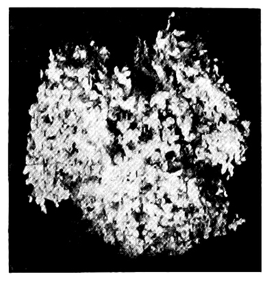
Fig. 23.—Abortion at fourth week.
(Glaister.)
Fig. 23.—Abortion at week 4.
(Glaister.)
It has been decided in Scotland that drugging or operating to procure abortion is criminal, though unsuccessful, but it is not certain whether the woman alone can be charged with taking drugs to procure abortion. Both in England and in Scotland, to make the procuring of abortion criminal, “there must be felonious intent,” for it may be necessary to cause abortion. It must be borne in mind that the law allows no discretionary power on the part of medical practitioners who, to save the life of the mother, may deem it advisable to induce premature delivery. This being the case, no medical man should attempt to induce premature labour without the consent of the relatives of the [Pg 160] woman, and the sanction of a medical colleague after consultation. This precaution is the more necessary as several medical men have been prosecuted, an event which would not have taken place had the precaution above suggested been observed. A medical man should also be very careful never to give any medicine “to bring on the courses” if he has the slightest suspicion of pregnancy, even as a “placebo” to satisfy an importunate patient, for should abortion be otherwise procured, his really harmless medicine may be accused with the result, and a grave suspicion be raised against him, to say the least.
It has been determined in Scotland that drugging or performing procedures to obtain an abortion is a crime, even if unsuccessful, but it’s unclear if the woman can solely be charged for using drugs to induce an abortion. In both England and Scotland, to classify the act of seeking an abortion as a crime, “there must be felonious intent,” as it may be necessary to cause an abortion. It’s important to note that the law does not give medical practitioners any discretionary power; they cannot induce premature delivery even to save the mother’s life without proper authorization. Given this, no doctor should attempt to induce premature labor without the consent of the woman’s relatives and the approval of a medical colleague after consultation. This precaution is particularly important because several doctors have faced prosecution, which likely would not have happened had the suggested precautions been followed. A doctor should also be very cautious never to prescribe any medicine “to bring on the courses” if there’s any suspicion of pregnancy, even as a “placebo” to appease a demanding patient, as if an abortion is procured by other means, that seemingly harmless medicine could be implicated, leading to severe suspicions against him, to say the least. [Pg 160]
The term abortion is understood in medicine to mean the expulsion of the contents of the fœcundated uterus before the sixth month of pregnancy, that is, before the child is considered viable. After this period it is said to be a premature labour.
The term abortion is understood in medicine to mean the expulsion of the contents of the fertilized uterus before the sixth month of pregnancy, which is before the baby is considered viable. After this period, it is referred to as premature labor.
In law, however, no distinction is made and the expulsion of the contents of the uterus at any period before the full time of pregnancy is considered an abortion; in popular language, a miscarriage.
In law, however, no distinction is made and the expulsion of the contents of the uterus at any time before the full term of pregnancy is considered an abortion; in everyday language, a miscarriage.

Fig. 24.—Abortion between
sixth and eighth week.
(Glaister.)
Fig. 24.—Abortion between
the sixth and eighth week.
(Glaister.)

Fig. 25.—Abortion at tenth
week.
(Glaister.)
Fig. 25.—Abortion at 10 weeks.
(Glaister.)
Abortion, when not produced by criminal means, generally occurs at or a little before the third month of utero-gestation, and then usually in first pregnancies, or during the latter part of the period of child-bearing. It is also more frequent among the rich than among the poor. Of the two thousand cases of pregnant women examined by Dr. Whitehead of Manchester, the sum of whose pregnancies was 8681, or 4.38 for each, rather less than 1 in 7 had aborted.
Abortion, when not caused by illegal methods, usually happens around or just before the third month of pregnancy, often in first pregnancies or later in the childbearing years. It is also more common among the wealthy than the poor. In a study of two thousand pregnant women conducted by Dr. Whitehead of Manchester, with a total of 8,681 pregnancies, or an average of 4.38 pregnancies per woman, just under 1 in 7 had experienced an abortion.
When abortion is criminally induced, it generally takes place between the fourth and fifth months, that is, about the time the woman becomes certain of her condition.
When abortion is illegally induced, it usually happens between the fourth and fifth months, which is around the time the woman becomes sure of her condition.
The Causes of Abortion are—
The Reasons for Abortion are—
- 1. Natural or Accidental.—
- (a) Maternal—belonging to the mother;
- (b) Fœtal—belonging to the ovum.
- 2. Violent.—
- (a) Mechanical;
- (b) Medicinal.
1. Natural or Accidental
(a) Maternal.—Among the maternal causes may be mentioned excessive lactation; any irritation of the rectum or bladder; loss of blood, which, by increasing the amount of carbonic acid in the blood, acts as an excitant to the spinal cord; excessive irritability and excitability of the uterus, &c. Certain states of the system conduce to [Pg 161] abortion—albuminuria, syphilis, certain fevers, scarlet fever, smallpox, &c. Abortion may become habitual in some women. Great joy or sudden sorrow have not infrequently been the cause of abortion. The tendency to abortion is greatest at the menstrual periods, that is, at the time when, had not the woman become pregnant, menstruation would have taken place. Slight causes acting at these times are very liable to produce abortion.
(a) Maternal.—Among the maternal causes, we can mention excessive breastfeeding; any irritation of the rectum or bladder; loss of blood, which increases the amount of carbon dioxide in the blood and acts as a stimulant to the spinal cord; excessive sensitivity and excitability of the uterus, etc. Certain conditions in the body contribute to [Pg 161] abortion—such as protein in the urine, syphilis, certain fevers, scarlet fever, smallpox, etc. Abortion can become a frequent occurrence for some women. Moments of great joy or sudden distress have often led to abortion. The likelihood of abortion is highest during menstrual periods, meaning at the time when, if the woman hadn't been pregnant, menstruation would have occurred. Minor triggers at these times are very likely to result in abortion.
(b) Fœtal.—The death of the ovum, or a diseased condition of its uterine coverings, or of the placenta, probably of an inflammatory nature.
(b) Fetal.—The death of the egg, or an unhealthy condition of its uterine linings, or of the placenta, likely due to inflammation.
2. Violent
(a) Mechanical.—Under this head may be mentioned the passage of certain instruments into the cavity of the womb, and the rupture by violence of the membranes which surround the fœtus; also the injection of fluids into the uterus. A medical man practising in Yorkshire informed Husband that so great was the dread of large families, that he knew of several ladies who, if they went a day over their monthly period, passed a catheter into the uterus, with the desired result. “It was wonderful,” he added, “how clever they were.” In India a twig of the Euphorbium nivulia, anointed with assafœtida, is used for the same purpose. “The fœtus is never delivered alive, but there is said to be no great danger to the woman” (Chevers). Women may use hairpins, knitting-needles, and the idea is to pass the instrument “until blood comes,” which is accepted as a sign that abortion will be sure to follow. In some cases it is by no means easy to procure abortion, and women have been known to undergo a considerable amount of violence without abortion taking place. In some women, however, on the other hand, the slightest violence—such, for instance, as slipping from a step or low chair—will cause them to abort.
(a) Mechanical.—This category includes the use of certain instruments to enter the uterus, as well as the forceful breaking of the membranes surrounding the fetus; it also involves the injection of fluids into the uterus. A doctor in Yorkshire told Husband that the fear of having large families was so intense that he knew several women who, if they missed their period by a day, would insert a catheter into their uterus to achieve their goal. “It was amazing,” he remarked, “how skilled they were.” In India, a branch of the Euphorbium nivulia, treated with assafœtida, is used for the same purpose. “The fetus is never born alive, but it is said there’s not much risk for the woman” (Chevers). Women may also use hairpins or knitting needles, aiming to insert the instrument “until blood comes,” which is seen as a sign that abortion is imminent. In some cases, procuring an abortion is quite difficult, and women have been known to endure significant violence without success. However, for some women, even the slightest trauma—like slipping from a step or a low chair—can trigger an abortion.
(b) Medicinal.—Certain drugs, among which may be mentioned ergot, savin, pennyroyal, rue, tansy, saffron, perchloride of iron, diachylon which contains lead, and others, have been used for the induction of abortion. In India unripe pineapple has a great reputation as an abortive (Medical Jurisprudence for India, Chevers). It is scarcely necessary to mention each drug individually, but it must be remembered that there is not one single internal medicament of which it can be consistently with experience asserted that, even when an abortion has followed its use, it must have produced this abortion, and that cause and effect are in such a case “indirect and necessary connection.” All the so-called abortives are most uncertain in their action, and their use is attended with considerable risk to the woman. In the case of diachylon profound lead poisoning may be the result. Be this as it may, they are more frequently used to induce abortion than mechanical procedure, from the fact that the latter requires some amount of anatomical knowledge and manipulative skill.
(b) Medicinal.—Certain drugs, including ergot, savin, pennyroyal, rue, tansy, saffron, perchloride of iron, and diachylon which contains lead, have been used to trigger abortions. In India, unripe pineapple is highly regarded for its abortive properties (Medical Jurisprudence for India, Chevers). It’s not necessary to list each drug individually, but it's important to note that there is not one single internal medicine that can be reliably said to cause an abortion, even if it has followed its use; the connection between cause and effect in such cases is “indirect and necessary.” All so-called abortives are very unpredictable in their effects, and using them carries significant risks for the woman. For instance, using diachylon can lead to severe lead poisoning. Nevertheless, these drugs are used more often to induce abortion than mechanical methods, simply because the latter requires some anatomical knowledge and technical skill.
The dangers of abortion from any cause are hæmorrhage, sepsis, and peritonitis. In mechanical interference, especially where proper precautions have not been taken to prevent them, sepsis and peritonitis from local injury and perforation are prone to occur. [Pg 162]
The risks of abortion from any cause include bleeding, infection, and inflammation of the abdominal lining. In cases of physical intervention, especially where proper precautions haven't been taken to prevent them, infection and abdominal inflammation from local injury and perforation are likely to happen. [Pg 162]
A medical man may be required to—(1) Examine into the nature and characters of the substances expelled from the womb; (2) Examine the woman stated to have aborted.
A doctor may need to—(1) Investigate the nature and characteristics of the substances expelled from the uterus; (2) Examine the woman reported to have had an abortion.
1. Examination of the Substances expelled from the Womb.—The substances expelled from the womb often become the subject of judicial inquiry, and the medical man may be required to give his opinion as to their probable nature.
1. Examination of the Substances Expelled from the Womb.—The substances expelled from the womb often become the focus of legal investigation, and a medical professional may need to provide their opinion on what those substances are likely to be.
Dr. Gallard has called attention to the following:
Dr. Gallard has pointed out the following:
1. During the last six months of pregnancy, abortion, even when it occurs spontaneously, goes through the two stages as at full time, i.e. the expulsion of the products of conception is, as a rule, preceded by rupture of the membranes, followed after a time by the expulsion of the placenta.
1. During the last six months of pregnancy, abortion, even if it happens naturally, goes through the two stages just like full-term pregnancies, i.e. the expulsion of the products of conception is usually preceded by the breaking of the membranes, followed after a while by the delivery of the placenta.
2. In the first three months this order of things is absent, for it is the rule to see the fœtus expelled entire en bloc without rupture of the membranes.
2. In the first three months, this situation doesn't occur, as it's typical for the fetus to be expelled completely en bloc without breaking the membranes.
3. If, then, we find during the first three months of pregnancy the products of an abortion in which the membranes have been ruptured and the embryo expelled alone, we must look for a pathological cause for this infraction of a general rule; and if no disease of the embryo or of the mother is found, we are justified in attributing the abortion to mechanical means used directly against the products of conception. Charpentier has shown that this rupture of the membranes is not an absolute proof of criminal abortion; but in eighteen cases of spontaneous abortion M. Leblond only found rupture of the membranes in one, and in this the membranes presented an abnormal friability.
3. If we find during the first three months of pregnancy that the membranes have ruptured and the embryo has been expelled on its own, we need to consider a medical reason for this violation of the usual situation. If there's no disease affecting the embryo or the mother, we can reasonably conclude that the abortion was caused by mechanical actions directly affecting the products of conception. Charpentier has pointed out that the rupture of membranes isn't definitive proof of a criminal abortion; however, in eighteen cases of spontaneous abortion, M. Leblond only found membrane rupture in one case, and in that instance, the membranes showed unusual fragility.
The questions may be asked—(1) Is it a fœtus?—(2) Is it a mole? If so, is a mole also a fœtus?—(3) Is it merely the coats of the uterus, and unconnected with pregnancy?
The questions can be asked—(1) Is it a fetus?—(2) Is it a mole? If so, is a mole also a fetus?—(3) Is it just the layers of the uterus, and unrelated to pregnancy?
1. Is it a Fœtus?—The development of the fœtus is given on pp. 35, 36 et seq.
1. Is it a Fetus?—The development of the fetus is detailed on pp. 35, 36 et seq.
2. Is it a Mole?—This question gives rise to another: Is a mole a fœtus? To this the answer must be in the affirmative. Moles, being the diseased appendages of the fœtus, vary in character, and have been described by obstetrical writers under the following heads: (a) Hydatiginous; (b) Carneous; (c) Fatty Moles.
2. Is it a Mole?—This question leads to another: Is a mole a fetus? The answer to this must be yes. Moles, being abnormal growths connected to the fetus, vary in type and have been categorized by medical writers as follows: (a) Hydatid moles; (b) Flesh moles; (c) Fatty moles.
(a) Hydatiginous Moles are a result of a diseased condition of the villi of the chorion. The villi become dropsical, and hang in masses like a bunch of grapes.
(a) Hydatid Moles are caused by a disease affecting the villi of the chorion. The villi become swollen and dangle in clusters like a bunch of grapes.
(b) Carneous Moles.—These are the result of hæmorrhage into the chorion. The blood becomes organised, and a fleshy mass is formed, to which in some cases a withered fœtus is attached.
(b) Carneous Moles.—These are caused by bleeding into the chorion. The blood gets organized, forming a fleshy mass, and in some cases, a shriveled fetus is attached to it.
(c) Fatty Moles.—Death of the fœtus and fatty degeneration of the placenta, or fatty degeneration of the placenta and death of the fœtus, produces this variety of mole. A withered fœtus with a mass of fatty placenta are expelled.
(c) Fatty Moles.—The death of the fetus and fatty degeneration of the placenta, or fatty degeneration of the placenta and the death of the fetus, results in this type of mole. A dried-up fetus along with a mass of fatty placenta is expelled.
3. Is it merely the Coats of the Uterus, and unconnected with Pregnancy?—Fleshy masses may be expelled from the womb, which may not be the result of sexual intercourse. The description just given of true moles will, it is hoped, assist in forming a correct diagnosis. [Pg 163] Considerable care will be required, for the honour of the woman accused depends upon the opinion given as to the nature of the substances submitted for examination. It must also not be forgotten that moles may be retained for many months in the uterus and be then expelled. The knowledge of this fact may rebut an accusation of infidelity against a wife. Polypi may be discharged from the womb; the presence of a pedicle will point to their true character. All substances expelled from the uterus should be carefully washed in water, and all clots removed. The examination of the woman may also help in the formation of the diagnosis. The absence of the signs of defloration or of recent delivery will be in her favour.
3. Is it just the Uterine Linings, unrelated to Pregnancy?—Fleshy masses can be expelled from the uterus that aren't a result of sexual intercourse. The description provided for true moles should help in making an accurate diagnosis. [Pg 163] A lot of care is necessary since the reputation of the woman accused relies on the opinion regarding the nature of the substances examined. It’s also important to remember that moles can remain in the uterus for many months before being expelled. Knowing this fact might counter an accusation of infidelity against a wife. Polyps can be discharged from the womb; the presence of a stalk will indicate their true nature. All substances expelled from the uterus should be thoroughly washed in water, and all clots should be removed. Examining the woman can also aid in forming the diagnosis. The absence of signs of defloration or recent delivery will be in her favor.
2. Examination of a Woman stated to have aborted.—This subject may be divided under two heads—(1) Has the woman been recently delivered? (2) What were the means used to procure the abortion?
2. Examination of a Woman Said to Have Aborted.—This topic can be divided into two parts—(1) Has the woman recently given birth? (2) What methods were used to induce the abortion?
It is by no means easy to answer the question whether an alleged abortion has really taken place or not. The signs of recent delivery are in most cases absent, for the woman can better hide her condition during the earlier than during the later months of utero-gestation; consequently suspicion may not have been aroused against her for some weeks or months after the event. The history of the case, with other attendant circumstances—milk in the breasts, change in the colour of the areola round the nipples, severe flooding, absence of the hymen, injuries to the os uteri, transverse condition of the os uteri in contradistinction to its circular form after delivery, &c.—will, in most cases, assist in forming a correct diagnosis; but it must be again repeated that few of the signs applicable to delivery at the full time are here available.
It’s definitely not easy to determine whether an alleged abortion actually occurred. Most of the signs of recent delivery are usually missing because a woman can conceal her condition better in the earlier months of pregnancy than in the later ones; as a result, suspicions might not arise for weeks or even months after the event. The case history, along with other relevant details—such as milk in the breasts, changes in the color of the areola around the nipples, heavy bleeding, absence of the hymen, injuries to the cervix, and the cervix being transverse instead of circular like it is after delivery, etc.—will generally help in making an accurate diagnosis. However, it must be emphasized again that few of the signs typically associated with full-term delivery are applicable here.
In all doubtful cases—
In any uncertain situations—
1. Examine into the general and present state of the health of the woman.
1. Look into the overall and current health of the woman.
2. Find out if there are any reasons which would occasion a pretext to use drugs which are not usually given to women during pregnancy.
2. Find out if there are any reasons that would create a justification to use drugs that are not typically given to women during pregnancy.
3. Learn if menstruation is regular and easy, or if the woman is in the habitual use of emmenagogues, for, if so accustomed, she may have used them ignorant of pregnancy.
3. Find out if menstruation is regular and easy, or if the woman frequently uses emmenagogues, because if she does, she might have used them without realizing she was pregnant.
4. If a woman ascribes her abortion to a fall, to an accident, or to violence used against her, carefully examine into the nature of these.
4. If a woman attributes her abortion to a fall, an accident, or violence against her, take a close look at the details of these situations.
5. Examine into the general causes of abortion, and also inspect the expelled substances.
5. Look into the general causes of abortion, and also check the expelled substances.
Where death is supposed to have followed the use of abortives, the alimentary canal must be examined for the signs of the action of irritants, or the presence of disease in the internal organs; but when death has resulted from an attempt to procure abortion by instrumental means, the neck of the womb is most frequently found covered by a number of small more or less irregular wounds, which may penetrate into the womb or lose themselves in the walls of the organ. Their course is indicated by infiltration, or a small extravasation of coagulated blood, the exact condition of which must, if possible, be ascertained, so as to decide when the wound was inflicted. [Pg 164]
Where death is believed to have occurred after using abortion methods, the digestive tract should be checked for signs of irritant action or diseases in the internal organs. However, if death happened from trying to induce abortion using instruments, the cervix is often found with several small, more or less irregular wounds that might penetrate into the uterus or blend into the walls of the organ. Their path is shown by swelling or a small leakage of clotted blood, and the exact condition of this must be determined if possible, to figure out when the injury occurred. [Pg 164]
The examiner must not forget that the wounds may extend to the fundus of the uterus, and in this case the autopsy shows that a blunt instrument, as a catheter or uterine sound, introduced through the os uteri into the retroverted uterus, glides by its own weight into the rent. The seat of the tear leads one to think that pregnancy was not far advanced when the attempt was made, and in fact the accident most frequently occurs in cases of suspected pregnancy. It must be remembered that the uterus is often punctured by the injudicious use of the uterine sound, but without any immediate dangerous symptoms. Wounds in the walls of the vagina indicate the use of instruments by an inexperienced hand; in the fundus of the uterus, to one at least accustomed to the introduction of instruments. Spontaneous rupture of the uterus is impossible during the early periods of pregnancy, just when the attempts at abortion are usually made. Rupture due to external violence is, as a rule, accompanied with outward signs of the violence used.
The examiner should keep in mind that the wounds might reach the top of the uterus, and in such cases, the autopsy reveals that a blunt instrument, like a catheter or uterine sound, inserted through the cervix into the tilted uterus, can slide in due to its own weight into the tear. The location of the tear suggests that the pregnancy was likely not far along when the attempt occurred, and in fact, this type of accident often happens in cases of suspected pregnancy. It’s important to remember that the uterus can be punctured by careless use of the uterine sound, but this may not show any immediate dangerous symptoms. Wounds in the vaginal walls indicate that instruments were used by someone inexperienced; in the top of the uterus, by someone at least somewhat familiar with instrument use. Spontaneous rupture of the uterus is not possible during early pregnancy, which is when attempts at abortion are usually made. Rupture from external force typically shows visible signs of the violence applied.
In all cases a careful examination of the structure of the uterus should be made. An examination of the ovaries for false or true corpora lutea should be made. The opinions on the character and differences of these bodies are so discordant as to destroy all confidence in their value as proof of conception or the reverse.
In every situation, a thorough examination of the uterus's structure should be conducted. An assessment of the ovaries for false or true corpora lutea should also be performed. The views on the nature and differences of these bodies are so varied that they undermine any confidence in their reliability as evidence of conception or its absence.
Taylor says: “The discovery of the ovum in the uterus in process of development could alone, in the present state of our knowledge, warrant an affirmative opinion on this point in a Court of Law, and this I believe to be the safest view at present of this much-contested question. On the other hand, the absence of a corpus luteum from the ovary would not in all cases warrant an opinion that conception had not taken place.”
Taylor says: “The discovery of the ovum in the uterus in process of development could alone, based on what we currently know, support a confident opinion on this point in a Court of Law, and I believe this to be the safest perspective at this time regarding this highly debated issue. Conversely, the lack of a corpus luteum in the ovary wouldn’t always justify an opinion that conception hasn’t occurred.”
Examine carefully for local sepsis and signs of inflammation of the uterus and its surrounding structures.
Examine closely for local infection and signs of inflammation in the uterus and its surrounding structures.
Recapitulation
Summary
In Medicine, Abortion occurs before the sixth month of pregnancy—premature labour after that period.
In Healthcare, Abortion happens before the sixth month of pregnancy—premature labor occurs after that period.
In Law, Abortion may take place any time before the full period of utero-gestation.
In Law, abortion can happen anytime before the full term of pregnancy.
Abortion may be due to—
Abortion may be because of—
- 1. Natural or Unavoidable Causes.
- (a) Maternal. (b) Fœtal.
- 2. Violence, with Criminal Intent.
- (a) Mechanical. (b) Medicinal.
According to the present state of English law, infanticide—murder of a new-born child—is not regarded as a specific crime, but is treated and tried by those rules of evidence which are applicable in cases of felonious homicide, but with this difference, that the law requires proof that the child was born alive. An old Statute (21 Jac. I. c. 27) made the concealment of the birth of a bastard child conclusive evidence of murder. As far as the legal estimation of the crime is concerned, it matters not whether the child was killed immediately on its entrance into the world, or within a few days afterwards. A fœtus not bigger than a man‘s finger, but having the shape of a child, is a child within the Statute (R. v. Colmer, 9 Cox, 506; R. v. Hewitt, 4 F. & F. 1101). An English judge, at a late trial, stated that if the jury were of the opinion that the prisoner had strangled her child before being wholly born, she must be acquitted of murder. The law also, on the score of humanity, presumes that every child is born dead until direct evidence to the contrary, from medical or other sources, is given. The onus of the proof of live birth, therefore, devolves on the prosecution. It may also be difficult to decide as to the maternity, and the woman accused will have to be examined as to the possibility of her recent delivery.
According to the current state of English law, infanticide—killing a newborn child—is not considered a specific crime. Instead, it is handled and judged under the same rules of evidence that apply to cases of serious homicide, with the key difference being that the law requires proof that the child was born alive. An old statute (21 Jac. I. c. 27) states that hiding the birth of an illegitimate child is sufficient evidence of murder. When it comes to how the law views the crime, it doesn't matter if the child was killed right after birth or a few days later. A fetus no larger than a person's finger, but shaped like a child, is considered a child under the statute (R. v. Colmer, 9 Cox, 506; R. v. Hewitt, 4 F. & F. 1101). In a recent trial, an English judge said that if the jury believed the defendant strangled her child before it was fully born, she should be found not guilty of murder. The law also assumes, for humane reasons, that every child is born dead until there is direct evidence to prove otherwise, from medical or other sources. Therefore, the burden of proof for a live birth rests with the prosecution. It might also be challenging to determine maternity, and the accused woman will need to be examined to establish the possibility of her having recently given birth.
Here let me repeat the advice given on page 148 as to the examination of a woman. Your duty is to request the woman to allow of the necessary examination, giving her the warning which every magistrate or coroner is bound to give to any person charged with a crime, before requiring an answer to a question which may be used in evidence against her at the subsequent trial. The innocent and the guilty may alike object to an examination, but the presumption is against the party declining, if several have voluntarily submitted. A young lady committed suicide rather than submit to an examination by two medical men under an order from the coroner. The medical men were guilty of a grave indiscretion, and both they and the coroner were acting ultra vires in attempting to force a woman to obtain evidence against herself (Taylor, vol. ii. p. 431).
Here, let me repeat the advice given on page 148 regarding the examination of a woman. Your responsibility is to ask the woman for permission to conduct the necessary examination, giving her the warning that every magistrate or coroner is required to provide to anyone charged with a crime before seeking an answer to a question that could be used as evidence against her in a later trial. Both innocent and guilty individuals may refuse an examination, but the assumption is against the one refusing if several others have willingly agreed. A young woman chose to take her own life rather than undergo an examination by two medical professionals under an order from the coroner. The medical professionals acted with serious indiscretion, and both they and the coroner were acting ultra vires by trying to force a woman to provide evidence against herself (Taylor, vol. ii. p. 431).
The decision as to recent delivery will, to a great extent, rest on the condition of the mother, and the apparent age of the child found dead. The discovery of the body of the child is not necessary to conviction, but the medical evidence as to the signs of respiration, of course, depends on the body being found and examined. In most cases of alleged infanticide tried in England, juries appear more inclined to fall back [Pg 166] on the minor offence—concealment of birth—than to convict of the capital offence; and this appears to be the only alternative if the body cannot be found, for, as we have just said, in law every child is held to be born dead. It must of course be shown that the woman has been recently delivered. In case of failure to prove the murder of the child, the Act (24 and 25 Vict. c. 100, sec. 60) enacts that “if any woman shall be delivered of a child, every person who shall, by any secret disposition of the dead body of the said child, whether such child died before, at, or after its birth, endeavour to conceal the birth thereof, shall be guilty of a misdemeanour.” The mere avowal of the birth is not sufficient to convict her; she must be proved to have done some act of disposal of the body after the child was dead (R. v. Turner, 8 C. & P. 755).
The decision about recent delivery will largely depend on the mother's condition and the apparent age of the deceased child. Finding the child's body isn’t necessary for a conviction, but medical evidence regarding signs of breathing obviously hinges on the body being found and examined. In most cases of alleged infanticide tried in England, juries seem more inclined to revert to the lesser offense—concealment of birth—rather than convict for the more serious crime; and this seems to be the only option if the body cannot be found, because, as we previously mentioned, the law treats every child as if it were born dead. It must be demonstrated that the woman recently gave birth. If the murder of the child cannot be proven, the Act (24 and 25 Vict. c. 100, sec. 60) states that “if any woman shall be delivered of a child, every person who shall, by any secret disposal of the dead body of the said child, whether such child died before, at, or after its birth, attempt to conceal the birth thereof, shall be guilty of a misdemeanour.” Simply admitting to the birth isn’t enough for a conviction; there must be proof that she took some action to dispose of the body after the child was dead (R. v. Turner, 8 C. & P. 755).
In Scotland, concealment of pregnancy is a statutory crime, chargeable when the child born is found dead or is not found at all, and there is no proof of its having been murdered. Pregnancy, up to a period when a child might be born alive, must be proved, and the words “during the whole period of her pregnancy” do not imply that the pregnancy must have continued for the full period of nine months. All that is necessary is that there should be such proof of duration of pregnancy as made a living birth possible. If the accused can bring forward a witness to whom she communicated her pregnancy, or called for assistance at the birth, or (it is believed) can prove that the child was born dead, she is entitled to an acquittal.
In Scotland, concealment of pregnancy is a crime defined by law. It can be charged when a child is born dead or not found at all, and there’s no evidence that it was murdered. The pregnancy must be proven until a point where the child could have been born alive, and the phrase “during the whole period of her pregnancy” doesn’t mean that the pregnancy has to last the full nine months. All that’s needed is enough proof that shows a living birth was possible. If the accused can bring in a witness who she shared her pregnancy with, or who was called to help during the birth, or (it’s believed) can show that the child was stillborn, she deserves to be acquitted.
It has also been said that a woman ought not to be convicted of “concealment of pregnancy,” if at the time of delivery the fœtus do not appear to have reached the seventh month of intra-uterine existence. The birth of a “child,” whether dead or alive, is essential; therefore, if the woman accused “can prove that that which she brought forth was not a ‘child,’ but an abortion, or a fœtus, which, from some accident, was in such a condition that, though there had been assistance, it could not have been in a condition to be called ‘a child,’ then the case is out of the Statute.” The Scotch Statute differs from the English on the “concealment of birth” in this, that so long as the woman makes known her pregnancy, the motive for doing so is not considered. Thus, if she make arrangements with anyone to conceal the birth, “the Statute is eluded by that very circumstance” (Alison). The Statute applies to married as well as to single women; but, in the former case, the penalty is seldom enforced unless foul play is suspected.
It has also been said that a woman shouldn’t be convicted of “concealment of pregnancy” if the fetus doesn’t seem to have reached the seventh month of pregnancy at the time of delivery. The birth of a “child,” whether dead or alive, is crucial; therefore, if the accused woman can prove that what she gave birth to was not a “child,” but rather an abortion or a fetus, which, due to some circumstance, was in such a condition that, even with help, it couldn’t be considered “a child,” then the case falls outside the Statute. The Scotch Statute differs from the English one regarding “concealment of birth” in that as long as the woman acknowledges her pregnancy, the reason for doing so is not taken into account. Thus, if she makes arrangements with anyone to hide the birth, “the Statute is evaded by that very circumstance” (Ally). The Statute applies to both married and single women; however, in the case of married women, the penalty is rarely enforced unless foul play is suspected.
DEFINITION OF THE TERM “LIVE BIRTH”
IN CRIMINAL CASES
“The entire delivery of a child.” There must be an independent circulation in the child before it can be accounted alive (R. v. Enoch, 5 C. & P. 539). The entire child must be actually born into the world in a living state (R. v. Poulton, 5 C. & P. 329). But the fact of the child being still connected with the mother by the umbilical cord will not prevent the killing from being murder [Pg 167] (R. v. Reeves, 9 C. & P. 25). To kill a child in its mother‘s womb is no murder, because the person killed must be “a reasonable creature in being, and under the King‘s peace.” But if the child be injured in the womb, and yet be born alive, and then die as a result of such injuries, it may be murder in the person who inflicted them (R. v. Senior, 1 Mood. C. C. 346).
“The complete delivery of a child.” A child must have its own independent circulation before it can be considered alive (R. v. Enoch, 5 C. & P. 539). The entire child must be actually born into the world in a living state (R. v. Poulton, 5 C. & P. 329). However, the fact that the child is still connected to the mother by the umbilical cord does not prevent the act from being classified as murder [Pg 167] (R. v. Reeves, 9 C. & P. 25). Killing a child in its mother‘s womb is not considered murder, because the person killed must be “a reasonable creature in being, and under the King‘s peace.” But if the child is harmed in the womb, is born alive, and then dies as a result of those injuries, it could be classified as murder for the person who caused the injuries (R. v. Senior, 1 Mood. C. C. 346).
A distinction must be drawn between medical or physiological life and legal life. A child may have breathed, as it not infrequently does, before it is completely born into the world; and this might, in a medical point of view, be considered as a live child, but it is not one legally. The entire delivery of the child is necessary in law; and “it must also be proved that the entire child has actually been born into the world in a living state, and the fact of its having breathed is not a conclusive proof thereof.” The inference unfortunately follows from this ruling, that a mother may kill her child without fear of punishment, if she do so before the entire body has slipped from her.
A distinction must be made between medical or physiological life and legal life. A child may have breathed, as often happens, before it is fully born into the world; from a medical standpoint, this might be seen as a live child, but legally it is not. The complete delivery of the child is required by law; and “it must also be proven that the entire child has actually been born into the world in a living state, and the fact that it has breathed is not definitive proof of that.” Unfortunately, this ruling implies that a mother may kill her child without facing punishment if she does so before the entire body has emerged from her.
DEFINITION OF THE TERM “LIVE BIRTH”
IN CIVIL CASES
The evidence of live birth in civil is somewhat different from that required in criminal cases. The viability of the child is determined in Scotland by its crying; in France, by its respiration; in Germany, “the LIVE BIRTH of a child is to be held proven when it has been heard to cry by witnesses of unimpeachable veracity present at its birth”; but in England, the pulsation of the child‘s heart, or any tremulous motion of the muscles, however slight, has been considered as satisfactory proof of live birth.[15]
The evidence of live birth in civil cases is somewhat different from what is needed in criminal cases. In Scotland, a child is considered viable if it cries; in France, it's determined by whether it breathes; in Germany, “the Live birth of a child is proven when it has been heard to cry by witnesses of unquestionable credibility present at its birth”; but in England, the heartbeat of the child, or any slight movement of the muscles, even if minimal, has been accepted as valid proof of live birth.[15]
According to Blackstone, “crying, indeed, is the strongest evidence, but it is not the only evidence”; and Coke remarks, “If it be born alive, it is sufficient though it be not heard to cry, for peradventure it may be born dumb.”
According to Blackstone, “crying, indeed, is the strongest evidence, but it is not the only evidence”; and Coke points out, “If it is born alive, it is enough even if it doesn’t cry, because it might have been born mute.”
Signs of Live Birth prior to Respiration, and independent of it.—(1) Negative.—Signs of intra-uterine death, i.e. putrefaction, or “intra-uterine maceration,” or of such imperfect development that it could not have been born alive. (2) Positive.—Injuries to the child showing that it must have been born alive.
Signs of Live Birth before Breathing, and separate from it.—(1) Negative.—Signs of death in the womb, i.e. decay, or “death in the womb,” or signs of such incomplete development that it couldn’t have been born alive. (2) Positive.—Injuries to the child indicating that it must have been born alive.
1. Negative.—Intra-uterine Putrefaction.—This condition differs in some remarkable points from putrefaction in air.
1. Nope.—Intra-uterine Putrefaction.—This condition has some notable differences from putrefaction that occurs in air.
The body is extremely flaccid and flattened, the bones of the cranium moving easily on one another. The skin of the hands and other parts of the body bear the evidence of prolonged soaking in fluid. In parts, the skin is whitish, or of a reddish-brown or coppery-red colour, without any trace of green, which is always present when putrefaction takes place in the air. The cuticle may be raised in blisters, and be easily detached from the true skin. The denuded patches are moist and greasy, and exude a stinking, reddish-coloured serous fluid. The face [Pg 168] is flattened, and the features distorted. In one case that Husband attended of intra-uterine death of the fœtus in a primipara, and where putrefaction was far advanced, the scalp burst during delivery, and the brain was poured out. Should, however, the child be exposed to the air, it may soon acquire the appearances proper to putrefaction in that medium. If the child, immediately after birth, be thrown into water, the putrefactive changes would be like those of intra-uterine decomposition. In this case the lungs must be examined for the evidence of death by drowning.
The body is very soft and flat, with the bones of the skull moving easily against one another. The skin on the hands and other parts shows signs of being soaked in fluid for a long time. In some areas, the skin appears whitish, reddish-brown, or coppery-red, without any hint of green, which is always present when decay happens in the air. The outer layer of skin may be raised in blisters and can be easily pulled away from the inner skin. The exposed patches are moist and oily, releasing a foul-smelling, reddish liquid. The face is flattened, and the features are distorted. In one case that Husband handled, concerning the intra-uterine death of a fetus in a first-time mother, there was considerable decay, and the scalp burst during delivery, spilling the brain out. However, if the child is exposed to the air, it can quickly take on the signs of decay typical for that environment. If the child is placed in water immediately after birth, the decaying changes would resemble those of intra-uterine decomposition. In this situation, the lungs need to be examined for signs of drowning. [Pg 168]
2. Positive.—Evidence that injuries found on the body could not have been inflicted during birth, or accidentally after birth. On this subject it is scarcely possible to give an opinion one way or the other. All the medical witness can fairly state is, that, from the condition of the lungs, respiration has or has not taken place; that, in the former case, it is not easy to state whether the injuries were the cause of death or inflicted after death.
2. Awesome.—Evidence shows that the injuries found on the body couldn't have been caused during birth or accidentally afterward. It's nearly impossible to form a definitive opinion on this matter. All the medical expert can reasonably say is that, based on the condition of the lungs, respiration either did or did not occur; and if it did occur, it's difficult to determine whether the injuries caused the death or were inflicted after death.
Appearances showing that a New-Born Child has breathed.—1. Walls of the Chest.—“The vaulting of the thorax is not of the slightest diagnostic value.” Casper quotes from Elsässer the following remarks: “It is irrefutable that the variations in the circumference of the thorax (and, of course, in its diameters) are so considerable that no certain normal mean for a thorax that has breathed, and for one that has not breathed, can be laid down. In most cases the measurements of the thorax are incapable of determining whether the lungs contain air or not. The reasons for these variations is, without doubt, to be referred to the congenital differences in the volume of the osseous thorax; partly, also, to the thickness of the soft parts, particularly of the subcutaneous fat and the thoracic muscles; partly, also, to the differences in the degree and amount of the dilatation of the thorax by respiration, with which the distension of the lungs also corresponds,” &c.
Signs that a Newborn Child has Breathed.—1. Chest Walls.—“The shape of the chest doesn’t really help with diagnosis.” Casper cites Elsässer’s remarks: “It's clear that the variations in the chest circumference (and, of course, in its diameters) are so significant that we can't establish a definite average for a chest that has breathed compared to one that hasn't. In most cases, measurements of the chest can’t determine whether the lungs are filled with air or not. The reasons for these variations are undoubtedly due to the natural differences in the size of the bony chest; partly, they are also due to the thickness of the soft tissues, especially the subcutaneous fat and thoracic muscles; partly, they also relate to the differences in how much the chest expands with breathing, which corresponds to the expansion of the lungs as well," etc.
2. Diaphragm.—The position of the diaphragm may be considered as a good diagnostic sign; for it is found that, in children born dead, the highest point of the concavity is between the fourth and fifth ribs, whereas in those born alive it is between the fifth and sixth. The position of the diaphragm may be affected by the gases produced during putrefaction, and also, in children who have breathed, from distension of the stomach and intestines with gas.
2. Diaphragm.—The position of the diaphragm can be seen as a useful diagnostic sign; it has been observed that in stillborn infants, the highest point of the concavity is located between the fourth and fifth ribs, while in those born alive, it is found between the fifth and sixth. The position of the diaphragm may also be influenced by gases generated during decomposition and, in infants who have breathed, by swelling of the stomach and intestines due to gas.
3. Stomach and Intestines.—With regard to the stomach, Tardieu has suggested that the presence of air-bubbles in the glairy mucus usually found in that organ is a sign of live birth, as it can only have arisen from the swallowing of saliva and mucus, aerated by repeated attempts at respiration, probably lasting from five to fifteen minutes. Air in the duodenum is strong evidence of live birth. Breslau of Prague, who has further investigated this subject, states that, in children born dead, or who have undergone prolonged intra-uterine putrefaction, there is never any accumulation of gas in the stomach or intestines, and that the presence of gas in these organs is contemporaneous with respiration, and is independent of the ingestion of food. The intestines of newly-born children do not float [Pg 169] in water, but rapidly sink in that fluid. As respiration proceeds, the coils of the intestines become distended with gas.
3. Digestive System.—Regarding the stomach, Tardieu has suggested that the presence of air bubbles in the slimy mucus typically found in that organ indicates live birth, as it can only result from swallowing saliva and mucus, aerated by repeated attempts at breathing, which likely last from five to fifteen minutes. Air in the duodenum is strong evidence of live birth. Breslau of Prague, who has further explored this topic, states that in infants who are stillborn or have experienced prolonged decomposition in the womb, there is never any gas buildup in the stomach or intestines, and that the presence of gas in these organs occurs simultaneously with breathing and is not dependent on food intake. The intestines of newborns do not float in water, but sink quickly in it. As breathing continues, the loops of the intestines become filled with gas. [Pg 169]
4. Kidneys and Bladder.—The presence of crystals of uric acid in the pelvis of the kidneys and even in the bladder has been suggested as a sign of live birth. Uric acid infarction, as it has been called, usually occurs in from two to ten days after birth, at a period when there are more important signs of live birth than this, even if infarction did not occur, as it does, in still-born infants.
4. Kidneys and bladder.—The presence of uric acid crystals in the kidney pelvis and even in the bladder has been suggested as a sign of live birth. Uric acid infarction, as it’s called, typically happens about two to ten days after birth, during a time when there are clearer signs of live birth, even if infarction didn’t occur, as it does with stillborn infants.
5. Lungs.
Lungs.
(a) Size.—In the fœtus, prior to respiration, the lungs do not fill the cavity of the chest, and the left lung is never found even partially covering the heart.
(a) Size.—In the fetus, before breathing starts, the lungs don’t occupy the chest cavity, and the left lung is never found even partially covering the heart.
After respiration they fill the thorax more or less completely, the amount of distension depending, of course, upon the completeness of the respiratory acts on the part of the child.
After breathing, they fill the chest fairly completely, with the degree of expansion depending, of course, on how fully the child breathes.
(b) Consistence.—Before respiration has taken place, the lungs feel firm, compact, and resistant, and are of the consistency of liver.
(b) Consistence.—Before breathing occurs, the lungs feel firm, compact, and tough, similar in texture to liver.
After respiration they are spongy, crepitant, and yielding when pressed between the fingers. They also present a marbled appearance. These signs of respiration are more or less modified by disease, and the atelectasis pulmonum of Jörg, jun.
After breathing, they are soft, crackling, and squishy when pressed between the fingers. They also have a marbled look. These signs of breathing can be altered by disease, along with the atelectasis pulmonum of Jörg, jun.
Casper denies the existence of atelectasis pulmonum as a distinct disease of newly-born children, and considers that “it is nothing else than the original fœtal condition, from which it differs in no anatomical respect”—an opinion supported by Meigs, who says “it, in fact, resembles exactly the fœtal lung.” It is simply the result of the child dying from some cause before respiration has had time to become fully established, and has possibly been confounded with hepatisation. It must also be remembered that cases are on record of infants having lived for some hours, and then died, yet the lungs sank as a whole, and when cut in pieces.
Casper denies that atelectasis pulmonum is a separate disease in newborns and believes that “it is simply the original fetal condition, differing from it in no anatomical way”—a view supported by Meigs, who states “it actually looks exactly like a fetal lung.” It's merely the result of the baby dying from some cause before breathing has fully started, and it might have been mistaken for hepatization. It's also important to note that there are documented cases of infants who lived for a few hours and then died, yet their lungs were completely collapsed, even when cut into pieces.
(c) Colour.—The colour of the fœtal lungs is “exceedingly various,” and it is by no means easy to convey the idea of colour by words. Speaking in general terms, the lungs of children who have not breathed are of a reddish-brown liver colour, this colour changing to a brighter red at their margins. In children who have breathed, the lungs are of a slaty-blue colour, more or less mottled with circumscribed red patches. This circumscribed mottling is never found in perfectly fœtal lungs. When the lungs are inflated artificially, they swell up and present a uniform cinnabar-red colour, destitute of insular marbling. The insular marbling of the lungs is characteristic of lungs that have breathed, and is due to the presence of blood in the arteries and veins surrounding the inflated lung tissue.
(c) Color.—The color of fetal lungs varies greatly, and it's not easy to describe color with words. Generally, the lungs of children who have not breathed are a reddish-brown, similar to liver color, which changes to a brighter red at the edges. In children who have breathed, the lungs appear slate-blue, often speckled with defined red patches. This kind of speckling is never seen in perfectly fetal lungs. When the lungs are artificially inflated, they expand and show a uniform cinnabar-red color, lacking any patchy marbling. The patchy marbling of the lungs is typical of lungs that have breathed and is caused by the presence of blood in the arteries and veins surrounding the inflated lung tissue.
(d) Buoyancy in Water.—Lungs which have respired float in water.
(d) Buoyancy in Water.—Lungs that have breathed float in water.
But the objection may be raised that lungs that have not respired may yet float from—
But the objection may be raised that lungs that have not respired may still float from—
- 1. The result of artificial respiration.
- 2. The result of putrefaction.
The following table is given by Tidy:
The following table is provided by Tidy:
| Lungs that have not Breathed. | Lungs that have Breathed. |
| 1. Dark in colour (black-blue, | 1. Light in colour (rose-pink, |
| maroon, or purple), resembling | pale pink, light red, or crimson), |
| liver. No mottling. | mottled. |
| 2. Air-vesicles not visible | 2. Air-vesicles distinctly |
| to the naked eye. | visible to the naked eye, or a |
| lens of low power (say a two-inch, | |
| or even a common reading-glass). | |
| 3. When squeezed or cut, do not | 3. Crepitate or crackle freely. |
| crepitate or crackle. | |
| 4. Contain but little blood, | 4. Contain a good deal of blood, |
| therefore little escapes on section. | which escapes freely on section. |
| 5. The blood present is not | 5. The blood present is freely |
| frothy, unless there be | mixed with air, and therefore |
| putrefaction. | appears frothy. |
| 6. Sink in water, unless putrid, | 6. Float in water; or, at all |
| and often not then. | events, the parts which have been |
| events, the parts which have been | |
| expanded, or have breathed, float. | |
| If fully expanded, they will buoy | |
| up the heart. | |
| 7. Bubbles of gas arising | 7. The air cannot be squeezed |
| from putrefaction may be | out by pressure. |
| squeezed out, and as they | |
| escape are usually noted to | |
| be of large size. |
Hydrostatic Lung Test
Hydrostatic Lung Test
(Docimasia pulmonum hydrostatica)
(Hydrostatic Lung Testing)
The value of this test, which is a test of respiration and not of live birth, is founded on the supposition that a lung in which respiration has taken place will float if placed in water, and that when this has not occurred it will sink. Admitting that a lung floats as a result of respiration, it has been objected that this is no proof of live birth, for respiration may take place in:
The significance of this test, which measures respiration rather than live birth, is based on the idea that a lung that has been used for breathing will float when put in water, while a lung that hasn’t will sink. Assuming that a lung floats because of respiration, some have argued that this doesn’t prove live birth, because respiration can happen in:
- 1. The womb, vagitus uterinus.
- 2. The maternal passages, vagitus vaginalis.
- 3. Cases when the head protrudes, the body not yet being born.
With regard to the two first objections, it will be sufficient to say that, in all the cases of so-called intra-uterine respiration, the respiratory acts have occurred in difficult or instrumental labours, where it is justifiable to suppose that, in the endeavour to remove the child, a certain amount of air may have been unavoidably admitted into the maternal passages. But the cases with which the medical jurist has to deal cannot be classed with these, for in all those brought under his notice delivery has been more or less rapid and unassisted.
Regarding the first two objections, it's enough to note that in all cases of so-called intra-uterine respiration, the breathing has happened during difficult or assisted deliveries, where it's reasonable to assume that some air might have accidentally entered the mother's passages while trying to remove the baby. However, the cases the medical jurist deals with don't fall into this category, as in all those presented to them, the delivery has been relatively quick and unassisted.
To the last objection the same reply may be given, that rapid delivery in doubtful cases must be considered as the rule, and that the time [Pg 171] which elapses between the birth of the head of the child and its complete delivery is so short as not to lead to any great error in diagnosis. It is true that the woman may faint with the child half born, and that respiration may thus take place; and it has not yet been decided how many inspirations a child must make to entirely inflate its lungs, or the length of time required to do so.
To the last objection, the same response can be given: that quick delivery in uncertain situations should be seen as the norm, and that the time that passes between the birth of the baby's head and its full delivery is so brief that it doesn’t cause significant diagnostic errors. It’s true that a woman might faint with the baby half born, which could allow for breathing to start; and it hasn’t been determined yet how many breaths a baby needs to fully inflate its lungs, or how long that takes.
N.B.—Any pressure exerted on the umbilical cord during the process of delivery gives rise to respiratory acts on the part of the fœtus. The presence of what Casper calls petechial ecchymoses beneath the pleuræ, upon the aorta, and even on the heart, are, as a rule, a proof that attempts at respiration have been made. These petechial ecchymoses are sometimes found on the same parts in the drowned. (See “Drowning.”)
N.B.—Any pressure on the umbilical cord during delivery causes the fetus to make respiratory movements. The existence of what Casper refers to as petechial ecchymoses under the pleura, on the aorta, and even on the heart usually indicates that attempts to breathe have occurred. These petechial ecchymoses can sometimes be seen in drowning victims as well. (See “Drowning.”)
How is the Hydrostatic Lung Test performed?
and What are the Objections to its Use?
How is the Hydrostatic Lung Test done?
and What are the Concerns about its Use?
As this test was first used, it consisted in placing the lungs, with or without the heart, in water, and then noting whether they sank or floated. A glass vessel, eighteen inches high and twelve in diameter, half filled with distilled water at 60° F., should be used. In summer, water at the ordinary temperature of the room will answer the purpose. To this rough test pressure is now added; the lung, or portions of it, are greatly compressed in a linen cloth, and then thrown into water as before. If the lungs thus compressed float, respiration is held to have taken place; should they sink, the contrary is presumed.
As this test was first used, it involved placing the lungs, with or without the heart, in water and observing whether they sank or floated. A glass container, eighteen inches high and twelve inches in diameter, should be half-filled with distilled water at 60° F. In the summer, room temperature water will suffice. This initial test is now joined by a pressure component; the lung, or parts of it, are tightly wrapped in a linen cloth and then submerged in water as before. If the compressed lungs float, it is assumed that respiration has occurred; if they sink, the opposite is presumed.
Pressure is used for the following reason: The air generated by putrefaction, and which may cause the lungs to float, is removed by pressure, but no amount of pressure, short of entirely destroying the lung tissue, will remove that which is the result of respiration or inflation; and between these the medical expert must decide from collateral evidence.
Pressure is used for the following reason: The gas produced by decay, which can make the lungs buoyant, is eliminated by pressure, but no level of pressure, without completely destroying the lung tissue, will eliminate what comes from breathing or inflation; and between these, the medical expert has to make a decision based on additional evidence.
In performing the test: (1) Try if the lungs will float with the heart and thymus gland attached to them. (2) If they will float without the heart, &c. (3) Try if portions will float with or without pressure.
In doing the test: (1) See if the lungs will float with the heart and thymus gland still attached. (2) Check if they will float without the heart, etc. (3) Test if pieces will float with or without pressure.
The following are the Objections to this Test:
The following are the objections to this test:
- 1. The lungs may sink as a result of disease.
- 2. Respiration, even in healthy lungs, may be so imperfect
- that they may sink.
- 3. Emphysema pulmonum neonatorum.
- 4. Putrefaction.
- 5. Artificial inflation.
1. That in consequence of disease the entire lungs, or portions of them, may sink, and yet respiration may have taken place. Disease of the lung may occur previously to birth or soon afterwards, but it is scarcely probable that the disease would attack every portion of the [Pg 172] lung. Parts, doubtless, small in proportion to the diseased part, may yet have been sufficiently inflated to float. The presence of disease is also not difficult of detection.
1. As a result of illness, the entire lungs or parts of them can collapse, and breathing can still occur. Lung disease can happen before birth or shortly after, but it's unlikely that the disease would affect every part of the lung. Some areas, although small compared to the diseased sections, may still have been inflated enough to stay buoyant. It's also not hard to detect the presence of disease.
2. That respiration, even in healthy lungs, may be so imperfect that they may sink. This objection can scarcely be considered valid against the general application of the test, for in these cases there is no known test by which respiration or its absence can be determined. They are, therefore, out of the pale of the test, as they are out of every other mode of investigation.
2. Respiration, even in healthy lungs, can be so inadequate that they might fail. This argument is hardly valid against the overall use of the test, because in these situations there isn't any known method to measure respiration or its absence. They are, therefore, beyond the scope of the test, just like they are with any other form of investigation.
3. Emphysema pulmonum neonatorum.—Emphysema is generally the result of excessive dilatation of the air cells of the lung, rupture of the cell walls, and infiltration of the intra-lobular areola tissue. This condition may be brought about by:
3. Emphysema pulmonum neonatorum.—Emphysema usually happens due to the over-expansion of the lung's air sacs, breaking of the cell walls, and filling of the tissue between the lobules. This condition can be caused by:
(a) Respiration. (b) Inflation.
(a) Breathing. (b) Inflation.
The fact of the matter is simply this, that the so-called emphysema pulmonum neonatorum, or emphysema of new-born children, is nothing more or less than incipient putrefaction, induced by certain unascertained conditions.
The truth is this: the so-called emphysema pulmonum neonatorum, or emphysema in newborns, is nothing more than early decay, caused by some unknown factors.
Casper sums up his conclusions on this subject in the following words:
Casper summarizes his thoughts on this topic with these words:
“That not one single well-observed and incontestable case of emphysema, developing itself spontaneously within the lungs of a fœtus born without artificial assistance, is known; and it is not, therefore, permissible in forensic practice to ascribe the buoyancy of the lungs of new-born children, brought forth in secrecy and without artificial assistance, to this cause.”
“Not a single well-documented and undeniable case of emphysema has been found to develop spontaneously in the lungs of a fetus born without medical intervention; therefore, in forensic practice, it is not acceptable to attribute the buoyancy of the lungs of newborns delivered in obscurity and without medical assistance to this cause.”
4. Putrefaction.—It must be admitted as proved that the lungs of new-born children in a state of decomposition will float in water. But this admission does not render the test valueless, for it must be remembered:
4. Putrefaction.—It must be acknowledged that the lungs of newborns in a state of decay will float in water. However, this acknowledgment does not make the test worthless, as it is important to remember:
(a) That air generated by putrefaction is found in bubbles under the pleuræ, or in the fissures between the lobuli of the lungs, and not in the air cells of the lungs.
(a) That gas produced by decay is found in bubbles under the pleura, or in the spaces between the lobules of the lungs, and not in the air sacs of the lungs.
(b) That gas as a result of putrefaction can easily be removed by compressing the lungs, or portions of them.
(b) That gas from decay can be easily eliminated by compressing the lungs or parts of them.
(c) That crepitation in putrefied lungs is absent, owing to the fact stated under (a).
(c) That crackling sound in decaying lungs is not present, due to the reason mentioned in (a).
(d) That the lungs are among those organs which putrefy late.
(d) That the lungs are one of the organs that decay last.
(e) That negative evidence may be obtained, if the lungs, in a highly putrescent body, sink in water. The tendency of putrefaction, as above stated, is to cause them to float.
(e) Negative evidence can be found if the lungs in a highly decomposed body sink in water. As mentioned earlier, the tendency of decomposition is to make them float.
5. Inflation.—In the first place, it is to be remarked that to inflate the lungs is by no means an easy task. Elsässer states “that in forty-five experiments performed on children born dead, without opening their thorax and abdomen, only one was attended with complete success, thirty-four with partial success, and ten with none whatever; and it must also be remembered that these experiments were conducted without disturbance, and with the greatest care.” Professor Gross states his opinion on this subject thus: “We are decidedly of opinion that artificial inflation of the lungs is a very difficult matter; and we believe that the complete distension of these organs can only be [Pg 173] effected where a tube is introduced into the mouth of the larynx.” In the cases that come before the medical expert, the question naturally arises, Who would inflate the lungs? Surely not the mother. If not the mother, who else? It has been suggested that some malicious person might inflate them to sustain a charge of infanticide. Is this probable?
5. Inflation.—First of all, it's important to note that inflating the lungs is definitely not an easy task. Elsässer points out that in forty-five experiments conducted on stillborn children, without opening their chest and abdomen, only one resulted in complete success, thirty-four saw partial success, and ten were unsuccessful at all; it's also worth mentioning that these experiments were carried out carefully and without interruption. Professor Gross shares his view on this matter: “We firmly believe that artificial inflation of the lungs is quite difficult, and we think that complete expansion of these organs can only be achieved if a tube is inserted into the mouth of the larynx.” When cases come before the medical expert, the question naturally arises: Who would inflate the lungs? Surely not the mother. If not the mother, then who? Some have suggested that a malicious person might inflate them to support a charge of infanticide. Is that likely?
The following points may be noticed on this subject:
The following points can be observed on this topic:
(a) Known difficulty in inflating the lungs.
(a) Recognized difficulty in inflating the lungs.
(b) Absence on the part of the mother of any preparation to save the life of her child.
(b) The mother's lack of any action to protect her child's life.
(c) Presence of air in the stomach and intestines, the result of attempted inflation.
(c) Presence of air in the stomach and intestines, caused by attempted inflation.
(d) Bright cinnabar-red colour of the lungs, without trace of mottling.
(d) Bright red color of the lungs, with no signs of mottling.
(e) Absence of frothy blood when the lungs are cut into.
(e) No frothy blood present when the lungs are sliced open.
(f) When, therefore, we observe the following phenomena, a sound of crepitation without any escape of blood-froth on incision, laceration of the pulmonary cells with hyperæmia, bright cinnabar-red colour of the lungs without any marbling, and perhaps air in the (artificially inflated) stomach and intestines, we may with certainty conclude that the lungs have been artificially inflated.
(f) When we observe the following signs: a crackling sound without any blood-froth escaping upon cutting, tearing of the lung cells with congestion, a bright red color of the lungs without any marbling, and possibly air in the (artificially inflated) stomach and intestines, we can confidently conclude that the lungs have been artificially inflated.
It may be further noted that natural respiration is accompanied with, first, the distension of the air cells of the lungs with air; and, second, with an increased flow of blood into the organs, beyond that necessary for their nourishment and growth. They thus increase in absolute weight, while their specific gravity is lessened.
It’s also important to mention that natural breathing involves, first, the expansion of the air sacs in the lungs with air; and, second, an increased blood flow to the organs that goes beyond what’s needed for their nourishment and growth. As a result, they gain weight, while their specific gravity decreases.
The objections just mentioned apply to the hydrostatic test as originally employed. It will now be necessary to notice those against the same test when modified by pressure. These are two in number:
The objections mentioned earlier apply to the hydrostatic test as it was originally used. Now, it's important to address the objections to the same test when it has been modified by pressure. There are two of these objections:
1. That no amount of pressure, short of entirely destroying the lung tissue, can expel the air from a lung that has been inflated, or from one in which respiration has taken place.
1. No amount of pressure, except for completely destroying the lung tissue, can push the air out of a lung that has been inflated or one that has already done some breathing.
2. Pressure is, therefore, no test of natural respiration or of artificial inflation.
2. Pressure is, therefore, not a valid test of natural breathing or of artificial ventilation.
In answer to the above, it will only be necessary to refer to what has been already said with regard to the difficulty of inflation, and the more probable event of the condition of the lungs being the result of respiration.
In response to the above, it’s only necessary to mention what has already been said about the challenges of inflation, and the more likely scenario that the condition of the lungs is caused by respiration.
Casper thus sums up the result of his views with regard to the probative value of the docimasia pulmonaris:
Casper sums up his views on the probative value of the docimasia pulmonaris as follows:
“That a child has certainly lived
during and after its birth—
“That a child has definitely lived
during and after its birth—
“1. When the diaphragm stands between the fifth and sixth ribs.
“1. When the diaphragm is located between the fifth and sixth ribs.
“2. When the lungs more or less completely occupy the thorax, or at least do not require to be sought for by artificial separation of the walls when cut through.
"2. When the lungs mostly fill the chest, or at least don’t need to be moved apart artificially when opened up."
“3. When the ground colour of the lungs is broken by insular marblings.
“3. When the base color of the lungs is interrupted by isolated streaks.”
“4. When the lungs are found by careful experiment to be capable of floating.
“4. When tests carefully show that the lungs can float.
The Lung Test is unnecessary when—
The Lung Test isn't needed when—
1. The umbilical cord has dropped off, and cicatrisation has followed.
1. The umbilical cord has fallen off, and healing has taken place.
2. Where food is found in the stomach.
2. Where food is located in the stomach.
3. Where there are evident signs of putrefaction in utero.
3. Where there are clear signs of decay in utero.
4. Also in the case of the birth of monsters, or where, from congenital malformation, the possibility of live birth is excluded.
4. Also in the situation of giving birth to monsters, or when, due to birth defects, the possibility of a live birth is ruled out.
Besides the hydrostatic test, the following have been proposed:
Besides the hydrostatic test, the following have been suggested:
Ploucquet‘s Test.—This test is based on the relative weight of the lungs, before and after respiration, to that of the entire body of the child. The variations found in practice between the relative weights render the test worse than useless.
Ploucquet’s Test.—This test relies on the comparison of the lungs' weight before and after breathing to the total weight of the child's body. The differences observed in practice between the relative weights make the test more of a hindrance than helpful.
Absolute Weight of the Lungs.—This test consists in a comparison of the weight of the lungs before and after respiration, and it may be stated here that the lungs, prior to respiration, vary in weight from about 400 to 650 grains; but so much depends on the maturity or immaturity of the child, and degree of respiration, that, like the last, the test is unworthy of confidence.
Lung Weight.—This test involves comparing the weight of the lungs before and after breathing. It's important to note that the lungs, before breathing, can weigh anywhere from about 400 to 650 grains. However, much depends on the maturity or immaturity of the child and the level of breathing, so, similar to the last test, this one isn't very reliable.
Wredin‘s Test.—Dr. Wredin, of Petrograd, states that the gelatinous substance found in the middle ear of infants before birth, gradually disappears, to be replaced by air on the subsequent establishment of respiration. Wendt, of Leipzig, from an examination of 300 cases, declares that the gelatinous substance can only be expelled by the establishment of full respiration. The value of this test has been questioned, as some observers have found that in different cases intervals of from a few hours to five weeks have occurred, before the replacement of the gelatinous material by air.
Wredin's Test.—Dr. Wredin from Petrograd says that the jelly-like substance found in the middle ear of infants before they are born gradually disappears and is replaced by air when they start breathing after birth. Wendt from Leipzig, after examining 300 cases, claims that the jelly-like substance can only be removed once full respiration is established. The reliability of this test has been questioned because some observers have noted that in different cases, it can take anywhere from a few hours to five weeks for the jelly-like material to be replaced by air.
Table Showing the Signs of
Maturity of Child At Birth
Table Showing the Signs of
Child Maturity at Birth
As regards:
About:
1. Average Length of Body.—Nineteen inches.
Average Body Length.—Nineteen inches.
2. Average Weight of Body.—About seven pounds.
2. Average Weight of Body.—Approximately seven pounds.
3. Eyes.—The pupillary membrane is not found in the mature child.
3. Eyes.—The pupillary membrane is not present in a mature child.
4. Navel.—Said to be exactly midway between the pubes and the ensiform cartilage.
4. Navel.—Said to be exactly halfway between the pubic area and the bottom of the breastbone.
5. External Genitals.—Testicles found in the scrotum, and the labia majora cover the vagina and clitoris.
5. External Genitals.—Testicles are located in the scrotum, and the labia majora cover the vagina and clitoris.
6. Os Femoris.—Ossification of the inferior femoral epiphysis. The osseous nucleus measures from three-quarters of a line to three lines in diameter.
6. Os Femoris.—The bone development of the lower femoral epiphysis. The bony center measures between three-quarters of a line and three lines in diameter.
CAUSE OF DEATH TO THE FŒTUS
Death may be due to—
Death may be caused by—
- I. Immaturity on the part of the fœtus.
- II. Complications occurring during or immediately after birth.
- III. Congenital disease in one or more of the fœtal organs.
- IV. Neglect or exposure, constituting “Infanticide by Omission.”
II. Complications occurring during or immediately after Birth.—(1) Unavoidable or inherent in the process of parturition. (2) Induced with criminal intent, constituting “infanticide by commission.”
II. Complications that occur during or immediately after birth.—(1) Unavoidable or inherent in the process of childbirth. (2) Caused with criminal intent, constituting “infanticide by commission.”
1. Unavoidable or Inherent in the Process of Parturition.—The immediate cause of death may be either maternal or fœtal. In the former, the presence of tumours in the pelvic passages, or disease of the bones, causing a narrowing of the canal, may lead to fatal compression of the head of the child. Death may also be due to protracted labour from debility on the part of the mother, or she may suddenly faint after delivery. A congested state of the brain may be present in these cases. In the latter (fœtal), pressure on the umbilical cord from malposition of the child during labour, or an abnormal increase in the size of the head, may cause death. There is also a greater mortality, both during and after delivery, among male than female children. The child may be also accidentally suffocated in the fæces of the mother, or in the fold of her dress; or it may be born while the woman is straining at stool, and be drowned in the contents of the pan. Husband once met with a case of accidental death of a child from suffocation in the drawers of the mother, who persisted, from motives of delicacy, in wearing those articles of dress during her confinement. Death may also result from strangulation, occasioned by the pressure of the funis round the child‘s neck. The death in this case can scarcely be considered as due to strangulation, as the child had never breathed, but it is probably the result of the arrest of the flow of blood along the cord, from the tightness of the folds round the neck. Some congestion of the brain may, however, be found resulting from the pressure on the vessels of the neck. Lastly, death may ensue from a fall on the floor in cases of sudden and quick labours, especially if the woman be in the erect posture at the time of delivery.
1. Unavoidable or Inherent in the Process of Childbirth.—The immediate cause of death can be either maternal or fetal. In the maternal case, tumors in the birth canal, or bone diseases that narrow the passage, can lead to fatal pressure on the baby's head. Death may also result from prolonged labor due to the mother's fatigue, or she might suddenly faint after giving birth. Congestion of the brain can be present in these situations. In the fetal case, pressure on the umbilical cord caused by the baby's position during labor, or an unusually large head, can be fatal. Additionally, there is a higher mortality rate during and after delivery for male babies compared to female babies. A baby might also be accidentally suffocated in the mother's feces or tangled in her clothing; or it could be born while the woman is straining to have a bowel movement and drown in the contents of the pan. There was once a case where a baby accidentally suffocated in the mother's dress drawers because she insisted on wearing this clothing for modesty during her labor. Death can also happen due to strangulation caused by the umbilical cord tightening around the baby's neck. In this situation, the death is not really due to strangulation since the baby never breathed, but likely results from blood flow being cut off along the cord because of the tightness around the neck. Some brain congestion may be observed, however, due to pressure on the neck vessels. Finally, death can occur from a fall to the floor during quick and sudden deliveries, particularly if the woman is standing at the time of birth.
2. Induced with Criminal Intent.—Infanticide by commission: was the death due to violence? The answer to this question is by no means easy. In all doubtful cases the attendant circumstances must be taken into consideration. A woman may unintentionally injure her child in her efforts to drag it from her. The presence of respiration, more or less complete, is strongly presumptive against the death being the result of accident. But even here considerable caution is necessary, for the injury may not be immediately fatal, although accidentally inflicted, sufficient time elapsing between its infliction and the death of the child to allow of respiration. Foreign bodies found in the mouth and fauces are also corroborative of death by violence. A case is recorded in which the child‘s fauces, upper portion of the œsophagus, the larynx, and the trachea were closely packed with a coarse green sand, and yet the lungs sank when the hydrostatic test was applied to them. There was nothing to show when the packing of the fauces was effected. [Pg 176]
2. Induced with Criminal Intent.—Infanticide by commission: was the death caused by violence? The answer to this question is definitely not straightforward. In all uncertain cases, the surrounding circumstances must be taken into account. A woman might unintentionally harm her child while trying to pull it away from her. The presence of breathing, whether full or partial, strongly suggests that the death was not accidental. However, even in this case, significant caution is needed, as the injury might not be immediately fatal, allowing enough time between the injury and the child's death for breathing to occur. Foreign objects found in the mouth and throat also support the conclusion of death by violence. There is a recorded case where the child's throat, upper part of the esophagus, larynx, and trachea were packed tightly with coarse green sand, yet the lungs sank when put through the hydrostatic test. There was no evidence indicating when the throat packing happened. [Pg 176]
Strangulation may be produced by the constriction of the umbilical cord round the neck, and for this reason marks round the child‘s neck cannot always be ascribed to intentional violence. Of 327 cases collected by Elsässer, in which the cord was from one to four times round the children‘s necks, there was not in a single instance any mark of the cord perceptible, even though in some cases the cord had to be cut to permit the completion of labour. With regard to marks round the neck of a new-born child, Casper remarks that it is possible “to mistake the folds of the skin, produced by the movements of the head, and which remain strongly marked in the solidified fat, and are very prominent, particularly in short necks, for the marks of the cord.” The mark left by the funis is broad, corresponds with the breadth of the cord, runs without interruption round the neck, and is everywhere quite soft, and never excoriated. Ecchymoses may be present, irregularly following the line made by the cord. On the other hand, “a mummified, parchment-like, unecchymosed depression points in every case to strangulation by a hard, rough body,” and this more especially if there be any abrasion of the cuticle or laceration of the skin. Death, sometimes ascribed to strangulation, is probably the result of suffocation, and happens thus: any pressure exerted on the cord cuts off the blood from the placenta to the fœtus, and gives rise to respiratory attempts on the part of the child, the child dying from suffocation, or from the engorgement of the lungs with liquor amnii drawn into them at every effort to breathe. An infant may be poisoned. This cause of death is very rare, but deaths have resulted from the use of poisonous gases. While on this subject it may be advisable to state here that ulcerations have been found in the stomach and intestines more or less accompanied with a collection of dark brown or black bloody fluid, which have given rise to suspicions of poisoning in infants to all outward appearances quite healthy. An infant may be thrown into water and drowned. No traces of this mode of death would be discoverable in the infant unless respiration had taken place prior to its immersion. The plea of accidental drowning in a cesspool or water-closet pan may be put forward; it is therefore well to examine the cord. Has a ligature been placed upon it? Has it been cut by a sharp instrument? The nature and character of the fluid found in the stomach should be noted.
Strangulation can occur when the umbilical cord wraps around the baby's neck, so marks on the newborn's neck can’t always be attributed to intentional harm. In a study by Elsässer of 327 cases where the cord was wrapped one to four times around the baby’s neck, there were no visible cord marks, even in cases where the cord had to be cut to finish the delivery. Casper notes that the folds of skin from head movements can look like cord marks, especially noticeable in babies with short necks due to the prominent fat deposits. The mark left by the cord is broad, matching its width, continuous around the neck, soft, and never raw. There may be bruises following the line of the cord, but “a dry, parchment-like, smooth depression always indicates strangulation by a hard object,” particularly if there's skin abrasion or tearing. Death, sometimes thought to be caused by strangulation, is likely due to suffocation: pressure on the cord cuts off blood flow from the placenta, triggering the baby's attempts to breathe which can lead to suffocation or lungs filling with amniotic fluid. An infant can also be poisoned. Though rare, there have been cases where deadly gases caused death. It’s worth mentioning that ulcers have been found in the stomach and intestines, often with dark brown or black fluid, raising suspicions of poisoning in infants who seemed healthy. An infant might also drown if thrown into water. No signs of drowning would be found unless the baby had breathed before immersion. Accidental drowning claims, such as in a cesspool or toilet, may arise, so it’s important to check the cord. Is there a ligature? Was it cut with something sharp? Pay attention to the nature of the fluid in the stomach.
Fractures of the skull may happen—
Fractures of the skull can occur—
1. In the Womb.—The parturient female may fall from a considerable height, and thus cause injury to her child. These cases are of no judicial importance, as the presence of intra-uterine putrefaction or an examination of the lungs will at once show that the child has not breathed. It must be borne in mind, however, that dislocations may take place in the womb, and this fact may be brought forward in defence. The history of the case, and the absence of any other signs of violence, will decide the truth or falsity of the plea.
1. In the Womb.—A pregnant woman can fall from a significant height, which may injure her baby. These situations aren't important in court since signs of intra-uterine decay or an examination of the lungs will quickly reveal that the baby hasn't taken a breath. However, it's important to remember that dislocations can occur in the womb, and this could be used as a defense. The details of the case and the lack of any other signs of violence will determine whether the defense is valid or not.
2. During Labour.—Fracture of the cranial bones during labour generally occurs in difficult and protracted labours, which, from this [Pg 177] very cause, seldom become the subject of judicial inquiry. In some cases the defective ossification of the bones of the skull may give rise to fractures, which may lead to dangerous mistakes. This deficiency, in the process of ossification is thus described by Casper: “If the bone in question is held up to the light, this is seen to shine through the opening, which is closed only by the pericranium. When the periosteal membrane is removed, the deficiency in the ossification is seen in the form of a round or irregularly circular opening, not often more than three lines in diameter, though frequently less; its edges are irregular and serrated: these edges are never depressed, as is the case in fractures; and neither they nor the parts in their neighbourhood are ever observed to be ecchymosed.” The child in these cases may breathe for a short time, and then die without any apparent cause.
2. During Labour.—Fractures of the cranial bones during labor usually happen in difficult and long labors, which, because of this reason, rarely become the focus of legal examination. In some instances, poor ossification of the skull bones can lead to fractures, resulting in serious errors. This deficiency in ossification is described by Casper as follows: “If the bone in question is held up to the light, it can be seen to shine through the opening, which is only closed by the pericranium. When the periosteal membrane is removed, the deficiency in ossification appears as a round or oddly shaped opening, typically no larger than three lines in diameter, and often smaller; its edges are jagged and irregular: these edges are never depressed, as in the case of fractures; and neither they nor the surrounding areas show any signs of bruising.” In these cases, the child may breathe for a short while and then die without any obvious reason.
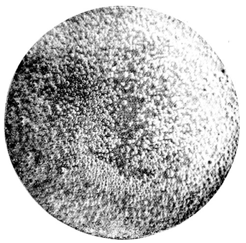
Fig. 26.—Photo-micrograph
of human milk, × 250.
(R. J. M. Buchanan.)
Fig. 26.—Photo-micrograph
of human milk, × 250.
(R. J. M. Buchanan.)
3. By Falls.—It is beyond doubt possible for a child to be born so precipitately as to fall on the floor and be severely injured, and that even fatally. In cases of alleged precipitate birth, to account for injuries found on the child, the following points should be remembered, and will assist in forming a diagnosis:
3. By Falls.—It is definitely possible for a child to be born so quickly that they fall to the floor and get seriously hurt, even fatally. In cases of supposedly rapid birth, to explain any injuries observed on the child, the following points should be kept in mind, and will help in making a diagnosis:
1. In Support of Premature Birth
and Unintentional Injury
(a) Rupture of the umbilical cord. In all cases it would be advisable to measure the length of the cord, and then the distance of the vulva from the ground, allowing of course for the woman not being quite erect at the time of delivery owing to a separation of the legs. A disproportion between the two measurements may or may not account for the rupture of the cord. The following measurements may be taken: usual length of cord, eighteen to twenty inches; distance of vulva from the [Pg 178] ground, twenty-six inches, but allowing for stooping, two-thirds of the above. To the length of the cord must be added about nine inches, the distance from the navel to the top of the head of the child. Thus, a fall of about thirty inches will put no strain on the cord. A case is on record of a rupture of the cord taking place while the woman was in a recumbent position, but in that case the labour was precipitate, and the cord very short and small.
(a) Rupture of the umbilical cord. In all cases, it's advisable to measure the length of the cord and then the height of the vulva from the ground, taking into account that the woman may not be fully upright during delivery due to her legs being apart. A mismatch between these two measurements may or may not explain the rupture of the cord. The following measurements can be taken: the usual length of the cord is eighteen to twenty inches; the distance from the vulva to the ground is twenty-six inches, but considering a stooped position, reduce that by a third. To the length of the cord, add about nine inches, which is the distance from the navel to the top of the child's head. Therefore, a fall of about thirty inches shouldn't put stress on the cord. There’s a recorded case of a cord rupture occurring while the woman was lying down, but in that instance, the labor was very quick, and the cord was very short and thin.
(b) Placenta not detached from the child.
(b) Placenta not separated from the baby.
(c) Fracture of the parietal bones; the fracture radiating into the frontal and squamous portion of the temporal bone. In experiments on twenty-five children dropped from a height of thirty inches, one parietal bone was found fractured in sixteen of the cases; both parietals, in six cases. The fractures in most cases occurred about the parietal protuberances. It must be remembered that the children were dead, and that it is easier to fracture the skull of a live infant than that of a dead one.
(c) Fracture of the parietal bones; the fracture spreading into the frontal and squamous parts of the temporal bone. In experiments on twenty-five children dropped from a height of thirty inches, one parietal bone was fractured in sixteen cases; both parietal bones were fractured in six cases. The fractures mostly occurred around the parietal protuberances. It should be noted that the children were deceased, and it is easier to fracture the skull of a living infant than that of a deceased one.
(d) Imperfect ossification of the bones of the skull.
(d) Incomplete bone formation in the skull.
(e) Absence of other injuries.
No other injuries present.
2. In Support of Criminal Violence
(a) The fact of the umbilical cord being divided by some sharp instrument and not torn. A caution must be here inserted, for Taylor mentions a case where rupture of the cord occurred in such a manner that it could not be decided whether it had been intentionally cut or torn.
(a) The umbilical cord was cut with a sharp instrument rather than torn. A warning should be noted here, as Taylor talks about a case where the cord ruptured in a way that made it unclear whether it had been cut on purpose or torn.
(b) Extensive fracture of one or more of the bones of the cranium.
(b) Extensive fractures of one or more of the bones in the skull.
(c) Fracture and dislocation of the neck.
Neck fractures and dislocations.
(d) Presence of incised wounds, and other evidence of violence.
(d) Presence of cut wounds and other signs of violence.
N.B.—In all doubtful cases, a guarded opinion should be given, stating simply that the dissection does not reveal anything contrary to the statements offered as to the cause of death.
N.B.—In any uncertain situations, a cautious opinion should be provided, simply stating that the dissection does not show anything that contradicts the explanations given regarding the cause of death.
III. Congenital Disease in one or more of the Fœtal Organs.—In all cases the presence of congenital disease must be sought for.
III. Congenital disease in one or more fetal organs.—In all cases, the presence of congenital disease needs to be checked for.
IV. Neglect or Exposure, constituting “Infanticide by Omission.”—Under this head may be mentioned the following:
IV. Neglect or Exposure, referred to as “Infanticide by Omission.”—Under this section, the following can be mentioned:
(a) Neglecting to place the child in such a position that it may breathe freely.
(a) Failing to put the child in a position that allows them to breathe easily.
(b) Neglecting to protect the child from extremes of cold or heat.
(b) Failing to protect the child from extreme cold or heat.
(c) Neglecting to feed it with the food appropriate to its age. (See Signs of Death from Starvation, pp. 132 et seq.)
(c) Failing to feed it the right food for its age. (See Signs of Death from Starvation, pp. 132 et seq.)
(d) Neglecting to tie the umbilical cord.
(d) Forgetting to tie the umbilical cord.
To give answers to these questions will in many cases be impossible, and each must be decided by such circumstances as present themselves in each individual case. For instance, if the body be found stiff, blanched, naked or nearly so, lying on the ground, the vessels of the interior gorged with blood, whilst the superficial vessels are contracted and can be seen only with difficulty; at the same time, the hydrostatic test shows that respiration has taken place, and in the absence of all external or internal causes—the probability is in favour of death by cold. In close relation with the present subject is the question—
To answer these questions is often impossible, and each one must be determined by the specific circumstances of each individual case. For example, if a body is found stiff, pale, and naked or almost so, lying on the ground, with the internal blood vessels full and the surface vessels contracted to the point of being difficult to see; at the same time, if the hydrostatic test indicates that breathing has occurred, and there are no external or internal causes present—the likelihood suggests death by hypothermia. A closely related question is—
How Long did the Child survive its Birth?—The answer to this question is by no means easy, and the data on which a decision can be based are not very reliable. The presence or absence of the vernix caseosa should be noticed. In still-born children the closed eyelids, when raised, do not remain open; in the live-born, on the other hand, the eyes remain half open even after repeated attempts to close them. Another guide to the determination of the length of time the child survived its birth may be found in the absence or presence of the meconium in the intestines. The meconium—so-called from its resemblance to inspissated poppy juice—is found in the large intestine as a dark-greenish pasty mass, more or less filling that portion of the bowel. In the upper portions of the intestines it varies from a light-yellowish or greyish to a greenish-brown colour, till in the large intestine it assumes the colour and consistence above mentioned. It is generally discharged by the infant in from four or five to forty-eight hours after birth. In breech presentations it may be passed during the process of delivery, although the child be still-born; but its entire absence from the intestines is presumptive of existence for some days after birth.
How Long Did the Child Survive After Birth?—The answer to this question is definitely not straightforward, and the data available for making a decision are not very trustworthy. The presence or absence of the vernix caseosa is important to note. In stillborn babies, the closed eyelids, when lifted, do not stay open; in contrast, live-born babies' eyes remain partially open even after multiple attempts to close them. Another clue for determining how long the child survived after birth can be found in the presence or absence of meconium in the intestines. Meconium—named for its resemblance to thickened poppy juice—appears in the large intestine as a dark greenish pasty substance, partially filling that section of the bowel. In the upper parts of the intestines, it varies in color from light yellowish or grayish to greenish-brown, and in the large intestine, it takes on the color and consistency mentioned above. It is usually expelled by the infant within four to five to forty-eight hours after birth. In breech births, it may be released during delivery, even if the baby is stillborn; however, its complete absence from the intestines suggests that the baby survived for several days after birth.

Fig. 27.—Photo-micrograph
of
starch granules, × 250 (potato).
(R. J. M. Buchanan.)
Fig. 27.—Microphoto of
starch grains, × 250 (potato).
(R. J. M. Buchanan.)
Table showing how long a
New-born Child has Lived.
Table showing how long a
Newborn Child has Lived.
2. Changes in the Umbilical Cord.—Mummification of the cord is not of the slightest value as a proof of extra-uterine life; but the separation of the cord which occurs between the fourth and seventh day, especially when cicatrisation has taken place, is a sure sign that the child must have lived four or five days at least. Two other appearances of some value may also be noted, namely:
2. Changes in the Umbilical Cord.—Mummification of the cord doesn’t serve as any proof of life outside the womb; however, the separation of the cord that happens between the fourth and seventh day, particularly when healing has occurred, is a definite indication that the baby must have lived for at least four or five days. Two other notable signs may also be observed, specifically:
(a) In fresh bodies, the appearance of a bright red ring about a line in breadth, which surrounds the insertion of the cord, and which is formed within the uterus.
(a) In new bodies, a bright red ring appears around a line in width, which surrounds where the cord is attached, and this forms within the uterus.
(b) A similar red ring, about two lines broad, around the insertion of the cord, accompanied with “thickening, inflammatory swelling of the portion of the skin affected, and slight purulent secretion from the umbilical ring itself.” This latter condition Casper considers as affording “irrefragable proof of the extra-uterine life of the child.”
(b) A similar red ring, about two lines wide, around the attachment of the cord, accompanied by “thickening, inflammatory swelling of the affected skin area, and a slight pus discharge from the umbilical ring itself.” Casper considers this condition to provide “irrefutable evidence of the child’s life outside the womb.”
3. Changes in the Circulatory System.
3. Changes in the Blood System.
(a) Ductus Arteriosus.—Arterial duct. A contracted condition of this duct is of no value as a proof that a child has survived its birth; for the duct is liable to become contracted, and even obliterated, before the birth of the child.
(a) Patent Ductus Arteriosus.—Arterial duct. A narrowed condition of this duct does not confirm that a child has survived birth; the duct can narrow and even close off before the child is born.
(b) Ductus Venosus.—Nothing certain is known as to the exact time when this duct closes; the condition of the vessel is, therefore, of no assistance in determining the possibility of the child having survived its birth. The duct has been found closed in a still-born child; and in one child, which lived for a quarter of an hour, both the ductus arteriosus and the foramen ovale were found closed. Cases are also on record in which these fœtal channels were found open after thirty days of extra-uterine life.
(b) Ductus Venosus.—It's unclear exactly when this duct closes, so its status doesn't help in figuring out if the child survived birth. The duct has been found closed in a stillborn baby; and in one baby that lived for fifteen minutes, both the ductus arteriosus and the foramen ovale were observed to be closed. There are also records of cases where these fetal passages were still open after thirty days of life outside the womb.
(c) Foramen Ovale.—What has been said of the preceding may be said with regard to the foramen ovale.
(c) Foramen Ovale.—What was mentioned earlier applies to the foramen ovale as well.
N.B.—To sum up, therefore, in the fewest words, any attempt at forming an opinion on the docimasia circulationis may result in a fatal error on the part of the medical witness, as it is impossible to determine with any accuracy by days the period of their closure. As a general statement, however, the following, according to Bernt and Orfila, is the order in which obliteration of the fœtal vessels takes place: (1) The umbilical arteries. (2) Ductus venosus. (3) Ductus arteriosus. (4) Foramen ovale.
N.B.—To summarize briefly, any attempt to form an opinion on the docimasia circulationis could lead to a serious mistake by the medical witness, as it’s impossible to determine the exact number of days for their closure. However, as a general guideline, the following order in which the fetal vessels become obliterated, according to Bernt and Orfila, is: (1) The umbilical arteries. (2) Ductus venosus. (3) Ductus arteriosus. (4) Foramen ovale.
Synopsis
Summary
1. Infanticide is not regarded as a specific crime.
1. Infanticide is not seen as a distinct crime.
2. To be tried by the same rules of evidence as apply to murder.
2. To be tried by the same evidence rules that apply to murder.
3. The law presumes that every child is born dead, till proof to the contrary is given.
3. The law assumes that every child is born dead until proven otherwise.
4. Onus of proving live birth devolves on the prosecution.
4. The burden of proving live birth falls on the prosecution.
The medical evidence, however, depends on the body being found and examined.
The medical evidence, however, relies on the body being discovered and examined.
The medical witness may be examined on one or more of the following points:
The medical witness can be questioned on one or more of the following points:
- (1) The recent delivery of the accused.
- (For “Signs of Recent Delivery,” see page 155 et seq.)
- (2) Maturity of the child found.
- (3) Was the child still-born or live-born?
- (4) Cause of death.
- (5) Lastly, as to the mental condition of the mother.
- Puerperal mania, &c.
6. In absence of proof of infanticide, the woman, in England, may be tried for concealment of birth, that is, disposing secretly of the body, whether the child be born dead or alive.
6. Without evidence of infanticide, a woman in England can be charged with concealment of birth, which means secretly disposing of the body, whether the child was born dead or alive.
7. In Scotland, a woman may be tried for concealment of pregnancy when the child is dead or missing, if she do not call for or make use of help or assistance in the birth; but the case is quashed, if the child be shown alive by the mother to others.
7. In Scotland, a woman can be charged with concealment of pregnancy when the child is dead or missing, if she doesn’t seek help or assistance during the birth; however, the case is dismissed if the mother shows the child alive to others.
CHAPTER XV
INHERITANCE—LEGITIMACY—IMPOTENCE AND STERILITY
—SURVIVORSHIP—MALPRACTICE AND NEGLIGENCE
—FAKE ILLNESSES—EXEMPTION FROM PUBLIC DUTIES—WILLS
INHERITANCE
This subject will be discussed under the following heads: (1) The child must be born alive. (2) The child must be born during the lifetime of the mother. (3) The child must be born capable of inheriting. (4) Tenancy by courtesy, and possessio patris.
This topic will be addressed under the following points: (1) The child must be born alive. (2) The child must be born while the mother is still alive. (3) The child must be able to inherit. (4) Tenancy by courtesy, and possessio patris.
1. The Child must be born alive.—This has been discussed in the preceding section.
1. The Child must be born alive.—This has been discussed in the preceding section.
2. The Child must be born during the lifetime of the Mother.—Death terminates the marriage contract. Would a child born after the death of the mother, and therefore not during marriage, be entitled to inherit?
2. The Child must be born during the lifetime of the Mother.—Death ends the marriage contract. Would a child born after the mother's death, and therefore not during the marriage, have the right to inherit?
On this point Lord Coke writes:—“If a woman, seised of lands in fee, taketh husband, and by him is bigge with childe, and in her travell dyeth, and the childe is ripped out of her body alive, yet shall he not be tenant by the curtesie, because the child was not born during the marriage nor in the life of the wife; but in the meantime her land descended.”
On this point, Lord Coke writes:—“If a woman owns land in fee, gets married, and becomes pregnant by her husband, and she dies during childbirth, and the child is delivered alive, he will not be a tenant by the courtesy, because the child wasn't born during the marriage or while the wife was alive; however, in the meantime, her land has been passed down.”
It appears from this that the husband is not entitled to the life-rent.
It seems from this that the husband isn't entitled to the life estate.
3. The Child must be born capable of inheriting.—Monsters cannot inherit according to law. Blackstone says: “A monster which hath not the shape of mankind hath no inheritable blood,” and cannot, therefore, inherit; but, “if it hath human shape, it may be an heir.”
3. The Child must be born able to inherit.—Monsters cannot legally inherit. Blackstone states: “A monster that does not have the shape of a human does not have inheritable blood,” and therefore cannot inherit; however, “if it has a human shape, it may be an heir.”
Buffon classes monsters under three divisions: (a) Monsters by excess of organs. (b) Monsters by defect of organs. (c) Monsters by alteration or wrong position of parts.
Buffon categorizes monsters into three groups: (a) Monsters with an excess of organs. (b) Monsters with a deficiency of organs. (c) Monsters with altered or improperly positioned parts.
A hermaphrodite inherits, or not, property according to the prevailing sex.
A hermaphrodite inherits property based on their recognized gender.
4. Tenancy by Courtesy and Possessio Patris.—“When a man marries a woman seised of an estate of inheritance, and has by her issue born alive, which was capable of inheriting her estate; in this case he shall, on the death of his wife, hold the lands for his life as tenant by the courtesy of England.”
4. Tenancy by Courtesy and Possessio Patris.—“When a man marries a woman who owns an inheritance, and they have living children together who can inherit that estate, then he will, upon her death, retain the lands for his lifetime as tenant by courtesy of England.”
There is yet another case bearing closely on this subject, known in law as possessio fratris. On this subject Mr. Amos writes: “In the event of a man twice married dying, and leaving a daughter by each marriage, his estate would be equally shared by the daughters of the two [Pg 185] marriages; but if we suppose that there is also a son by the second marriage, born in a doubtful state, the legal effect of his momentarily surviving birth would be to disinherit the daughter of the first marriage entirely, and transfer the whole of the estate to the daughter of the second marriage, she being sister to the male heir, while the daughter of the first marriage is only half-blood.”
There’s another case closely related to this topic, known in legal terms as possessio fratris. On this matter, Mr. Amos writes: “If a man who was married twice dies, leaving a daughter from each marriage, his estate would be equally divided between the daughters from both [Pg 185] marriages. However, if we consider that there is also a son from the second marriage, born under uncertain circumstances, the legal outcome of his surviving birth would be to completely disinherit the daughter from the first marriage and assign the entire estate to the daughter from the second marriage, as she is the sister of the male heir, whereas the daughter from the first marriage is only a half-sibling.”
In both of these cases proof of live birth, as before mentioned, is of the slenderest kind.
In both of these cases, evidence of live birth, as mentioned earlier, is very limited.
A fœtus in the womb (en ventre sa mère) may—(a) Have a legacy or estate made over to it. (b) A guardian assigned to it. That these conditions may take effect, it must be born alive. (c) Be an executor. To exercise this post partum function, the child must in England have attained the age of twenty-one.
A fetus in the womb (en ventre sa mère) may—(a) have a legacy or estate given to it. (b) A guardian appointed to it. For these conditions to take effect, it must be born alive. (c) Be an executor. To carry out this post partum role, the child must be at least twenty-one years old in England.
LEGITIMACY
Every child born in wedlock is presumed to have the husband of the woman as its father; but this presumption may be denied for the following reasons:
Every child born to a married couple is assumed to be the husband’s child; however, this assumption can be challenged for the following reasons:
1. Absence or death of the reputed father.
1. The absence or death of the well-known father.
2. Impotence or disease in the reputed father, preventing matrimonial intercourse.
2. Impotence or illness in the assumed father that prevents sexual relations.
3. In the case of a premature delivery in a newly-married woman.
3. In the case of an early delivery in a newly married woman.
4. Want of access.
Lack of access.
5. The paternity of the child may be disputed when the woman marries immediately after the death of her husband.
5. The paternity of the child can be contested if the woman marries right after her husband's death.
In Scotland, a child is held to be legitimate if born ten lunar months after the death or absence of its alleged father; and the absence of the supposed father must continue till within six lunar months of the birth of the child, to prove its illegitimacy.
In Scotland, a child is considered legitimate if born ten lunar months after the death or absence of its alleged father; and the absence of the supposed father must last until within six lunar months of the child's birth to prove its illegitimacy.
In the same country, a child born before marriage is rendered legitimate by the subsequent marriage of the parents. This is not the case in England.
In the same country, a child born before marriage becomes legitimate once the parents get married. This is not the case in England.
A child born during wedlock is legitimate, although the date of conception may be before marriage. A child born after the death of its mother is held to be legitimate. A child may, as Taylor remarks, be conceived before marriage, and born after the death of the mother, and yet be legitimate, though neither conceived nor born in wedlock.
A child born to parents who are married is considered legitimate, even if conception happened before the marriage. A child born after the mother's death is also considered legitimate. As Taylor points out, a child can be conceived before marriage and born after the mother has died, and still be deemed legitimate, even if neither conception nor birth occurred within the bounds of marriage.
The Code Napoleon prohibits the contraction of a second marriage until ten months after the death of the first husband; and this is also the case in Germany. The Anglo-Saxon law prohibits remarriage for twelve months. In Britain no time is fixed by law.
The Napoleonic Code prevents a second marriage until ten months after the death of the first husband; the same rule applies in Germany. Anglo-Saxon law prohibits remarriage for twelve months. In Britain, there’s no legal time frame set.
Duration of Pregnancy.—The consideration of this subject is of importance in its relation to the legitimacy of a child.
Duration of Pregnancy.—Thinking about this topic is important regarding the legitimacy of a child.
The natural period of human gestation is usually stated at forty weeks, ten lunar or nine calendar months, or 280 days. In Prussia, the period is extended to 302 days, and in the Code Napoleon to 300; in Scotland, ten months is held as the limit.
The typical human gestation period is generally considered to be forty weeks, which is ten lunar months or nine calendar months, equating to 280 days. In Prussia, this period is extended to 302 days, while the Code Napoleon sets it at 300 days; in Scotland, ten months is regarded as the maximum.
The duration of human gestation is subject to considerable variation; in [Pg 186] some females it is always protracted; in others, always premature. Several modes of calculation are adopted by women:
The length of human pregnancy can vary greatly; for some women, it’s consistently longer, while for others, it’s often shorter. Women use different methods to track their pregnancies:
1. Ascertained date of impregnation from one coïtus.
1. Determined the date of conception from one sexual encounter.
2. Supposed sensations of female at time of conception.
2. Assumed feelings of a woman at the time of conception.
3. Suppression of the catamenia. This is open to the objection, that causes other than that of impregnation may arrest them. The catamenia may be stopped by cold or other causes for two or three months, and then, before their return, pregnancy may occur, thus upsetting all calculations. The usual mode of calculation is from two weeks after the last menstruation, and the period so fixed is corrected by the time at which quickening occurs.
3. Stopping the period. This raises the issue that reasons other than pregnancy can interrupt it. The period can be delayed by cold or other factors for two or three months, and then, before it resumes, pregnancy can happen, throwing off all estimates. The standard way to calculate is from two weeks after the last period, and that timeframe is adjusted based on when the baby starts moving.
4. Period of quickening. (a) Quickening supposed when pregnancy is absent. (b) Pregnancy without quickening. (c) Variations in the time of its occurrence.
4. Period of quickening. (a) Quickening is thought to occur when pregnancy is not present. (b) Pregnancy can exist without quickening. (c) There are variations in the timing of its occurrence.
Whichever may be the mode of calculation adopted, it may be stated that, as a rule, the period of human gestation is from 275 to 280 days, and that cases of alleged pregnancy beyond 300 days must be received with considerable caution.
Whichever method of calculation is used, it can generally be said that the typical length of human gestation is between 275 and 280 days, and cases claiming pregnancy longer than 300 days should be viewed with significant skepticism.
The pregnancy of the Countess of Gloucester was held, in the reign of Edward II., to be legitimate, although her husband had been dead one year and seven months at the date of the application.
The Countess of Gloucester’s pregnancy was considered legitimate during the reign of Edward II, even though her husband had been dead for one year and seven months at the time of the application.
Premature Births.—The question may be asked, At what period of gestation may a child be born viable—that is, capable of living and attaining to maturity? Seven months, or 210 days, is considered as the limit; but cases have been recorded of children born at six months being reared. The Roman law admitted the legitimacy of seven-months’ children. (For the Signs of Immaturity, see “Table of the Development of the Embryo,” pp. 35, 36.)
Premature Births.—The question might be asked, at what point in pregnancy can a baby be born viable—that is, able to live and grow to adulthood? Seven months, or 210 days, is regarded as the limit; however, there are documented cases of babies born at six months who survived. Roman law recognized the legitimacy of seven-month-olds. (For the Signs of Immaturity, see “Table of the Development of the Embryo,” pp. 35, 36.)
Superfœtation.—The term is used to imply the conception of a second embryo in a woman already pregnant, and the birth of two children at one time, differing considerably in their maturity, or of two births, a considerable period of time elapsing between each. The possibility of this occurrence has been doubted.
Superfœtation.—This term refers to the conception of a second embryo in a woman who is already pregnant, resulting in the birth of two children at the same time, with significant differences in their maturity, or it can refer to two births with a significant time gap between them. The possibility of this happening has been questioned.
Churchill, in his work on Midwifery, writing on this subject says: “In conclusion, I would say—(1) That the theory of superfœtation is unnecessary to explain the birth of a mature fœtus and a blighted ovum, of a mature and immature fœtus born together or within a month of each other, or of fœtuses of different colours, as they may reasonably be supposed to be the product of one act of generation, or of two nearly contemporaneous. (2) That, in cases of double uterus, it is possible for a second conception to take place, and—judging from the subsequent birth of the second child in the only case on record—at a later period than the first. (3) That, in the remaining cases, where one mature child succeeded the birth of another after a considerable interval, we have no proof of a double uterus in any, and positive proof that in one case it was single; and that to the explanation of these cases no theory as yet advanced is adequate, that of superfœtation being opposed by physical difficulties which are unsurmountable in the present state of our knowledge.”
Churchill, in his work on Midwifery, writes on this topic: “In conclusion, I would say—(1) The theory of superfetation is unnecessary to explain the birth of a fully developed fetus and a blighted ovum, of a full and underdeveloped fetus born together or within a month of each other, or of fetuses of different colors, as they can reasonably be considered to result from one act of conception, or two nearly simultaneous ones. (2) In cases of a double uterus, a second conception can occur, and—based on the birth of the second child in the only case recorded—it can happen at a later time than the first. (3) In the other cases where one mature child follows the birth of another after a significant gap, we have no evidence of a double uterus in any, and clear evidence that in one case it was single; and no existing theory adequately explains these cases, with the theory of superfetation being hindered by physical challenges that are insurmountable given our current understanding.”
The late Dr. Matthews Duncan has, however, shown that the mouth of the [Pg 187] womb is not completely closed by conception, and the communication between the vagina and ovary is not destroyed for some months after impregnation, and that there is no impediment to the ascent of the spermatozoa. Galabin[16] records an instance of extra-uterine and uterine pregnancy occurring at the same time, the extra-uterine fœtus being advanced in development as compared with that in the uterus, and regards the condition as one of superfœtation.
The late Dr. Matthews Duncan has shown that the opening of the womb is not completely sealed after conception, and the connection between the vagina and ovary remains intact for several months following impregnation, allowing the sperm to move freely. Galabin[16] documents a case where both extra-uterine and uterine pregnancies occurred simultaneously, with the extra-uterine fetus being further along in development than the one in the uterus, considering this situation as a case of superfecundation.
The late Dr. Milne, while admitting this form of pregnancy as possible, though very rare, remarks: “This variety we should not think due so much to mechanical hindrances as to the absence of proper ovules. It would imply extraordinary vigour were perfect ovulation to be achieved for any length of time after impregnation.”
The late Dr. Milne, while acknowledging that this type of pregnancy is possible, though very rare, states: “This variety should not be attributed so much to mechanical barriers as to the lack of proper ovules. It would require extraordinary strength for perfect ovulation to occur for any significant time after conception.”
IMPOTENCE AND STERILITY
Evidence in relation to the above subjects may be required in actions for nullity of marriage, divorce, legitimacy, inheritance, pregnancy, and criminal assault.
Evidence related to the above topics may be needed in cases concerning annulment of marriage, divorce, legitimacy, inheritance, pregnancy, and criminal assault.
Impotence.—By impotence is meant the incapacity for sexual intercourse, and applies both to the male and female; but the term is more especially used in reference to the former.
Impotence.—Impotence refers to the inability to engage in sexual intercourse and applies to both men and women; however, the term is primarily used in relation to men.
Sterility.—Sterility denotes the incapacity for procreation of children; is also applicable to both sexes, but more usually in reference to the female. A person may be impotent without being sterile, although the former is usually regarded as implying the latter. On the other hand, a person may be sterile without being impotent, the former not necessarily denoting the latter. In reference to nullity of marriage, if natural sexual relations are not and cannot be consummated, the marriage will be declared null and void, provided that such inability of consummation was unknown to the person bringing the action for nullity before marriage. Impotence is sufficient ground for bringing an action for nullity, provided it was present at the date of the marriage, that it is irremediable, and that the person bringing the action was not informed of it previously. Should, however, the marriage have been consummated and impotence develop later, there will be no grounds for such an action.
Sterility.—Sterility means the inability to have children; it applies to both men and women, but is usually more commonly associated with women. A person can be impotent without being sterile, even though impotence is generally thought to imply sterility. Conversely, someone can be sterile without being impotent, as sterility doesn't necessarily mean impotence. In the context of marriage nullity, if natural sexual relations are not and cannot be completed, the marriage will be declared null and void, as long as the inability to consummate was unknown to the person requesting annulment before marriage. Impotence is a valid reason to seek annulment if it existed at the time of marriage, is permanent, and the person seeking annulment was not informed of it beforehand. However, if the marriage has been consummated and impotence occurs later, there are no grounds for annulment.
Impotence and Sterility in the Male.—This may arise from some organic defect of the organs or functional disorder. In reference to the former there are certain abnormalities of the male organs which have to be considered. Monorchids, men in whom one testis is absent from the scrotum, are not necessarily impotent or sterile; nor are cryptorchids, where both testes are undescended. In many of these cases spermatozoa are absent from the seminal fluid with consequent sterility; on the other hand, procreation has taken place, proving that cryptorchids are not necessarily sterile.
Impotence and Sterility in Males.—This can stem from some organic defect in the reproductive organs or a functional disorder. In terms of organic defects, there are specific abnormalities in male organs that need to be addressed. Monorchids, men who have one testis missing from the scrotum, are not automatically impotent or sterile; the same goes for cryptorchids, where both testes have not descended. In many of these cases, sperm cells are absent from the seminal fluid, leading to sterility; however, there are instances of successful reproduction, indicating that cryptorchids are not necessarily sterile.
Absence of the penis may be the result of want of development, injury, [Pg 188] disease, or operation. The penis may be present but attached in its whole length to the scrotum; this may be remedied by operation.
Absence of the penis can occur due to lack of development, injury, [Pg 188] disease, or surgery. The penis might be there but fully attached to the scrotum; surgery can fix this.
Epispadias, so often associated with ectopion vesicæ, as a rule renders an individual impotent and sterile. On the other hand, hypospadias does not necessarily bar procreation; it will depend largely upon the position of the urethral opening, and the possibility of its being remedied by operation.
Epispadias, which is often linked with ectopic bladder, usually makes a person impotent and unable to have children. In contrast, hypospadias doesn't automatically prevent reproduction; it largely depends on where the urethral opening is located and whether it can be fixed through surgery.
Removal or destruction of both testes renders a man sterile eventually, but not necessarily impotent.
Removal or destruction of both testes makes a man sterile over time, but it doesn't automatically mean he will be impotent.
Functional disorder due to disease may give rise to impotence, although the organs may remain anatomically perfect. Diseases such as diabetes and influenza, neurasthenia, tabes dorsalis, myelitis, mumps, and orchitis, and injuries to the head may be causative factors in impotence.
Functional disorders caused by diseases can lead to impotence, even if the organs are structurally normal. Conditions like diabetes and influenza, as well as neurasthenia, tabes dorsalis, myelitis, mumps, and orchitis, along with head injuries, can be contributing factors to impotence.
The capacity for sexual intercourse is influenced to a varying degree by age. Sexual capacity is regarded as coincident with puberty. In judging the sexual capacity of a youth, age is of less moment than the degree of physical development. I have seen a male child of five years of age with as complete development of the sexual organs as an adult, and with a deep voice. At the other extreme of life it is impossible to lay down any definite limit to sexual capacity. Although it is regarded as diminishing with age, yet there are many instances of procreative power in men of an advanced age.
The ability to have sexual intercourse is affected by age to different extents. Sexual capability is typically seen as starting at puberty. When evaluating a young person's sexual capacity, age matters less than their level of physical development. I've seen a five-year-old boy with fully developed sexual organs and a deep voice, resembling an adult. On the other end of the age spectrum, it's hard to establish a clear limit to sexual capacity. While it's generally thought to decrease with age, there are numerous examples of men in their later years still being capable of procreation.
The principal points for consideration in reference to impotence and sterility in the male are: (a) Does the condition prevent the secretion of semen? (b) Does it prevent the conveyance of semen to the vagina?
The main points to consider regarding male impotence and sterility are: (a) Does the condition stop the production of semen? (b) Does it stop the delivery of semen to the vagina?
Impotence and Sterility in the Female.—As in the male, these may be associated with organic defect or functional disorder. The external organs may be absent, with or without the internal. The vagina may be wanting through lack of development, or it may be obstructed by mal-development or the result of disease. Again, the external organs may be present, but the internal absent in whole or part. The hymen may be imperforate, or unusually tough. Diseases of the uterus often give rise to sterility. Vaginismus, in which attempts at coïtus cause painful spasm, may prevent intercourse. In reference to functional disorders are to be noted extreme debility, constant leucorrhœa, dysmenorrhœa, menorrhagia, and amenorrhœa, all of which may be associated with or causative factors in sterility. Emotional psychical conditions may prevent sexual intercourse in women. General diseases, however, do not necessarily prevent intercourse, as the woman may remain a passive agent, neither is bodily deformity always a barrier to the act.
Impotence and Sterility in Women.—Just like in men, these issues can be linked to either an organic defect or a functional disorder. The external organs might be missing, with or without the internal ones. The vagina could be underdeveloped or blocked due to malformation or disease. Additionally, the external organs might be present, but the internal ones could be absent in part or entirely. The hymen may be imperforate or unusually tough. Diseases of the uterus often lead to sterility. Vaginismus, where attempts at intercourse cause painful spasms, can make sexual activity difficult. Regarding functional disorders, factors like extreme fatigue, constant vaginal discharge, painful periods, heavy menstrual bleeding, and the absence of menstruation can all contribute to sterility. Emotional and psychological conditions may also make sexual intercourse challenging for women. However, general illnesses don’t necessarily prevent intercourse, as the woman might simply remain a passive participant, and bodily deformities aren't always an obstacle to the act.
The advent of sexual capacity in women is regarded as coincident with the onset of the menses, about fourteen years of age, but pregnancy has been known to take place prior to the first menstrual period. The age at which the menses first appear varies in no small degree. It has been known to occur during the first year of life, and pregnancy has been [Pg 189] known to occur as early as the eighth year. The menopause in women is regarded as coincident with loss of procreative power. Women as a rule cease to menstruate at forty-five years of age, but in not a few the function persists until fifty, in exceptional cases to a more advanced age. It is rare for a woman to bear children after the menopause, but exceptions have been known to take place.
The start of sexual maturity in women is seen as happening when they begin their periods, around the age of fourteen, but it's possible for pregnancy to occur before the first menstrual cycle. The age when periods first start can vary significantly. There have been cases of periods beginning during the first year of life, and pregnancy has been documented as early as age eight. Menopause in women is considered to occur when they lose the ability to have children. Generally, women stop menstruating by the age of forty-five, but some continue to have periods until fifty, and in rare cases, even longer. It’s uncommon for a woman to have children after menopause, but there have been exceptions.
In the case of a husband seeking a nullity of marriage on the grounds of impotence or sterility in his wife, the question at issue is not whether she can bear children, but can she permit sexual intercourse? Many conditions which cause sterility in the woman need not render her impotent, and unless the latter obtains a nullity of marriage would not be allowed. Further, the conditions which render the woman impotent must be permanent and irremediable.
In a situation where a husband is looking to annul his marriage due to his wife's impotence or sterility, the main concern isn't whether she can have children, but whether she can engage in sexual intercourse. Many issues that lead to a woman being sterile don’t necessarily make her impotent, and unless he proves the latter, an annulment of the marriage wouldn't be granted. Additionally, the factors that make a woman impotent must be permanent and cannot be fixed.
SURVIVORSHIP
The question of survivorship is not infrequently raised when a mother and her new-born infant are found dead, or where several persons have perished by a common accident. In the first case the mother is generally presumed to have lived longest; and this presumption may be borne out by the fact of the delivery being premature, or if there be considerable disproportion between the size of the child and the maternal passages. As pointed out before, important civil rights may depend upon the question as to the live birth of an infant; and the husband‘s rights to be tenant to the courtesy will, of course, depend upon the view taken as to the probable survivorship or not of the child.
The question of who survived often comes up when a mother and her newborn baby are found dead, or when several people die in the same accident. In the first situation, it's usually assumed that the mother lived longer; this assumption can be supported by the fact that the baby was born prematurely, or if there’s a significant size difference between the baby and the mother's body. As mentioned earlier, important legal rights can be based on whether the baby was born alive or not, and the husband’s rights to be a tenant to the courtesy will clearly depend on whether the child is likely to have survived or not.
With regard to the second question, much will depend upon the relative ages and strength of the individuals. Sex will also have to be taken into consideration. In the case of one or more persons found dead, either from wounds or other causes, the fact of some being warm and others cold, the presence of the rigor mortis in one and absence in the other, will point to the probable survivorship. The severity of the wounds and injuries to large arterial trunks must also be considered. (See test case, Underwood v. Wing, 1 Jur. N.S. 169.) In this case a man, his wife, and three children were washed overboard and drowned, one child, however, being seen alive a few minutes after the others were submerged. The question at issue was, Did the husband survive the wife, or the wife the husband? and on this Wightman, J., in summing up, said: “We may guess, or imagine, or fancy, but the law of England requires evidence, and we are of opinion that there is no evidence upon which we can give a judicial opinion that either survived the other; in fact, we think it unlikely that both did die at the same moment of time, but there is no evidence to show who was the survivor.” Verdict for the plaintiff.
Regarding the second question, much will depend on the ages and strength of the individuals involved. Gender will also need to be considered. In cases where one or more people are found dead, whether from wounds or other causes, the fact that some bodies are warm while others are cold, along with the presence of rigor mortis in one body and its absence in another, can indicate who might have survived. The severity of wounds and injuries to major arteries must also be taken into account. (See test case, Underwood v. Wing, 1 Jur. N.S. 169.) In this case, a man, his wife, and three children were washed overboard and drowned, although one child was seen alive a few minutes after the others went under. The question at hand was whether the husband survived the wife or vice versa. In summation, Wightman, J., stated: “We may guess, or imagine, or fancy, but the law of England requires evidence, and we believe that there is no evidence on which we can give a judicial opinion that either survived the other; in fact, we think it unlikely that both died at the same moment, but there is no evidence to show who was the survivor.” Verdict for the plaintiff.
MALPRAXIS AND NEGLECT OF DUTY
A medical man is liable to a civil action for damages who, by a culpable want of care and attention, or by the absence of a competent degree of skill and knowledge, causes injury to a patient. And it is not [Pg 190] necessary that the patient should have employed or was to have paid him, provided always that there be no negligence or carelessness on the part of the patient. Lord Chief-Justice Tindall remarks: “Every person who enters into a learned profession undertakes to bring to the exercise of it a reasonably fair and competent degree of skill.” It has also been decided that if the defendant acted honestly, and used his best skill to cure, and it does not appear that he thrust himself in the place of a competent person, it makes no difference whether he was at the time a regular physician or surgeon or not (R. v. Van Butchell; R. v. Williamson, &c.). A surgeon does not undertake to perform a cure, nor does he profess to bring the highest professional skill into the consideration of the case; but he does undertake to bring a fair and reasonable amount. The degree of skill required by law is good common sense, or such knowledge as the operator had, joined with a good purpose to help the afflicted, even if such interference rendered the patient a cripple for life. “It would be dreadful,” says Hullock, B., “if every time an operation was performed an individual was liable to have his practice questioned.” “So, if a physician or surgeon give his patient a potion or plaster to cure him, which, contrary to expectation, kills him, this also is neither murder nor manslaughter, but misadventure.” A medical man is only liable for gross negligence, not for every slip he may make; but the distinction between criminal and actionable negligence cannot be defined; but it appears that the negligence must be so gross as to come under the legal meaning of the word “felonious.” (See p. 82.)
A medical professional can be sued for damages if their careless behavior or lack of adequate skill and knowledge causes harm to a patient. It's not necessary for the patient to have hired or intended to pay them, as long as the patient was not negligent or careless. Lord Chief Justice Tindall notes: “Anyone who enters a learned profession agrees to bring a reasonable level of skill to their work.” It has also been determined that if the defendant acted in good faith and used their best skills to provide treatment, and it doesn't appear they tried to take on the role of a properly qualified person, it doesn't matter whether they were a licensed doctor or surgeon at the time (R. v. Van Butchell; R. v. Williamson, etc.). A surgeon does not promise to guarantee a cure, nor do they claim to offer the highest level of professional skill; rather, they promise to apply a fair and reasonable level of skill. The standard of skill required by law is essentially common sense or the knowledge the practitioner had, along with a genuine desire to help the patient, even if their actions unintentionally result in the patient being permanently disabled. “It would be terrible,” says Hullock, B., “if a practitioner were questioned every time they performed a procedure.” “So, if a doctor or surgeon gives a patient a medication or treatment that, unexpectedly, leads to the patient's death, this is neither murder nor manslaughter, but a tragic accident.” A medical professional is only liable for gross negligence, not for every mistake they make; however, the line between criminal and actionable negligence is not clearly defined, but it seems that the negligence must be so significant that it fits the legal definition of “felonious.” (See p. 82.)
FEIGNED DISEASES—MALINGERING
Human ingenuity is not wanting among those who, for private ends, pretend to be suffering from disease. The soldier or sailor, anxious to escape the dangers of active service, finds a ready means of evading his duties by shamming; the prisoner, in order to lighten the burden of his punishment, does the same. A man declares himself impotent to save the expense of keeping an alleged bastard child, or to avoid punishment for rape. Beggars appeal to the public by feigning some painful disease, and incautious benevolence becomes the dupe of the clever impostor.
Human creativity is evident among those who, for personal gain, pretend to be suffering from illness. A soldier or sailor, looking to avoid the dangers of active duty, easily fakes an ailment to dodge his responsibilities; similarly, a prisoner does this to lessen the severity of his punishment. A man might claim he's unable to father a child to save on the costs of supporting an alleged illegitimate child or to escape punishment for rape. Beggars trick the public by faking some painful condition, and unsuspecting kindness often falls for the schemes of these clever frauds.
Any attempt at classification is here out of the question, nor does it appear necessary to give a long list of diseases which have been feigned, or the means that have been employed by artists in deception. To give some general hints for guidance is all that will be attempted here, leaving matters of detail to the acumen of the medical examiner, who, if in active practice, will have many opportunities of testing his powers of discernment:
Any attempt at classification is not possible here, nor does it seem necessary to provide an extensive list of fabricated diseases or the tactics used by artists to deceive. The goal here is to offer some general guidance, leaving the specifics to the expertise of the medical examiner, who, if actively practicing, will have plenty of chances to test their discernment skills.
1. Never be satisfied with one visit, but pay a second at a short interval, and unannounced.
1. Never be satisfied with just one visit; make a second one shortly after and without giving notice.
2. Have the patient carefully watched in the interval of your visits.
2. Have the patient monitored closely during the time between your visits.
4. Note the discrepancies in the statements of the patient as to his symptoms and their known occurrence in real disease.
4. Note the differences in the patient's statements about his symptoms and how they typically appear in actual illnesses.
5. Sometimes ask questions the reverse of his statements, or take his statements for granted, when in all probability he will contradict himself.
5. Sometimes ask questions that are the opposite of his statements, or take his statements at face value, because he will likely contradict himself.
6. Remove all bandages and other dressings.
6. Take off all bandages and other dressings.
7. The administration of sham physic, or the suggestion of some heroic mode of treatment; the application of the actual cautery may have a beneficial effect.
7. Giving a fake treatment or suggesting some extreme method of care; using actual cautery might have a positive effect.
8. Pay little attention to the reports of bystanders, or of the culprit‘s fellow-prisoners.
8. Pay little attention to what bystanders or the culprit's fellow prisoners say.
9. Anæsthetics may be employed, if necessary, for the purpose of detection.
9. Anesthetics may be used, if needed, for the purpose of detection.
10. The motives of deception should be inquired into, and borne in mind, in the examination of all cases.
10. The reasons behind deception should be investigated and kept in mind when examining all cases.
EXEMPTION FROM PUBLIC DUTIES
The existence of certain diseases may be claimed as a bar to active service, both in a civil and in a military capacity; and the opinion of a medical man may be required as to the fitness or unfitness of the individual for the service from which he claims exemption. In giving certificates of this nature, the medical practitioner cannot be too guarded in wording them; and each case must be treated on its merits, so that strict justice may be done.
The presence of certain illnesses can be cited as a reason to be excused from active duty, both in civilian and military roles; and a doctor's opinion may be needed to determine if the person is fit or unfit for the service they seek to be exempt from. When providing such certificates, the doctor must be very careful with their wording; and each case should be evaluated individually to ensure fairness.
Among the diseases which may incapacitate a man for active employment may be mentioned—syphilis; hernia; phthisis; affections of the eyes, attended with dimness of vision, or colour blindness; varicose veins; and some other diseases.
Among the illnesses that can prevent a person from working actively are syphilis, hernia, tuberculosis, eye conditions that cause blurred vision or color blindness, varicose veins, and several other diseases.
WILLS
Although a medical man, as a rule, should refuse to draw up a will, still there are occasions when his doing so may save much litigation and expense. The following directions may therefore be of use:
Although a medical professional typically should not draft a will, there are times when doing so can prevent a lot of legal disputes and costs. The following guidelines may be helpful:
1. Let the wishes of the testator be expressed in the plainest and simplest words, avoiding all expressions that seem to admit of another meaning than the one intended.
1. Let the wishes of the person making the will be expressed in clear and straightforward words, avoiding any phrases that could be interpreted in a way other than the one intended.
2. All alterations in the will should be initialled.
2. All changes to the will should be initialed.
3. Do not scratch out a word with a knife, and no alteration must be made after the will is executed.
3. Do not cross out a word with a knife, and no changes can be made after the will is executed.
4. Two witnesses are necessary, who must both be present and sign the following attestation at the end of the will, or on each sheet if more than one sheet of paper be used: “Signed by the testator (or testatrix, as the case may be) in the joint presence of us, who thereupon signed our names in his (or her) and each other‘s presence.”
4. Two witnesses are required, and they must both be present and sign the following statement at the end of the will, or on each page if more than one page is used: “Signed by the testator (or testatrix, as applicable) in our joint presence, and we then signed our names in his (or her) and each other’s presence.”
5. Add address of witnesses.
5. Include witnesses' addresses.
6. A clause appointing an executor should be inserted thus: “And I appoint J. B. executor of this my will.”
6. A clause naming an executor should be added like this: “And I appoint J. B. as executor of this my will.”
7. Begin, “This is the last will of me, W. B. of S.”; and end, “and I
revoke all former wills and codicils.”
Dated this ___________ day of
______________ one thousand, &c.
7. Start with, “This is my last will, W. B. of S.”; and finish with, “and I cancel all previous wills and codicils.”
Dated this ___________ day of ______________ one thousand, &c.
In the whole range of medical jurisprudence there is no subject more interesting, more difficult, or more important than the diagnosis of insanity, and its relation to the criminal responsibility of individuals. It is impossible, in the short space at our disposal, to do more than to offer a few remarks which may assist the student in the elucidation of some of the most important cases which may engage his attention.
In the entire field of medical law, there's no topic that's more fascinating, more challenging, or more significant than diagnosing insanity and how it relates to people's criminal responsibility. Given the limited time we have, I can only provide a few comments that might help the student understand some of the most crucial cases they may encounter.
Legal Definitions.—Three forms of mental disorder are recognised in law:
Legal Definitions.—The law recognizes three types of mental disorders:
1. A nativitate, vel dementia naturalis—idiocy or imbecility.
1. From birth or natural dementia—idiocy or imbecility.
2. Dementia accidentalis, vel adventitia—acquired general insanity, either temporary or permanent, lunacy.
2. Dementia accidentalis, or adventitia—acquired general insanity, which can be temporary or permanent, madness.
Under the term lunacy are included the mania, monomania, and dementia of medical writers. Another term frequently used in legal proceedings, the meaning of which it is not easy to give, is “non compos mentis,” unsoundness of mind. According to the late Forbes Winslow, “unsoundness of mind is not lunacy” in the legal acceptance of the phrase. This term was first used in a Statute passed in the reign of Henry VIII., relating to the punishment of treasonable offences, and is defined by the early law text-books to be strictly one who gaudet lucidis intervallis—a definition not psychologically exact. The phrase “unsoundness of mind” was first used by the late Lord Eldon to designate a state of mind not exactly idiotic, and not lunatic with delusions, but a condition of intellect occupying a place between the two extremes, and unfitting the person for the government of himself and the management of his affairs.
The term lunacy covers conditions like mania, monomania, and dementia as described by medical writers. Another term often used in legal situations, which is hard to define, is “non compos mentis,” meaning unsoundness of mind. According to the late Forbes Winslow, “unsoundness of mind is not lunacy” in the legal sense. This term was first introduced in a statute during the reign of Henry VIII about the punishment for treason, and early legal textbooks define it as someone who gaudet lucidis intervallis—a definition that isn't very accurate from a psychological perspective. The phrase “unsoundness of mind” was first used by the late Lord Eldon to describe a mental state that isn't quite idiotic or delusional lunacy, but a mental condition that falls between these two extremes, making the person unable to govern themselves or manage their affairs.
The above definition has been acted upon by other judges—Lyndhurst, Brougham, &c. As a rule, a medical witness will consult his own interest in not attempting to define insanity, bearing in mind the philosophic caution of Polonius, who, when addressing Hamlet‘s mother, says—
The above definition has been applied by other judges—Lyndhurst, Brougham, etc. Generally, a medical expert will look out for their own interests by avoiding a definition of insanity, keeping in mind the wise caution of Polonius, who, when speaking to Hamlet’s mother, says—
- “Your noble son is mad:
- Mad call I it; for, to define true madness,
- What is‘t but to be nothing else but mad?”
Since the trial and acquittal of MacNaughton on the ground of insanity, the doctrine of the knowledge of abstract right and wrong has been changed to a knowledge of right and wrong in relation to the particular act of which the person is accused, and also at the time of committing it.
Since the trial and acquittal of MacNaughton on the grounds of insanity, the understanding of knowledge of abstract right and wrong has shifted to a knowledge of right and wrong in relation to the specific act the person is accused of, and also at the time it was committed.
It has also been held that, on the assumption that a person labours under partial delusion only, and is not in other respects insane, he must be considered in the same situation as to responsibility as if the facts, with respect to which the delusion exists, were real. For example, if, under the influence of delusion, he supposes another man to be in the act of attempting to take his life, and he kills that man, as he supposes, in self-defence, he would be exempt from punishment. If his delusion were that the deceased had inflicted a serious injury on his character and fortune, and he killed him in revenge for such supposed injury, he would be liable to punishment. “Here,” says Maudsley, “is an unhesitating assumption that a man, having an insane delusion, has the power to think and act in regard to it reasonably, ... that he is, in fact, bound to be reasonable in his unreason, sane in his insanity.” Yet this was the doctrine laid down by the judges in answer to certain questions propounded by the House of Lords after the acquittal of MacNaughton (see Maudsley‘s Responsibility in Mental Disease, pp. 88 et seq.).
It has also been established that if a person has only a partial delusion and is otherwise sane, he should be regarded as having the same level of responsibility as if the facts related to the delusion were true. For instance, if he believes that someone is trying to kill him due to this delusion and he kills that person, thinking he’s acting in self-defense, he would not face punishment. However, if his delusion is that the deceased has seriously harmed his reputation and finances, and he kills him out of revenge for that perceived harm, he would be subject to punishment. “Here,” Maudsley states, “is an unhesitating assumption that a man with an insane delusion can think and act regarding it reasonably, ... that he is, in fact, expected to be reasonable in his unreason, sane in his insanity.” Yet this was the principle established by the judges in response to specific questions posed by the House of Lords after MacNaughton’s acquittal (see Maudsley‘s Responsibility in Mental Disease, pp. 88 et seq.).
As laid down by English lawyers, madness absolves from all guilt in criminal cases. Where the deprivation of the understanding and memory is total, fixed, and permanent, it excuses all acts; so, likewise, a man labouring under adventitious insanity is, during the frenzy, entitled to the same indulgence, in the same degree, as one whose disorder is fixed and permanent (Beverley‘s Case, Co. 125, Co. Litt. 247, 1 Hale 31). “But the difficulty in these cases is to distinguish between a total aberration of intellect and a partial or temporary delusion merely, notwithstanding which the patient may be capable of discerning right from wrong; in which case he will be guilty in the eye of the law, and amenable to punishment.”[17]
According to English lawyers, insanity removes all criminal guilt. If someone has a complete and permanent loss of understanding and memory, it excuses all their actions. Similarly, a person experiencing temporary insanity during a mental episode is entitled to the same leniency as someone whose condition is permanent (Beverley’s Case, Co. 125, Co. Litt. 247, 1 Hale 31). “However, the challenge in these cases is to distinguish between a total loss of intellect and a partial or temporary delusion, even if the person may still be able to tell right from wrong; in which case, they would be considered guilty by law and subject to punishment.”[17]
Lord Hale, who first pointed out the distinction to be drawn between total and partial insanity, offered the following as the best test he [Pg 194] could suggest: “Such a person, as labouring under melancholy distempers, hath yet as great understanding as ordinarily a child of fourteen years hath, is such a person as can be guilty of felony.” (On this subject, see R. v. Ld. Ferrers, 19 St. Tr. 333; R. v. Arnold, 16 St. Tr. 764, &c.)
Lord Hale, who was the first to point out the difference between total and partial insanity, proposed the following as the best test he could suggest: “A person who suffers from severe depression but still has as much understanding as an average fourteen-year-old is someone who can be guilty of a felony.” (On this subject, see R. v. Ld. Ferrers, 19 St. Tr. 333; R. v. Arnold, 16 St. Tr. 764, &c.)
To excuse a man from punishment on the ground of insanity, it appears that it must be distinctly proved that he was not capable of distinguishing right from wrong, and that he did not know, at the time of committing the crime, that the offence was against the laws of God and nature (R. v. Offord, 5 C. & P. 186).
To excuse someone from punishment due to insanity, it seems that it must be clearly proven that he couldn't tell right from wrong, and that he wasn't aware, at the time of committing the crime, that the act was against the laws of God and nature (R. v. Offord, 5 C. & P. 186).
I shall here quote from Macdonald‘s Criminal Law of Scotland: “Insanity or idiocy exempts from prosecution. But there must be an alienation of reason such as misleads the judgment, so that the person does not know ‘the nature of the quality of the act’ he is doing, or if he does know it, that he does not know he is doing what is wrong. If there be this alienation, as connected with the act committed, he is not liable to punishment, though his conduct may be otherwise rational. For example, if he kill another when under an insane delusion as to the conduct and character of the person—e.g. believing that he is about to murder him, or is an evil spirit,—then it matters not that he has a general notion of right and wrong. For, in such a case, ‘as well might he be utterly ignorant of the quality of murder.’ He does the deed, knowing murder to be wrong, but his delusion makes him believe he is acting in self-defence, or against a spirit. Nor does it alter the effect of the fact of insanity at the time, that the person afterwards recovers.... But the alienation of reason must be substantial. Oddness or eccentricity, however marked, or even weakness of mind, will not avail as a defence. Even monomania may be insufficient as a defence, where the delusion and the crime committed have no connection, or where the person, though having delusions, was yet aware that what he did was illegal.”
I will now quote from Macdonald's Criminal Law of Scotland: “Insanity or idiocy exempts someone from prosecution. However, there needs to be a true loss of reason that affects judgment, so that the person doesn’t understand ‘the nature of the quality of the act’ they are committing, or if they do understand it, they don’t realize that what they’re doing is wrong. If this loss of reason is present in relation to the act committed, they are not liable for punishment, even if their behavior seems otherwise rational. For instance, if someone kills another person while experiencing an insane delusion about that person’s actions and character—e.g. believing that they are about to kill them or that they are an evil spirit—then it doesn’t matter that they have a general understanding of right and wrong. In such a case, ‘they might as well be completely ignorant of the nature of murder.’ They commit the act knowing that murder is wrong, but their delusion leads them to think they are acting in self-defense or against a spirit. It also doesn’t change the impact of the insanity at the time that the person recovers afterward… But the loss of reason must be significant. Quirkiness or eccentric behavior, no matter how pronounced, or even mental weakness, will not be enough as a defense. Even a single delusion may not be adequate as a defense if there’s no connection between the delusion and the crime committed, or if the person, despite having delusions, was still aware that their actions were illegal.”
Mere moral insanity—where the intellectual faculties are sound, and the person knows what he is doing, and that he is doing wrong, but has no control over himself, and acts under an uncontrollable impulse—does not render him irresponsible (R. v. Burton, 3 F. & F. 772). Some medical writers contend that there are two forms of insanity—moral and intellectual. The law only recognises the latter, owing probably to the difficulty of distinguishing between so-called moral insanity and moral depravity. Taylor says: “Further, until medical men can produce a clear and well-defined distinction between moral depravity and moral insanity, such a doctrine, employed as it has been for the exculpation of persons charged with crime, should be rejected as inadmissible.”
Moral insanity—where a person's reasoning abilities are intact, and they understand what they are doing and know it's wrong, but lack control and act on an uncontrollable impulse—does not make them irresponsible (R. v. Burton, 3 F. & F. 772). Some medical professionals argue that insanity has two types—moral and intellectual. The law typically only acknowledges the intellectual type, likely because it's hard to differentiate between what’s called moral insanity and just moral depravity. Taylor states: “Moreover, until medical experts can clearly and definitively distinguish between moral depravity and moral insanity, this idea, which has been used to excuse people accused of crimes, should be dismissed as unacceptable.”
The day may not be far distant when the term “moral depravity” will be unknown, and future generations, ceasing to believe in absurd superstitions, will come to look on crime as the result of disease of the brain, and learn to treat, instead of to punish, the morally diseased. (For a full discussion of this subject the reader is referred to the works of Dr. Henry Maudsley.)
The day might not be too far off when the term “moral depravity” will be a thing of the past, and future generations, stopping their belief in ridiculous superstitions, will view crime as a brain disorder, learning to treat rather than punish those with moral issues. (For a complete discussion of this topic, the reader is directed to the works of Dr. Henry Maudsley.)
The fact of the sanity or insanity of the prisoner at the time the crime was committed is left to the jury to decide, guided by the [Pg 195] previous and contemporaneous acts of the party; and it has been laid down by Lord Moncreiff in Scotland, and Lord Westbury in England, that the mental soundness or unsoundness of any individual is to be decided by the jury on the ordinary rules of every-day life, and that on these principles they are as good judges as medical men. The whole tendency of legal practice, when dealing with the plea of insanity, is to entirely ignore the medical evidence. On the question of medical evidence in cases of insanity, Doe J., of New Hampshire, remarks: “At present, precedents require the jury to be instructed by experts in new medical theories, and by judges in old medical theories,” and that in this “the legal profession were invading the province of medicine, and attempting to install old exploded medical theories in the place of facts established in the progress of scientific knowledge. If the tests of insanity are matters of law, the practice of allowing experts to testify what they are should be discontinued; if they are matters of fact, the judge should no longer testify without being sworn as a witness, and showing himself qualified to testify as an expert.”
The jury decides whether the prisoner was sane or insane when the crime was committed, using the [Pg 195] previous and current actions of the individual as guidance. According to Lord Moncreiff in Scotland and Lord Westbury in England, the jury should assess someone's mental soundness using everyday life standards, making them just as capable as medical professionals in this regard. The trend in legal practice regarding the insanity defense tends to overlook medical evidence completely. Commenting on medical evidence in insanity cases, Judge Doe of New Hampshire states: “Right now, precedents require the jury to be guided by experts on new medical theories and by judges on old medical theories,” suggesting that “the legal profession is encroaching on the field of medicine, trying to replace well-established scientific facts with outdated medical theories. If insanity is a legal question, the practice of having experts testify about it should stop; if it’s a factual matter, judges should no longer testify without being sworn in and demonstrating their qualifications as expert witnesses.”
Lunacy—What Constitutes? (8 and 9 Vict. c. 100, secs. 90 and 114).—Imbecility and loss of mental power, whether arising from natural decay, or from paralysis, softening of the brain, or other natural cause, and although unaccompanied with frenzy or delusion of any kind, constitute unsoundness of mind, amounting to lunacy within the meaning of 8 and 9 Vict. c. 100 (R. v. Shaw, 1 C.C. 145).
Lunacy—What Constitutes? (8 and 9 Vict. c. 100, secs. 90 and 114).—Imbecility and loss of mental ability, whether due to natural aging, paralysis, brain deterioration, or other natural causes, even if not accompanied by any craziness or delusions, are considered unsoundness of mind, which qualifies as lunacy under the definition in 8 and 9 Vict. c. 100 (R. v. Shaw, 1 C.C. 145).
The above is the last definition of lunacy up to 1875; but as the law on this subject is so constantly changing, the student will find it best to consult the Law Reports from time to time. (See the account in the case of R. v. Treadaway, Law Reports. Also the Lancet, on the same case, vol. i. 1877.)
The above is the last definition of insanity up to 1875; however, since the law on this topic is always evolving, it’s advisable for students to check the Law Reports regularly. (See the account in the case of R. v. Treadaway, Law Reports. Also the Lancet, on the same case, vol. i. 1877.)
For some valuable remarks on the subject of the irresponsibility of madmen, the student is referred to the works of Maudsley, Pritchard, Ray, Hoffbauer, Georget, and others.
For some useful insights on the issue of the irresponsibility of the insane, the student is directed to the works of Maudsley, Pritchard, Ray, Hoffbauer, Georget, and others.
The following suggestions are offered for consideration on this subject:
The following suggestions are provided for your consideration on this topic:
1. Was the act an isolated event in the life of the culprit? Has it the appearance of spontaneity, or was it the culminating point of a life spent in so-called criminal acts?
1. Was the act a one-time thing for the culprit? Did it seem spontaneous, or was it the peak of a life spent in so-called criminal activities?
2. Absence of a motive for the committal of the deed.—The absence of an apparent motive is no proof of an unsound mind; the moving principle may be “the conscious impulse to the illegal gratification of a selfish desire.”
2. Absence of a motive for committing the act.—The lack of an obvious motive is not evidence of an unstable mind; the driving force may be “the conscious urge to satisfy a selfish desire illegally.”
3. The presence or absence of a well-concerted plan of action is a diagnostic sign of little value.—Casper remarks that “only in one case can the examination of the systematic planning of the deed afford any information, and that is when these plans and preparations themselves evince the stamp of a confused intellect, and betray the hazy consciousness, the mental darkness, in which the culprit was involved.” [Pg 196]
3. The presence or absence of a well-thought-out action plan is not very informative.—Casper notes that “only in one instance can the analysis of the systematic planning of the act provide any clarity, and that is when these plans and preparations clearly show the mark of a disorganized mind, revealing the unclear awareness and mental confusion in which the offender was caught.” [Pg 196]
4. A dominant delusion may be so concealed as to be for a time undiscoverable.—The case of the man who gave no indication of his madness till he was asked to sign the order for his release, when he signed Christ, is an example how carefully a delusion may be concealed even during a most careful examination. Questions directed to this point showed that he laboured under all the errors which such a delusion might suggest.
4. A dominant delusion can be so hidden that it may go undetected for a while.—The case of the man who showed no signs of his madness until he was asked to sign the order for his release, at which point he signed Christ, illustrates how effectively a delusion can be hidden even during a thorough examination. Questions aimed at this issue revealed that he was troubled by all the misconceptions that such a delusion could cause.
5. It may “easily be conceived that insane persons, whose unreason affects only one train of thought more or less restricted, yet labour in other respects under disorders of feeling which influence their conduct and their actions and behaviour without materially affecting their judgment: and that many of such deranged persons, who often conduct themselves tolerably well in a lunatic asylum, and while living among strangers with whom they have no relations, and against whom they have no prejudices or imaginary reason of complaint; subjected, besides, to the rules of the house and to an authority that nobody attempts to dispute; would, nevertheless, if restored to liberty and residing in the midst of their families, become insupportable, irritable at the slightest contradiction, abusive, impatient of the least remark on their conduct, and liable to be provoked by trifles to the most dangerous acts of violence. If, under such circumstances, a lunatic should commit any act of injury or serious damage to another, would it be just to punish him; because it cannot be made apparent that the action has any reference to, or connection with, the principal delusion which is known to cloud his judgment, it being apparent that his moral faculties have undergone a total morbid perversion?”
5. It can be easily understood that people with mental illness, whose irrational thoughts are limited to a specific area, still deal with emotional disorders that affect their actions and behavior without significantly impairing their judgment. Many of these individuals may behave relatively well in a psychiatric hospital and while living among strangers with whom they have no relationships, and who they have no biases or unfounded complaints against; additionally, they are subjected to the rules of the facility and an authority that no one challenges. However, if they were to regain their freedom and return to their families, they might become unbearable, overly sensitive to even minor disagreements, verbally abusive, and quick to react violently over trivial matters. If, in such a situation, a person with mental illness were to harm someone or cause serious damage, would it be fair to punish them? It’s not clear that their actions are connected to their main delusion that clouds their judgment, especially since it seems their moral reasoning has completely deteriorated.
6. Insanity with Lucid Intervals.—Haslam, Ray, and others appear to deny the possibility of lucid intervals; but M. Esquirol, on the other hand, fully recognises the existence of this form of insanity. In a legal sense, a temporary cessation of the insanity constitutes a lucid interval, but the cessation must be complete, and not merely a remission of the symptoms. The interval must be of some duration; and when continuous insanity has been proved, the onus of proving a lucid interval in civil cases rests with the party trying to support the validity of a deed executed during the alleged interval. “If you can establish,” says Sir W. Wynne, “that the party afflicted habitually by a malady of the mind has intermissions, and if there was an intermission of the disorder at the time of the act, that being proved is sufficient, and the general habitual insanity will not affect it, but the effect of it is this—it inverts the order of proof and presumption; for, until proof of habitual insanity, the presumption is that the party agent, like all human creatures, was rational; but when an habitual insanity in the mind of the person who does the act is established, then the party who would take advantage of the fact of an interval of reason must prove it.” In civil cases the law recognises the validity of wills made during lucid intervals, and has even taken the reasonableness of a will as a proof of a lucid interval.
6. Insanity with Lucid Intervals.—Haslam, Ray, and others seem to reject the idea of lucid intervals; however, M. Esquirol, on the other hand, fully acknowledges the existence of this type of insanity. Legally, a temporary halt in insanity counts as a lucid interval, but the halt must be complete, not just a reduction in symptoms. The interval must last for a certain amount of time; and when continuous insanity has been established, the burden of proving a lucid interval in civil cases falls on the party trying to validate a deed executed during the supposed interval. “If you can prove,” says Sir W. Wynne, “that someone who regularly suffers from a mental illness has moments of clarity, and if there was a moment of clarity at the time of the act, that proof is enough, and the ongoing issue of insanity won't change that. However, this changes the burden of proof and assumption; because, until the habitual insanity is proven, we assume the acting party, like any other person, was rational. But once habitual insanity is established in the person performing the act, then the party aiming to leverage the fact of a moment of reason must provide evidence of it.” In civil cases, the law recognizes the validity of wills made during lucid intervals and even considers the reasonableness of a will as evidence of a lucid interval.
The classification of insanity adopted here is that given by Ray, and is sufficient for all practical purposes:
The classification of insanity used here is the one provided by Ray, and it's adequate for all practical purposes:
| I N S A N I T Y. |
Defective development of the faculties. |
Idiocy | 1. Resulting from |
| congenital defect. | |||
| 2. Resulting from an | |||
| obstacle to the | |||
| development of the | |||
| faculties supervening | |||
| in infancy. | |||
| Imbecility | 1. Resulting from | ||
| congenital defect. | |||
| 2. Resulting from an | |||
| obstacle to the | |||
| development of the | |||
| faculties supervening | |||
| in infancy. | |||
| Lesion of the faculties subsequent to their development. |
Mania | 1. Intellectual— | |
| (a) General. | |||
| (b) Partial. | |||
| 2. Affective— | |||
| (a) General. | |||
| (b) Partial. | |||
| Dementia | 1. Consecutive to mania, | ||
| or injuries of | |||
| the brain. | |||
| 2. Senile, peculiar to | |||
| old age. |
DEFECTIVE DEVELOPMENT
OF THE FACULTIES
Under this heading may be included idiocy, cretinism, imbecility, feeble-mindedness, and moral imbecility.
Under this heading, we can include idiocy, cretinism, imbecility, feeble-mindedness, and moral imbecility.
Idiocy is congenital, and was defined by Esquirol thus: Idiocy is not a disease, but a condition in which the intellectual faculties are never manifested, or have never been developed sufficiently to enable the idiot to acquire such an amount of knowledge as persons of his own age, and placed in similar circumstances with himself, are capable of receiving. Idiocy commences with life, or at an age which precedes the development of the intellectual and affective faculties, which are from the first what they are doomed to be during the whole period of existence. Since the days of Esquirol, much improvement has been made in the care and treatment of the idiot; and it appears that he is capable of some, though in most cases slight, mental culture. The cases in which improvement takes place probably belong to imbecility, leaving the idiot in the same condition as described by Esquirol.
Idiocy is congenital and was defined by Esquirol as follows: Idiocy is not a disease, but a state in which intellectual abilities are never expressed or have never developed enough for the individual to acquire as much knowledge as others their age, who are in similar circumstances. Idiocy begins at birth, or at an age before the intellectual and emotional abilities develop, which remain unchanged throughout their entire life. Since Esquirol's time, there have been significant improvements in the care and treatment of individuals with idiocy, and it seems that they can achieve some, although often minimal, level of mental development. The cases that show improvement likely fall under the category of imbecility, while the idiot remains in the same condition described by Esquirol.
Cretinism differs from idiocy in being endemic; it is also more curable, or at least more susceptible of improvement, than the latter. In the idiot the malady is congenital; the cretin, on the other hand, may to all appearances be free from disease for a time. “Every cretin is an idiot, but every idiot is not a cretin; idiocy is the more comprehensive term, cretinism is a special kind of it.” The enlarged thyroid gland, high-arched palate, and brown or yellow colour of the skin, are characteristic of the cretin. Local causes are at work in the production of cretinism; e.g. defective function of the thyroid gland.
Cretinism is different from idiocy in that it is widespread; it's also more treatable, or at least more likely to improve, than idiocy. In idiots, the condition is present from birth; however, a cretin may seem healthy for a period. “Every cretin is an idiot, but not every idiot is a cretin; idiocy is the broader term, while cretinism is a specific type of it.” The enlarged thyroid gland, high-arched palate, and brown or yellow skin color are typical features of a cretin. Local factors contribute to the development of cretinism; for example, poor functioning of the thyroid gland.
The derivation of the word idiot, from the Greek, ίδιώτης— a private person, or an ill-informed ordinary fellow—is peculiar. A person suffering from any form of mental unsoundness, and thereby rendered incapable of taking care of himself or of his property, was formerly called by English law “an idiot,” and this word was not infrequently joined with “fatuus” in old writs.
The origin of the word idiot comes from the Greek, ίδιώτης— a private person, or an uninformed ordinary person—which is interesting. A person experiencing any kind of mental instability, and thus unable to manage themselves or their belongings, used to be referred to by English law as “an idiot,” and this term was often paired with “fatuus” in old legal documents.
Imbecility.—This is a minor form of idiocy, and may or may not be congenital. It admits of considerable degrees of intensity. Imbeciles exhibit mental defection, rendering them incapable of managing themselves or their affairs, and imbecile children are incapable of being taught to do so.
Imbecility.—This is a less severe form of idiocy and can be either present at birth or not. It has various levels of severity. Imbeciles show mental deficits, making them unable to manage themselves or their lives, and imbecile children cannot be taught to do so.
Feeble-mindedness is a lesser degree of mental defection than imbecility. It may exist from birth or an early age. Such persons require care and control for the protection of themselves and others. They may be incapacitated from acquiring the knowledge imparted in ordinary schools.
Feeble-mindedness is a milder form of mental deficiency compared to imbecility. This condition can be present from birth or develop at a young age. Individuals with feeble-mindedness need care and supervision to ensure their safety and the safety of those around them. They may be unable to learn the information typically taught in regular schools.
Moral imbeciles exhibit moral defects which render them vicious in behaviour, and they often exhibit criminal tendencies, which are not affected by punishment.
Moral imbeciles have moral flaws that make their behavior harmful, and they often show criminal tendencies that punishment doesn't change.
CARE OF MENTALLY DEFECTIVE PERSONS
The Mental Deficiency Act of 1913 provides for their care. Such a person may be either sent to an institution or placed under special guardianship by the parent or guardian, if an idiot or imbecile; or by the parent when, though not an idiot or imbecile, the person affected be under the age of twenty-one years. If in addition to being a defective, the person is neglected, abandoned, or without means of support; or cruelly treated, guilty of a criminal offence, or liable to be sent to an industrial school, or under imprisonment, detained in an industrial school, inebriate reformatory, or institution for lunatics, or habitual drunkard within the meaning of the Inebriates Act; or in whose case proper notice has been given by the Local Education Authority; or who is in receipt of relief at the time of giving birth to an illegitimate child, or pregnant of such child.
The Mental Deficiency Act of 1913 provides for their care. A person affected may be sent to an institution or placed under special guardianship by a parent or guardian if they are classified as an idiot or imbecile; or by the parent if, although not an idiot or imbecile, the person is under the age of twenty-one. If the person is not only classified as defective but is also neglected, abandoned, lacking support, cruelly treated, guilty of a crime, at risk of being sent to an industrial school, imprisoned, detained in an industrial school, inebriate reformatory, institution for mentally ill individuals, or a habitual drunkard as defined by the Inebriates Act; or if proper notice has been given by the Local Education Authority; or if they are receiving aid at the time of giving birth to an illegitimate child, or are pregnant with such a child.
Certificates required.—In the case of a parent or guardian who desires to place a mentally defective person under guardianship, two medical certificates are necessary, one of which must be from a medical man approved by the Local Authority or Board. If the person be not an idiot or imbecile, the certificates must be signed by a Judicial Authority, after such inquiry as he thinks fit. A defective to be dealt with otherwise than by parent or guardian, is so under an order by a Judicial Authority on a petition presented under the Act, an order of a Court if guilty of a criminal offence, or an order of the Secretary of State if detained in prison, a criminal lunatic asylum, or reformatory.
Certificates required.—If a parent or guardian wants to establish guardianship for someone with a mental disability, two medical certificates are needed, one of which must come from a medical professional approved by the Local Authority or Board. If the individual is not classified as an idiot or imbecile, the certificates need to be signed by a Judicial Authority after conducting whatever inquiry they see fit. A person who is to be managed in a way other than by a parent or guardian is done so under an order from a Judicial Authority based on a petition filed under the Act, an order from a Court if found guilty of a criminal offense, or an order from the Secretary of State if they are detained in prison, a criminal lunatic asylum, or a reformatory.
The order of a Judicial Authority may be obtained by petition of any relative or friend, or an officer of the Local Authority authorised under the Act for the purpose. Two medical certificates must accompany the petitions, one of which must be signed by a medical man approved by the Local Authority or Board; or, when a medical examination cannot be carried out, a certificate to that effect must be presented, and a [Pg 199] statutory declaration made by the petitioner and one other person, who may be one of the medical certifiers, stating the class to which the defective belongs. Upon receiving the certificates the Judicial Authority interviews the defective. When the petition is presented by a parent or guardian, the Judicial Authority, if satisfied, may issue an order for the defective to be placed in an institution or appoint a guardian. If the petitioner be not parent or guardian, consent in writing of one or other must be obtained, without which the order must not be made, unless the parent or guardian withhold their consent unreasonably or are not to be found. If the Judicial Authority be not satisfied, he may postpone the order, or refuse it.
A Judicial Authority order can be requested by any relative or friend, or an officer of the Local Authority authorized under the Act for this purpose. Petitions must include two medical certificates, one of which must be signed by a medical professional approved by the Local Authority or Board; if a medical examination isn't possible, a certificate stating that must be provided, along with a [Pg 199] statutory declaration made by the petitioner and another person, who may be one of the medical certifiers, indicating the classification of the individual with a disability. Once the certificates are received, the Judicial Authority interviews the individual. If a parent or guardian presents the petition and the Judicial Authority is satisfied, they may issue an order for the individual to be placed in an institution or appoint a guardian. If the petitioner is neither a parent nor a guardian, written consent from one of them must be obtained; otherwise, the order cannot be issued unless the parent or guardian unreasonably withholds consent or cannot be located. If the Judicial Authority is not satisfied, they may postpone or deny the order.
When the order is made by a Court, the Court must be satisfied, on medical evidence, that the person is a defective.
When the court issues an order, it must be convinced, based on medical evidence, that the person is considered defective.
Two medical certificates are necessary when the Secretary of State makes an order.
Two medical certificates are required when the Secretary of State issues an order.
The order remains in force for a year, may be renewed for a second year, and then for periods of five years.
The order is valid for a year, can be renewed for another year, and then for five-year periods.
GENERAL SYMPTOMS OF INSANITY
The onset of insanity may be gradual or sudden. More commonly the onset is gradual, and manifested by alterations of emotion and conduct, which may for a considerable period precede any impairment of intelligence. Periods of depression may alternate with periods of excitement. Irritability and instability of temper manifest themselves, and lead relatives and friends to become suspicious of the change that is the herald of serious mental impairment. Lack of interest in environment, business, or the usual pleasurable pursuits, also a tendency to personal seclusion manifest themselves, and changeability of the affections, more often to those nearly related, are not uncommon. Sooner or later the capacity to conduct business and allied pursuits becomes enfeebled, and the power of judgment lessened; depression begets apprehension and a dread of impending ruin in this world or in the world to come. Marked indecision and vacillation of action is quite common. Delusions follow, mostly of persecution, in the form of attempts to cause ruin or poisoning. Delusions associated with the special senses are common, particularly of hearing, supposed voices urging the committal of certain actions, or expressive of derision; of vision, by which objects are seen which are non-existent; of taste, imparting the idea of poisoning; of touch and pain, invoking peculiar sensations; of smell, conveying the idea that food, the body, &c., exhale disgusting odours.
The onset of insanity can be gradual or sudden. More often, it's gradual and shows changes in emotions and behavior, which can happen for a long time before any decline in intelligence becomes apparent. Periods of depression can alternate with periods of excitement. People may become irritable and unstable, leading family and friends to notice the changes that indicate serious mental decline. A lack of interest in the environment, work, or usual enjoyable activities, along with a tendency to isolate oneself, also emerges. Emotional fluctuations, especially towards those who are close, are common. Eventually, the ability to manage work and similar activities weakens, and judgment becomes impaired; depression can lead to anxiety and a fear of impending doom, whether in this life or the next. Marked indecisiveness and wavering actions are quite common. Delusions often follow, mostly involving feelings of persecution, such as beliefs that others are trying to ruin or poison them. Sensory delusions are also frequent, particularly auditory ones, where supposed voices urge them to act in certain ways or mock them; visual ones, where they see things that aren’t there; taste ones suggesting poisoning; tactile and pain ones creating unusual sensations; and olfactory ones that imply food or the body exudes disgusting smells.
Associated with the onset of insanity, and remaining permanently, are three special distortions of perceptions—viz. illusions, hallucinations, and delusions. So long as the first two can be reasoned upon and rejected, judgment remains. At one or other time the afflicted person becomes so affected by them that they become realities, and are accepted as true and existent; then the judgment is perverted, and the person is said to suffer from a delusion.
Linked to the onset of insanity and persisting permanently are three specific distortions of perception—namely, illusions, hallucinations, and delusions. As long as the first two can be reasoned through and dismissed, judgment remains intact. Eventually, the affected individual becomes so influenced by them that they are accepted as real and true; at that point, judgment is distorted, and the person is said to suffer from a delusion.
Illusions.—An illusion is a false perception, a perversion of [Pg 200] the senses, a mockery, false show, counterfeit appearance. The false perception is, however, invoked by some external appearance.
Illusions.—An illusion is a false perception, a distortion of the senses, a trick, a false display, a fake appearance. This false perception is, however, triggered by some external appearance.
Hallucinations.—Hallucinations are perverted sensations and perceptions, for the production of which no external impulse is present. The person may complain of seeing horrible reptiles around, which are not present. So long as the reasoning faculties are capable of dispelling the alleged reality of the hallucination and rejecting it, it remains but an hallucination.
Hallucinations.—Hallucinations are distorted sensations and perceptions that occur without any external stimulus. A person might report seeing terrifying reptiles that aren't really there. As long as their reasoning abilities can dismiss the supposed reality of the hallucination and reject it, it remains just a hallucination.
If, however, it becomes accepted as a reality and the person becomes obsessed thereby, it becomes a delusion.
If, however, it gets accepted as reality and the person becomes obsessed with it, it turns into a delusion.
Delusions.—A delusion is a chimerical thought, an affection of the mind. It implies a disordered intellect. Delusions generally concern the insane person, his power, soul, &c. A delusion is a perverted idea of the mind in which there is belief in non-existent things or occurrences. Delusions may be based upon previous hallucinations, or arise out of erroneous conceptions.
Delusions.—A delusion is a false belief or thought that affects the mind. It indicates a disordered way of thinking. Delusions usually involve a person suffering from a mental disorder, relating to their abilities, identity, etc. A delusion is a distorted idea where someone believes in things or events that aren't real. Delusions can stem from past hallucinations or come from mistaken perceptions.
MANIA
Mania is the result of a morbid condition of the brain, and to express which “the term raving madness may be used with propriety, as an English synonym for mania. All maniacs display this symptom occasionally, if not constantly, and in greater or less degree.” Like other diseases, mania observes the same pathological laws. There is a period of incubation, during which the true state of the patient is in most cases misunderstood, or not appreciated. Mental exaltation may exist from the first onset of the disease, or the attack may be ushered in by a stage of gloom or despondency. The general health shows signs of impairment, the liver becoming sluggish, and the bowels confined or relaxed. In some cases a febrile condition of the system is among the premonitory symptoms of an attack of mania. The physical health is not usually much affected during the paroxysm.
Mania is caused by an abnormal condition of the brain, and for this, the term "raving madness" can be used accurately as a synonym for mania. All individuals with mania show this symptom occasionally, if not all the time, and to varying degrees. Like other illnesses, mania follows the same pathological rules. There is a period of incubation, during which the true condition of the patient is often misunderstood or not recognized. Mental excitement can be present from the very beginning of the illness, or the onset may start with a phase of sadness or despair. Overall health shows signs of decline, with the liver becoming sluggish and the bowels either constipated or loose. In some cases, a feverish state in the body is one of the early signs that an episode of mania is coming. Physical health usually isn't much affected during the manic episode.
Dr. Conolly remarks that “even acute mania is not always accompanied by the ordinary external signs of excitement. It would seem as if we had yet to learn the real symptoms of cerebral irritation. Certainly, in recent cases of mania—cases which have lasted more than six weeks, and in young persons in whom I have seen the maniacal attack pass into dementia—I have known the most acute paroxysms of mania exist, rapid and violent talking, continual motion, inability to recognise surrounding persons and objects, a disposition to tear and destroy clothes and bedding, without any heat of the scalp or of the surface, without either flushing or paleness of the face, with a clean and natural appearance of the tongue, and a pulse no more than eighty or eighty-five.”
Dr. Conolly notes that “even severe mania doesn’t always come with the typical outward signs of excitement. It seems like we still need to understand the true symptoms of brain irritation. Certainly, in recent cases of mania—cases that have lasted more than six weeks, and in young people where I’ve seen the manic episode transition into dementia—I have observed the most intense episodes of mania with rapid and aggressive speech, constant movement, inability to recognize people and objects around them, a tendency to tear and destroy clothes and bedding, all without any heat in the scalp or on the skin, without any flushing or paleness in the face, with a clean and natural looking tongue, and a pulse no more than eighty or eighty-five.”
This may occur in some cases, but in the majority there is always some amount of physical derangement; the system, however, gradually becoming tolerant of the undue excitement to which it is subjected.
This can happen in some cases, but most of the time there is always some level of physical disruption; the body, however, gradually becomes tolerant of the excess stimulation it experiences.
General Intellectual Mania.—By many medical writers general intellectual mania is divided into mania and melancholia. The mind in the former type of the disease is involved in the most chaotic confusion possible, and there is also considerable bodily derangement. The moral faculties become more or less affected, and the patient‘s social and domestic relations are greatly altered. At one time he is subject to violent fits of immoderate laughter, at another he is gloomy and taciturn; sometimes quiet and tractable, at others wild and excited, necessitating close confinement. He is haunted by wild delusions, which at times take entire possession of him, and under the influence of which he acts in the most extraordinary manner. In the latter—melancholia, or mania with depression—delusion may be absent, or, rather, for a time undetectable. The sufferer is gloomy, and troubled with unhappy thoughts, which sometimes lead him to self-destruction. He is sleepless, and rejects his food as unnecessary. He may be aroused for a short time by questions addressed to him, his replies to which are usually given correctly, most frequently in monosyllables; but the moment his questioner leaves him he relapses into his former gloomy state.
General Intellectual Mania.—Many medical writers classify general intellectual mania into two types: mania and melancholia. In the first type of the disorder, the mind is filled with chaotic confusion, and there are significant physical issues as well. The individual’s moral judgments become affected, leading to major changes in their social and family relationships. At times, they might have violent episodes of uncontrollable laughter, while at other moments they are gloomy and silent; sometimes they're calm and compliant, and at other times they're wild and agitated, requiring strict supervision. They are tormented by intense delusions that can completely take over, causing them to act in strange ways. In the latter type—melancholia, or mania with depression—delusions may not be present, or may be hard to detect for a while. The person feels sad and is plagued by negative thoughts, which can sometimes drive them to consider self-harm. They struggle with insomnia and often refuse to eat, deeming food unnecessary. They may respond briefly to questions, usually giving correct answers, often in one-word replies; however, once the person asking questions leaves, they return to their previous gloomy state.
Partial Intellectual Mania.—The term monomania, first suggested by Esquirol, is now generally given to this variety of insanity. The patient, in the simplest form of this disorder, becomes possessed of some single notion, which is alike contradictory to common sense and to his own experience. Thus, he may fancy himself made of glass; and influenced by this idea, he walks with care, and in dread of being broken by contact with other bodies. In the case of an inmate at the City of London Asylum, the presence of a weasel in the stomach was stated by one woman. Esquirol mentions the case of a woman with hydatids in her womb, who believed that she was pregnant with the devil. Most of these strange fancies appear to be dependent on errors of sensation.
Partial Intellectual Mania.—The term monomania, first introduced by Esquirol, is now commonly used to describe this type of insanity. In its simplest form, the patient becomes fixated on a single idea that is both contrary to common sense and their own experiences. For example, they might believe they are made of glass; influenced by this thought, they walk cautiously, fearing they might shatter upon contact with others. One case from the City of London Asylum involved a woman who claimed to have a weasel in her stomach. Esquirol also noted a woman with hydatids in her womb who thought she was pregnant with the devil. Many of these bizarre beliefs seem to stem from sensory errors.
Monomaniacs are ready enough to declare their predominant idea; yet at times, and that without the occurrence of a lucid interval, they will as carefully conceal it. “In the simplest form of monomania, the understanding appears to be, and probably is, perfectly sound on all subjects but those connected with the hallucination. When, however, the disorder is more complicated, involving a longer train of morbid ideas, we have the high authority of Georget for believing that, though the patient may reason on many subjects unconnected with the particular illusion on which the insanity turns, the understanding is more extensively deranged than is generally suspected.”
Monomaniacs are quick to share their main obsession; however, there are times when, even without experiencing a clear moment of clarity, they will carefully hide it. “In the simplest case of monomania, the understanding seems to be, and likely is, completely sound regarding all topics except those related to the hallucination. But when the condition is more complex, involving a longer series of unhealthy ideas, we have the strong opinion of Georget that, although the patient might be able to reason about many subjects unrelated to the specific illusion at the center of their insanity, their understanding is more seriously impaired than is usually recognized.”
MORAL MANIA
Pinel first drew attention to this form of madness. Pritchard defines it as “consisting in a morbid perversion of the natural feelings, affections, inclinations, temper, habits, and moral dispositions, without any notable lesion of the intellect or knowing and reasoning faculties, and particularly without any maniacal hallucinations.” [Pg 202]
Pinel was the first to highlight this type of madness. Pritchard describes it as “a warped distortion of natural feelings, affections, inclinations, temperament, habits, and moral inclinations, without any significant impairment of intellect or cognitive and reasoning abilities, and especially without any maniacal hallucinations.” [Pg 202]
It is divided into—General Moral Mania. Partial Moral Mania.
It is divided into—General Moral Mania. Partial Moral Mania.
General Moral Mania.—“There are many individuals,” says Pritchard, “living at large, and not entirely separated from society, who are affected in a certain degree with this modification of insanity. They are reputed persons of a singular, wayward, and eccentric character. An attentive observer will often recognise something remarkable in their manners and habits, which may lead him to entertain doubts as to their entire sanity; while circumstances are sometimes discovered on inquiry which add strength to this suspicion. In many instances it has been found that a hereditary tendency to madness has existed in the family, or that several relatives of the person affected have laboured under other diseases of the brain. The individual himself has been discovered to have suffered, in a former period of life, an attack of madness of a decided character. His temper and disposition are found to have undergone a change, or to be not what they were previously to a certain time; he has become an altered man, and the difference has perhaps been noted from the period when he sustained some reverse of fortune which deeply affected him, or the loss of some beloved relative. In other instances, an alteration in the character of the individual has ensued immediately on some severe shock which his bodily constitution has undergone. This has been either a disorder affecting the head, a slight attack of paralysis, or some febrile or inflammatory complaint, which has produced a perceptible change in the habitual state of his constitution. In some cases, the alteration in temper and habits has been gradual and imperceptible; and it seems only to have consisted in an exaltation and increase of peculiarities which were always more or less natural and habitual. Persons labouring under this disorder are capable of reasoning, or supporting an argument upon any subject within their sphere of knowledge that may be presented to them; and they often display great ingenuity in giving reasons for the eccentricities of their conduct, and in accounting for, and justifying, the state of moral feeling under which they appear to exist. In one sense, indeed, their intellectual faculties may be termed unsound—they think and act under the influence of strongly excited feelings; and persons accounted sane are, under such circumstances, proverbially liable to error, both in judgment and conduct.” (For interesting cases of this form of madness, see Ray‘s Jurisprudence of Insanity.)
General Moral Mania.—"There are many people," says Pritchard, "who live freely and aren't completely isolated from society, but who are somewhat affected by this kind of insanity. They are seen as individuals with unique, quirky, and eccentric traits. A careful observer might notice something unusual about their behavior and habits, which could raise doubts about their full sanity; circumstances often come to light upon investigation that strengthen this suspicion. In many cases, it has been found that a family history of mental illness exists, or that several relatives of the affected person have experienced other brain-related issues. The individual themselves may have had a definite episode of madness at some point in their past. Their temperament and disposition may have changed, or they may not be like they were before a certain time; they have become a changed person, with the difference possibly noted from when they faced some misfortune that impacted them deeply, or the loss of a cherished family member. In other cases, a change in the person's character has followed a significant shock to their physical health. This could be a condition affecting the head, a minor stroke, or some feverish or inflammatory illness that has resulted in a noticeable shift in their usual state. In some instances, the change in mood and behavior has been gradual and subtle; it seems to consist only of an enhancement and increase in quirks that were always somewhat characteristic and habitual. People struggling with this disorder can reason and argue about any topic within their knowledge base that is presented to them; they often show great ingenuity in explaining the reasons behind their odd behavior and justifying the moral feelings they seem to operate under. In one sense, their intellectual capabilities may be considered unsound—they think and act under the influence of intense feelings; and those deemed sane are notoriously prone to errors in judgment and behavior under similar circumstances." (For interesting cases of this form of madness, see Ray's Jurisprudence of Insanity.)
Partial Moral Mania.—In the case of the unfortunate sufferers from this malady, one or two only of the moral powers are perverted.
Partial Moral Mania.—In the case of the unfortunate individuals affected by this condition, only one or two of their moral faculties are distorted.
This division admits of several subdivisions:—
This division can be broken down into several subcategories:—
Kleptomania.—A marked propensity to theft. “There are persons,” says Rush, “who are moral to the highest degree as to certain duties, but who, nevertheless, lie under the influence of some vice. In one instance, a woman was exemplary in her obedience to every command of the moral law except one—she could not refrain from stealing. What made this vice more remarkable was, that she was in easy circumstances, and not addicted to extravagance in anything. Such was the propensity [Pg 203] to this vice that, when she could lay her hands on nothing more valuable, she would often, at the table of a friend, fill her pockets secretly with bread. She both confessed and lamented her crime.”
Kleptomania.—A strong tendency to steal. “There are people,” says Rush, “who are highly moral regarding certain duties, but who still fall under the influence of some vice. In one case, a woman was exemplary in following every command of the moral law except one—she couldn’t stop stealing. What made this vice even more surprising was that she was financially stable and didn’t have a tendency toward extravagance in anything. Her urge to steal was so strong that, when she couldn’t find anything more valuable, she would often sneak bread into her pockets at a friend’s table. She both confessed to and regretted her wrongdoing.” [Pg 203]
Pyromania.—This consists in an insane impulse to set fire to everything—houses, churches, and property of every kind and description.
Pyromania.—This is an uncontrollable urge to set fire to anything and everything—houses, churches, and all kinds of property.
Erotomania and Nymphomania.—This is known as amorous madness, and consists in an inordinate and uncontrollable desire for sexual intercourse. The unfortunate victims of this disease often express the greatest disgust and repugnance for their conduct.
Erotomania and Nymphomania.—This is referred to as love madness, characterized by an excessive and uncontrollable desire for sexual intercourse. The unfortunate individuals affected by this condition often feel intense disgust and revulsion towards their behavior.
Homicidal Mania—In this form of madness the propensity to homicide is very great, and in most cases uncontrollable. In the case of the notorious Deeming, hanged in Australia in 1892 for the murder of his wife, an appeal was made from the finding of the Colonial Court by which he was tried to the Privy Council, on the ground of his being affected with homicidal mania. The plea was not sustained. (See the case of Henrietta Cornier, given by Pritchard, Ray, and others.)
Homicidal Mania—In this type of madness, the urge to commit murder is very strong and often uncontrollable. In the case of the infamous Deeming, who was executed in Australia in 1892 for killing his wife, there was an appeal made from the ruling of the Colonial Court that tried him to the Privy Council, claiming he was suffering from homicidal mania. This argument was not accepted. (See the case of Henrietta Cornier, discussed by Pritchard, Ray, and others.)
The following suggestions may be of assistance in forming a diagnosis as to the existence or non-existence of this form of madness:—
The following suggestions might help in figuring out whether this type of madness exists or not:—
1. Previous history of the individual.—Melancholy, eccentric, morose, &c.
1. Previous history of the individual.—Sad, unconventional, gloomy, etc.
2. Absence of motive.—Gain, jealousy, revenge, hatred, &c.
2. Lack of motive.—Profit, jealousy, revenge, hatred, etc.
3. A number of victims are often sacrificed at one time.—The murderer, on the other hand, seldom sheds more blood than is necessary for his success.
3. A number of victims are often sacrificed at one time.—The killer, on the other hand, rarely spills more blood than what's needed for his success.
4. Proceedings of the murderer before and after the crime.—Absence of attempts at concealment or escape on the part of the madman.
4. Proceedings of the murderer before and after the crime.—No attempts were made by the madman to hide or escape.
5. Character of the victims.—Not infrequently, in the case of madmen, their victims are those whom, when sane, they loved most, and to whom they were most attached.
5. Character of the victims.—Often, in the case of the insane, their victims are those whom, when they were sane, they loved the most and to whom they were most attached.
Suicidal Monomania, or the Propensity to Suicide.—Much discussion has arisen on this subject. Suicide is not always the result of unsoundness of mind. Some, like M. Esquirol, are inclined to consider suicide as always a manifestation of insanity. In the present day, the dislike of coroners‘ juries to bring in any other verdict but that of “suicide whilst in a state of unsound mind” is proverbial.
Suicidal Monomania, or the Propensity to Suicide.—There has been a lot of discussion around this topic. Suicide isn’t always a sign of mental illness. Some, like M. Esquirol, tend to view suicide as always being a sign of insanity. Nowadays, it’s well known that coroners' juries are often reluctant to issue any verdict other than “suicide while in a state of unsound mind.”
MELANCHOLIA
This condition is associated with mental depression and delusions. In its simple form, marked depression of spirits, apprehension of evil, sleeplessness, loss of appetite, and impaired alimentation with constipation are evident.
This condition is linked to depression and delusions. In its basic form, noticeable sadness, fear of bad outcomes, trouble sleeping, loss of appetite, poor nutrition, and constipation are apparent.
Delusions of ruin, of the committal of acts contrary to the laws of God and man—“the unpardonable sin”—a marked inaptitude to carry on the ordinary duties of life, indecision, and often unutterable misery, are commonly exhibited. The delusions are fixed and may be multiple. They may comprise persecution, by friends or others; that things are happening which powerfully influence the person‘s life and body, or, as is often the case, concern religious matters, and everlasting punishment.
Delusions of destruction, of committing acts that go against the laws of God and society—“the unpardonable sin”—a clear inability to handle everyday responsibilities, indecision, and often intense misery, are frequently displayed. The delusions are persistent and can be varied. They may include feeling persecuted by friends or others; believing that events are occurring that deeply affect the person's life and well-being, or, as is often the case, focusing on religious issues and eternal punishment.
Suicidal tendencies are often present, and depend largely upon the [Pg 204] misery associated with the condition. Melancholics often conceal this tendency, or may exhibit it in varied ways so as to hide the method which has been definitely decided upon. Thus a person so afflicted may be found in possession of poison at one time, a pistol at another, a knife at another, when the real intention is that of drowning. Thus it is necessary to keep an extremely careful watch on melancholics. Homicidal tendencies are not common. In some cases the melancholia is combined with marked agitation. The face depicts misery, the eyebrows raised, and the person moves about incessantly, picking up objects and replacing them, moaning and uttering the same phrases expressive of misery and hopelessness, wringing the hands, and rocking the body to and fro.
Suicidal thoughts are often present and largely depend on the pain associated with the condition. People with melancholic depression often hide these thoughts or show them in different ways to conceal their specific plans. For instance, someone struggling may have poison at one moment, a gun at another, or a knife at yet another time, while their true intention might be to drown themselves. Therefore, it's crucial to monitor individuals with melancholia very closely. Homicidal thoughts are not common. In some cases, melancholia is accompanied by noticeable agitation. The person's face shows distress, their eyebrows are raised, and they move around restlessly, picking things up and putting them down again, moaning and repeating phrases that express their pain and hopelessness, wringing their hands, and rocking their body back and forth.
In other cases stupor is predominant, and the person sits in silence and in the same attitude. Some resent interference, others are easily persuaded by their attendants to do certain acts, but when done relapse again into stupor. They manifest extreme apathy. Suicidal tendencies are a pronounced feature of such cases.
In other cases, stupor is dominant, and the person sits silently and in the same position. Some resist interference, while others can be easily convinced by their caregivers to perform certain actions, but once they do, they fall back into stupor. They show a high level of apathy. Suicidal tendencies are a noticeable characteristic in these cases.
Melancholia and mania may alternate periodically, with lucid intervals intervening. The term circular insanity has been applied to this alternate character of the disease.
Melancholia and mania can take turns, with clear-headed periods in between. The term circular insanity has been used to describe this alternating nature of the condition.
DEMENTIA OR FATUITY
Dementia consists in a failure of the mental faculties, not congenital, but coming on during life. “A man,” says Esquirol, “in a state of dementia is deprived of advantages which he formerly enjoyed. He was a rich man who has become poor. The idiot, on the contrary, has always been in a state of want and misery.” In this state there is always more or less coherence, and maniacal paroxysms are not infrequent. In mania, incoherence may be present, but then it is characterised by sustained and violent excitement. In dementia, on the other hand, there is apparent torpor and exhaustion of the mental faculties. Closely allied to this form of mental unsoundness is that interesting disease known as “general paralysis of the insane,” or perhaps a better term, progressive paralysis of the insane. It is considered by some to precede the psychical derangement, a contrary opinion being held by others. General paralysis may accompany any of the forms of mental derangement, but it is generally preceded by a stage of melancholy. As the paralytic affection becomes more marked, there is a concurrent loss of memory and incapability of mental association, and all sense of duty is lost; the patient becomes careless as to his person, and dirty in his habits. He expresses himself as possessed of great property, and boasts of the wonderful deeds that he can or has accomplished. Gradually he sinks into a state of complete mental and physical decay. He cannot give expression to his thoughts, and has to be fed, the food being pushed into his mouth. The symptom which first attracts the attention, and which is perhaps the first order of sequence, is a modification in the articulation. “This is neither stammering nor hesitation of speech. It more closely [Pg 205] resembles the thickness of speech observable in a drunken man. It depends upon loss of power over the co-ordinate action of the muscles of vocal articulation.” If the tongue be now examined, it will be found that when it is protruded it is not inclined to one side, but that it is tremulous, and is protruded and withdrawn in a convulsive manner. Griesinger was the first to call attention to the fact, and his statement has since been confirmed, “that this motory disorder is at the commencement not so much paralytic as convulsive in its nature.” The gait becomes unsteady, the patient walks stiffly, and stumbles over the slightest unevenness in the floor. Step by step the paralysis progresses, till at last the unfortunate sufferer takes to his bed, on which he may lie for months. Sometimes, especially during the earlier stages, he may suffer from terrible delusions, from maniacal paroxysms, or from epileptic fits, the latter possessing certain peculiarities. The tongue during the fit is seldom bitten, which is so commonly the case in epilepsy; and the convulsions are not so general, being limited more to one side than to the other. It is also remarkable that each fit is in most cases followed by an increase of the mental derangement.
Dementia is a decline in mental abilities that isn’t present from birth but develops over time. “A man,” says Esquirol, “in a state of dementia loses the advantages he once had. He was a wealthy man who has become poor. The idiot, on the other hand, has always lived in a state of need and despair.” In this condition, there is usually some level of coherence, and episodes of mania are not uncommon. In mania, incoherence can occur, but it is marked by persistent and intense excitement. In contrast, dementia shows clear signs of lethargy and exhaustion of mental faculties. A closely related condition is the fascinating illness known as “general paralysis of the insane,” or perhaps a more accurate term, progressive paralysis of the insane. Some believe this condition precedes psychological impairment, while others disagree. General paralysis can occur alongside any form of mental disorder, but it typically begins with a phase of depression. As the paralysis becomes more noticeable, there is a simultaneous decline in memory and the ability to connect thoughts, and any sense of responsibility fades; the patient becomes neglectful of personal care and develops poor hygiene. They often express delusions of great wealth and boast about remarkable achievements they can or have performed. Gradually, they descend into complete mental and physical decline. They struggle to express their thoughts and may need to be fed, with food pushed into their mouth. The first noticeable symptom, and possibly the initial sign, is a change in speech clarity. “This is neither stuttering nor speech hesitation. It more closely resembles the slurred speech of a drunken person. It arises from a loss of control over the coordinated action of the muscles involved in speech.” If the tongue is examined at this point, it will tremble when extended and move in a convulsive manner. Griesinger was the first to highlight this observation, which has since been confirmed: “this motor disorder is, at the outset, not so much paralytic as convulsive in nature.” The patient’s walking becomes unsteady; they walk stiffly and trip over the slightest unevenness in the ground. Gradually, the paralysis worsens until the unfortunate individual is confined to bed, possibly for months. Sometimes, especially in the earlier stages, they may experience severe delusions, manic episodes, or seizures, each having some distinct characteristics. Unlike typical epilepsy, the tongue is rarely bitten during these episodes, and the convulsions are not as widespread, often affecting one side more than the other. Additionally, it is notable that each episode usually leads to an increase in mental disturbance.
Pritchard recognises four stages of dementia or fatuity:—
Pritchard identifies four stages of dementia or mental decline:—
First Stage.—Forgetfulness and impaired memory. This is common to old age. In most cases passing events produce little, if any, impression, whilst the past is remembered with tolerable freshness.
First Stage.—Forgetfulness and memory problems. This is common in old age. In most cases, recent events make little, if any, impact, while the past is remembered fairly clearly.
Second Stage.—Incoherence and unreason, characterised by a total loss of the reasoning faculty.
Second Stage.—Confusion and irrationality, marked by a complete loss of the ability to think logically.
Third Stage.—Incomprehension. The person so affected is quite incapable of comprehending the meaning of the simplest question; and should he attempt to reply, his answer is generally remote from the subject.
Third Stage.—Incomprehension. The person affected is completely unable to understand even the simplest question, and if they try to respond, their answer is usually unrelated to the topic.
Fourth Stage.—Inappetency. The animal instincts are lost. The unfortunate sufferer lives, and that is all, being scarcely conscious of life. Organic life is all that is left.
Fourth Stage.—Loss of Appetite. The basic animal instincts are gone. The unfortunate person exists, and that's about it, barely aware of life. Only the biological functions remain.
DELIRIUM TREMENS. SIMPLE DELIRIUM.
SOMNAMBULISM. SLEEP-DRUNKENNESS.
Delirium Tremens.—A temporary form of insanity, the result of excessive indulgence in spirituous liquors. The drunkard, under the effects of intoxication, “can derive no privilege from a madness voluntarily contracted, but is answerable to the law equally as if he had been in full possession of his faculties at the time” (1 Hale 32; Co. Litt. 247). The intoxication of the defendant may be taken as a mitigating circumstance, showing that the deed was unpremeditated. A person rendered incapable of using his reason by intoxication brought about by others, is not liable for his actions.
Delirium Tremens.—A temporary state of insanity resulting from heavy drinking. A drunk person, while impaired, “cannot claim any excuse for a madness they voluntarily chose, and is responsible to the law just as if they were fully aware of their actions at the time” (1 Hale 32; Co. Litt. 247). The defendant’s intoxication may be seen as a mitigating factor, indicating that the act was not premeditated. A person who loses their ability to think clearly due to someone else's influence is not accountable for their actions.
Simple Delirium.—Acts performed during attacks of certain diseases—fever, sunstroke, &c.—accompanied with delirium, do not render the individual liable to punishment; and wills made during the continuance of the disorder, if they contain no statement inconsistent with the known wishes and desires of the party during health, are valid, the law looking more to the good sense of the will as a proof of a lucid interval, than to the proved existence of such lucid interval. [Pg 206]
Simple Delirium.—Actions taken during episodes of certain illnesses—fever, sunstroke, etc.—that involve delirium do not make the person subject to punishment; and wills created while the illness is ongoing, if they don’t include any statements that contradict the known wishes and desires of the person when they are healthy, are considered valid. The law focuses more on the logic of the will as evidence of a clear state of mind than on the verified existence of such a clear state of mind. [Pg 206]
Somnambulist, &c.—This is an abnormal mental state, closely allied to that artificially produced and known under the names of mesmerism, hypnotism, electro-biology, &c. It is commonly known as “sleep-walking.” In this condition the mind appears to become enslaved by one train of ideas to the exclusion of all others; the somnambulist, thus deeply bent on the accomplishment of a definite end, takes no heed of those objects which are in no way connected with the dominant ideas in his mind. Hence, he walks safely past dangers which, when awake, would disconcert his judgment and weaken his will. Somnambulism appears also to be closely connected with epilepsy. In 1878, a man named Fraser was tried in Glasgow for the murder of his child by beating it against the wall. He was acquitted on the ground of being unconscious of the nature of his act by reason of somnambulism. He had sprung from an epileptic and insane stock; his mother died in an epileptic fit, and some of his other relatives were insane. Thus it appears, if the somnambulism be proved, the accused is exonerated from any responsibility connected with the act for which he is being tried. So also, if a person be suddenly aroused from a deep sleep—somnolentia or sleep-drunkenness—the question may be raised as to his responsibility for an act committed at the moment of awakening (R. v. Milligan). There cannot be a doubt but that if a person be suddenly aroused whilst dreaming, he may unconsciously commit acts, the outcome of his dream, which, unless the possibility of this condition be recognised, may entail severe punishment on him. This state is closely allied to that mental condition which sometimes occurs in epileptics immediately after a fit. But in this, as in cases of somnambulism, the facts of the case would have to be most carefully scrutinised.
Sleepwalker, etc.—This is an unusual mental state, closely related to those artificially induced and known as mesmerism, hypnotism, electro-biology, etc. It’s commonly referred to as “sleepwalking.” In this state, the mind seems to become fixated on one line of thought, ignoring all others; the sleepwalker, focused on achieving a specific goal, pays no attention to things that don’t relate to their dominating thoughts. As a result, they can navigate past dangers that would disrupt their judgment and weaken their will when fully awake. Sleepwalking also seems to be closely linked to epilepsy. In 1878, a man named Fraser was tried in Glasgow for murdering his child by slamming it against a wall. He was acquitted on the basis that he was unaware of the nature of his actions due to sleepwalking. He came from a family with a history of epilepsy and mental illness; his mother died from an epileptic seizure, and other relatives had mental health issues. Thus, if sleepwalking is established, the accused may be cleared of any responsibility for the act they are charged with. Likewise, if someone is abruptly awakened from deep sleep—somnolentia or sleep drunkenness—there may be questions about their responsibility for actions taken at the moment of waking (R. v. Milligan). It’s undeniable that if a person is suddenly awoken while dreaming, they might unintentionally act out scenes from their dream, which, unless this possibility is acknowledged, could lead to harsh consequences for them. This state is closely connected to a mental condition that sometimes happens to epileptics right after a seizure. But in both this case and in sleepwalking incidents, the specifics of the situation would need to be examined very carefully.
The following hints may be of use as a guide in determining the responsibility or not of the accused:—
The following tips may help you figure out whether the accused is responsible or not:—
1. The person must be shown to have a general tendency to deep and heavy sleep, out of which he can only be aroused by a violent and convulsive effort.
1. The person must demonstrate a general tendency toward deep and heavy sleep, from which they can only be awakened by a violent and convulsive effort.
2. Are there any circumstances which, happening before the individual went to sleep, would produce a train of disturbed thought not entirely composed by sleep?
2. Are there any situations that occurred before the person went to sleep that could lead to a stream of unsettling thoughts that weren't entirely due to sleep?
3. Did the act occur during the usual hours for sleep?
3. Did the act happen during regular sleeping hours?
4. Was the cause of the awakening sudden, and does the act bear throughout the character of unconsciousness?
4. Was the reason for the awakening sudden, and does the act consistently reflect a state of unconsciousness?
5. What were the subsequent acts of the accused in relation to the deed? Did he try to evade responsibility? This must not have too much stress laid upon it, for the wretchedness of the sudden discovery may so overcome him, that he may seek to shelter himself from the consequences of an act for which he is legally but not morally responsible.
5. What were the later actions of the accused concerning the deed? Did he try to escape accountability? This shouldn't be emphasized too much, as the shock of the sudden revelation may overwhelm him, leading him to try to protect himself from the repercussions of an action he is legally but not morally responsible for.
THE RESTRAINT OF THE INSANE
AND DIRECTIONS
FOR SIGNING
MEDICAL CERTIFICATES.
No person can be put under restraint unless the conditions required by the Lunacy Acts are fulfilled. The Acts of Parliament for this purpose [Pg 207] are the Lunacy Act of 1890 (53 Vict. c. 53) and that of 1891 (54 and 55 Vict. c. 65). Lunatics may be put under restraint by the following procedures, according to the particular case:—
No one can be held against their will unless the requirements set by the Lunacy Acts are met. The relevant laws for this purpose are the Lunacy Act of 1890 (53 Vict. c. 53) and the one from 1891 (54 and 55 Vict. c. 65). People considered lunatics may be restrained through the following procedures, depending on the specific situation:—
- Reception Order on Petition.
- Urgency Order.
- Order after Inquisition.
- Summary Reception Order.
- Order for Lunatics Wandering at Large, and for Pauper Lunatics.
- Reception Order by Two Commissioners.
Reception Order on Petition.—This is usually the procedure for private patients. The order for petition may be obtained from a specially appointed Justice of the Peace, Judge of County Courts, or Magistrate. A petition for the order must be presented to the Judicial Authority by the husband, wife, or relative of the alleged lunatic; if any other person apply, the reasons for this must be given. A petitioner must be twenty-one years of age or over, and must have seen the alleged lunatic within fourteen days before its presentation. A statement of particulars and two medical certificates must accompany the petition. The proceedings are private, and no one except the petitioner, the alleged lunatic, and any one person appointed by him, and the two medical men who have signed the certificates, may be present, unless by permission of the Judicial Authority. If the Judicial Authority be satisfied, he may make the order at once, even without seeing the patient, or he may appoint a time within seven days for inquiries and consideration. He may visit the alleged lunatic.
Reception Order on Petition.—This is usually the process for private patients. The order for the petition can be obtained from a specially appointed Justice of the Peace, Judge of County Courts, or Magistrate. A petition for the order must be presented to the Judicial Authority by the husband, wife, or relative of the person believed to be of unsound mind; if someone else applies, they must provide the reasons for this. The petitioner must be at least twenty-one years old and must have seen the person in question within fourteen days before the petition is presented. A statement of particulars and two medical certificates must accompany the petition. The proceedings are private, and only the petitioner, the person believed to be of unsound mind, one person appointed by them, and the two doctors who signed the certificates may be present, unless permitted by the Judicial Authority. If the Judicial Authority is satisfied, they may issue the order immediately, even without seeing the patient, or they may schedule a time within seven days for inquiries and consideration. They may visit the person believed to be of unsound mind.
At the time of consideration of the petition he may adjourn it for not more than fourteen days, or he may make the order at the time. He may summon further witnesses, or dismiss the petition, giving his reasons for so doing in writing.
At the time he reviews the petition, he can postpone it for up to fourteen days, or he can make the decision then and there. He may call more witnesses or reject the petition, providing his reasons in writing.
A reception order is valid for seven days from its date, unless the lunatic is certified by a medical man to be unfit for removal, when the order is extended until a medical certificate of fitness for removal is obtained, which is valid for three days.
A reception order is valid for seven days from the date it was issued, unless a doctor certifies that the person is unfit to be moved, in which case the order is extended until a medical certificate stating they are fit for removal is obtained, and that certificate is valid for three days.
The medical men signing the certificates must not be in partnership, as principal and assistant, or have any direct or indirect interest in the patient or his keeping (16 and 17 Vict. c. 96, sec. 4). They must make separate visits at different times for the purpose of examination. Each medical man must have examined the person within seven clear days before the presentation of the petition.
The doctors signing the certificates can't be in a partnership, whether as partners or as principal and assistant, and they shouldn't have any direct or indirect interest in the patient or their care (16 and 17 Vict. c. 96, sec. 4). They need to make separate visits at different times for the examination. Each doctor must have examined the individual within seven clear days before the petition is presented.
Each medical man must write clearly and in the proper place on the certificate: (1) The facts observed by himself as evidence of insanity, and (2) the facts observed by others as evidence of insanity. The name of his informant must be given.
Each doctor must write clearly and in the right section of the certificate: (1) The facts he saw himself as proof of insanity, and (2) the facts observed by others as proof of insanity. The name of his informant must be included.
Neither of the certifying medical practitioners may be the father or father-in-law, brother or brother-in-law, sister or sister-in-law, partner or assistant of the other of them.
Neither of the certifying medical practitioners can be the father or father-in-law, brother or brother-in-law, sister or sister-in-law, partner, or assistant of the other.
Great care should be taken to follow carefully the marginal directions on the certificate form. The most trivial omission will invalidate the certificate. The omission of the name of the street and number of the house is sufficient to set it aside. A medical man should remember that, although his certificate may have passed the scrutiny of the Commissioners, it is liable to be made the subject of discussion in a Court of law, and in cross-examination he will have to support the statements therein made.
Great care should be taken to carefully follow the instructions on the certificate form. Even the smallest mistake can invalidate the certificate. Forgetting the name of the street and the number of the house is enough to dismiss it. A doctor should remember that, even though their certificate may have passed the check by the Commissioners, it could still be discussed in a court of law, and during cross-examination, they will have to back up the statements made in it.
The following certificate properly filled up by Dr. Millar of Bethnal House Asylum, is given in his book on Hints on Insanity, and may be taken as an example of a correct certificate at that time, the present form being slightly different:—
The following certificate, properly filled out by Dr. Millar of Bethnal House Asylum, is included in his book on Hints on Insanity, and can be considered an example of a correct certificate from that period, although the current format is slightly different:—
MEDICAL CERTIFICATE
PROPERLY FILLED UP
MEDICAL CERTIFICATE
FILLED OUT CORRECTLY
| 1. Here set forth the | I, the undersigned, |
| qualification entitling the | John Millar, being a (¹) |
| person certifying to practise | Licentiate of the |
| as a physician, surgeon, or | Royal College of Physicians, |
| apothecary. | Edinburgh, |
| and being in actual practice as | |
| 2. Physician, surgeon, or | a (²) Physician, hereby certify |
| apothecary, as the case | that I, on the third day of |
| may be. | November, One thousand eight |
| hundred and eighty-eight_, a | |
| 3. Here insert the street and | (³) 600 - Cambridge Road, |
| number of the house (if | Bethnal Green, in the county of |
| any), or other like particular. | Middlesex, separately from any |
| other medical practitioner, | |
| personally examined | |
| James Thompson, sen., of | |
| 4. Insert residence, and | (⁴) 600 Cambridge Road, |
| profession or occupation | Bethnal Green, gentleman, |
| (if any). | and that the said James Thompson, |
| sen., is a person (⁵) | |
| 5. Lunatic, or an idiot, | of unsound mind, and a proper |
| or a person of unsound mind. | person to be taken charge of, and |
| detained under care and treatment; | |
| and that I have formed this opinion | |
| upon the following grounds, viz.:— | |
| 6. Here state the facts. | 1. Facts indicating insanity |
| observed by myself (⁶)— | |
| He is incoherent in his | |
| conversation, violent in his | |
| conduct, and quite unable to | |
| take care of himself. | |
| 7. Here state the information, | 2. Other facts (if any) |
| and from whom. | indicating insanity |
| communicated to me by | |
| others (⁷)— | |
| His son, James Thompson, jun., | |
| informs me that he has | |
| threatened to commit suicide, | |
| and has twice attempted it with | |
| a razor. | |
| (Signed) Name—JOHN MILLAR | |
| Place of abode—Bethnal House, Bethnal Green. | |
| Dated this third day of November, One thousand eight hundred and eighty-eight. |
|
Table relating to “Facts” of Insanity,
Table on “Facts” of Insanity,
(compiled from Millar.)
(compiled from Millar.)
| Facts offering no Evidence of Insanity. |
| 1. Refuses to take her medicine and resists |
| in every way; closes her teeth; threatens |
| to strike every one near her; obliged to |
| use the strait-waistcoat. |
| 2. Violent in her temper, and very abusive. |
| 3. Moody and irritable temperament, and of |
| weak memory in many particulars. |
| 4. General restlessness of manner; considers |
| himself heavily involved in debt to many |
| thousand pounds; says he has been ruined |
| by the Government, and that he intends |
| prosecuting the Admiralty for £5000 damages. |
| Vague and Irrelevant Facts. |
| 1. She is suspicious of her husband; says he |
| keeps bad company; she is most irritable and |
| jealous, and takes stimulating drinks to a |
| dangerous and exciting extent. |
| 2. Obstinate; has the manner and appearance |
| of an insane person; complained of her head; |
| refused her food, and would not go |
| downstairs; melancholy. |
| 3. He has imperfect sight; good hearing and |
| taste; he is unable to speak; his gait is |
| ape-like, and the skull-bones seem to have |
| fallen together from the want of cerebral |
| development. He will occasionally slap his |
| face and strike his hands; sometimes makes |
| a howling noise. |
| 4. She is very good-tempered; but day and |
| night she talks almost incessantly; |
| occasionally sings. She says she comes from |
| Otaheite, and relates stories of thos |
| around her doing absurd things. |
| Good Facts. |
| 1. She states that she is a lost person and |
| without hope of forgiveness; that she will |
| be taken to prison, and die a miserable |
| death; that the devil whispers in her ear |
| that she has committed the unpardonable sin. |
| 2. Great taciturnity; complete seclusion |
| from society; aversion to cleanliness; |
| wandering about the streets at improper hours. |
| 3. He states that he is a Prince of France; |
| that he possesses a palace, and has recently |
| had two fortunes left him (he cannot tell by |
| whom)—one of £400,000, the other of |
| £600,000; that he is going to Liverpool, a |
| distance of 150 miles, with a horse and |
| cart, which will take him four hours to go and |
| eight to return. |
| 4. Inability to hold any rational |
| conversation; her manner and conduct are |
| totally at variance with her usual habits. |
1. Incoherence, perversion of facts, delusion. Fancies that he possesses large amounts of money which people have secreted from him.
1. Incoherence, twisting of facts, delusion. Ideas that he has a lot of money that others are hiding from him.
2. Says her sister lives in Chiselhurst, and she fears she is dying. She took great notice of my feet, and remarked that they were very large. Query by Commissioner—Are these delusions? Her sister does not live at Chiselhurst, and is perfectly well; my feet are not large.
2. She says her sister lives in Chiselhurst, and she worries that she is dying. She paid a lot of attention to my feet and mentioned that they were really big. Question by Commissioner—Are these delusions? Her sister does not live in Chiselhurst, and is perfectly fine; my feet are not big.
3. General restlessness of manner; considers himself heavily involved in debt to many thousands of pounds, whereas his debts do not amount to a few hundreds; says he has been ruined by the Government, whereas he has only been dismissed from his appointment on account of his incapacity; and he intends prosecuting the Admiralty for £5000 damages, he having no real ground of action. (This was twice sent back for correction, the first correction being—By these statements I was satisfied that the patient was of unsound mind, and by his general conduct during examination. Finally amended as given above.)
3. General restlessness in his behavior; believes he is deeply in debt for many thousands of pounds, whereas his debts only amount to a few hundred; claims he has been ruined by the Government, whereas he has only been let go from his job due to his incompetence; and he plans to sue the Admiralty for £5000 in damages, even though he has no real grounds for his case. (This was sent back for corrections twice, the first correction being—By these statements I was convinced that the patient was mentally unstable, and by his overall behavior during the examination. Finally amended as given above.)
(a)—a Justice of the Peace for ____, or His Honour the Judge of the County Court of ____, or ____ Stipendiary Magistrate for ____.
(a)—a Justice of the Peace for ____, or His Honor the Judge of the County Court of ____, or ____ Stipendiary Magistrate for ____.
(b) Full postal address and rank, profession, or occupation.
(b) Complete mailing address and title, job, or occupation.
(c) At least twenty-one.
At least 21.
(g) Some day within 14 days before the date of the presentation of the petition.
(g) Any day within 14 days before the date the petition is presented.
(h) Here state the connection or relationship with the patient.
(h) Here, describe the connection or relationship with the patient.
(k) Full Christian and surname.
Full Christian name and surname.
53 Vict. c. 5.—Sched. 2, Form 1.
53 Vict. c. 5.—Schedule 2, Form 1.
PETITION FOR AN ORDER FOR RECEPTION
OF A PRIVATE PATIENT
PETITION FOR AN ORDER TO ADMIT A PRIVATE PATIENT
In the Matter of_______________________________
a person alleged to be of unsound mind.
In this matter of_______________________________
a person claimed to be mentally unstable.
To (a)____________________________________
To (__A_TAG_PLACEHOLDER_0__)
____________________________________
Below is a short piece of text (5 words or fewer). Modernize it into contemporary English if there's enough context, but do not add or omit any information. If context is insufficient, return it unchanged. Do not add commentary, and do not modify any placeholders. If you see placeholders of the form __A_TAG_PLACEHOLDER_x__, you must keep them exactly as-is so they can be replaced with links. ____________________________________
____________________________________
Please provide the text you would like me to modernize.
The Petition of__________________________________________
The Petition of ________________
of (b)______________________________________________
of (b)______________________________________________
______________________________________________
______________________________________________
in the County of_________________________________________
in the County of _________________________________________
1. I am___________(c) years of age.
I am___________(c) years old.
2. I desire to obtain an Order for the Reception of
2. I want to get an Order for the Reception of
________________________________________________
Understood! Please provide the text for me to modernize.
as a person of unsound mind, in the Haydock Lodge Licensed House,
situate at Newton-le-Willows, Lancashire.
as a person of unsound mind, in the Haydock Lodge Licensed House,
located in Newton-le-Willows, Lancashire.
3. I last saw the said__________________________________
3. I last saw the mentioned__________________________________
at______________________________________________________
at
on the (g)_______day of________________ 19_______
on the (g)_______day of________________ 19_______
4. I am (h)_______________________________ of the
said________________________________________________
4. I am (h)_______________________________ of the
said________________________________________________
(or if the Petitioner is not connected with or related
to the or Patient, state as follows:)
(or if the Petitioner is not connected with or related
to the Patient, state as follows:)
I am not related to or connected with the said_________________
________________________________________________________
I am not related to or connected with the mentioned_________________
________________________________________________________
The reasons why this Petition is not presented by a relation
or connection are as follows:
The reasons this Petition is not presented by a relative
or connection are as follows:
5. I am not related to or connected with either of the persons
signing the certificates which accompany this petition as (where
the petitioner is a man) husband, father, father-in-law, son,
son-in-law, brother, brother-in-law, partner, or assistant (or
where the petitioner is a woman) wife, mother, mother-in-law,
daughter, daughter-in-law, sister, sister-in-law, partner, or assistant.
5. I have no relationship or connection to either of the people
signing the certificates that come with this petition as (if the
petitioner is a man) husband, father, father-in-law, son,
son-in-law, brother, brother-in-law, partner, or assistant (if the
petitioner is a woman) wife, mother, mother-in-law,
daughter, daughter-in-law, sister, sister-in-law, partner, or assistant.
6. I undertake to visit the said_____________________________
__________________________personally, or by some one specially
appointed by me, at least once in every six months while under
care and treatment under the order to be made on this petition.
6. I promise to visit the person named_____________________________
__________________________in person, or through someone I specifically
assign, at least once every six months while they are under
care and treatment as ordered in this petition.
7. A statement of particulars relating to the said________________
______________________________________accompanies this petition.
7. A statement of details regarding the mentioned________________
______________________________________is included with this petition.
If it is the fact, add: 8. The said_____________________
has been received in the Haydock Lodge Licensed House,
Newton-le-Willows, under an Urgency Order dated the____________
________________________________________________________
If it is the fact, add: 8. The said_____________________
has been received in the Haydock Lodge Licensed House,
Newton-le-Willows, under an Urgency Order dated the____________
________________________________________________________
The petitioner therefore prays that an order may be made in
accordance with the foregoing statement.
The petitioner requests that a ruling be made based on the statement provided above.
Signed (k)_______________________________________
Signed (k)_______________________________________
Date of Presentation of the Petition, this____________
Date of Submission of the Petition, this____________
day of___________________ 19_________
day of___________________ 19_________
53 Vict. c. 5, s. 31.
53 Vict. c. 5, s. 31.
When neither Certificate is signed by the
Usual Medical Attendant.
When neither Certificate is signed by the
Regular Doctor.
(a) Name
of
Patient.
Patient's Name
I, the undersigned, hereby state that it is not practicable to
obtain a Certificate from the usual Medical attendant of (a)
__________________________________________________________________
for the following reason, viz.:—
I, the undersigned, hereby state that it is not practical to
obtain a Certificate from the usual Medical attendant of (a)
__________________________________________________________________
for the following reason:—
(b) To be
signed
by the
petitioner.
To be signed by the petitioner.
(Signed) (b)______________________________
(Signed) (b)______________________________
_______________ 19____________
_______________ 19____________
Form 2.
Form 2.
STATEMENT OF PARTICULARS REFERRED
TO IN THE ANNEXED PETITION
DETAILS IN THE ATTACHED PETITION
If any particulars are not known the fact is to be so stated.
If any details are unknown, it should be clearly stated.
[Where the patient is in the petition or order described
as an idiot,
omit the particulars marked ►]
[Where the patient is in the petition or order described
as an idiot, omit the particulars marked ►]
| The following is a Statement of Particulars | __________________ | |
| relating to the said | __________________ | |
| Name of patient, with Christian name at | __________________ | |
| length | __________________ | |
| Sex and Age | __________________ | |
| ► Married, single, or widowed | __________________ | |
| ► Rank, profession, or previous occupation | __________________ | |
| (if any) | __________________ | |
| ► Religious persuasion | __________________ | |
| Residence at or immediately previous to | __________________ | |
| the date hereof | __________________ | |
| ► Whether first attack | __________________ | |
| Age on first attack | __________________ | |
| When and where previously under care | __________________ | |
| and treatment as a lunatic, idiot, or | __________________ | |
| person of unsound mind | __________________ | |
| ► Duration of existing attack | __________________ | |
| Supposed cause | __________________ | |
| Whether subject to epilepsy | __________________ | |
| Whether suicidal | __________________ | |
| Whether dangerous to others, and in | __________________ | |
| what way | __________________ | |
| Whether any near relative has been | __________________ | |
| afflicted with insanity | __________________ | |
| Names, Christian names, and full postal | __________________ | |
| addresses, of one or more relatives | __________________ | |
| of the patient | __________________ | |
| Name of the person to whom notice of | __________________ | |
| death to be sent, and full postal | __________________ | |
| address, if not already given | __________________ | |
| ‡Name and full Postal Address of the | __________________ | |
| usual Medical Attendant of the Patient | __________________ | |
| ‡When the Certificate is not signed by the
usual Medical Attendant, the Certificate on the other side must be filled out. |
||
petitioner
or person
signing an
urgency order
is not the
person who
signs the
statement, add
the following
particulars
concerning
the person who
signs the
statement.
| Signed (a) | |
| Name, with Christian | |
| Name at length | ____________________________________ |
| Rank, Profession or | |
| Occupation (if any) | ____________________________________ |
| How related to, or | |
| otherwise connected | |
| with the Patient | ____________________________________ |
53 Vict. c. 5. s. 7 (4).
53 Vict. c. 5. s. 7 (4).
When a previous Petition has been dismissed.
When a previous petition has been dismissed.
(a) Name of
Patient.
(a) Patient's name.
(b) Name
of asylum,
hospital,
licensed
house,
or single
charge.
Name of asylum, hospital, licensed house, or single charge.
(c) Justice
of the
Peace
for ——,
or Judge of
County
Court
of —, or
Stipendiary
Magistrate
for ——
(c) Justice
of the
Peace
for ——,
or Judge of
County
Court
of —, or
Stipendiary
Magistrate
for ——
I, the undersigned, hereby state that a former Petition for
I, the undersigned, hereby declare that a previous Petition for
Reception of (a)___________________________________________
Reception of (a)___________________________________________
into (b)__________________________________________________
into (b)__________________________________________________
was presented to __________________________________________
was given to __________________________________________
(c)______________________________________________________
(c)______________________________________________________
in the month of________________ 19_______, and dismissed.
in the month of ________________ 19_______, and dismissed.
Herewith is a copy
(furnished by the Commissioners in Lunacy)
of the statement sent to them of the reasons for its dismissal.
Here is a copy
(provided by the Commissioners in Lunacy)
of the statement they sent regarding the reasons for its dismissal.
Note.—This Copy is to be obtained from the Commissioners
in Lunacy by the Petitioner at his own expense.
Note.—This copy must be obtained from the Commissioners
in Lunacy by the petitioner at their own expense.
An Order for Reception of a Lunatic is to be obtained upon a private application by Petition to a Judge of County Courts, or Stipendiary Magistrate, or Metropolitan Police Magistrate, or specially appointed Justice of the Peace. The petition is to be presented, if possible, by the husband or wife, or by a relative (i.e. a lineal ancestor or lineal descendant, or lineal descendant of an ancestor not more remote than great-grandfather or great-grandmother) of the Lunatic, and is to be accompanied by a Statement of Particulars and two Medical Certificates on separate sheets of paper. One of the Medical Certificates accompanying the Petition must, if practicable, be by the usual Medical Attendant of the Lunatic; if not by him, the reason must be stated (see Form above). If a previous Petition has at any time been dismissed, the facts relating to its dismissal are to be stated in the fresh Petition (see Form above); and the Petitioner must obtain from the Commissioners in Lunacy a Copy of the Statement sent to them of the reasons for its dismissal, and present this copy with his Petition. The Reception Order (which will not remain in force for more than seven days after its date), the Petition, the Statement of Particulars, and the Medical Certificates must be sent to the Superintendent or Proprietor of the Asylum, Hospital, or House where the Patient is to be received. [Pg 214]
An Order for the Reception of a person with mental health issues can be obtained through a private request by petitioning a County Court Judge, Stipendiary Magistrate, Metropolitan Police Magistrate, or a specially appointed Justice of the Peace. The petition should ideally be presented by the husband or wife, or a relative (i.e., a direct ancestor or descendant, or a direct descendant of an ancestor not further removed than a great-grandparent) of the individual, and must include a Statement of Particulars and two Medical Certificates on separate sheets of paper. One of the Medical Certificates submitted with the petition should, if possible, come from the usual medical practitioner of the individual; if not, the reason needs to be explained (see Form above). If any previous petition has been denied, the reasons for its dismissal must be included in the new petition (see Form above); and the petitioner must obtain a copy of the statement sent to the Commissioners in Lunacy detailing the reasons for the dismissal and include this copy with their petition. The Reception Order (which will only be valid for up to seven days from its date), the petition, the Statement of Particulars, and the Medical Certificates must be sent to the Superintendent or Proprietor of the Asylum, Hospital, or House where the individual will be received. [Pg 214]
(a) Insert residence of patient.
Insert patient’s residence.
(b) County, city, or borough, as the case may be.
(b) County, city, or borough, depending on the situation.
(c) Insert profession or occupation, if any.
(c) Insert profession or occupation, if applicable.
53 Vict. c. 5.—Sched. 2, Form 8.
53 Vict. c. 5.—Sched. 2, Form 8.
CERTIFICATE OF MEDICAL PRACTITIONER
MEDICAL PRACTITIONER CERTIFICATE
In the Matter of_________________________________________
In the Matter of_________________________________________
of (a)___________________________________________________
of (a)___________________________________________________
in the (b)_____________________ of_________________________
in the (b)_____________________ of_________________________
(c) _____________________________________________________
(c) _____________________________________________________
_______________________________________an alleged lunatic.
an alleged crazy person.
I, the undersigned, ________________________________________
do hereby certify as follows:
I, the undersigned, ________________________________________
now certify the following:
1. I am a person registered under the Medical Act, 1858,
and I am in the actual practice of the medical profession.
1. I am a person registered under the Medical Act of 1858,
and I am currently practicing in the medical field.
(d) Insert the place of examination, giving the name of the street with number or name of house, or should there be no number, the Christian and surname of occupier.
(d) Provide the location of the examination by including the street name along with the house number or name. If there is no number, include the first name and last name of the occupant.
(e) County, city, or borough, as the case may be.
(e) County, city, or borough, depending on the situation.
(f) Omit this where only one certificate is required.
(f) Skip this if only one certificate is needed.
(g) A lunatic or an idiot, or a person of unsound mind.
(g) A crazy person or a fool, or someone who is not mentally stable.
(h) If the same or other facts were observed previous to the time of the examination, the certifier is at liberty to subjoin them in a separate paragraph.
(h) If the same or other facts were noted before the time of the examination, the certifier can add them in a separate paragraph.
(i) The names and Christian names (if known) of informants to be given, with their addresses and descriptions.
(i) The names and first names (if known) of informants should be provided, along with their addresses and descriptions.
(k) Strike out this clause in case of a patient whose removal is not proposed.
(k) Remove this clause if a patient’s removal is not being considered.
(l) Insert full postal address.
Insert full address.
2. On the_______________________ day of ________________ 19__
2. On the _______________________ day of ________________ 19__
at (d) _______________________________________________
at (d) _______________________________________________
in the (e)___________________ of _____________________
in the (e)___________________ of _____________________
_________________(separately from any other practitioner) (f)
Here is the paragraph: _________________(separately from any other practitioner) (f)
I personally examined the said ___________________________________
I personally looked over the mentioned ___________________________________
and came to the conclusion that ____ he is (g) ____________
and came to the conclusion that ____ he is (g) ____________
and a proper person to be taken charge of and detained under
and a suitable person to be taken into custody and held under
care and treatment.
care and treatment.
3. I formed this conclusion on the following grounds, viz.:—
3. I came to this conclusion for the following reasons:—
(a) Facts indicating insanity observed by myself at the
(a) Facts indicating insanity observed by me at the
time of examination (h), viz.:—
examination time (h), namely:—
_____________________________________________________________
Understood. Please provide the text for modernization.
_____________________________________________________________
I'm ready for the text. Please provide it.
_____________________________________________________________
_____________________________________________________________
_____________________________________________________________
Please provide the text you'd like me to modernize.
_____________________________________________________________
Understood! Please provide the text you would like me to modernize.
_____________________________________________________________
Please provide the text you would like me to modernize.
(b) Facts communicated by others (i), viz.:
(b) Information shared by others (i), namely:
_____________________________________________________________
Understood! Please provide the short text you would like me to modernize.
_____________________________________________________________
Please provide the text you would like me to modernize.
_____________________________________________________________
I'm sorry, but there is no text provided for me to modernize. Please provide a short piece of text.
_____________________________________________________________
Understood. Please provide the text you would like me to modernize.
_____________________________________________________________
Understood. Please provide the text for modernization.
_____________________________________________________________
Please provide the text you would like to have modernized.
4. The said __________________________________________________
The mentioned __________________________________________________
appeared to me to be[18] ..................in a fit condition of
appeared to me to be[18] ..................in good shape of
bodily health to be removed to an asylum, hospital, or licensed house (k).
bodily health to be moved to a mental health facility, hospital, or licensed home (k).
5. I give this certificate having first read the section of the
5. I provide this certificate after having read the section of the
Act of Parliament printed below.
Parliament Act printed below.
Dated this __________ day of ________ 19__
Dated this __________ day of ________ 19__
53 Vict. c. 5, ss. 4, 11, 16, 28, 29.
53 Vict. c. 5, ss. 4, 11, 16, 28, 29.
Extract from section 317 of the Lunacy Act, 1890.
Extract from section 317 of the Lunacy Act, 1890.
Any person who makes a wilful misstatement of any material
fact in any medical or other certificate or in any statement or
report of bodily or mental condition under this Act, shall be
guilty of a misdemeanour.
Any person who deliberately makes a false statement about any important fact in any medical or other certificate, or in any statement or report about their physical or mental condition under this Act, will be guilty of a misdemeanor.
(a) A Justice for —, specially appointed under the Lunacy Act, 1890, or the Judge of the County Court of —, or the Stipendiary Magistrate for—
(a) A Justice for —, specifically appointed under the Lunacy Act, 1890, or the Judge of the County Court of —, or the Stipendiary Magistrate for —
(b) Address and occupation.
Address and job title.
(d) Name of petitioner.
(d) Petitioner's name.
53 Vict. c. 5.—Sched. 2, Form 3.
53 Vict. c. 5.—Schedule 2, Form 3.
ORDER FOR RECEPTION OF A PRIVATE PATIENT, TO BE MADE
BY A JUSTICE APPOINTED UNDER THE LUNACY ACT, 1890,
JUDGE OF COUNTY COURTS, OR STIPENDIARY MAGISTRATE
ORDER FOR THE ADMISSION OF A PRIVATE PATIENT, TO BE MADE
BY A JUSTICE APPOINTED UNDER THE MENTAL HEALTH ACT, 1890,
COUNTY COURT JUDGE, OR STIPENDIARY MAGISTRATE
I, the undersigned _______________________________________________
being (a) _________________________________________________
__________________________________________________________________
upon the petition of _____________________________________________
of (b) ____________________________________________________
in the Matter of _________________________________________________
a person of unsound mind, accompanied by the Medical Certificates
of _______________________________________________________________
and ______________________________________________________________
hereto annexed, and upon the undertaking of the said (d)
__________________________________________________________________
to visit the said ________________________________________________
personally or by some one specially appointed by the said (d)
__________________________________________________________________
once at least in every six months while under care and treatment
under this Order, hereby authorise you to receive the
said _____________________________________________________________
as a patient into your Licensed House (e).
I, the undersigned _______________________________________________
being (a) _________________________________________________
__________________________________________________________________
upon the request of _____________________________________________
of (b) ____________________________________________________
in the Matter of _________________________________________________
a person not of sound mind, along with the Medical Certificates
of _______________________________________________________________
and ______________________________________________________________
attached here, and based on the commitment of the said (d)
__________________________________________________________________
to personally visit the said ________________________________________________
or have someone specially appointed by the said (d)
__________________________________________________________________
at least once every six months while under care and treatment
under this Order, I hereby authorize you to admit the
said _____________________________________________________________
as a patient into your Licensed House (e).
And I declare that I have [or have not] personally
seen the said ____________________________________________________
before making this Order.
And I state that I have [or have not] personally
seen the mentioned ____________________________________________________
before making this Order.
Dated this __________________ day of ________________ 19___
Dated this __________________ day of ________________ 19___
Urgency Orders.—Where it is urgent that an alleged lunatic (not a pauper) must be put under restraint as soon as possible, he may be received upon an urgency order, without petitioning a Judicial Authority, accompanied by a statement of particulars and one medical certificate. The order should be made by the husband, wife, or a relative of the alleged lunatic; if this be not possible, the reasons must be stated. It may be signed before or after the medical certificate, and before or after a petition order has been made. If before, it must be noted in the petition; if after, a copy must accompany the petition. No person may sign an urgency order if under twenty-one years of age, and must have seen the alleged lunatic within two days before the date of the order.
Urgency Orders.—If it's urgent to put someone who is believed to be insane (not a homeless person) under restraint as soon as possible, they can be received on an urgency order without needing to petition a Judicial Authority, provided there is a statement of particulars and one medical certificate. The order should be made by the husband, wife, or a relative of the person in question; if that’s not possible, the reasons must be explained. It can be signed before or after the medical certificate and before or after a petition order has been issued. If signed before, it must be mentioned in the petition; if signed after, a copy must be included with the petition. No one under the age of twenty-one may sign an urgency order, and they must have seen the alleged lunatic within two days prior to the date of the order.
The urgency order remains in force seven days from its date, during which period the procedure for a “Judicial Order on Petition” is carried out. If the petition has been presented and the order is for [Pg 216] some reason or other deferred, then the urgency order remains in force. The medical examination for certification must have been made not more than two days before reception, and reasons must be given why “it is expedient” that the alleged lunatic should be put under control “forthwith.”
The urgency order stays in effect for seven days from its date, during which time the process for a “Judicial Order on Petition” is carried out. If the petition has been submitted and the order is postponed for any reason, then the urgency order continues to remain in effect. The medical examination for certification must have been conducted no more than two days before receipt, and reasons must be provided for why “it is necessary” for the alleged mentally ill person to be placed under control “immediately.”
53 Vict. c. 5.—Sched. 2.
53 Vict. c. 5.—Sched. 2.
FORM OF URGENCY ORDER FOR THE RECEPTION OF
A PRIVATE PATIENT, WITH MEDICAL CERTIFICATE
AND STATEMENT ACCOMPANYING URGENCY ORDER
FORM OF URGENCY ORDER FOR THE RECEPTION OF
A PRIVATE PATIENT, WITH MEDICAL CERTIFICATE
AND STATEMENT ACCOMPANYING URGENCY ORDER
Forms 4, 2, 8 and 9.
Forms 4, 2, 8, and 9.
(a) House, or hospital, or asylum, or as a single patient.
House, hospital, asylum, or patient.
(b) Name of Patient.
Name of Patient.
(c) Lunatic, or an idiot, or a person of unsound mind.
(c) Crazy, or an idiot, or someone who is not mentally stable.
(d) Some day within two days before the date of the order.
(d) Some day within two days before the order date.
(e) Husband, wife, father, father-in-law, mother, mother-in-law, son, son-in-law, daughter, daughter-in-law, brother, brother-in-law, sister, sister-in-law, partner, or assistant.
(e) Husband, wife, father, father-in-law, mother, mother-in-law, son, son-in-law, daughter, daughter-in-law, brother, brother-in-law, sister, sister-in-law, partner, or assistant.
(If not the husband or wife, or a relative of the patient, the person signing to state as briefly as possible—1. Why the order is not signed by the husband or wife, or a relative of the patient. 2. His or her connection with the patient, and the circumstances under which he or she signs.)
(If not the husband or wife, or a relative of the patient, the person signing should briefly state—1. Why the order is not signed by the husband or wife, or a relative of the patient. 2. Their connection to the patient, and the circumstances under which they are signing.)
(f) Superintendent of ____ the ____ asylum, ____ hospital or resident licensee of the ____ house (describing the asylum, hospital, or house by situation and name.)
(f) Superintendent of ____ the ____ asylum, ____ hospital or resident licensee of the ____ house (describing the asylum, hospital, or house by situation and name.)
Lunacy, Nos. 4 & 2.
Lunacy, Nos. 4 & 2.
(33 Vict. c. 5, s. 11.)
(33 Vict. c. 5, s. 11.)
I, the undersigned, being a Person Twenty-one years of age,
hereby authorise you to receive as a Patient into your (a)
House (b) _______________________________________________
_______________________________________________________
as a (c) __________________________________whom I last saw at
________________________________________________________
on the (d) __________________ day of_______________ 19______
I, the undersigned, being 21 years old,
hereby authorize you to admit as a Patient into your (a)
House (b) _______________________________________________
_______________________________________________________
as a (c) __________________________________who I last saw at
_______________________________________________________
on the (d) __________________ day of _______________ 19______
I am not related to or connected with the Person signing
the Certificate which accompanies this Order in any of the
ways mentioned in the Margin. (e) Subjoined (or annexed)
hereto is a Statement of Particulars relating to the said ____________
I am not related to or connected with the person signing
the certificate that comes with this order in any of the
ways mentioned in the margin. (e) Attached (or included)
is a statement of particulars related to the said ____________
Name and Christian Name _______________________
at length_______________________
Name and First Name _______________________
in detail_______________________
Rank, Profession, or Occupation ___________________
(if any)_______________________
Rank, Job Title, or Role ___________________
(if any)_______________________
Full Postal Address _____________________________
Full Mailing Address _____________________________
How related to or connected _____________________
with the patient___________________________
How related to or connected _____________________
with the patient___________________________
Dated this _______________ day of __________ 19____
Dated this _______________ day of __________ 19____
Form 2.
Form 2.
STATEMENT OF PARTICULARS REFERRED
TO IN THE ANNEXED ORDER
Details in the attached order
If any particulars are not known the fact is to be so stated.
If any details are unknown, that should be clearly stated.
[Where the patient is in the petition or order described
as an idiot,
omit the particulars marked ►]
[Where the patient is in the petition or order described
as an idiot, omit the particulars marked ►]
| The following is a Statement of Particulars | __________________ | |
| relating to the said | __________________ | |
| Name of patient, with Christian name at | __________________ | |
| length | __________________ | |
| Sex and Age | __________________ | |
| Married, single, or widowed | __________________ | |
| ► Rank, profession, or previous occupation | __________________ | |
| (if any) | __________________ | |
| ► Religious persuasion | __________________ | |
| Residence at or immediately previous to | __________________ | |
| the date hereof | __________________ | |
| ► Whether first attack | __________________ | |
| Age on first attack | __________________ | |
| When and where previously under care | __________________ | |
| and treatment as a lunatic, idiot, or | __________________ | |
| person of unsound mind | __________________ | |
| ► Duration of existing attack | __________________ | |
| Supposed cause | __________________ | |
| Whether subject to epilepsy | __________________ | |
| Whether suicidal | __________________ | |
| Whether dangerous to others, and in | __________________ | |
| what way | __________________ | |
| Whether any near relative has been | __________________ | |
| afflicted with insanity | __________________ | |
| Names, Christian names, and full postal | __________________ | |
| addresses, of one or more relatives | __________________ | |
| of the patient | __________________ | |
| Name of the person to whom notice of | __________________ | |
| death to be sent, and full postal | __________________ | |
| address, if not already given | __________________ | |
| Name and full Postal Address of the | __________________ | |
| usual Medical Attendant of the Patient | __________________ | |
| Signed (a) | ____________________ | |
When the Petitioner or person signing an Urgency Order is NOT
the person who signs the Statement, add the following particulars
concerning the person who signs the Statement.
When the Petitioner or the person signing an Urgency Order is NOT
the person who signs the Statement, add the following details
about the person who signs the Statement.
| Name, with Christian | |
| Name at length | ____________________________________ |
| Rank, Profession or | |
| Occupation (if any) | ____________________________________ |
| How related to, or | |
| otherwise connected | |
| with the Patient | ____________________________________ |
(a) Insert residence of patient.
Insert patient's residence.
(b) County, city, or borough, as the case may be.
County, city, or borough, as relevant.
(c) Insert profession or occupation, if any.
(c) Insert profession or job, if applicable.
(d) Insert the place of examination, giving the name of the street, with number or name of house, or should there be no number, the Christian and surname of occupier.
(d) Insert the location of the examination, including the street name, house number or name, or if there is no number, the first and last name of the occupant.
(e) County, city, or borough, as the case may be.
County, city, or borough.
(f) A lunatic, an idiot, or a person of unsound mind.
(f) A crazy person, an idiot, or someone who is not of sound mind.
(g) If the same or other facts were observed previous to the time of the examination, the certifier is at liberty to subjoin them in a separate paragraph.
(g) If the same or different facts were noted before the time of the examination, the certifier can add them in a separate paragraph.
(h) The names and Christian names (if known) of informants to be given, with their addresses and descriptions.
(h) The names and first names (if known) of informants should be provided, along with their addresses and descriptions.
Lunacy, Nos. 8 & 9.
Lunacy, Issues 8 & 9.
(53 Vict. c. 5, ss. 11, 28, 29, 32 and 33.)
(53 Vict. c. 5, ss. 11, 28, 29, 32 and 33.)
53 Vict. c. 5.—Sched. 2, Form 8.
53 Vict. c. 5.—Sched. 2, Form 8.
CERTIFICATE OF MEDICAL PRACTITIONER
Certificate of Medical Practitioner
In the Matter of ___________________________________
of (a) _____________________________________________
in the (b)________________________ of ________________
(c) ________________________________________________
an alleged lunatic.
In the Matter of ___________________________________
of (a) _____________________________________________
in the (b)________________________ of ________________
(c) ________________________________________________
an alleged mentally ill person.
I, the undersigned __________________________________
do hereby certify as follows:—
I, the undersigned __________________________________
hereby certify the following:—
1. I am a person registered under the Medical Act, 1858,
and I am in the actual practice of the medical profession.
1. I am a person registered under the Medical Act of 1858,
and I am currently practicing in the medical field.
2. On the _____________ day of _____________ 19 ____
at (d) ____________________________________________
in the (e) ________________ of ______________________
I personally examined the said ________________________
and came to the conclusion that ______ he is (f)___________
and a proper person to be taken charge of and detained under
care and treatment.
2. On the _____________ day of _____________ 19 ____
at (d) ____________________________________________
in the (e) ________________ of ______________________
I personally evaluated the said ________________________
and concluded that ______ he is (f)___________
and a suitable person to be taken care of and kept under
supervision and treatment.
3. I formed this conclusion on the following grounds, viz.:—
3. I came to this conclusion for the following reasons:—
(a) Facts indicating Insanity observed by myself
at the time of examination (g), viz.:—
(a) Facts indicating Insanity observed by me
at the time of examination (g), namely:—
______________________________________________
______________________________________________
______________________________________________
______________________________________________
______________________________________________
______________________________________________
______________________________________________
______________________________________________
(b) Facts communicated by others (h), viz:—
______________________________________________
______________________________________________
______________________________________________
______________________________________________
(b) Facts shared by others (h), namely:—
______________________________________________
______________________________________________
______________________________________________
______________________________________________
(i) If an urgency certificate is required, it must be added here.—Form No. 9.
(i) If an urgency certificate is needed, it should be included here.—Form No. 9.
53 Vict. c. 5.—Form 9.
53 Vict. c. 5.—Form 9.
(i) STATEMENT ACCOMPANYING
URGENCY ORDER
(i) STATEMENT WITH
URGENCY ORDER
I certify that it is expedient for the welfare of the said
_______________________ (or for the public safety, as
the case may be) that the said ________________________________
should be forthwith placed under care and treatment.
I confirm that it is important for the well-being of the individual mentioned
_______________________ (or for public safety, as
the situation requires) that the individual ________________________________
should be immediately placed under care and treatment.
My reasons for this conclusion are as follows: __________________
_______________________________________________________
_______________________________________________________
_______________________________________________________
_______________________________________________________
My reasons for this conclusion are as follows: __________________
_______________________________________________________
_______________________________________________________
_______________________________________________________
_______________________________________________________
(k) Strike out this clause in case of a private patient whose removal is not proposed.
(k) Remove this clause for a private patient whose discharge is not being considered.
4. The said ___________________________________
appeared to me to be [‡ _____________] in a fit condition
of bodily health to be removed to an asylum, hospital,
or licensed house(k).
4. The mentioned ___________________________________
seemed to me to be [‡ _____________] in a suitable condition
of physical health to be transferred to an asylum, hospital,
or licensed facility(k).
(‡) Or not to be.
Or not to be.
5. I give this certificate having first read the section of the Act
of Parliament printed below.
5. I issue this certificate after reading the section of the Act
of Parliament printed below.
Dated this ________________ day of ________________
One thousand nine hundred and ______________
Dated this ________________ day of ________________
Nineteen hundred and ______________
(l) Insert full postal address.
Insert full postal address.
Extract from section 317 of the Lunacy Act, 1890.
Extract from section 317 of the Lunacy Act, 1890.
Any person who makes a wilful misstatement of any
material fact in any medical or other certificate,
or in any statement or report of bodily or mental
condition under this Act, shall be guilty of a misdemeanour.
Any person who intentionally makes a false statement about any
important fact in any medical or other certificate,
or in any statement or report regarding physical or mental
condition under this Act, will be guilty of a misdemeanor.
[Pg 220] Orders after Inquisition.—This constitutes a legal investigation as to whether or not a person is capable of managing his or her own affairs, and whether restraint is necessary. It is conducted before a judge, with a jury if the alleged lunatic demand one, unless the judge is satisfied by personal examination that the lunatic is not mentally competent to understand the demand for a jury. In such a case the medical man is only concerned as a witness.
[Pg 220] Orders after Inquisition.—This is a legal investigation to determine if a person is capable of managing their own affairs and whether they need any form of restraint. It is held before a judge, with a jury if the person accused of being mentally unstable requests one, unless the judge is convinced through a personal evaluation that the individual is not mentally competent to make that request. In such instances, the medical professional only serves as a witness.
According to circumstances the alleged lunatic may be kept under restraint, or remain at liberty with the control of his or her affairs under a “Committee of Estate”; or, if declared sane, set free and with control of his estate.
Depending on the situation, the supposedly insane person may be kept under restraint or allowed to live freely while their affairs are managed by a “Committee of Estate”; or, if found sane, they can be released and take control of their own estate.
Summary Reception Orders.—When a lunatic is not under proper control, and if without relations or friends, and there be no one who will sign a petition for detention, or when a lunatic is uncared for, cruelly treated, and is found so by a medical practitioner, his duty is to inform a constable, relieving officer, or overseer of the parish of the fact. The official will then make a statement on oath to a Judicial Authority, who will direct two medical practitioners to make the necessary examinations, and if satisfied he will issue an order for the removal of the individual to an asylum. The procedure followed will then be the same as for a “petition for reception.” Under a summary reception order the Judicial Authority may place the person under the care of a relation or friend, or the visitors of the asylum in which the person is intended to be, or is placed.
Summary Reception Orders.—When someone with a mental illness isn't under proper control and has no family or friends willing to petition for their detention, or if they're being neglected or mistreated and a medical professional finds this to be the case, that professional must notify a constable, relieving officer, or parish overseer. The official will then provide a sworn statement to a Judicial Authority, who will direct two medical practitioners to conduct the necessary examinations. If satisfied, the authority will issue an order for the individual to be moved to an asylum. The process will then follow the same steps as a “petition for reception.” Under a summary reception order, the Judicial Authority can place the individual in the care of a family member or friend, or the visitors of the asylum where the person is meant to go or has been placed.
Lunatics Wandering at Large.—Every constable, relieving officer, or overseer of a parish who knows of a person, whether pauper or not, who is deemed to be a lunatic and wandering at large, shall apprehend and take such person before a Justice, or if the Justice receive information on oath, he may have the person apprehended and brought before him. The Justice has the person medically examined, and if certified a lunatic to the satisfaction of the Justice, he may issue an order for detention; if the medical man certify that the person is not fit for removal, the removal is postponed until the person is certified fit for it.
Lunatics Wandering at Large.—Every police officer, social worker, or parish overseer who knows of someone, whether they're a welfare recipient or not, who is considered to be mentally ill and wandering unsupervised, must detain that person and bring them before a judge. If the judge receives information under oath, they can have the person taken into custody and brought before them. The judge then orders a medical examination, and if the person is declared mentally ill to the judge's satisfaction, the judge can issue a detention order. If the doctor states that the person cannot be moved, the transfer will be delayed until they're cleared for it.
The above proceedings are not necessary if it be considered a matter of public safety and for the good of the alleged lunatic that immediate detention be carried out. The constable, relieving officer, or overseer of the parish may remove such person to the workhouse of the union in which the person is, and detain him for not more than three days. Before the expiration of that time the necessary proceedings under the Lunacy Act must be taken.
The above procedures aren't needed if it's seen as a matter of public safety and for the well-being of the supposed mentally ill person that they be detained immediately. The constable, relieving officer, or parish overseer can take that person to the union workhouse where they're located and keep them there for up to three days. Necessary actions under the Lunacy Act must be taken before that time is up.
Reception Order by two Commissioners.—Any two or more Commissioners in Lunacy may visit a pauper lunatic or an alleged lunatic not detained in a workhouse or lunatic asylum, and if satisfied after certification by a medical man that the person is a lunatic, order removal to an asylum.
Reception Order by two Commissioners.—Any two or more Commissioners in Lunacy can visit a pauper lunatic or a person who is believed to be a lunatic and is not being held in a workhouse or lunatic asylum. If they find, after certification by a doctor, that the person is indeed a lunatic, they can order their transfer to an asylum.
Pauper Lunatics.—A medical officer of a Poor Law Union who has knowledge that a pauper within his district is alleged to be a lunatic, [Pg 221] shall notify the relieving officer or overseer of the parish where the pauper resides of the fact, who within three days shall notify a Justice, who will interview the alleged lunatic and call in a medical practitioner to examine and certify. If the Justice be satisfied that the person be a lunatic, he makes an order for removal to an asylum. One medical certificate only is necessary.
Pauper Lunatics.—A medical officer of a Poor Law Union who knows that a pauper in his district is said to be a lunatic, [Pg 221] must inform the relieving officer or overseer of the parish where the pauper lives about this. Within three days, the overseer must notify a Justice, who will meet with the alleged lunatic and bring in a medical practitioner to assess and certify the situation. If the Justice is convinced that the person is a lunatic, he will issue an order for their transfer to an asylum. Only one medical certificate is required.
Escape of Lunatics.—An escaped lunatic may be retaken at any time within fourteen days without a fresh order.
Escape of Lunatics.—An escaped mental health patient may be recaptured at any time within fourteen days without a new order.
Discharge of Lunatics.—The reception order remains in force for periods of one, two, and three years, and then for periods of five years. At the end of any of these periods the Lunacy Commissioners may continue the detention of the lunatic, if satisfied by certification from the medical man of the institution, or the usual medical attendant of the lunatic, that the patient remains of unsound mind and further detention is necessary.
Discharge of Lunatics.—The reception order is valid for one, two, or three years, and then for five-year periods. At the end of any of these periods, the Lunacy Commissioners may keep the person detained if they receive a certification from the medical professional at the institution or the regular doctor of the individual, confirming that the patient is still mentally unwell and that further detention is necessary.
The petitioner of the reception order may request the discharge of a patient. The discharge may be refused if the medical man in charge certifies the lunatic dangerous and unfit to be at large, unless two of the visitors to the asylum or the Commissioners visiting the asylum or house give their consent in writing.
The person who requested the admission order can ask to have a patient released. The release can be denied if the doctor in charge certifies that the patient is dangerous and not fit to be out in the community, unless two of the visitors to the asylum or the Commissioners visiting the asylum or facility provide their consent in writing.
Two Commissioners—one medical, the other legal—may order the discharge of any patient.
Two Commissioners—one from the medical field and the other from the legal field—can approve the discharge of any patient.
When a patient recovers, the medical attendant of the institution notifies this to the petitioner or person responsible for the payment on account of the patient. Should the patient not be removed within seven days of such notice, the patient may be discharged “forthwith.”
When a patient gets better, the hospital staff informs the person who requested care or is responsible for paying for the patient. If the patient isn't picked up within seven days of that notice, they may be discharged immediately.
PROCEDURE IN SCOTLAND
PROCESS IN SCOTLAND
Idiots and imbeciles under eighteen years of age may be received into training schools without the legal procedure which obtains in England and Wales. They are sent to these institutions as lunatics, however, under the usual legalities, in order that the Government grant may be obtained. When over eighteen years of age, if sent to institutions other than training schools, they are classed as lunatics, and the necessary legal procedure is followed.
Idiots and imbeciles under eighteen can be admitted to training schools without the legal process that applies in England and Wales. However, they are sent to these facilities as mentally ill individuals under standard legal requirements to secure government funding. Once they turn eighteen, if they are sent to institutions other than training schools, they are categorized as mentally ill, and the necessary legal procedures are carried out.
Insane persons whose malady is not confirmed may be cared for privately for a period not exceeding six months. The certificate of one medical practitioner only is required.
Insane individuals whose condition isn't officially diagnosed can be taken care of privately for a maximum of six months. Only one medical practitioner's certificate is needed.
Insane persons, pauper or not, can be placed in an asylum by order of the Sheriff, by petition and two medical certificates. The inspector of the poor acts as petitioner for paupers.
Insane individuals, regardless of whether they are poor, can be admitted to a mental health facility by the Sheriff’s order, following a petition and two medical certificates. The poor inspector serves as the petitioner for those who are destitute.
In cases of urgency a certificate of emergency from one medical practitioner is required, along with a request from the petitioner, to the superintendent of the asylum.
In urgent situations, a certificate of emergency from a medical professional is needed, along with a request from the person making the petition, to the superintendent of the asylum.
A person who is prodigal or facile can be restrained from alienating his property by guardians appointed by the Court.
A person who is wasteful or easygoing can be prevented from squandering their property by guardians assigned by the Court.
PROCEDURE IN IRELAND
PROCESS IN IRELAND
The procedure differs in pauper and private cases.
The process is different in cases involving poor individuals and private cases.
Pauper patients, not dangerous.—In order to detain such a pauper there must be a declaration of insanity and destitution, with the names and addresses and descriptions of two relatives of the person, given before a magistrate. A magistrate and a clergyman or poor-law guardian must certify that they have personally inquired into the case. One medical certificate is necessary. The applicant must remove the patient when called upon.
Poor patients are not dangerous.—To hold a poor person, there must be a declaration of insanity and poverty, along with the names, addresses, and descriptions of two relatives of the individual, presented before a magistrate. A magistrate and either a clergyman or poor-law guardian must verify that they have personally looked into the situation. One medical certificate is required. The applicant must take the patient away when requested.
Paying patients who are not dangerous, for admission to a district asylum, are under more stringent regulations. A declaration that they are unable to pay the expenses necessary for support in a licensed house must be made before a magistrate; further, that there is no friend who can undertake this; and a statement of the length of time the patient has resided in the country. A magistrate and clergyman must certify that they have investigated the case. One medical certificate must be signed by two practitioners. A guarantee for the payment must be given, and also to remove the patient when called upon. The sanction of an inspector of lunatics must be given.
Paying patients who are not dangerous seeking admission to a district asylum face stricter rules. They must declare before a magistrate that they cannot cover the costs of staying in a licensed facility, that there is no friend who can take on this responsibility, and provide a statement about how long they have lived in the country. A magistrate and a clergyman must confirm that they have looked into the situation. One medical certificate needs to be signed by two practitioners. A guarantee for payment must be provided, as well as an agreement to transfer the patient when required. The approval of a lunacy inspector is also needed.
For admission into licensed houses, charitable institutions, and single care, an order by a relative or connection of the lunatic is required with two medical certificates, unless urgent, when one is sufficient, but a second must follow within fourteen days.
To get into licensed facilities, charitable organizations, and individual care, you need a request from a family member or someone close to the person with mental health issues, along with two medical certificates. If it's an emergency, one certificate is enough, but a second one must be provided within fourteen days.
LIABILITIES OF PERSONS SIGNING
LUNACY CERTIFICATES
A medical practitioner is bound to certify as to the sanity of a person. If, however, he undertakes to fill up the certificates necessary for the detention of an alleged lunatic, he becomes responsible to the authorities for the correctness of the certificates, and if he make a wilful misstatement of facts he is guilty of a misdemeanour. The medical practitioner may have an action brought against him by the lunatic when recovered or discharged. The most vindictive feelings may be entertained against the medical man. In such a case, if the medical man proves his “good faith” and the exercise of “reasonable care” in his examination and certification, he receives the protection of the Court.
A doctor is required to confirm whether someone is sane. However, if they agree to fill out the necessary certificates for the detention of a suspected mentally ill person, they become responsible to the authorities for the accuracy of those certificates. If the doctor deliberately misstates any facts, they can be charged with a misdemeanor. The person who was detained may file a lawsuit against the doctor once they are recovered or released. There might be strong negative feelings toward the doctor in such cases. However, if the doctor can demonstrate their “good faith” and the use of “reasonable care” in their examination and certification, they are protected by the Court.
LIABILITIES OF PERSONS RECEIVING
INSANE PATIENTS
In the case of Nottidge v. Ripley and Nottidge, the Lord Chief Baron having been understood to intimate an opinion that no person ought to be so confined unless he is dangerous to himself or others, the Commissioners pointed out that the scope of the Lunacy Acts is not thus limited. They said:
In the case of Nottidge v. Ripley and Nottidge, the Lord Chief Baron seemed to suggest that no one should be confined unless they pose a danger to themselves or others. The Commissioners clarified that the Lunacy Acts are not limited in that way. They stated:
“The object of these Acts is not, as your Lordship is aware, so much to confine lunatics, as to restore to a healthy state of mind such of them [Pg 223] as are curable, and to afford comfort and protection to the rest. Moreover, the difficulty of ascertaining whether one who is insane be dangerous or not is exceedingly great, and in some cases can only be determined after minute observation for a considerable time.
“The purpose of these Acts is not, as you know, just to confine the mentally ill, but to help those who can be treated return to a healthy state of mind, and to provide comfort and protection for those who cannot be cured. Additionally, determining whether someone who is insane poses a danger is extremely challenging and, in some cases, can only be figured out after careful observation over a lengthy period of time. [Pg 223]
“It is of vital importance that no mistake or misconception should exist, and that every medical man who may be applied to for advice on the subject of lunacy, and every relative and friend of any lunatic, as well as every magistrate and parish officer (each of whom may be called upon to act in cases of this sort), should know and be well assured that, according to law, any person of unsound mind, whether he be pronounced dangerous or not, may legally and properly be placed in a county asylum, lunatic hospital, or licensed house, on the authority of the preliminary order and certificates prescribed by the Acts.
“It’s really important that there’s no misunderstanding, and that every medical professional consulted about mental illness, along with every relative and friend of someone with a mental health issue, as well as every magistrate and parish officer (all of whom might need to act in such cases), should know for sure that, according to the law, any person deemed mentally unfit, whether they are considered dangerous or not, can legally and rightfully be placed in a county asylum, mental hospital, or licensed facility, based on the preliminary order and certificates required by the Acts."
“Upon the whole, it appears that the power to restrain and confine a lunatic is limited at common law to cases in which it would be dangerous, either as regards others or himself, for the lunatic to be at large; but that the power to place and detain a lunatic in a registered hospital or licensed or other house, under an order and medical certificates duly made and obtained in accordance with the Lunacy Acts, is not so limited.”
“Overall, it seems that the authority to restrain and confine someone with a mental illness is, under common law, only applicable in situations where it would be dangerous for them or others if they were free. However, the ability to place and keep someone with a mental illness in a registered hospital or licensed facility, following an official order and properly obtained medical certificates in line with the Lunacy Acts, is not restricted in the same way.”
The terms of the Lunacy Act, 1890, are as follows:
The terms of the Lunacy Act, 1890, are as follows:
“Subject to the exceptions in this Act mentioned a person shall not be received or detained as a lunatic, as a single patient, unless under a reception order by a judicial authority.” “Every person who, except under the provisions of the Act, receives or detains a lunatic or an alleged lunatic in an institution for lunatics, or for payment takes charge of, receives to board or lodge, or detains a lunatic or alleged lunatic in an unlicensed house, shall be guilty of a misdemeanour, and in the latter case shall also be liable to a penalty not exceeding fifty pounds.” “Except under the provisions of this Act, it shall not be lawful for any person to receive or detain two or more lunatics in any house, unless the house is an institution for lunatics or a workhouse.” “Any person who receives or detains two or more lunatics in any house except as aforesaid shall be guilty of a misdemeanour.”
“Subject to the exceptions mentioned in this Act, a person cannot be admitted or held as a lunatic or an individual patient unless there is a reception order from a judicial authority.” “Anyone who, except under the provisions of this Act, admits or holds a lunatic or someone suspected to be a lunatic in a mental institution, or who for payment takes responsibility for, boards, or detains a lunatic or suspected lunatic in an unlicensed facility, shall be guilty of a misdemeanor, and in the latter case, may also face a fine up to fifty pounds.” “Except as provided in this Act, it is not lawful for any person to admit or hold two or more lunatics in any house unless it is an institution for lunatics or a workhouse.” “Any person who admits or holds two or more lunatics in any house outside of the aforementioned provisions shall be guilty of a misdemeanor.”
It is therefore unlawful to receive a lunatic or alleged lunatic except by reception order. Only one can be received into a private house, and a reception order is required. No medical man should receive a lunatic into residence without the necessary reception order. All the statutory regulations are demanded for a single case, and the private house is subject to visitation and inspection.
It is therefore illegal to take in a person deemed insane or supposedly insane without a reception order. Only one person can be admitted into a private home, and a reception order is required. No medical professional should take in a person with mental health issues without the proper reception order. All legal regulations apply to each individual case, and the private residence is subject to inspection and visitation.
It is also important to remember that if any one receive a person not insane at the time, but who subsequently becomes insane, he renders himself liable to prosecution, unless he procure the necessary medical certificates and order (R. v. Wilkins).
It’s also important to remember that if someone accepts a person who isn’t insane at the time, but later becomes insane, they could be subject to prosecution unless they obtain the required medical certificates and orders (R. v. Wilkins).
Is a Lunatic a competent Witness?—Mr. Fitzjames Stephen maintains (Criminal Law) that madmen are competent witnesses in relation to testimony as in relation to crime. If they understand the nature of an oath, and the character of the proceedings in which they [Pg 224] are engaged, they are competent witnesses whatever be the nature or degree of their mental disorder. An idiot shall not be allowed to give evidence (Co. Litt. 6 b; Gilb. Ev. 144); a lunatic during a lucid interval may do so (Id. Com. Dig. Testm. {A}). When a lunatic is tendered as a witness, it is for the judge to examine and ascertain whether he is of competent understanding to give evidence, and is aware of the nature and obligation of an oath; if satisfied that he is, the judge should allow him to be sworn and examined (R. v. Hill, 2 Den. 255; 20 L.J. [M.C.] 222).
Is a Lunatic a Competent Witness?—Mr. Fitzjames Stephen argues in his book Criminal Law that people with mental illness can be competent witnesses just as they can be involved in criminal matters. If they comprehend the meaning of an oath and the context of the proceedings they are part of, they are considered competent witnesses, regardless of the nature or severity of their mental disorder. A person deemed an idiot cannot provide testimony (Co. Litt. 6 b; Gilb. Ev. 144); however, a lunatic may testify during a clear moment (Id. Com. Dig. Testm. {A}). When a lunatic is proposed as a witness, it is up to the judge to evaluate and determine if the person has the understanding necessary to provide evidence and recognizes the significance and responsibility of an oath; if the judge is convinced of their competency, they should permit the individual to be sworn in and examined (R. v. Hill, 2 Den. 255; 20 L.J. [M.C.] 222).
The Civil Rights of Lunatics.—If an individual be suffering from such mental disease as to render him incompetent to manage his own affairs, the law steps in to protect him and his property from injury. But the power so used does not necessarily imply that he is deprived of his personal freedom, but merely such restraint as is necessary for his protection.
The Civil Rights of Lunatics.—If a person is experiencing a mental illness that makes them unable to handle their own affairs, the law intervenes to protect them and their property from harm. However, this intervention does not automatically mean that they lose their personal freedom; it only involves the necessary restrictions for their protection.
Many lunatics, under the protection of the Court, live in their own houses with large establishments. A person so protected by the law is said to be subject to an “interdiction.” In these cases a commission is usually granted by the Court of Chancery, and a writ known under the name of “de lunatico inquirendo” issued, after certain legal matters of detail are settled, and affidavits from medical men certifying to the insanity of the party have been filed.
Many individuals classified as mentally ill, under the protection of the Court, live in their own homes with considerable support. A person who has this legal protection is said to be under an “interdiction.” In these situations, a commission is typically granted by the Court of Chancery, and a writ known as “de lunatico inquirendo” is issued, once specific legal details are addressed and affidavits from medical professionals confirming the person's mental illness have been submitted.
The tests of insanity in these cases differ from those required in criminal cases, where the knowledge of right from wrong is imperatively demanded. The mental defect must not be the result of ignorance or want of education, and at one time commissions were only issued when it was shown that lunacy and idiocy alone existed, imbecility or mere weakness of mind not being deemed sufficient to deprive a man of his civil rights, or to place him under the protection of the Court.
The tests for insanity in these cases are different from those in criminal cases, where understanding right from wrong is absolutely necessary. The mental defect can't be due to ignorance or lack of education, and there was a time when commissions were only granted if it was proven that only lunacy or idiocy was present; imbecility or just weakness of mind was not seen as enough to take away a person's civil rights or to put them under the Court's protection.
To so great an absurdity did this lead, that the man suffering from a delusion sufficient to be comprehended under the legal term “lunacy” was protected, whereas the feeble-minded were left without interference, though needing it more. The cost of these commissions sometimes reached almost fabulous sums. The expense has been somewhat lessened by recent enactments, and the process simplified—the Lord Chancellor having it in his power to direct an inquiry before two Commissioners, thus dispensing with a jury. (See the 16 and 17 Vict. c. 70, and 25 and 26 Vict. c. 86.)
This led to such a ridiculous situation that a person deemed legally insane received protection, while those who were merely weak-minded were left without help, even though they needed it more. The costs of these commissions often reached astronomical amounts. Recent laws have reduced these expenses somewhat and simplified the process—the Lord Chancellor can now direct an inquiry before two Commissioners, removing the need for a jury. (See the 16 and 17 Vict. c. 70, and 25 and 26 Vict. c. 86.)
In Scotland, however, the law is far more simple. The cognition proceeds on a brieve or writ addressed to the Lord President of the Court of Session, and directs him to inquire “whether the person sought to be cognosced is insane, who is his nearest agnate, and whether such agnate is of lawful age.” “And such person shall be deemed insane if he be furious or fatuous, or labours under such unsoundness of mind as to render him incapable of managing his affairs.” “The trial is before a judge of the Supreme Court and a special jury. If the insanity be proved, the nearest agnate—relation by the father‘s side—is by law entitled to the guardianship.” No one not a near relative can institute these proceedings.
In Scotland, the law is much simpler. The process starts with a brieve or writ sent to the Lord President of the Court of Session, directing him to investigate “whether the person being examined is insane, who their closest relative is, and whether that relative is of legal age.” “A person will be considered insane if they are furious or foolish, or suffer from such mental instability that makes them unable to manage their affairs.” “The trial takes place before a judge of the Supreme Court and a special jury. If insanity is proven, the closest relative—relation on the father’s side—has the legal right to guardianship.” Only immediate family members can initiate these proceedings.
In Scotland also, the trial by jury may be avoided by applying by petition to the Court of Session for the appointment of a judicial [Pg 225] factor or curator bonis. Of this appointment the alleged lunatic is informed, which, if he please, he may oppose; medical evidence is received, and on this the Court rests its decision—the usual course being to remit the case to some competent person to make inquiry, take evidence, and report. The Commissioner is usually the Sheriff.
In Scotland, individuals can skip the trial by jury by submitting a petition to the Court of Session to appoint a judicial [Pg 225] factor or curator bonis. The person alleged to be insane is notified about this appointment, and they can contest it if they wish. Medical evidence is collected, and the Court bases its decision on this information—the typical procedure involves referring the case to someone qualified to investigate, gather evidence, and provide a report. The Commissioner is generally the Sheriff.
Examination of the Insane.—A few words of caution need here be said. Medical men will consult their own dignity and that of their profession by remembering that in cases of alleged insanity, as in fact in all other cases when their opinion is sought, they are not justified in taking sides. Their evidence will be the more valuable in proportion to the care they take in examining into the facts of the case, and the good sense and judgment shown in their examination of the patient. To distinguish between the mistakes, the result of ignorance and want of education, and those the result of a feeble mind, is of primary importance. It is no sign of insanity in an uneducated farmer that he knows not the pons asinorum. All cases should be tested by considering the surroundings and possible degree of culture of a person placed under like conditions as the party under examination. Has he shown himself capable of an average amount of culture? or is his mental condition inferior to what one might legitimately expect under the influences to which he has been subjected? The medical examiner should also direct his attention to this important point, setting aside all legal and medical theories of insanity, viz.—“Is the case of such mental disorder as to create an incapacity for managing affairs.”
Examination of the Insane.—A few words of caution are necessary here. Medical professionals should consider their own dignity and that of their profession by remembering that in cases of claimed insanity, as in all situations where their opinion is requested, they should not take sides. Their testimony will be more valuable the more thoroughly they examine the facts of the case and the good judgment they demonstrate in assessing the patient. It is crucial to differentiate between mistakes caused by ignorance and lack of education, and those arising from a weak mind. Just because an uneducated farmer does not know the pons asinorum does not mean he is insane. All cases should be evaluated by considering the individual's environment and potential level of education, compared to others in similar circumstances. Has the individual demonstrated an average level of education? Or is their mental condition below what would be reasonably expected given their experiences? The medical examiner should also focus on this important issue, setting aside all legal and medical theories of insanity, asking—“Is the case of such mental disorder as to create an incapacity for managing affairs?”
TESTAMENTARY CAPACITY
A medical practitioner may be called upon to give evidence as to the capacity of a testator to make a valid will. An ordinary person witnessing a will does so to fact only, but if a medical man do so it implies that he was of the opinion that the testator was fit to make a will and of a sound and disposing mind. In making an examination of a person for fitness to make a will, the medical man must endeavour to find out if the testator understands the nature of his action, and all the details associated with it; also if he knows the nature and amount of his property, and the claims or otherwise of those who may become beneficiaries under it. Further, has he such a delusion as may influence his will in disposing of his property, and bring about a disposal of it which, if the mind had been sound, would not have been made.
A doctor may be called to provide evidence regarding a person's ability to create a valid will. A regular person witnessing a will does so only as a fact, but if a doctor does this, it suggests that they believe the person was capable of making a will and was of sound mind. When examining someone for their ability to make a will, the doctor must try to determine if the person understands the nature of their actions and all the details involved; whether they know what their property consists of and who may benefit from it. Additionally, do they have any delusions that could affect their decisions about their property, leading to choices that wouldn’t have been made if they were of sound mind?
A person may have a delusion or delusions without interfering with the making of a will. If the “disposing mind” be left intact, testamentary capacity is upheld. In severe illness and old age the mind may be so disturbed, without true insanity being present, that a person is rendered incapable of making a will. In all such cases the medical man should be sure of his ground before granting, if requested, that the patient is capable or otherwise. It is a good plan, when examining a patient as to testamentary capacity, to have the will produced, and [Pg 226] privately read it out to the person and ask if it be correct, then to have the person repeat the dispositions of the will, and see if they coincide with the contents of the document.
A person can have a delusion or delusions and still be able to make a will. As long as their “disposing mind” is intact, they have testamentary capacity. During serious illness or old age, a person's mind might be so affected, without actual insanity being present, that they can't make a will. In these situations, the doctor should be certain of their assessment before confirming, if asked, whether the patient is capable or not. It's a good idea, when evaluating a patient’s testamentary capacity, to have the will available, privately read it to the person, and ask if it’s correct. Then, the person should be asked to repeat the terms of the will to see if they match the document. [Pg 226]
People who are aphasic may make wills which are valid. Difficulties arise in cases of sensory aphasia.
People who are aphasic can create valid wills. Issues come up in cases of sensory aphasia.
RESTRAINT OF HABITUAL DRUNKARDS
An habitual drunkard, as defined by law, is “a person who, not being amenable to any jurisdiction in lunacy, is notwithstanding, by reason of habitual intemperate drinking of intoxicating liquor, at times dangerous to himself or herself, or to others, or incapable of managing himself or herself or his or her own affairs.”
An habitual drunkard, as defined by law, is “a person who, not being subject to any legal authority regarding mental health, is nonetheless, due to regular excessive drinking of alcohol, at times a danger to themselves or others, or unable to manage themselves or their own affairs.”
Before placing such a person under restraint in a licensed retreat the person‘s consent must be obtained. The patient must make an application to a Justice of the Peace, and supported by a declaration from two persons stating that the applicant is an habitual drunkard within the meaning of the Act. If the justice be satisfied, he can make out an order for detention. The order is valid for any time mentioned in the application not exceeding two years. The patient can be detained, forcibly if necessary, and if escaped may be arrested and taken back.
Before putting someone like that in a licensed facility, you need to get their consent. The individual must apply to a Justice of the Peace, with a statement from two people saying that the applicant is a habitual drunkard as defined by the law. If the justice agrees, they can issue a detention order. This order is valid for the time specified in the application, not exceeding two years. The patient can be held against their will if necessary, and if they escape, they can be arrested and returned.
DEFINITION OF A POISON—SALE OF POISONS—CLASSIFICATION OF POISONS—ACTION OF POISONS—GENERAL EVIDENCE OF POISONING—GENERAL TREATMENT IN CASES OF POISONING —GENERAL METHODS OF EXAMINATION FOR POISON
DEFINITION OF A POISON—SALE OF POISONS—CLASSIFICATION OF POISONS—ACTION OF POISONS—GENERAL EVIDENCE OF POISONING—GENERAL TREATMENT IN CASES OF POISONING—GENERAL METHODS OF EXAMINATION FOR POISON
Toxicology is that division of Forensic Medicine which takes into consideration the modes and actions of poisons upon the living body, the treatment of their effects upon the body, and the methods of detecting them when occasion requires.
Toxicology is the branch of Forensic Medicine that looks at how poisons affect the living body, how to treat the effects they have on the body, and the ways to detect them when needed.
Definition of a Poison.—Neither the law nor medicine defines a poison. The popular definition is to be avoided, viz., that, a poison is a substance capable of acting injuriously on the body when taken or administered in a small dose.
Definition of a Poison.—Neither the law nor medicine defines a poison. The common understanding is that a poison is a substance that can harm the body when ingested or given in a small amount.
Husband defined a poison as “any substance which, introduced into the system or applied to the body, is injurious to health and destroys life, irrespective of temperature or mechanical means.”
Husband defined poison as “any substance that, when introduced into the body or applied to it, harms health and can lead to death, regardless of temperature or mechanical methods.”
Taylor and Stevenson define a poison as “a substance which when absorbed into the blood is, by its direct action, capable of seriously affecting health or destroying life.”
Taylor and Stevenson define a poison as “a substance that, when absorbed into the bloodstream, directly affects health or can be fatal.”
There are substances, however, which do not require absorption into the blood in order to exert their deleterious action, e.g. the corrosive acids and alkalies; but, although the chief action is a local one in most cases, some absorption does take place and is evidenced in systemic effects, and are so classified as poisons differing from powdered glass, which acts mechanically only and is not a poison in the true sense of the word.
There are substances, however, that don't need to be absorbed into the bloodstream to cause harm, like corrosive acids and alkalis. While the primary effect is usually local, some absorption does occur, leading to systemic effects. These are classified as poisons, which is different from powdered glass, which only works mechanically and isn't considered a true poison.
Winter Blyth considers that “a substance of definite chemical composition, whether mineral or organic, may be called a poison if it is capable of being taken into any living organism, and causes, by its own inherent chemical nature, impairment or destruction of function.”
Winter Blyth considers that “a substance with a specific chemical composition, whether mineral or organic, can be called a poison if it can be taken into any living organism and causes, due to its own chemical properties, impairment or destruction of function.”
According to Luff a poison is “a substance which, either by its direct action upon the skin or mucous membranes, or after its absorption into the blood, is capable of injuriously affecting health or destroying life.”
According to Luff, a poison is “a substance that can harm health or kill, either through direct contact with the skin or mucous membranes, or after being absorbed into the bloodstream.”
The law does not recognise the manner in which the substance acts, nor the result; the legal standpoint is the intent of the administrator qua administrator. The law is as follows: “Whosoever shall administer or cause to be administered or taken by any person any poison or other destructive thing with intent to commit murder shall be guilty of a felony” (24 and 25 Vict. c. 100, sec. 11).
The law doesn’t consider how the substance works or its effects; it focuses solely on the intent of the person administering it as an administrator. The law states: “Anyone who administers or causes someone else to take any poison or other harmful substance with the intent to commit murder shall be guilty of a felony” (24 and 25 Vict. c. 100, sec. 11).
Section 22. “Whosoever shall unlawfully apply or administer to or cause to be taken by, or attempt to apply or administer to, or attempt to cause to be administered to or taken by, any person, any chloroform, laudanum, or other stupefying or overpowering drug, matter, or thing, with intent, in any of such cases, thereby to enable himself or any other person to commit, or with intent, &c., to assist any other person in committing any indictable offence, shall be guilty of felony.”
Section 22. “Anyone who unlawfully gives, applies, or causes someone to take, or attempts to give, apply, or cause someone to be administered, any chloroform, laudanum, or other drug that induces stupor or overpowering effects, with the intention of enabling themselves or someone else to commit an indictable offense, or with the intent to assist someone else in committing such an offense, shall be guilty of felony.”
Section 23 enacts that, “Whosoever shall unlawfully administer to, or cause to be administered to, or taken by any other person, any poison or other destructive or noxious thing so as thereby to endanger the life of such person, or so as thereby to inflict upon such person any grievous bodily harm, shall be guilty of a felony.”
Section 23 states that, “Anyone who unlawfully gives, or causes to be given, or allows another person to take any poison or other harmful substance that endangers that person's life, or causes serious bodily harm to that person, will be guilty of a felony.”
Section 24. “Whosoever shall unlawfully or maliciously administer to or cause to be administered to or taken by any other person any poison or other destructive or noxious thing with intent to injure or aggrieve or annoy such person shall be guilty of a misdemeanour.”
Section 24. “Anyone who unlawfully or intentionally gives or causes someone else to take any poison or other harmful or dangerous substance with the aim of hurting, causing distress, or annoying that person will be guilty of a misdemeanor.”
Section 25. “If upon the trial of any person charged with the felony above mentioned the jury shall not be satisfied that such person is guilty thereof, but shall be satisfied that he is guilty of the misdemeanour above mentioned, then, and in every such case, the jury may acquit the accused for such felony and find him guilty of a misdemeanour.”
Section 25. “If during the trial of someone accused of the felony mentioned above the jury is not convinced that the person is guilty of that felony, but believes they are guilty of the misdemeanor mentioned above, then in every such case, the jury can acquit the accused of the felony and find them guilty of the misdemeanor.”
Administration of Noxious Drugs.—The law throws on the medical witness the responsibility of the definition of a noxious thing, and whether it was given in excess, or liable to cause annoyance or injury to health. At a Bodmin Assize, Lord Chief-Justice Cockburn, after consultation with Mr. Justice Hawkins, delivered an important judgment on the subject. A man was charged with having administered cantharides with criminal intent. The judges ruled that there must not only be an administration of a noxious drug with a guilty intent, but the drug must have been administered in such quantities as to be noxious, whereas the dose here given was too small to be seriously deleterious. Distinction was drawn between a drug like cantharides, which is only noxious when given in excess, and strychnine, a well-established poison. Acquittal was therefore directed. In the case of R. v. Cramp, the prisoner was charged with having administered half an ounce of oil of juniper with intent to procure abortion. He was convicted, but appealed on the legal ground that the substance must be noxious in itself, and not only when given in excess. Lord Coleridge ruled that “if a person administers with intent to produce miscarriage something which as administered is ‘noxious,’ he administers a ‘noxious thing.’”
Administration of Noxious Drugs.—The law places the responsibility on the medical witness to define what constitutes a noxious thing and whether it was administered in excessive amounts or could cause annoyance or harm to health. During a Bodmin Assize, Lord Chief-Justice Cockburn, after consulting with Mr. Justice Hawkins, delivered a significant judgment on this issue. A man faced charges for having administered cantharides with criminal intent. The judges concluded that there must be not only the administration of a noxious drug with a guilty intent, but also that the drug must have been given in amounts that are harmful, while the dose given in this case was too small to be seriously harmful. A distinction was made between a drug like cantharides, which is only harmful in excessive doses, and strychnine, which is a well-known poison. Therefore, the court directed an acquittal. In the case of R. v. Cramp, the defendant was charged with administering half an ounce of oil of juniper with the intent to cause an abortion. He was convicted but appealed on the legal basis that the substance must be inherently harmful, not just harmful in excessive amounts. Lord Coleridge ruled that “if a person administers with the intent to induce miscarriage something which as administered is ‘noxious,’ he administers a ‘noxious thing.’”
The Sale of Poisons.—The law, by the Pharmacy Act, 1868, and its amendments, restricts the sale of poisons to pharmaceutical [Pg 229] chemists, chemists and druggists, and registered medical practitioners. The Acts define and indicate by schedule “Poisons within the meaning of the Act.” The Schedule of Poisons, as amended in the “Poisons and Pharmacy Act, 1908,” and “Additions to Schedule 1913,” is:
The Sale of Poisons.—The law, according to the Pharmacy Act of 1868 and its updates, limits the sale of poisons to pharmacists, chemists and druggists, and licensed medical practitioners. The Acts specify what constitutes "Poisons within the meaning of the Act." The Schedule of Poisons, as updated in the “Poisons and Pharmacy Act, 1908,” and “Additions to Schedule 1913,” is:
SCHEDULE OF POISONS
LIST OF TOXINS
[As amended by Orders in Council]
[As updated by Orders in Council]
Part I
Part I
Arsenic, and its medicinal preparations.
Arsenic and its medical uses.
Aconite, aconitine, and their preparations.
Aconite, aconitine, and their uses.
Alkaloids.—All poisonous vegetable alkaloids not specifically
named in this schedule, and their salts, and all
poisonous derivatives of vegetable alkaloids.
Alkaloids.—All poisonous plant alkaloids not specifically
listed in this schedule, along with their salts, and all
poisonous derivatives of plant alkaloids.
Atropine, and its salts, and their preparations.
Atropine, its salts, and their formulations.
Belladonna, and all preparations or admixtures (except belladonna
plasters) containing 0.1 or more per cent. of belladonna
alkaloids.
Belladonna, and any products or mixtures (except belladonna
plasters) that contain 0.1 percent or more of belladonna
alkaloids.
Cantharides, and its poisonous derivatives.
Cantharides and its toxic derivatives.
Coca, any preparation or admixture of, containing 1 or more
per cent. of coca alkaloids.
Coca, any preparation or mixture of it, containing 1 or more
percent of coca alkaloids.
Corrosive sublimate.
Corrosive sublimate.
Cyanide of potassium, and all poisonous cyanides and their
preparations.
Cyanide of potassium and all toxic cyanides and their
formulations.
Emetic tartar, and all preparations or admixtures containing 1 or
more per cent. of emetic tartar.
Emetic tartar and all preparations or mixtures containing 1% or more of emetic tartar.
Ergot of rye, and preparations of ergots.
Ergot of rye and its various preparations.
Nux vomica, and all preparations or admixtures containing 0.2 or
more per cent. of strychnine.
Nux vomica, and all formulations or mixtures containing 0.2% or
more of strychnine.
Opium, and all preparations or admixtures containing 1 or more
per cent. of morphine.
Opium, and any products or mixtures that contain 1 percent or more
of morphine.
Picrotoxin.
Picrotoxin.
Prussic acid, and all preparations or admixtures containing 0.1 or
more per cent. of prussic acid.
Prussic acid and any products or mixtures containing 0.1% or more of prussic acid.
Savin, and its oil, and all preparations or admixtures containing
savin or its oil.
Savin, its oil, and any mixtures or products that include
savin or its oil.
Part II
Part II
Almonds, essential oil of (unless deprived of prussic acid).
Almonds, essential oil of (unless free from prussic acid).
Antimonial wine.
Antimony wine.
mixtures of.
Carbolic acid, and liquid preparations of carbolic acid,
and its homologues containing more than 3 per cent.
of those substances, except preparations for use as
sheep wash or for any other purpose in connection with
agriculture or horticulture, contained in a closed
vessel distinctly labelled with the word “poisonous,”
the name and address of the seller, and a notice of the
special purposes for which the preparations are intended.
[Pg 230]
Carbolic acid, along with liquid formulations of carbolic acid,
and its related compounds containing more than 3 percent.
of those substances, excluding formulations intended for
sheep wash or any other use related to
agriculture or horticulture, must be contained in a closed
vessel clearly labeled with the word “poisonous,”
the name and address of the seller, and a notice of the
specific purposes for which the preparations are meant.
[Pg 230]
Chloral hydrate.
Chloral hydrate.
Chloroform, and all preparations or admixtures containing more
than 20 per cent. of chloroform.
Chloroform, and any mixtures or formulations that contain more than 20 percent chloroform.
Coca, any preparation or admixture of, containing more than 0.1
per cent. but less than 1 per cent. of coca alkaloids.
Coca, any preparation or mixture containing more than 0.1
percent but less than 1 percent of coca alkaloids.
Diethyl-barbituric acid, and other alkyl, aryl, or metallic
derivatives of barbituric acid, whether described
as veronal, proponal, medinal, or by any other
trade name, mark, or designation; and all poisonous
urethanes and ureides. [Added March 12, 1913.]
Diethyl-barbituric acid and other alkyl, aryl, or metallic
derivatives of barbituric acid, whether called
veronal, proponal, medinal, or any other
brand name, label, or designation; along with all toxic
urethanes and ureides. [Added March 12, 1913.]
Digitalis.
Foxglove.
Mercuric iodide.
Mercury iodide.
Mercuric sulphocyanide.
Mercury thiocyanate.
Oxalic acid.
Oxalic acid.
Poppies, all preparations of, excepting red poppy petals
and syrup of red poppies (Papaver rhœas).
Poppies, all preparations of, except for red poppy petals
and syrup of red poppies (Papaver rhœas).
Precipitate, red, and all oxides of mercury.
Precipitate, red, and all forms of mercury oxides.
Precipitate, white.
White precipitate.
Strophanthus.
Strophanthus.
Sulphonal, and its homologues, whether described as trional,
tetronal, or by any other trade name, mark, or
designation. [Added March 12, 1913.]
Sulphonal and its similar compounds, whether called trional,
tetronal, or any other brand name, mark, or
label. [Added March 12, 1913.]
All preparations or admixtures which are not included in
Part I. of this schedule, and contain a poison within
the meaning of the Pharmacy Acts, except preparations
or admixtures the exclusion of which from this
schedule is indicated by the words therein relating to
carbolic acid, chloroform, and coca, and except such
substances as come within the provisions of Section 5
of this Act.
All preparations or mixtures not listed in
Part I of this schedule, which contain a poison as defined by
the Pharmacy Acts, are excluded, except for preparations
or mixtures specifically exempted from this
schedule by terms related to
carbolic acid, chloroform, and coca, and also excluding any
substances that fall under the provisions of Section 5
of this Act.
Memorandum.—Special importance attaches to the last paragraph of Part II. of the schedule, as the effect of that paragraph is to include in Part II. many preparations and admixtures of vegetable drugs which contain poisonous alkaloids, although the drugs containing them are not specified in the schedule.
Memo.—The last paragraph of Part II of the schedule is particularly important, as it allows for the inclusion of various preparations and mixtures of plant-based drugs that contain toxic alkaloids, even if the specific drugs are not mentioned in the schedule.
With those in Part I. a registration of the sale is compulsory, the purchaser must be known to or introduced by some person known to the vendor, and the purpose for which the poison is required, the date, the name and amount of the poison sold, the name and address of the buyer, and the entry must be signed by the purchaser and introducer. All substances in Parts I. and II. must be labelled by the vendor with a label bearing the name of the poison, the name and address of the seller, and the word “Poison”; but with those in Part II. no registration as in Part I. is required. In the sale of arsenic both the seller and purchaser must sign the entry, and the introducer must witness it. No arsenic can be legally sold to a person under the age of twenty-one years; nor may it be sold in quantities of less than 10 lbs. unless mixed with soot or indigo—one ounce of the former, or half an ounce of the latter, to each pound of arsenic. If quantities of over 10 lbs. be sold, and the soot or indigo would render it unfit for use in the way desired, then they may be omitted (Arsenic Act, 1851).
With the substances in Part I, registering the sale is mandatory. The buyer must be known to or introduced by someone familiar to the seller. The purpose for which the poison is needed, the date, the name and amount of the poison sold, the name and address of the buyer, and the entry must be signed by both the buyer and the introducer. All substances in Parts I and II must be labeled by the seller with a label that includes the name of the poison, the name and address of the seller, and the word “Poison”; however, registration as required in Part I is not necessary for those in Part II. When selling arsenic, both the seller and the buyer must sign the entry, which the introducer must witness. No arsenic can legally be sold to anyone under the age of twenty-one; it also cannot be sold in quantities of less than 10 lbs unless mixed with soot or indigo—one ounce of soot or half an ounce of indigo for each pound of arsenic. If larger quantities over 10 lbs are sold and the soot or indigo would make it unsuitable for its intended use, then they may be left out (Arsenic Act, 1851).
| (a) Corrosives—Local corrosion. | ||
| (b) Irritants—Gastro-intestinal irritation. | ||
| (c) Neurotics—Altered action of the nervous system. | ||
| 1. INORGANIC |
||
| Corrosive—Sulphuric acid, &c. | ||
| Irritant—Arsenic, &c. | ||
| 2. ORGANIC |
||
| Irritant—Savin, Cantharides. | ||
| Affecting Brain—Opium. | ||
| Affecting Spinal Cord—Strychnia. | ||
| Affecting Heart—Digitalis. | ||
| Affecting Lungs—Carbonic acid. | ||
| (Dude. | ||
| IRRITANTS |
||
| Mineral | Acid poisons—Sulphuric acid, &c. | |
| Alkaline poisons—Caustic soda, &c. | ||
| Non-metallic—Phosphorus, Iodine, &c. | ||
| Metallic—Arsenic, Antimony, &c. | ||
| Vegetable—Savin, Elaterium, &c. | ||
| Animal—Cantharides. | ||
| NEUROTICS |
||
| Cerebral—Opium, Hydrocyanic acid, Alcohol. | ||
| Spinal—Strychnia, Nux vomica. | ||
| Cerebro-spinal—Conium, Belladonna, Aconite. | ||
| Cerebro-cardiac—Calabar bean, Digitalis. | ||
| (Taylor.) | ||
The subjoined classification is based upon that adopted by the late Professor Sir Douglas Maclagan. Where the poison acts in such a manner as to place it in two or more groups, I have fully described it in one, merely drawing attention to it under the others:
The classification below is based on the one used by the late Professor Sir Douglas Maclagan. When a poison fits into two or more groups, I have fully described it in one, only noting it under the others:
| DIVISION I | |||
| CHEMICAL |
|||
| Corrosive, | |
Acids. | |
| Alkalies. | |||
| Caustic salts. | |||
| Vulnerant, | Needles. | ||
| DIVISION II |
|||
| VITAL |
|||
| Irritant, | Metalloid, | Phosphorus, Iodine. | |
| “ | Metallic, | Arsenic, Antimony, Mercury, &c. | |
| “ | Vegetable, | Gamboge, Elaterium, Colchicum, Squill. | |
| “ | Animal, | Cantharides, Ptomaines. [Pg 232] | |
| Narcotic, | Somniferous, | Opium. | |
| “ | Deliriant, | Hyoscyamus, Belladonna. | |
| “ | Inebriant, | Alcohol, Cocculus Indicus, Chloroform, | |
| Ether, Cannabis Indica. | |||
| Sedative, | Cardiac, | Digitalis, Aconite, &c. | |
| “ | Cerebral, | Ether, Chloroform, Hydrocyanic acid. | |
| “ | Neural, | Conium, Aconite. | |
| Excitomotory, | Strychnia, Ergot. | ||
| Irrespirable Gases, | Carbonic acid, Carbon monoxide, | ||
| Coal gas, Chlorine. | |||
| Toxicohæmic or Septic, | Snake venom, Ptomaines, “Toxins.” | ||
Action of Poisons.—Amid the difficulties which surround this subject, three points appear to have been clearly made out: (1) That it is necessary for all poisons to enter the blood before their specific action can be produced. (2) That poisons possess an elective affinity for certain tissues and organs. Thus, arsenic, however introduced into the system, as a rule attacks the stomach; and this peculiarity of action closely allies it to the poisons of typhoid, scarlet fever, smallpox, &c., which appear to have, respectively, an elective affinity for the glands of the intestines, the throat, and the skin. (3) That the habitual use of a poison in medicinal doses does not ensure a perfect toleration on the part of the system with regard to the action of the poison, for sooner or later a complete cachexia is produced, showing that the poisonous effect of the drug is not arrested.
Action of Poisons.—Despite the challenges surrounding this topic, three points seem to be clearly established: (1) All poisons must enter the bloodstream to produce their specific effects. (2) Poisons have a selective affinity for certain tissues and organs. For example, arsenic, regardless of how it enters the body, typically targets the stomach; this specific action connects it to the poisons associated with typhoid, scarlet fever, smallpox, etc., which seem to have specific affinities for the intestinal glands, throat, and skin, respectively. (3) Regular use of a poison in medicinal doses does not guarantee perfect tolerance from the body regarding the poison's effects, as eventually a complete state of cachexia occurs, indicating that the harmful effects of the drug are not being mitigated.
Besides the above, there are also certain conditions connected with the action of poisons: (1) The poison is absorbed and distributed by the blood. (2) A portion is eliminated by the fluid secretions and excretions. (3) Another portion is for a time deposited in the tissues and organs of the body. These processes are of necessity simultaneous.
Besides the above, there are also certain conditions related to how poisons work: (1) The poison is absorbed and spread through the blood. (2) Some is removed through bodily fluids and waste. (3) Another part is temporarily stored in the body's tissues and organs. These processes happen at the same time.
The channels of entrance and exit are as follows: Of entrance we have—(1) The blood-vessels as a result of wounds—more important in a physiological than a medico-legal question. (2) The skin and cellular membrane.—Absorption by the skin is modified by the condition of the part, and also by the form in which the drug is applied. Thus the skin of the arm-pits and groins is more absorbent than the palms of the hands. Watery solutions are not so effective as oily preparations, and the application of the drug in fine powder is more effectual than a watery solution of it. This is explained by the presence of a natural oily, unctuous substance on the skin, which prevents the direct contact of the watery solution, but if the solution be allowed to evaporate on the part, the substance thus left in minute division is then readily absorbed. The danger of allowing strong solutions of corrosive sublimate to evaporate on the head in the treatment of certain skin eruptions is thus explained. (3) The lungs and air-passages.—Absorption by these organs is most active, hence the intense rapidity in the action of aerial poisons. (4) The stomach and intestines.—Poisons introduced into the stomach or intestines take longer to arrive at the special organs on which they act than by the other channels of entrance. They are absorbed by the capillaries into the mesenteric veins, and before passing to the heart, [Pg 233] by which they enter the general circulation, they pass through the liver, where they are in part excreted in the bile or deposited in the gland. The absorbing power of the stomach is modified by its fulness or emptiness, and poisons not soluble in water may be rendered so by the gastric secretion.
The ways substances can enter or exit the body are as follows: 1. The blood vessels as a result of wounds—more relevant for physiological concerns than legal ones. 2. The skin and cellular membrane.—The skin's ability to absorb varies based on the condition of the area and the form of the drug used. For instance, the skin in the armpits and groin absorbs better than the palms of the hands. Watery solutions aren’t as effective as oily ones, and using the drug in fine powder is more effective than using a watery solution. This is due to a natural oily substance on the skin that hinders direct contact with the watery solution. However, if that solution dries on the skin, the remaining particles can be absorbed easily. This explains why applying strong solutions of corrosive sublimate on the head to treat certain skin conditions can be dangerous. 3. The lungs and air passages.—Absorption through these organs is very effective, which is why the effects of airborne poisons can be so rapid. 4. The stomach and intestines.—Poisons that enter through the stomach or intestines take longer to reach the specific organs they affect compared to other entry points. They are absorbed by capillaries into the mesenteric veins, and before reaching the heart, [Pg 233] where they join the general circulation, they pass through the liver, where some are excreted in bile or stored in the gland. The stomach's absorption ability changes based on whether it’s full or empty, and poisons that aren't water-soluble can be made soluble by gastric secretions.
The avenue of entrance may materially modify the action, and some poisons which act rapidly when entering by a wound, are inert when taken into the stomach. Snake poisons when given by the mouth are entirely harmless. Hydrogen sulphide is more toxic when inhaled than when taken in solution. This, though true in some cases, does not always occur; and the inertness of these poisons, it has been suggested, may be due to the elimination of them being as rapid as their absorption, so that a poisonous dose never enters the circulation. The intestines absorb more rapidly than the stomach, and this must be borne in mind when administering powerful drugs per anum.
The way a substance enters the body can significantly change its effects. Some poisons that act quickly when they enter through a wound are harmless when swallowed. Snake venom is completely safe when taken by mouth. Hydrogen sulfide is more toxic when breathed in than when ingested in solution. While this is true in some cases, it doesn't apply all the time. It's been suggested that the reason these poisons are inactive may be because they're eliminated from the body as quickly as they are absorbed, so a toxic dose never actually gets into the bloodstream. The intestines absorb substances faster than the stomach, and this should be kept in mind when giving strong drugs rectally.
Of the channels of exit we have: (1) The kidneys. (2) The lungs. (3) The bile. (4) The milk. (5) The saliva. (6) Mucous membrane. (7) The skin.
Of the ways out, we have: (1) The kidneys. (2) The lungs. (3) The bile. (4) The milk. (5) The saliva. (6) Mucous membrane. (7) The skin.
We know not the cause, but certain poisons appear to select a particular route for their exit—thus iodide of potassium leaves by the urine; mercury and its salts by the saliva; arsenic and eserine, the active principle of the Calabar bean, in small quantities, by the stomach, &c. We are, however, prepared to show that all poisons must enter the blood before they produce their effects, and that almost simultaneously with the entrance of the poison into the blood a process of elimination begins, and that fatal effects depend upon absorption taking place more rapidly than elimination. On the amount also of the poison absorbed do its fatal effects depend, and not on the quantity actually taken. Whilst absorption and elimination are both going on, some of the poison is being deposited in the organs and tissues of the body. As proofs of these statements it has been shown that poisons have been detected in the blood, and that urine, saliva, and milk, fluids secreted from it, may contain portions of the poison taken, and produce dangerous symptoms when given to other animals. Poisons applied to the brain tissue, or to nerve trunks, do not produce symptoms, and the action of a poison may be arrested for a time by compressing by a ligature the main vessels of the limb under the skin of which the poison has been injected. After death no trace of the poison may be detected, the quantity taken being just sufficient to produce a fatal result, or elimination may be so rapid that, although death was directly due to the poison, any remains of its existence cannot be made out. This occurred in the case of Dr. Alexander, who died from an accidental dose of arsenic, all the arsenic being eliminated in seventeen days—in another fatal case, in seven days. (Taylor)
We don’t know the reason, but certain poisons seem to take specific routes to exit the body—like iodide of potassium leaving through urine; mercury and its compounds exiting through saliva; arsenic and eserine, the active substance from the Calabar bean, leaving in small amounts through the stomach, etc. However, we are ready to demonstrate that all poisons must enter the bloodstream before they can have an effect, and almost immediately after the poison enters the blood, a process of elimination starts. The deadly effects depend on the absorption happening more quickly than the elimination. The severity of the effects also relies on the amount of poison absorbed, not just on how much was actually taken. While both absorption and elimination are happening, some poison gets stored in the organs and tissues of the body. Evidence supporting these statements shows that poisons have been found in the blood and that urine, saliva, and milk, fluids secreted from it, can contain parts of the ingested poison and cause harmful symptoms when given to other animals. Poisons applied to brain tissue or nerve pathways don’t cause symptoms, and the effects of a poison can be temporarily halted by compressing the main blood vessels of the limb under the skin where the poison was injected. After death, no traces of the poison may be found; the amount ingested might be just enough to cause death, or elimination can happen so quickly that, even though death was directly caused by the poison, any remaining traces cannot be detected. This happened in the case of Dr. Alexander, who died from an accidental arsenic overdose, with all the arsenic eliminated in seventeen days—in another fatal case, in seven days. (Taylor)
| Physical. | |
Dilatation of the pupil in poisoning by | |
| belladonna, hyoscyamus, &c. | |||
| Contraction of the pupil in poisoning by | |||
| opium, Calabar bean. | |||
| Physical. | |
Taste.— | Bitter taste of the secretions. Strychnia, |
| picrotoxin. The milk of animals fed on wormwood | |||
| may become bitter; on colchicum, poisonous. | |||
| Smell.— | Prussic acid, tobacco, conium, &c. | ||
| Colour.— | Skin blackened by nitrate of silver, given | ||
| internally. | |||
N.B.—By the aid of the spectroscope the salts of lithium and thalium have been detected in the liver and other tissues.
N.B.—With the help of the spectroscope, lithium and thallium salts have been found in the liver and other tissues.
Recapitulation of the Mode of Action of Poisons,
and the Causes which Modify their Action.
Summary of How Poisons Work,
and the Factors that Change their Effects.
| MODE OF ACTION | ||
|---|---|---|
| I. LOCAL | ||
| 1. Corrosion of the part | |
Strong acid, alkali, &c. |
| to which the poison | ||
| is applied. | ||
| 2. Inflammation as the | |
Arsenic, cantharides, &c. |
| result of irritants | ||
| applied to a part. | ||
| 3. Effects on the nerves | |
Dilatation of the pupil by belladonna, |
| of motion and sensation. | tingling of the tongue and skin by | |
| aconite, paralysis by conium. | ||
| II. REMOTE | ||
|---|---|---|
| 1. Common—not to be distinguished from the effects of | ||
| injury or disease. | ||
| 2. Specific—peculiar to the poison itself. | ||
| (1) General—affecting the whole system.—Antimony. | ||
| (2) Partial—acting on a particular organ.—Antimony | ||
| MODIFYING CAUSES |
||
| 1. Quantity. | |
1. Quantity of the poison increases its rapidly |
| fatal action. | ||
| 2. Action changed by the size of the dose. Thus, | ||
| oxalic acid in large doses acts as a corrosive; in | ||
| small doses on the heart, brain, or spinal cord. | ||
| 2. Form. | |
Solubility increases the activity of poisons. |
| Chemical Combinations.—Baryta is poisonous, | ||
| sulphate of baryta is inert. | ||
| Mixture.—Dilution may retard or accelerate | ||
| the action of a poison.[19] | ||
| 3. Point of application—Skin, lungs, mucous and serous membranes. [Pg 235] | ||
| 4. Condition of the body. |
|
Habit—generally lessens the action of |
| poisons, e.g. arsenic-eater, morphine | ||
| taker, morphine taker. | ||
| Idiosyncrasy—increases or may lessen the | ||
| action of poisons. | ||
| Disease—generally lessens, but in some | ||
| cases increases the action of poisons. | ||
GENERAL EVIDENCE AND
DIAGNOSIS OF POISONING
It will now be necessary to consider briefly the general evidences of poisoning, in order to determine whether a death alleged to be due to poison is not really the result of disease. For convenience of description, this subject will be divided into five sections:
It’s now important to take a quick look at the common signs of poisoning, so we can figure out if a death that’s claimed to be from poison is actually caused by a disease. To make this easier to discuss, this topic will be split into five sections:
- 1. Evidence from the Symptoms.
- 2. Evidence from the Post-mortem Appearances.
- 3. Evidence from Chemical Analysis.
- 4. Evidence from Experiments on Animals.
- 5. Moral Evidence.
1. Evidence from the Symptoms.—As a general rule the symptoms come on suddenly while the person is in apparent health, except in cases of slow poisoning, when the poison may be so administered by frequently repeated and small doses as to simulate disease, and the physician is more easily misled than when a single large dose is given. In cases of suspected homicide this suddenness in the accession of the symptoms is particularly to be noticed, and we may have to decide as to the probabilities of accident, suicide, or homicide. Here collateral evidence must be our guide. The slowness, obscurity, and irregularity of the symptoms are more in favour of homicide than either accident or suicide. But it must also be borne in mind that the invasion of many diseases is sudden, as is the case with cholera, gastritis, and some others.
1. Evidence from the Symptoms.—Generally, the symptoms appear suddenly while the person seems to be in good health, except in cases of slow poisoning, where the poison can be given in small, repeated doses to mimic an illness, making it easier for the doctor to be misled compared to a single large dose. In suspected homicide cases, this sudden onset of symptoms is particularly noteworthy, and we may need to determine the likelihood of accident, suicide, or homicide. Here, additional evidence must guide us. The slow, unclear, and irregular onset of symptoms leans more towards homicide than either accident or suicide. However, it’s also important to remember that the onset of many diseases, like cholera and gastritis, can be sudden.
Certain conditions of the system more or less modify the effects of some poisons. Thus, sleep delays the action of arsenic; and this may also be the case with other poisons. Intoxication has also been said to exert a retarding power over the action of certain poisons. This is probably more apparent than real, the fact being that the symptoms in the cases observed are masked.
Certain conditions in the system can change how some poisons affect the body. For example, sleep slows down the effects of arsenic, and it might do the same for other poisons. It's also been said that intoxication can slow down the effects of some poisons. However, this is likely more of an illusion than a reality, as the symptoms in the cases observed are hidden.
Much more important, however, is the influence of disease. Large doses of opium are well borne in mania, delirium tremens, dysentery, and tetanus; whereas it is well known that even small doses of mercury in cases of Bright‘s disease of the kidney, or in children recovering from any of the eruptive fevers, have produced dangerous salivation.
Much more important, though, is the impact of disease. High doses of opium are tolerated in conditions like mania, delirium tremens, dysentery, and tetanus; however, it’s well-known that even small doses of mercury in cases of Bright’s disease of the kidney, or in children recovering from any of the eruptive fevers, can cause dangerous salivation.
The symptoms of poisoning go on from bad to worse in a steady course; but there may be remissions, followed, under treatment, by their entire disappearance, no ill effect remaining. Remissions are most likely to [Pg 236] occur in slow poisoning with the metallic irritants, from fear of detection or cunning on the part of the poisoner to imitate the progress of disease. In nervous affections, all the symptoms must be taken into consideration, and these will be found to differ from those of any known poison. The history of the case should also have due attention paid to it.
The symptoms of poisoning gradually worsen; however, there can be periods of relief, and with treatment, they may completely disappear without any lasting effects. These periods of relief are most common in cases of slow poisoning from metallic irritants, where the poisoner might try to avoid detection by mimicking the natural progression of an illness. In cases involving nervous disorders, all symptoms need to be considered, and they will likely differ from those associated with any known poison. The patient’s history should also be carefully reviewed.
In poisoning, the symptoms appear soon after food or drink has been taken. This is open to the objection that apoplexy has occurred immediately after a meal. The probative value of the above statement is, however, increased if several persons have been similarly affected after partaking of the same dish, especially if the symptoms followed within a short time—under four hours—of the meal. But it must also be remembered that all persons are not affected alike by the same poison. Again, the diagnostic value is weakened if it can be proved that the person or persons affected have taken nothing in the way of food for two or three hours previously.
In cases of poisoning, symptoms show up soon after consuming food or drink. However, it's worth noting that someone can have a stroke immediately after eating. The significance of this point increases if several people experience similar symptoms after eating the same dish, especially if the symptoms appear within a short time—less than four hours—after the meal. It's also important to remember that not everyone reacts the same way to the same poison. Additionally, the diagnostic value decreases if it's shown that the affected person or people haven't eaten anything for two or three hours prior.
The flesh of animals poisoned by accident, or intentionally, may seriously affect those who eat it.—As a case in point may be mentioned the injurious effects produced in some persons who had partaken of the Canadian partridges imported to this country some years ago, and which had probably eaten some poisonous berries during the severe winter of that year.
The meat of animals that are accidentally or intentionally poisoned can seriously harm those who consume it.—For example, there were harmful effects experienced by some people who ate Canadian partridges that were imported to this country a few years ago, likely because the birds had eaten poisonous berries during that harsh winter.
Poisons may be introduced into the system otherwise than by the mouth; that is, they may be placed in the vagina or rectum, or inhaled when volatile poisons are used. Sometimes a poison has been introduced into the medicine, or a poisonous draught substituted for the one prescribed. In any case, where suspicious symptoms suddenly occur, the poison has most probably been taken in from half an hour to an hour previously, and it is of special importance to note the period of time that may have elapsed between the accession of the symptoms and the last meal, or administration of medicine.
Poisons can enter the body in ways other than through the mouth; for example, they can be inserted into the vagina or rectum, or inhaled if they are volatile. Sometimes a poison might be mixed into medication, or a harmful drink could replace the one that was prescribed. If suspicious symptoms appear suddenly, it's likely that the poison was taken about half an hour to an hour beforehand. It's especially important to keep track of the time that has passed between the onset of symptoms and the last meal or medication taken.
When called in to a case of suspected poisoning, and in many cases where no suspicion at the time arises, the medical attendant should pay attention to the following points:
When called in for a suspected poisoning case, and in many situations where there's no suspicion at the time, the medical professional should focus on the following points:
1. The time of the occurrence of the symptoms, and their character.
1. When the symptoms appeared and what they were like.
2. The time that has elapsed between their commencement and the last meal, dose of medicine, &c.
2. The time that has passed between when they started and the last meal, dose of medicine, etc.
3. Have the symptoms continued without intermission or remission, and in an aggravated form, till death?
3. Have the symptoms persisted without interruption or improvement, and in a more severe form, until death?
4. The order of their occurrence.
4. The sequence in which they happen.
5. The previous health or illness of the patient.
5. The patient's previous health or illness.
6. Have the symptoms any relation to a particular meal or article of food, &c.?
6. Do the symptoms relate to a specific meal or type of food, etc.?
7. If patient has vomited, have the vomited matters, especially the first, been carefully preserved?
7. If the patient has vomited, has the vomit, especially the first, been carefully preserved?
8. Preserve all vomited matters, food, medicines, &c.
8. Keep all vomited substances, food, medicines, etc.
9. How many were at the meal, and was what was taken common to all, or only taken by a few?
9. How many people were at the meal, and was what was eaten shared by everyone, or just by a few?
2. Evidence from Post-mortem Appearances.—The morbid [Pg 237] appearances found in cases of poisoning will be treated more in detail when each poison, or group of poisons, comes to be separately considered. A caution may be given here against allowing the post-mortem signs of disease or external injury to exclude the idea of poisoning; for death may to all appearance be the result of disease or injury, and yet be caused by poison. An attention to the post-mortem appearances is important in all cases; for in many instances, where the symptoms were unknown to the experts at the time the inspection was made, they were subsequently found to correspond with the morbid changes which the autopsy revealed. The normal appearance of the stomach is white or nearly so, except during digestion, when it is reddened; yet we may sometimes come across cases in which the mucous membrane of this organ may be found so reddened as to lead to a suspicion of poisoning. The knowledge of this fact, and the absence of symptoms, will prevent an error in diagnosis. Ulceration from disease and from irritant poisoning must be distinguished. In that due to disease, the ulcers formed are, as a rule, small and circumscribed; in those from poison, there is diffused inflammatory redness over other parts of the stomach, and even in the intestines; and the poison, as in the case of arsenic, may be found adhering to the sides of the ulcer. Ulceration is more frequently the result of disease than of the action of poisons. Perforation of the stomach or intestines may be due to ulceration or to corrosion. The condition of the mouth and gullet will help the diagnosis. The appearance of the ulcer and the parts around it, together with the hints just given, must guide the diagnosis. Of post-mortem softening little need be said, beyond stating that it very rarely occurs, and is of course not preceded by symptoms. (For the diagnosis between inflammatory redness of the intestines and post-mortem staining, see page 45.)
2. Evidence from Post-mortem Appearances.—The abnormal [Pg 237] findings observed in poisoning cases will be discussed in more detail when each poison or group of poisons is considered separately. It’s important to note that the post-mortem signs of illness or external injury should not eliminate the possibility of poisoning; death may outwardly appear to be due to illness or injury but could actually be caused by poison. Paying attention to the post-mortem findings is crucial in all cases; often, when the symptoms were unknown to the professionals at the time of the examination, they were later found to align with the harmful changes revealed during the autopsy. The normal appearance of the stomach is white or nearly so, except during digestion when it appears reddened; however, there may be instances where the mucous membrane of this organ appears so reddened that it raises suspicion of poisoning. Understanding this fact, along with the absence of symptoms, can help prevent misdiagnosis. Distinguishing between ulceration due to disease and that from irritant poisoning is essential. In disease-related cases, the ulcers tend to be small and localized; in contrast, those caused by poison often show widespread inflammatory redness in other areas of the stomach and even in the intestines; the poison, such as arsenic, may adhere to the ulcer's edges. Ulceration is generally more often caused by disease than by poison. Perforation of the stomach or intestines can result from either ulceration or corrosion. The state of the mouth and esophagus will assist with the diagnosis. The appearance of the ulcer and the surrounding areas, along with the guidelines provided earlier, must inform the diagnosis. Regarding post-mortem softening, there is little to say, except that it occurs very rarely and, of course, is not preceded by symptoms. (For distinguishing between inflammatory redness of the intestines and post-mortem staining, see page 45.)
3. Evidence from Chemical Analysis.—The objects of a chemical analysis are to determine: (1) The presence and nature of the poison. (2) The proportion or quantity of the poison taken. (3) The solution of certain questions connected with the administration of the poison.
3. Evidence from Chemical Analysis.—The goals of a chemical analysis are to determine: (1) The presence and type of the poison. (2) The amount or quantity of the poison ingested. (3) The answers to specific questions related to how the poison was administered.
The detection of poison in the body is of course the most important proof of poisoning; but it may be suggested that the poison was introduced after death, which, to say the least, is a most ingenious line of defence, but which, at the same time, must be held to be highly improbable, and impossible if found deposited in one or more of the solid organs. Again, granting that poison has been taken, is it the cause of death? This question may arise when injuries are found on the body, and it then becomes a matter of importance to know something of the symptoms which preceded death, and the morbid appearances found after death. The case of the girl who took arsenic to escape a beating by her father is a case in point. The father was tried for causing the death of the girl by undue severity, but it was subsequently shown that arsenic self-administered was the true cause of death. The poison may disappear from the body. This disappearance may be effected by vomiting, purging, or by the urine, or the poison may become absorbed [Pg 238] and decomposed. The person poisoned may live sufficiently long to allow of the entire elimination of the poison, and yet die of the induced exhaustion. (See case of Dr. Alexander, ante.)
Detecting poison in the body is obviously the most crucial evidence of poisoning; however, one might argue that the poison was introduced after death, which, to say the least, is a very clever defense, but it must also be considered highly unlikely and impossible if found in one or more solid organs. Furthermore, if it's confirmed that poison has been ingested, is it what caused the death? This question may come up when there are injuries found on the body, and it then becomes important to understand the symptoms that occurred before death and the abnormal conditions observed after death. A relevant example is the case of the girl who took arsenic to avoid a beating from her father. The father was put on trial for causing her death through excessive punishment, but it was later proven that the arsenic she took herself was the real cause of death. The poison may disappear from the body, which can happen through vomiting, diarrhea, or urination, or the poison may be absorbed and broken down. The poisoned person might survive long enough for the poison to be completely eliminated and still die from the resulting exhaustion. (See case of Dr. Alexander, ante.)
Some poisons, especially those which are sparingly soluble, are with difficulty removed from the stomach, even by the most incessant and violent vomiting. This is notably the case with arsenic, which adheres to the mucous coat of the stomach with considerable tenacity. But even after all traces of the poison have left the stomach, it may be detected in the solid viscera.
Some poisons, especially those that dissolve poorly, are hard to remove from the stomach, even with constant and violent vomiting. This is particularly true for arsenic, which sticks tightly to the stomach's mucous lining. However, even after all traces of the poison are gone from the stomach, it can still be found in the solid organs.
Temporary deposit of poison in the organs or tissues (Taylor): (1) The Liver. (2) The Kidneys. (3) The Spleen. (4) The Heart. (5) The Lungs. (6) The Muscles. (7) The Brain. (8) The Fat. (9) The Bones.
Temporary deposit of poison in the organs or tissues (Taylor): (1) The Liver. (2) The Kidneys. (3) The Spleen. (4) The Heart. (5) The Lungs. (6) The Muscles. (7) The Brain. (8) The Fat. (9) The Bones.
With regard to arsenic, the following table, taken from Taylor, is of importance, as showing the amount of the poison which may be found in the liver at certain intervals:
With respect to arsenic, the following table, taken from Taylor, is important as it shows the amount of the poison that may be found in the liver at specific intervals:
| After taking the Poison. | Total Weight of Arsenic. | |
| In | 5½ to 7 hours | 0.8 grains. |
| 8¾ hours | 1.2 | |
| 15 hours | 2.0 | |
| 17 to 20 hours | 1.3 | |
| 10½ days | 1.5 | |
| 14 days | 0.17 | |
| 17 days | nil | |
Is it necessary that the poison should be found in the body or in the evacuations to lead to a conviction for poisoning? On this point, Christison was of opinion that if the symptoms, post-mortem, appearances, and moral evidence are very strong, it is not necessary that the poison be found in order to establish a charge of poisoning. This opinion was also supported by the late Dr. Geoghehan, Professor of Medical Jurisprudence in the Royal College of Surgeons, Ireland, and was also virtually acted upon in the case of Palmer, where the non-detection of strychnia was strongly dwelt upon by the counsel for the defence, but without success. Many of the vegetable poisons almost defy detection, except by the symptoms, post-mortem appearances, and some experiments on animals of doubtful value. The detection of poison in the food taken, or in the vomited matters, is of great importance; but it is of still greater importance if it can be found in the urine, drawn from the bladder, this being a proof that it has passed through the system. Here again a caution is necessary—for it must be remembered that poisoning may be feigned or imputed—the poison being mixed with the food and evacuations, and an innocent person accused.
Is it necessary for the poison to be found in the body or in the waste to convict someone of poisoning? Christison believed that if the symptoms, post-mortem findings, and circumstantial evidence are very strong, it’s not essential for the poison to be found to support a poisoning charge. This view was also backed by the late Dr. Geoghehan, who was a Professor of Medical Jurisprudence at the Royal College of Surgeons in Ireland, and it was effectively applied in the case of Palmer, where the defense's focus on the absence of strychnine did not succeed. Many plant-based poisons are nearly impossible to detect, except through symptoms, post-mortem findings, and some questionable experiments on animals. Finding poison in the food or in vomit is very important; however, it is even more significant if it can be detected in urine extracted from the bladder, as this shows it has circulated through the body. Once again, caution is needed—it's important to remember that poisoning can be feigned or falsely attributed, with the poison being mixed into food and waste, leading to the wrongful accusation of an innocent person.
The following suggestions should be carefully considered by every analyst when substances are sent to him for examination:
The following suggestions should be carefully considered by every analyst when substances are sent to them for examination:
He should carefully note when and from whom the substances were received; in what state they were received—secured, or exposed—the number of articles, and whether properly labelled. He must make the [Pg 239] analysis himself, and state where it was made. The character and nature of the substances examined should be noted, and he must be prepared to give an outline of the methods or processes used for their determination. He must also be able to guarantee the purity of his reagents, and be prepared to answer the following questions:
He should carefully note when and from whom he received the substances; the condition they were in upon receipt—secured or exposed—the number of items, and whether they were properly labeled. He must conduct the analysis himself and specify where it was done. The characteristics and nature of the substances examined should be recorded, and he must be ready to provide an overview of the methods or processes used to determine their properties. He must also ensure the purity of his reagents and be prepared to answer the following questions:
- 1. Is the poison free or combined?
- 2. What is the strength and quantity found?
- 3. Could the poisonous substance exist naturally?
- 4. How much of the poison found is a fatal dose?
- 5. If no poison is found, is there anything noxious
- or injurious to health?
The analyst may have the following submitted to him for examination: (1) Substances found on the accused, or in the room, or on the person of the deceased. (2) Articles of food. (3) Vomited matters, urine, &c. (4) Contents of the stomach. (5) Solid organs of the body.
The analyst may receive the following items for examination: (1) Substances found on the accused, in the room, or on the deceased. (2) Food items. (3) Vomit, urine, etc. (4) Stomach contents. (5) Solid organs of the body.
He may also have his results called in question for the following reasons: (1) Purity of his reagents. (2) Faulty processes. (3) Hasty conclusions. (4) Experiments on animals.
He might also have his results questioned for the following reasons: (1) Purity of his reagents. (2) Flawed processes. (3) Rushed conclusions. (4) Experiments on animals.
Death may undoubtedly be due to the action of a poison, and yet its presence may fail to be detected, due to—(1) The nature of the poison-strychnia, hydrocyanic acid, &c. (2) Vomiting and purging. (3) Absorption and elimination. (4) Decomposition—phosphorus, chloral hydrate, chloroform. (5) Smallness of the dose.
Death can certainly result from poisoning, but it might go unnoticed because of—(1) the type of poison, like strychnine or hydrocyanic acid; (2) vomiting and diarrhea; (3) how the body absorbs and gets rid of substances; (4) decomposition from substances like phosphorus, chloral hydrate, or chloroform; (5) the size of the dose.
(For directions for conducting a post-mortem examination in cases of poisoning, see page 61.)
(For directions on how to conduct a post-mortem examination in cases of poisoning, see page 61.)
4. Evidence from Experiments on Animals.—The evidence derived from experiments on animals with the contents of the stomach and vomited matters must not be too implicitly trusted, as these may give rise to vomiting and other symptoms when no poison is present. All animals are not alike affected with man by the same poisons; and it appears that the dog and the cat are the only animals that at all approach man with regard to the effects produced. Experiments on the lower animals are useless to decide—(1) The fatal dose of any poison. (2) The rate of absorption, deposition, or elimination of poisons. (3) The rapidity of the action of certain poisons.
4. Evidence from Experiments on Animals.—The evidence obtained from experiments on animals regarding stomach contents and vomited materials shouldn't be fully trusted, as they can cause vomiting and other symptoms even in the absence of poison. Not all animals react to poisons in the same way as humans do, and it seems that dogs and cats are the only animals that closely resemble humans in terms of the effects of certain poisons. Experiments on lower animals are not helpful for determining—(1) The lethal dose of any poison. (2) The rate at which poisons are absorbed, stored, or eliminated. (3) The speed of action of specific poisons.
In the case, however, of some vegetable poisons, the effects produced on animals by a portion of the substance taken by the person suspected of having been poisoned, may afford corroborative evidence of poisoning. In the case of Lamson, executed for poisoning his brother-in-law with aconite, experiments on animals formed the chief evidence against the accused.
In the case of certain plant toxins, the effects seen in animals after a person suspected of poisoning ingests a part of the substance can provide supporting evidence of poisoning. In Lamson's case, where he was executed for poisoning his brother-in-law with aconite, tests on animals were the main evidence used against him.
5. Moral Evidence.—The moral evidence of poisoning is generally furnished by the common witnesses of the Crown; but the value of this kind of evidence, in many cases, can only be fully appreciated by a medical witness. To render this part of the subject as complete as possible, a few remarks may not be out of place. The suspicious conduct of the prisoner before and after the event, the recent purchase of [Pg 240] poison, the mode of administration, the object of the prisoner in getting rid of his supposed victim, and the fact of several persons being alike affected, should be carefully noted down. The anxiety evinced during the illness of the deceased, and the hurry in the funeral arrangements, showing an over-anxiety to remove all traces of his guilt, are suspicious. The probability of suicide is weakened by the state of the mind and the nature of the dying declarations of the deceased. In the case of a person indicted for poisoning, evidence to show motive in another case is admissible. (R. v. Geering, 18 L.J. [M.C.] 215; R. v. Heeson or Johnson; R. v. Garner, 3 F. & F. 681.)
5. Moral Evidence.—The moral evidence of poisoning is usually provided by the common witnesses of the Crown; however, the value of this type of evidence can often be fully understood only by a medical expert. To make this part of the topic as comprehensive as possible, a few comments may be relevant. The suspicious behavior of the accused before and after the incident, the recent purchase of poison, the method of administration, the accused's intent in eliminating their supposed victim, and the fact that several people were similarly affected should all be carefully documented. The anxiety shown during the deceased's illness and the rush in the funeral arrangements, which indicate an eagerness to eliminate any evidence of guilt, are also suspicious. The likelihood of suicide is diminished by the deceased's mental state and the nature of their dying statements. In cases involving someone charged with poisoning, evidence that reveals motive from another case is permissible. (R. v. Geering, 18 L.J. [M.C.] 215; R. v. Heeson or Johnson; R. v. Garner, 3 F. & F. 681.)
Lastly, it remains to be considered—
Lastly, it's worth thinking about—
What is the duty of a Medical Man who suspects the Action of Poison in a Patient on whom he is in attendance?
What should a doctor do if he suspects that poison is affecting a patient he is caring for?
In the case of R. v. Wooler, Baron Martin, who tried the case, in his charge to the jury, stated that, in his opinion, the medical men in attendance ought, “when the idea of poisoning struck them, to have communicated their suspicion to the husband, if they did not suspect him; and if they did suspect him, they ought to have gone before a magistrate.” Suppose they had acted as the learned judge suggested, and spoken to the husband, who, had he been guilty, would in all probability have desisted from his terrible design for a time, then a great criminal would have been let loose on society, and without punishment. Then, again, had they applied to the magistrates, the delay caused by the indecision of the magistrates how to act in so delicate a case would have allowed the criminal to remove all traces of his design, and the means of testing their suspicions would have been lost; and, along with this, would have been lost the professional character and fortunes of the authors of the investigation. “There is a third course,” said the late Sir R. Christison, “and in my opinion it is the fittest of all: When the medical attendant is satisfied of the fact of poisoning, he should communicate his conviction to the patient himself. His predicament, in every other way, is so embarrassing, that he ought not to be deterred by the chance of injury to his patient from making so dreadful a disclosure.” (See an account of the same course being adopted in the case of Mr. Blandy by his physician, Dr. Addington, reported in Howell‘s State Trials, vol. xviii.)
In the case of R. v. Wooler, Baron Martin, who presided over the trial, told the jury that he believed the medical professionals present should have, “when the thought of poisoning occurred to them, informed the husband of their suspicion, if they did not suspect him; and if they did suspect him, they should have gone to a magistrate.” If they had followed the learned judge's advice and talked to the husband, who, if he had been guilty, would most likely have stopped his deadly plan for a while, a serious criminal would have been allowed to roam free in society without facing punishment. Furthermore, if they had approached the magistrates, the time lost due to the magistrates' hesitation on how to handle such a sensitive situation would have given the criminal the opportunity to erase any evidence of his actions, and they would have lost the chance to test their suspicions; along with that, the professional reputation and future of the investigators would have been at stake. “There is a third option,” said the late Sir R. Christison, “and in my view, it is the best: When the medical professional is certain that poisoning has occurred, he should share his belief with the patient. His situation is so complicated in every other way that he shouldn't hold back due to the potential harm to his patient when making such a terrible revelation.” (See an account of the same approach being taken in the case of Mr. Blandy by his physician, Dr. Addington, reported in Howell‘s State Trials, vol. xviii.)
Table giving the names of Diseases, the Symptoms of which resemble those the result of Irritant Poisons, together with such points of difference as may assist in distinguishing the one from the other:
Table listing the names of diseases whose symptoms are similar to those caused by irritant poisons, along with key differences that can help differentiate between the two:
Irritant Poison.—Symptoms of violent irritation in one or more portions of the alimentary canal. Pricking and burning of the tongue and mouth, and intense thirst, frequently accompanied with great constriction in the throat. Great abdominal pain and tenderness. Vomiting and purging are also usually present. The skin is hot and cold at intervals; the pulse small, frequent, and irregular. In the last stage the skin may become icy cold. An acrid, metallic, or burning taste in the mouth precedes the vomiting. The vomit and alvine discharges are generally mixed with blood. Death occurs in from six hours to two days and a half. [Pg 241]
Irritating poison.—Symptoms include severe irritation in one or more parts of the digestive tract. There is a sharp and burning sensation in the tongue and mouth, along with intense thirst, often accompanied by a tight feeling in the throat. There is significant abdominal pain and tenderness. Vomiting and diarrhea are usually present as well. The skin alternates between hot and cold; the pulse is weak, rapid, and irregular. In the final stage, the skin may become icy cold. A sharp, metallic, or burning taste in the mouth occurs before vomiting. The vomit and stool typically contain blood. Death can happen within six hours to two and a half days. [Pg 241]
Cholera.—Extreme and sudden prostration. The breath is cold to the hand in the last stages. The body is cold, shrivelled, and livid, or of a leaden hue. Vomiting and purging are present; the former is never bloody, the latter resembling rice-water. The thirst is intense, and in this particular alone resembles the effects of irritant poison. Death in from one to two days, or even less.
Cholera disease.—Severe and sudden weakness. The breath feels cold to the touch in the final stages. The body is cold, shriveled, and has a bluish or leaden color. Vomiting and diarrhea occur; the vomiting is never bloody, while the diarrhea looks like rice-water. Thirst is extreme, and in this aspect, it resembles the effects of an irritant poison. Death can occur in one to two days, or even sooner.
Summary of the General Evidence of Poisoning,
in a Tabular Form
Summary of General Evidence of Poisoning,
in Table Format
| Poison. | Natural Causes. |
| 1. The symptoms come on suddenly, | 1. Many diseases come on |
| and rapidly progress. | suddenly—cholera, gastritis, |
| &c—and run a rapid course to | |
| a fatal termination. | |
| 2. The symptoms begin while | 2. Some acute diseases begin |
| the person is in sound health. | under like circumstances. |
| 3. The symptoms of poisoning | 3. This is also the case with |
| go on from bad to worse in a | many common diseases. |
| steady increase. | |
| 4. Uniformity in the nature of | 4. The uniformity of the symptoms |
| the symptoms. | is common to many diseases; |
| but in some cases the absence | |
| of uniformity may be a proof | |
| of disease. | |
| 5. The symptoms come on | 5. Apoplexy, colic, cholera, and |
| immediately after a meal. | some other diseases may follow |
| a meal. But the fact that some | |
| hours have elapsed since the | |
| last meal is against the | |
| probability of poisoning. | |
| 6. Several persons are attacked, | 6. As a general principle it may |
| after partaking of the same | be stated that there is no |
| meal, with the same symptoms. | disease likely to attack |
| several persons at once, but | |
| there are cases on record of | |
| this having occurred. | |
| 7. Poison found in the food, | 7. Poison may be mixed with food, |
| vomited matters, urine, &c. | &c, in cases of imputed poisoning. |
English Cholera.—In this disease all the symptoms of irritant poisoning are present. Pain in the belly, and vomiting. But in this disease the vomit and alvine discharges are never bloody, most frequently bilious. An acrid taste in the mouth and throat succeeds the vomiting. This is due to the acrid nature of the vomited matters. The stools contain bile in English cholera; in irritant poisoning, sometimes blood. Death is rare within three days.
English Cholera.—This disease shows all the signs of irritant poisoning. There's stomach pain and vomiting. However, in this condition, the vomit and bowel movements are never bloody, and are usually bilious instead. You experience an acrid taste in your mouth and throat after vomiting. This results from the harsh nature of what was vomited. The stools in English cholera contain bile; in cases of irritant poisoning, there can sometimes be blood. Death is uncommon within three days.
Gastritis.—Acute idiopathic gastritis is so rare in this country as scarcely to need description. Most of the cases recorded of acute gastritis have been found to be due to irritants. We must, therefore, consider the period and order of the occurrence of the symptoms in relation to the last meal. Costiveness of the bowels would point to the presence of gastritis or enteritis, violent purging and vomiting to irritant poisoning.
Stomach inflammation.—Acute idiopathic gastritis is so rare in this country that it hardly needs to be described. Most of the cases of acute gastritis that have been reported are due to irritants. Therefore, we need to look at the timing and sequence of the symptoms in relation to the last meal. Constipation would suggest the presence of gastritis or enteritis, while severe diarrhea and vomiting would indicate irritant poisoning.
Enteritis.—Though more common than gastritis, enteritis is a rare disease. The bowels are generally confined. Tubercular and[Pg 242] aphthous inflammation of the intestines may simulate irritant poisoning, especially chronic poisoning by arsenic. The post-mortem and a chemical analysis will reveal the true cause of death.
Enteritis.—While it occurs more often than gastritis, enteritis is still a rare condition. The intestines are typically restricted. Tubercular and [Pg 242] aphthous inflammation of the intestines can mimic irritant poisoning, particularly from long-term exposure to arsenic. An autopsy and a chemical analysis will uncover the actual cause of death.
Peritonitis.—In the early stages of the disease vomiting is rare, and constipation is the rule, with marked tenderness over the whole abdomen. The morbid appearances in the peritoneum are seldom caused by irritants.
Peritonitis.—In the early stages of the disease, vomiting is uncommon, and constipation is typical, with noticeable tenderness throughout the entire abdomen. The abnormal changes in the peritoneum are rarely caused by irritants.
Perforation of the Stomach.—The symptoms supervene immediately after a meal; the pain, which is very acute, gradually extending over the abdomen. In most cases the patient has suffered for some time previously from dyspepsia.
Stomach Perforation.—The symptoms start right after a meal; the pain, which is very sharp, gradually spreads across the abdomen. In most cases, the patient has been experiencing dyspepsia for some time before this.
Hernia.—Examine the seat of pain, the cause will be soon detected. But an omental hernia may be present, giving rise to twisting pain at umbilicus.
Hernia.—Look at the location of the pain; the cause will soon be identified. However, an omental hernia could be present, which may cause twisting pain around the belly button.
Intussusception of the Bowels.—Pain, sudden and confined to one spot below the stomach. Vomiting is present without purging, thus differing from diarrhœa and cholera. After a time the vomit becomes fæcal.
Bowel intussusception.—Sudden pain that's focused in one area below the stomach. Vomiting occurs without diarrhea, distinguishing it from diarrhea and cholera. Eventually, the vomit becomes fecal.
Colic.—May be confounded with poisoning by the salts of lead. If lead be taken in large doses, it produces the symptoms common to irritant poisons added to those of colic. In chronic lead poisoning, the blue line round the gums, the aspect of the patient, and history of the case, will point to the true cause of the symptoms. Lead colic is also generally accompanied with extreme depression of spirits.
Colic pain.—It may be mistaken for lead poisoning. If someone takes a lot of lead, it causes symptoms similar to those of irritant poisons along with colic symptoms. In chronic lead poisoning, the blue line around the gums, the patient's appearance, and the medical history will indicate the actual cause of the symptoms. Lead colic is usually accompanied by severe depression.
Rupture of Internal Organs.—Rupture of the stomach, duodenum, gall-bladder, and impregnated uterus, is of rare occurrence. The autopsy will show the true cause of death.
Internal Organ Rupture.—Rupture of the stomach, duodenum, gallbladder, and pregnant uterus is uncommon. An autopsy will reveal the actual cause of death.
Table giving the names of Diseases the Symptoms of which resemble those the result of Narcotic Poisoning, together with such points of difference as may assist in distinguishing the one from the other:
Table listing the names of diseases whose symptoms are similar to those caused by narcotic poisoning, along with key differences to help distinguish between the two:
Narcotic Poisoning.—Giddiness, headache, drowsiness, and considerable difficulty in keeping awake. Paralysis of the muscles, convulsions, ending in profound coma and death. The symptoms of narcotic poisoning begin not later than an hour, or at most two hours, after the poison is taken, except in the case of poisonous fungi and spurred rye, when a day or two may elapse. The symptoms of narcotic poisoning advance gradually. The person may, in most cases, be roused from the deepest lethargy. The pupil in opium poisoning is, as a rule, contracted. Recovery seldom occurs after twelve hours; in most cases, death takes place in six or eight hours—the shortest time being three hours.
Drug Overdose.—Dizziness, headache, sleepiness, and significant difficulty staying awake. Muscle paralysis, convulsions, leading to deep coma and death. Symptoms of narcotic poisoning begin no later than an hour, or at most two hours, after taking the poison, except in the case of poisonous mushrooms and spurred rye, which may take a day or two to show effects. The symptoms of narcotic poisoning progress gradually. In most cases, the person can be awakened from the deepest lethargy. In opium poisoning, the pupil is typically contracted. Recovery is rare after twelve hours; in most cases, death occurs within six to eight hours—the shortest time being three hours.
Apoplexy.—In some cases apoplexy is preceded by warning symptoms—headache and giddiness. As a rule apoplexy is a disease of old age, and of stout, plethoric people. If the symptoms do not come on for some hours after food or drink has been taken, this disease is to be suspected; but it may occur at or immediately after a meal, too soon to be the result of the action of narcotics—ten to thirty minutes always elapsing before these poisons act. Apoplexy generally comes on suddenly, coma at once present. It is seldom possible to rouse the person when the sopor of apoplexy is fully developed. The pupils in apoplexy are usually unequal or dilated; but should the effusion of blood take place into the pons Varolii, the pupils may be contracted, hence closely simulating opium poisoning. Apoplexy may last for days, or death may occur in an hour.
Stroke.—Sometimes, apoplexy is preceded by warning signs like headache and dizziness. Generally, apoplexy affects older individuals and those who are overweight and have high blood volume. If the symptoms don't start for several hours after eating or drinking, apoplexy should be suspected; however, it can also happen at or right after a meal, which is too soon to be caused by narcotics—there's usually a delay of ten to thirty minutes before these substances take effect. Apoplexy typically occurs suddenly, with immediate unconsciousness. It's rare to wake someone up once they've entered a deep state of apoplexy. The pupils in apoplexy are often unequal or dilated; however, if bleeding occurs in the pons Varolii, the pupils may be constricted, which can mimic opium poisoning. Apoplexy can last for days, or death can happen within an hour.
Epilepsy.—Loss of consciousness and presence of convulsions mark this disease; and in these it resembles poisoning by prussic acid. Epilepsy is in most cases a chronic disease. Warnings—aura epileptica—are often present. The fit begins violently and abruptly. The paroxysm generally lasts for some time, and death rarely occurs during the first attack. [Pg 243]
Epilepsy.—This condition is characterized by loss of consciousness and convulsions, similar to poisoning by prussic acid. In most cases, epilepsy is a chronic condition. Warnings—aura epileptica—are often experienced beforehand. The seizure starts suddenly and intensely. The episode usually lasts for a significant duration, and death is uncommon during the first seizure. [Pg 243]
Table showing Points
of Difference in the Action of
Corrosive and Irritant Poisons
Table Showing Differences in the Effects of
Corrosive and Irritant Poisons
| Corrosives. | Irritants. |
| 1. Destruction of the parts to | 1. Irritation of the parts to |
| which they are applied. No | which they are applied |
| remote action on the system. | producing inflammation. |
| Remote action present in | |
| most of the irritants. | |
| 2. Symptoms supervene immediately | 2. Symptoms may rapidly |
| they are swallowed, and consist | supervene after they are |
| of a burning, scalding pain | taken, or some delay may |
| elt in the mouth, gullet, and | occur, due to the state of |
| stomach. | concentration or dilution |
| of the poison. Pain in | |
| the stomach and bowels, | |
| more or less severe, is | |
| always present with the | |
| other signs of irritation. | |
| 3. Death may result from— | 3. Death may result from— |
| (1) Shock. | (1) Shock. |
| (2) Extensive destruction of | (2) Irritation, causing |
| the parts touched. | |
| (3) Starvation. | (3) Protracted suffering. |
| (4) Suffocation, the result of | (4) Starvation. |
| œdema, or spasms due to | |
| acid in larynx. | |
| 4. Post-mortem appearances: | 4. Post-mortem |
| corrosion and extensive | appearances: irritation |
| destruction of tissue. | and signs of inflammation, |
| ulceration, &c. |
GENERAL TREATMENT OF CASES
OF POISONING
The principal modes of procedure are as follows:
The main ways to proceed are as follows:
1. To remove the poison from the digestive tract, or neutralise it or render it insoluble by the administration of suitable antidotes.
1. To eliminate the poison from the digestive system, neutralize it, or make it insoluble by using appropriate antidotes.
2. To overcome the effects of that which has been absorbed.
2. To deal with the effects of what has been absorbed.
3. To promote its elimination.
To support its removal.
4. To alleviate dangerous symptoms, and endeavour to keep the patient alive until the effects of the poison have passed off.
4. To relieve dangerous symptoms and try to keep the patient alive until the poison wears off.
To forcibly empty the stomach, emetics may be administered or other means used to induce vomiting, or the stomach pump or tube may be employed.
To empty the stomach forcibly, emetics can be given, or other methods can be used to induce vomiting, or the stomach pump or tube can be used.
Suitable emetics are sulphate of zinc in thirty-grain doses dissolved in warm water, a dessert-spoonful of mustard in half a pint of warm water, ipecacuanha wine in tablespoonful doses, copious draughts of hot water. Tartar emetic and sulphate of copper should be avoided, except in cases of phosphorus poisoning, when the latter may be used. The hypodermic injection of ⅒ grain of apomorphine is a very useful emetic, especially in cases of narcotic poisoning. Irritation of the fauces with the finger or a feather promotes vomiting, and may be useful in the absence of an emetic.
Appropriate emetics include sulfate of zinc in thirty-grain doses dissolved in warm water, a dessert spoonful of mustard in half a pint of warm water, ipecacuanha wine in tablespoon doses, and large amounts of hot water. Tartar emetic and copper sulfate should be avoided, except in cases of phosphorus poisoning, where copper sulfate may be used. A hypodermic injection of ⅒ grain of apomorphine is a very effective emetic, especially in cases of narcotic poisoning. Stimulating the throat with a finger or a feather can induce vomiting and may be helpful when an emetic is not available.
The stomach tube or pump is most useful in that it is under the control of the operator and enables him to thoroughly wash out the stomach. [Pg 244] After passing the tube, previously lubricated with vaseline or glycerine, through the œsophagus down to the stomach, a pint of warm water should be first injected before withdrawing any of the contents. By alternate injection of water and its withdrawal, the stomach may be efficiently cleansed, and at the same time solutions of suitable antidotes may be passed into it. It must be remembered that neither the stomach pump, syphon tube, nor emetics, are to be used in cases of poisoning with corrosives, with the exception of carbolic acid. The tube should be used with great caution in cases of irritant poisoning.
The stomach tube or pump is especially useful because it's controlled by the operator and allows for a thorough cleaning of the stomach. [Pg 244] After inserting the tube, which has been lubricated with vaseline or glycerine, through the esophagus and down into the stomach, you should first inject a pint of warm water before removing any contents. By alternating between injecting water and withdrawing it, the stomach can be effectively cleaned, and suitable antidote solutions can also be introduced. It's important to remember that neither the stomach pump, siphon tube, nor emetics should be used in cases of poisoning with corrosives, except for carbolic acid. The tube needs to be handled very carefully in cases of irritant poisoning.
Antidotes are remedies which counteract the effects of poisons. They act mechanically, e.g. flour and water, chalk mixture; chemically, as magnesia and chalk in mineral acid poisoning, alkaline sulphates in lead and barium poisoning; and physiologically, as antagonists, such as morphine and atropine, atropine and physostigmine, strychnine and chloral hydrate.
Antidotes are treatments that counteract the effects of poisons. They work mechanically, like a mixture of flour and water or chalk; chemically, as with magnesia and chalk in cases of mineral acid poisoning, and alkaline sulfates in lead and barium poisoning; and physiologically, as antagonists, such as morphine and atropine, atropine and physostigmine, and strychnine and chloral hydrate.
To counteract the effects of the portion of the poison absorbed, special treatment is necessary: purges and diuretics may be called for; artificial respiration may be necessary; cardiac depressants require cardiac stimulants; the cold douche as a restorative and external warmth are desirable in certain cases. Sedatives may be administered to alleviate convulsive seizures. Tracheotomy may be required in cases with laryngeal complications; and pain, exhaustion, excessive vomiting or purging are to be treated by appropriate remedies.
To counteract the effects of the poison that has been absorbed, special treatment is necessary: purges and diuretics may be needed; artificial respiration might be required; cardiac depressants need cardiac stimulants; a cold shower can be restorative, and external warmth is beneficial in certain situations. Sedatives can be given to relieve convulsive seizures. A tracheotomy may be needed in cases with throat complications; and pain, exhaustion, excessive vomiting, or purging should be treated with appropriate remedies.
EXAMINATION OF THE CONTENTS OF THE
STOMACH, VISCERA, ETC., FOR POISON
The number and condition of the vessels received should be noted and copies made of any affixed labels.
The quantity and condition of the vessels received should be recorded, and copies should be made of any attached labels.
The appearance, smell, colour, and reaction to test paper of the contents should be noted, and their weights and volumes determined. All jars, wrappers, labels, and seals should be preserved for future reference.
The look, smell, color, and reaction to test paper of the contents should be observed, and their weights and volumes should be measured. All jars, wrappers, labels, and seals should be kept for future reference.
The stomach contents should be carefully examined in reference to their nature, colour, and smell, and the presence or absence of any abnormal constituents. The mucous membrane of the stomach should be examined with the naked eye and by the aid of a lens, the surface washed with distilled water, and the washings added to the contents.
The stomach contents should be closely examined for their nature, color, and smell, as well as for any unusual substances that might be present or absent. The stomach's mucous membrane should be inspected both with the naked eye and with a lens, the surface rinsed with distilled water, and the rinse water combined with the contents.
There is often some clue as to the nature of the poison afforded, and the investigation should be made for it first; the presence or absence of other poisons, however, must be determined. If there be no clue, then a systematic examination must be carried out. The poison must be looked for not only in the contents of the stomach, but in the viscera as well; it must be remembered that poison may be introduced into the stomach after death.
There’s usually a hint about the type of poison involved, so that should be the first thing to investigate; however, it’s also important to check for other poisons. If there’s no hint, then a thorough examination needs to be conducted. The poison should be searched for not just in the stomach contents, but also in the organs; it's important to keep in mind that poison can be introduced into the stomach after death.
Other metallic poisons may be tested for, with or without destruction of organic matter. To destroy the organic matter, the moist method is the one in general use: After reducing the contents of the stomach or the viscus to a pulp, they are mixed with distilled water to the consistency of thin gruel, and placed in a flask with some crystals of potassium chlorate—half an ounce to each pound of the liquid. Pure hydrochloric acid is added, and the flask gently heated on a water bath, a mixture of chlorine and oxides of chlorine is given off and gradually breaks up the organic matter, converting at the same time any mineral poison present into the chloride. This procedure is followed until the material becomes quite limpid, more chlorate or hydrochloric acid may be added if necessary. It is then transferred to an evaporating dish and heated on the water bath until the smell of chlorine disappears. It is then filtered while hot, to allow chloride if present to pass through; a stream of sulphur dioxide is passed through the filtrate when cold, to reduce any metals present to a lower state of oxidation. Silver chloride will not pass through the filter in this process, so it has to be dealt with in a special manner.
Other metallic poisons can be tested for, with or without breaking down organic matter. To break down the organic matter, the moist method is typically used: After turning the stomach contents or the organ into a pulpy mixture, they are blended with distilled water to make a consistency similar to thin porridge, and placed in a flask with some potassium chlorate crystals—half an ounce for every pound of the liquid. Pure hydrochloric acid is added, and the flask is gently heated in a water bath, releasing a mix of chlorine and chlorine oxides that gradually decomposes the organic matter while also converting any mineral poison present into chloride. This process continues until the material becomes clear; more chlorate or hydrochloric acid can be added if needed. It is then moved to an evaporating dish and heated in the water bath until the chlorine smell fades. The mixture is filtered while hot to allow any chloride to pass through; a stream of sulfur dioxide is then passed through the filtrate when it cools to reduce any metals present to a lower oxidation state. Silver chloride won’t pass through the filter in this process, so it needs to be handled separately.
In making a systematic analysis, volatile poisons must be tested for first, then ascertain the presence or absence of alkaloids, after which the inorganic poisons must be dealt with.
In conducting a systematic analysis, volatile poisons should be tested for first, then determine if alkaloids are present or not, after which, the inorganic poisons should be addressed.
CORROSIVE POISONS
THE MINERAL ACIDS
General Characters.—The mineral acids have no remote effects on the system; their action is purely local. They are seldom used for the purpose of homicide, except in the case of young children. By suicides they are more frequently employed.
General Characters.—Mineral acids don’t have any distant effects on the body; their impact is purely local. They’re rarely used for murder, except when it comes to young children. However, they are more commonly used in suicides.
The Symptoms common to the action of these acids supervene immediately the acid is swallowed, and consist in a sensation of burning in the mouth and gullet. Dreadful pain is felt in the stomach, attended with constant eructations, and vomiting of a brownish or blackish matter, mixed with blood. Mucus, together with, in severe cases, portions of the mucous membrane of the stomach, may be detected in the vomited matters, which have an intensely acid reaction, changing the colour and destroying the texture of cloth or other material on which they may fall. The act of swallowing is attended with intense pain, and not infrequently is rendered quite impossible. The thirst is intense, the bowels are confined, and the urine is diminished in quantity. The pulse is small and weak, and the skin clammy and cold. Respiration is performed with difficulty, and the countenance expresses the most intense anxiety. Sometimes, when the upper part of the windpipe is implicated, there is more or less cough and difficulty of speech. Death may even result from suffocation—the skin, in this case, presenting a cyanosed appearance. The mouth is excoriated, the lips shrivelled and blistered. In children, when the acid has been poured far back into the mouth, by forcing the bottle backwards before emptying it of its contents, the mouth may more or less escape injury, and the signs in it of corrosive poisoning be absent. The teeth may become loose, and fall out of the mouth. The mental faculties remain clear, death generally coming on suddenly, the patient dying convulsed or suffocated. The period at which death ensues is very variable, and considerable power for locomotion may be retained by the sufferer, though, as a rule, he is found writhing in exquisite agony on the floor or elsewhere. Some cases recover, leaving the coats of the stomach more or less injured, and the general health greatly impaired.
The Symptoms that often occur from these acids appear right away after swallowing, starting with a burning sensation in the mouth and throat. There’s intense pain in the stomach, accompanied by constant burping and vomiting of a brownish or blackish material mixed with blood. You might see mucus and, in severe cases, pieces of the stomach lining in the vomit, which is extremely acidic and can change the color and damage any cloth or material it touches. Swallowing becomes very painful and can sometimes be impossible. Thirst is intense, constipation occurs, and urine output decreases. The pulse is weak and small, the skin is cold and clammy. Breathing is difficult, and the face shows severe anxiety. Sometimes, if the upper part of the windpipe is affected, there can be coughing and trouble speaking. Death can occur from suffocation, with the skin appearing blue. The mouth gets irritated, and the lips may become shriveled and blistered. In children, if the acid is poured far back in the mouth by tilting the bottle before emptying it, they may avoid injury in the mouth, and signs of corrosive poisoning may be absent. Teeth may become loose and fall out. The mind usually stays clear, and death often happens suddenly, with the patient either convulsing or suffocating. The timing of death varies greatly, and the person may still have some ability to move, although they are typically found in severe pain on the floor or elsewhere. Some cases do recover, although the stomach lining may be damaged to some extent, and overall health may be significantly affected.
Post-mortem Appearances.—The body externally is healthy. The lips and external parts of the body, which have come in contact with the acid, are charred. The mucous membrane of the mouth, shrivelled and eroded, is whitish, yellowish, or brownish, sometimes [Pg 247] appearing “as if it had been smeared with white paint” or thin arrowroot. Many of the appearances above described will depend upon the rapidity with which death has followed the swallowing of the poison. The mouth, gullet, and trachea may alone show any signs of the corrosive action of the poison, and it is important to remember that death may be due to sulphuric acid, and yet the acid may never have reached the stomach. In one or two cases where the poison was poured into the mouth during sleep, and in the case of children in whom the mouth was held open, there were no signs of the poison on the lips, and the mouth even escaped in one or two cases. The stomach, in some cases more or less contracted and perforated by the corroding action of the acid, may contain a dark grumous liquid, the acidity of which will depend upon the treatment adopted, or the length of time that may have elapsed from the swallowing of the acid to the fatal termination. The stomach also appears as if carbonised, this being due to the action of the acid on the effused blood; no such appearance being produced when sulphuric acid is poured into the dead stomach. Corrosive action may be found in the duodenum. In cases where the patient has survived from sixteen to twenty hours, the small intestines have been found inflamed. The blood, Casper states, is never fluid in acute poisoning by sulphuric acid, but always “syrupy at least, and sometimes ropy; it has a cherry-red colour, and acid reaction.” Sulphuric acid is also said to possess powerful antiseptic properties, and that bodies of those who have died from its effects remain long fresh.
Post-mortem Appearances.—The body looks healthy on the outside. The lips and outer parts that came into contact with the acid are burned. The mucous membrane of the mouth, shriveled and damaged, appears whitish, yellowish, or brownish, sometimes looking “as if it had been smeared with white paint” or thin arrowroot. Many of the appearances described above depend on how quickly death follows swallowing the poison. The mouth, esophagus, and trachea may show signs of the corrosive effects of the poison, and it’s important to remember that death can be caused by sulfuric acid even if the acid never reached the stomach. In one or two cases where the poison was poured into the mouth during sleep, and in children where the mouth was held open, there were no signs of poison on the lips, and the mouth even looked normal in some instances. The stomach, in some cases somewhat contracted and perforated by the corrosive action of the acid, may contain a dark, thick liquid; its acidity will depend on the treatment used or the time that passed from swallowing the acid to death. The stomach may also appear charred, which is due to the acid acting on the spilled blood; no such charred appearance occurs when sulfuric acid is poured into a dead stomach. Corrosive damage may also be found in the duodenum. In cases where the patient has survived from sixteen to twenty hours, the small intestines have shown inflammation. The blood, according to Casper, is never fluid in acute poisoning by sulfuric acid, but always “syrupy at least, and sometimes ropy; it has a cherry-red color and an acidic reaction.” Sulfuric acid is also said to have strong antiseptic properties, and bodies of those who have died from its effects can remain fresh for a long time.
There are two things which may render the diagnosis difficult—(1) Gastric ulcer. (2) Post-mortem digestion of the stomach.
There are two things that can make the diagnosis challenging—(1) Gastric ulcer. (2) Post-mortem digestion of the stomach.
Gastric ulcers vary in size from that of a threepenny-piece to that of a florin, or larger. In shape they are round or oval, and present the appearance of shallow but level pits, with sharp, smooth, vertical edges—appearing as if they had been punched out. The peritoneal opening is smaller than that on the internal surface of the stomach. The absence of injury to the mouth and gullet will distinguish gastric ulcer and post-mortem softening from the action of corrosive poisons.
Gastric ulcers can range in size from a threepenny piece to a florin or even larger. They can be round or oval shaped and look like shallow, flat pits with sharp, smooth, vertical edges, as if they were punched out. The opening in the peritoneum is smaller than the one on the stomach's internal surface. The lack of damage to the mouth and esophagus helps to differentiate gastric ulcers and post-mortem softening from the effects of corrosive poisons.
General Treatment.—Chalk, carbonate of magnesia, bicarbonate of soda, the plaster from the walls or ceiling of the apartment stirred in water, and followed by diluent drinks—barley water, linseed tea, &c. The use of the stomach pump is contra-indicated.
General Treatment.—Chalk, magnesium carbonate, baking soda, and plaster from the walls or ceilings of the room mixed with water, followed by diluting drinks—like barley water, linseed tea, etc. The use of a stomach pump is not recommended.
SULPHURIC ACID
Forms.—Sulphuric acid occurs in two forms—concentrated and diluted.
Forms.—Sulfuric acid comes in two forms—concentrated and diluted.
Characters.—Concentrated sulphuric acid or oil of vitriol (specific gravity, 1.800 to 1.845), as it is found in commerce, is a heavy, oily, colourless, or slightly brownish-coloured liquid, not fuming when exposed to the atmosphere; but, when added to water, causing a rapid increase of temperature, which may crack the glass [Pg 248] vessel in which the mixture is made. Sulphuric acid chars and destroys the texture of organic bodies placed in it. Dilute sulphuric acid is a colourless, strongly acid liquid, reddening litmus, and charring paper dipped into it when subsequently dried, care being taken not to scorch the paper.
Characters.—Concentrated sulfuric acid, or oil of vitriol (specific gravity, 1.800 to 1.845), as sold commercially, is a heavy, oily, colorless, or slightly brownish liquid that doesn’t give off fumes when exposed to the air. However, when mixed with water, it heats up quickly, which can crack the glass container holding the mixture. Sulfuric acid burns and destroys the structure of organic materials placed in it. Dilute sulfuric acid is a colorless liquid with a strong acidity, turning litmus red and charring paper that has been dipped in it when dried, with caution taken not to burn the paper. [Pg 248]
Symptoms, &c.—The symptoms and post-mortem signs have been already described, p. 246.
Symptoms, &c.—The symptoms and post-mortem signs have been previously described, p. 246.
Chemical Analysis and Tests.—The acid will have to be examined under the following heads: (1) Simple, concentrated acid. (2) Dilute acid. (3) Mixed with organic liquids, food, vomit, &c. (4) On the clothes of the person injured.
Chemical Analysis and Tests.—The acid needs to be analyzed under these categories: (1) Simple, concentrated acid. (2) Dilute acid. (3) Mixed with organic liquids, food, vomit, etc. (4) On the clothes of the affected person.
I. Concentrated Acid
I. Strong Acid
1. Chars Organic Matter.—A piece of wood or paper placed in the strong acid rapidly becomes blackened.
1. Chars Organic Matter.—A piece of wood or paper put in strong acid quickly turns black.
2. Heat when added to Water.—Equal quantities of acid and water added together produce intense heat.
2. Heat when added to Water.—Equal amounts of acid and water mixed together create a lot of heat.
3. Evolution of Sulphurous Acid.—When boiled with chips of wood, copper cuttings, or mercury, fumes of sulphurous acid are evolved, detected by their sulphur-like odour, and by their power of first bluing and then bleaching starched paper dipped in iodic acid.
3. Evolution of Sulphurous Acid.—When boiled with wood chips, copper shavings, or mercury, fumes of sulphurous acid are released, recognizable by their sulfur-like smell and their ability to first blue and then bleach starched paper dipped in iodic acid.
II. Dilute Acid
II. Diluted Acid
1. Chars Paper.—This only occurs when the paper is dried by the aid of heat, subsequently to moistening it in the dilute acid.
1. Chars Paper.—This only happens when the paper is dried using heat after being soaked in a dilute acid.
2. Precipitation of Sulphate of Barium.—A solution of the nitrate or of the chloride of barium is precipitated by sulphuric acid in the form of a white insoluble powder, unaffected by nitric or hydrochloric acid, even when heat is applied. This test is so delicate, that a liquid containing ¹/₂₅₀₀₀th part by weight of the acid is precipitated by either of the test solutions.
2. Precipitation of Barium Sulfate.—A solution of barium nitrate or barium chloride forms a white insoluble powder when mixed with sulfuric acid, which is not affected by nitric or hydrochloric acid, even with heat applied. This test is so sensitive that a liquid containing ¹/₂₅₀₀₀th part by weight of the acid will precipitate with either of the test solutions.
3. Action of Heat.—The dilute acid is entirely volatilised by heat.
3. Action of Heat.—The dilute acid is completely evaporated by heat.
III. Mixed with Organic Liquids, &c.
III. Blended with Organic Liquids, etc.
In tea, coffee, or beer the mode of applying the tests are the same, the mixture being previously filtered, or the acid separated from the organic mixture by dialysis, or the following cautions are necessary:
In tea, coffee, or beer, the method of applying the tests is the same. The mixture should first be filtered, or the acid removed from the organic mixture through dialysis, or the following precautions are necessary:
Objection A.—Alum, or any acid sulphate, would give all the reactions with the nitrate and chloride of barium.
Objection A.—Alum, or any acidic sulfate, would produce all the reactions with barium nitrate and barium chloride.
Answer A.—Evaporate a portion of the doubtful liquid; if pure acid, there will be no residue, sometimes only the slightest trace of sulphate of lead.
Answer A.—Evaporate some of the questionable liquid; if it’s pure acid, there won’t be any residue, sometimes just the slightest trace of lead sulfate.
Objection B.—Erroneous estimation of free sulphuric acid, in consequence of the presence of some saline or neutral sulphates.
Objection B.—Incorrect measurement of free sulfuric acid due to the presence of certain saline or neutral sulfates.
Calculate in the following manner—
Calculate like this—
- BaCO₃ + H₂SO₄ = BaSO₄ + H₂O + CO₂.
- BaCO₃ = 197.
- H₂SO₄ = 98.
- CO₂ = 44.
In the above equation, 98 parts of H₂SO₄ take the place of 44 parts of CO₂. If, therefore, 100 grains of BaCO₃ renders the liquid neutral, the amount of free SO₄ present will be represented by the increased weight of the precipitate in the proportion of 54 to 98, the difference between 44 and 98 the equivalent of CO₂ + SO₄.
In the equation above, 98 parts of H₂SO₄ replace 44 parts of CO₂. So, if 100 grains of BaCO₃ makes the liquid neutral, the amount of free SO₄ will be indicated by the increased weight of the precipitate in the ratio of 54 to 98, which is the difference between 44 and 98, the equivalent of CO₂ + SO₄.
To extract the acid from organic mixtures, digestion with alcohol is required: filter and neutralise the filtrate with caustic potash, evaporate to dryness, and dissolve the residue in distilled water acidulated with hydrochloric acid; the previous tests may then be applied. Extraction with alcohol leaves behind combined acid in the form of sulphates. The presence of free acid in the filtrate or original mixture may be detected by the change of tropæolin solution from yellow to crimson or ruby colour when added to it; or on adding a few drops of the mixture or filtrate to a solution of potassio-tartrate of iron B.P. in water, so as to make it of a yellow tinge, and to which potassium sulphocyanide has been added, the liquid changes from a yellow to a port-wine colour.
To extract the acid from organic mixtures, you need to digest it with alcohol: filter and neutralize the filtrate with caustic potash, evaporate it until dry, and then dissolve the residue in distilled water mixed with hydrochloric acid; you can then carry out the previous tests. Extracting with alcohol leaves behind combined acid as sulfates. You can detect the presence of free acid in the filtrate or original mixture by observing the change of tropæolin solution from yellow to crimson or ruby color when it's added; or by adding a few drops of the mixture or filtrate to a potassio-tartrate of iron B.P. solution in water, making it a yellow tint, and adding potassium sulfocyanide, which will change the liquid from yellow to a port-wine color.
IV. Stains on the Clothes, &c.
IV. Stains on the Clothes, etc.
1. The strong acid changes the colour of black woollen cloth to a dirty brown, the edges of the spots assuming a reddish tint after a few days. The dilute acid on the same cloth produces a red stain, which in time becomes brown.
1. The strong acid turns black wool fabric into a dirty brown color, with the edges of the spots taking on a reddish hue after a few days. The diluted acid on the same cloth creates a red stain that eventually changes to brown.
2. The spots made by the strong acid remain damp for some time—strong sulphuric acid, having a great affinity for water, continually absorbs moisture from the atmosphere.
2. The spots left by the strong acid stay wet for a while—strong sulfuric acid, which has a strong attraction to water, constantly draws moisture from the air.
3. The spot should be cut out, boiled in distilled water, or digested with alcohol, filtered and tested for free sulphuric acid.
3. The spot should be removed, boiled in distilled water, or soaked in alcohol, filtered, and tested for free sulfuric acid.
4. A portion of the cloth not touched by the acid should be tested, in order to show that the sulphuric acid found is not due to sulphates present in the cloth.
4. A part of the cloth that hasn't come into contact with the acid should be tested to demonstrate that the sulfuric acid detected is not from sulfates already in the cloth.
5. An acid sulphate—bisulphate of potash—gives a reddish stain to black cloth like that produced by the dilute acid. Test for this salt by incineration.
5. An acid sulfate—potash bisulfate—leaves a reddish stain on black cloth similar to that caused by dilute acid. Test for this salt by burning it.
Fatal Period.—Average time before death ensues is from two to twenty-four hours. The shortest time was one hour, but in children death may be instantaneous. Life, however, may be prolonged for some weeks, or even months.
Fatal Period.—The average time until death occurs is between two to twenty-four hours. The shortest recorded time was one hour, but in children, death can happen immediately. However, life can be extended for several weeks or even months.
Fatal Dose.—One drachm in a healthy adult has proved fatal; on the other hand, however, four ounces have been swallowed without being fatal.
Fatal Dose.—One drachm in a healthy adult has been deadly; however, four ounces have been consumed without causing death.
Treatment.—As before mentioned.
Treatment.—As previously mentioned.
NITRIC ACID
Forms.—Strong nitric acid, and dilute nitric acid.
Forms.—Concentrated nitric acid and diluted nitric acid.
Characters.—This acid is commonly known as aqua fortis, or red spirit of nitre. It is seldom used as a poison.
Characters.—This acid is commonly known as aqua fortis, or red spirit of nitre. It's rarely used as a poison.
The strong acid varies in colour from a pale yellow to a deep orange. The colour is due to admixture with peroxide of nitrogen. On cloth and articles of dress it produces yellow stains, which are darkened by the application of an alkali. If poured on copper cuttings, reddish fumes of nitrous acid are given off. Dilute nitric acid is a colourless acid liquid, not precipitated by nitrate of barium or nitrate of silver, showing absence of sulphuric and hydrochloric acids. All its alkaline salts are soluble in water.
The strong acid ranges in color from light yellow to dark orange. This color comes from mixing with nitrogen peroxide. On fabric and clothing, it leaves yellow stains, which get darker when an alkali is applied. If it's poured on copper shavings, reddish fumes of nitrous acid are released. Dilute nitric acid is a clear acid liquid that doesn't form a precipitate with barium nitrate or silver nitrate, indicating there's no sulfuric or hydrochloric acid present. All of its alkaline salts dissolve in water.
Symptoms.—The symptoms have been before described, and are similar to those produced by sulphuric acid, though not quite so severe. The vapour of this acid has caused death in eleven hours by congestion of the bronchial tubes and lungs; care should, therefore, always be taken not to inhale the fumes given off by the acid in the manufacture of gun-cotton, &c.
Symptoms.—The symptoms have been previously described, and are similar to those caused by sulfuric acid, although not quite as intense. The vapor from this acid has resulted in death within eleven hours due to congestion of the bronchial tubes and lungs; therefore, caution should always be exercised to avoid inhaling the fumes released by the acid during the production of gun cotton, etc.
Post-mortem Appearances.—Same as the mineral acids generally, but the tissues touched are turned yellow, and if bile be in the stomach it is turned green. The stomach is rarely perforated.
Post-mortem Appearances.—Similar to the mineral acids in general, but the affected tissues turn yellow, and if there is bile in the stomach, it turns green. The stomach is rarely perforated.
Chemical Analysis.—Nitric, like sulphuric, acid will be examined under four heads; but being a volatile acid, easily decomposed, and also having its nature changed by contact with organic substances, its presence on clothes may fail to be detected after a few weeks. The colour of the stain on cloth may also remain, although the acid has been entirely removed by washing. 1. Strong, concentrated acid. 2. Dilute acid. 3. Mixed with organic liquids, &c. 4. On the clothes of the person injured.
Chemical Analysis.—Nitric acid, like sulfuric acid, will be examined under four categories; however, since it is a volatile acid that breaks down easily and its nature can change when it comes into contact with organic materials, its presence on clothing may go undetected after a few weeks. The stain's color on the fabric might still be visible, even if the acid has been completely washed away. 1. Strong, concentrated acid. 2. Dilute acid. 3. Mixed with organic liquids, etc. 4. On the clothing of the injured person.
I. Concentrated Acid
I. Concentrated Acid
1. Volatility.—When exposed to the atmosphere, strong nitric acid gives off colourless or orange-coloured acid fumes. Heated in a watch-glass, it is evaporated without residue.
1. Volatility.—When strong nitric acid is exposed to the air, it releases colorless or orange acid fumes. When heated in a watch glass, it evaporates leaving no residue.
2. Action on Organic Matter.—The acid leaves on woollen clothes a yellow-coloured stain, which is darkened by the addition of an alkali. The colour of the stain is due to the formation of picric acid.
2. Action on Organic Matter.—Acidic substances leave a yellow stain on wool clothing, which darkens when an alkali is added. The color of the stain comes from the formation of picric acid.
3. Action on Metals.—Gently heated in a test tube with copper filings, orange-coloured fumes are given off, which redden but do not bleach litmus paper. Starch paper treated with iodide of potassium becomes purple.
3. Action on Metals.—When gently heated in a test tube with copper filings, orange-colored fumes are released, which turn litmus paper red but do not bleach it. Starch paper treated with potassium iodide turns purple.
4. Solution of Gold.—If a small portion of gold leaf be put into the acid, no effect is produced; but on the addition of concentrated hydrochloric acid, the metal is rapidly dissolved.
4. Solution of Gold.—If you add a small piece of gold leaf to the acid, nothing happens; however, when you add concentrated hydrochloric acid, the metal dissolves quickly.
II. Dilute Acid
II. Diluted Acid
2. It does not char paper when the paper is heated, as is the case with sulphuric and hydrochloric acids.
2. It doesn’t burn paper when heated, unlike sulfuric and hydrochloric acids.
3. If a piece of filtering paper be dipped into a solution of the acid saturated with carbonate of potash, dried and ignited, it will burn like touch-paper.
3. If you dip a piece of filter paper into a solution of the acid saturated with potassium carbonate, dry it, and then ignite it, it will burn like kindling.
4. The acid liquid, saturated with carbonate of potash and evaporated, deposits fluted prisms which do not effloresce or deliquesce on exposure. Neutralised with soda, the crystals are of a rhombic form.
4. The acidic liquid, saturated with potassium carbonate and evaporated, deposits fluted prisms that do not effloresce or dissolve when exposed. When neutralized with soda, the crystals take on a rhombic shape.
5. A crystal, so formed, moistened with distilled water on a plate, and then heated with strong sulphuric acid and allowed to cool, gives with—
5. A crystal that's formed, dampened with distilled water on a plate, heated with strong sulfuric acid, and then allowed to cool, results in—
(1) A piece of green sulphate of iron—a dark green ring round the crystal.
(1) A piece of green sulfate of iron—a dark green ring around the crystal.
(2) A small portion of morphia—a rich orange colour, a yellow liquid being formed.
(2) A small amount of morphia—a bright orange color, creating a yellow liquid.
(3) A small portion of brucine—a blood-red colour.
(3) A small amount of brucine—a deep red color.
6. If an aqueous solution of diphenylamine be added to a solution containing nitric acid, in a test tube, and pure sulphuric acid be poured down the side of the tube to form a layer at the bottom, a blue colour forms at the junction of the two liquids.
6. If you add an aqueous solution of diphenylamine to a solution with nitric acid in a test tube, and then carefully pour pure sulfuric acid down the side of the tube to create a layer at the bottom, a blue color will appear at the meeting point of the two liquids.
III. Mixed with Organic Liquids, &c.
III. Combined with Organic Liquids, etc.
Due to the measures employed by way of treatment the vomited matters may be neutral and yet nitric acid be present. The method adopted with viscid and turbid organic mixtures is to dilute them with pure water, and then filter. If the filtrate be acid, it is neutralised with carbonate of potash, evaporated, and then set aside to crystallise, and the crystals purified by digesting them in ether or alcohol. The crystals are again dissolved and re-crystallised. The tests just mentioned should then be employed. It should be remembered that nitric acid has a strong tendency to combine with solid organic structures, and to become decomposed. The parts of the body stained by the acid should, therefore, be digested in water at a gentle heat, the liquid cooled, filtered, and neutralised with carbonate of potash, and then examined for nitre.
Because of the treatment methods used, the vomited materials may appear neutral even if nitric acid is present. The approach for handling thick and cloudy organic mixtures is to dilute them with pure water and then filter them. If the liquid that passes through the filter is acidic, it's neutralized with potassium carbonate, evaporated, and then set aside to crystallize. The resulting crystals are purified by soaking them in ether or alcohol. The crystals are dissolved again and re-crystallized. The tests previously mentioned should then be performed. It’s important to note that nitric acid tends to bond with solid organic materials and can break down. Therefore, body parts stained by the acid should be soaked in water at a low heat, then cooled, filtered, neutralized with potassium carbonate, and examined for nitre.
IV. Stains on the Clothes, &c.
IV. Stains on the Clothes, etc.
Macerate the piece of cloth in distilled water by the aid of heat. If the solution be acid, neutralise with carbonate of potash, and filter. Test the solution as before mentioned.
Soak the piece of cloth in distilled water using heat. If the solution is acidic, neutralize it with potassium carbonate and filter. Test the solution as mentioned earlier.
The action of the dilute solution of caustic potash on the following stains on cloth is characteristic: Nitric acid stain becomes of a clear orange tint. Iodine stain disappears. Bile stain remains unchanged.
The effect of a diluted solution of caustic potash on the following stains on fabric is distinctive: the nitric acid stain turns a clear orange color. The iodine stain vanishes. The bile stain stays the same.
Fatal Dose.—Two drachms; recovery has taken place after half a fluid ounce of the strong acid has been taken.
Fatal Dose.—Two drachms; recovery has occurred after half a fluid ounce of the strong acid was consumed.
Treatment.—As before mentioned when speaking of the acids generally.
Treatment.—As mentioned earlier when discussing the acids in general.
HYDROCHLORIC ACID
Forms.—Strong and the dilute acid. It is known as muriatic acid, or spirits of salts.
Forms.—Strong and dilute acid. It’s known as muriatic acid, or spirits of salts.
Characters.—Strong hydrochloric acid is either colourless or has a bright yellow tint, due to the presence of the perchloride of iron. It fumes in the air, and gives rise to dense white fumes when a glass rod moistened with ammonia is held over the surface of the acid. The dilute acid is colourless.
Characters.—Strong hydrochloric acid is either colorless or has a bright yellow shade because of the iron perchloride. It produces fumes in the air and creates thick white vapors when a glass rod dipped in ammonia is held above the acid's surface. The dilute acid is colorless.
Symptoms.—Poisoning with muriatic acid is so rare that the symptoms have not been well studied, but they do not appear to differ much from those produced by the action of the other acids. It does not stain the skin, but may darken the mucous membranes. The fumes are apt to attack the air-passages.
Symptoms.—Poisoning with muriatic acid is so uncommon that the symptoms haven't been thoroughly researched, but they don't seem to differ much from those caused by other acids. It doesn't stain the skin, but it can darken the mucous membranes. The fumes are likely to irritate the airways.
Chemical Analysis.—The acid will have to be examined under the following heads: 1. Simple, concentrated acid. 2. Dilute acid. 3. Mixed with organic liquids, food, &c. 4. On the clothes of the person injured.
Chemical Analysis.—The acid needs to be analyzed under these categories: 1. Pure, concentrated acid. 2. Diluted acid. 3. Mixed with organic liquids, food, etc. 4. On the clothing of the injured person.
I. Concentrated Acid
I. Strong Acid
1. Action on Organic Matter.—The strong acid tinges most organic tissues, when immersed in it, a yellow colour. The stains on black cloth are at first distinctly red, becoming reddish-brown after a few days.
1. Action on Organic Matter.—The strong acid colors most organic tissues a yellow when immersed in it. The stains on black fabric initially appear distinctly red, turning reddish-brown after a few days.
2. Action on Metals.—This acid does not act on copper or mercury, even when boiled with them, and this distinguishes it from the other acids.
2. Action on Metals.—This acid doesn’t react with copper or mercury, even when heated with them, which sets it apart from the other acids.
3. Hydrochloric acid, added to peroxide of manganese and then warmed, gives off chlorine gas, detected by its greenish-yellow colour and suffocating odour. The vapour thus produced bleaches litmus paper, and causes a blue coloration on starch paper moistened with iodide of potassium.
3. When hydrochloric acid is added to manganese peroxide and heated, it releases chlorine gas, which can be recognized by its greenish-yellow color and choking smell. The vapor produced bleaches litmus paper and creates a blue stain on starch paper that has been dampened with potassium iodide.
II. Dilute Acid
II. Diluted Acid
1. Decomposes alkaline carbonates, chlorides being formed which, when heated in the solid state with strong sulphuric acid and peroxide of manganese, evolve chlorine gas, known by the before-mentioned tests.
1. Breaks down alkaline carbonates, forming chlorides that, when heated in solid form with strong sulfuric acid and manganese peroxide, produce chlorine gas, which can be identified by the previously mentioned tests.
2. Precipitation of Chloride of Silver.—A white curdled precipitate of chloride of silver is thrown down when a solution of nitrate of silver is added to hydrochloric acid. This precipitate becomes grey on exposure to light. If a portion of the precipitate be added to ammonia it will dissolve; another portion, when boiled with nitric acid, is unaffected; and a third portion, ignited in a capsule, becomes converted into a horny mass. [Pg 253]
2. Precipitation of Silver Chloride.—A white, curdy precipitate of silver chloride forms when a solution of silver nitrate is added to hydrochloric acid. This precipitate turns gray when exposed to light. If you add some of the precipitate to ammonia, it will dissolve; another part, when boiled with nitric acid, remains unchanged; and a third part, when heated in a capsule, transforms into a hard mass. [Pg 253]
In any case where there is a doubt as to whether the hydrochloric acid exists in the free state, or is only present in the form of chlorides, the following test should be resorted to, which will not only discriminate between the two forms, but give the relative amount of each present:
In any situation where it's unclear whether hydrochloric acid is present in its free form or just as chlorides, you should use the following test. This will not only differentiate between the two forms but also indicate the relative amount of each one present:
Take two equal measures of the acid liquid. Precipitate one with nitrate of silver, after the addition of nitric acid, and weigh the precipitate. Evaporate the second portion of the acid liquid to dryness, and dry the residue in a water bath; dissolve this residue in distilled water, and treat the solution with nitrate of silver as before, weighing any precipitate which occurs. The weight of chloride of silver obtained from the first portion of the liquid will give all the hydrochloric acid present, both in the free state and in combination; while the weight of the silver precipitate in the second portion of liquid only gives the chlorides, all free—hydrochloric acid having passed off during the process of evaporation.
Take two equal amounts of the acidic liquid. Precipitate one portion with silver nitrate after adding nitric acid, and weigh the precipitate. Evaporate the second portion of the acidic liquid until it’s dry, then dry the residue in a water bath; dissolve this residue in distilled water, and treat the solution with silver nitrate as before, weighing any resulting precipitate. The weight of silver chloride obtained from the first portion of the liquid will indicate all the hydrochloric acid present, both in its free form and in combination; meanwhile, the weight of the silver precipitate from the second portion of liquid will only show the chlorides, since all free hydrochloric acid will have evaporated during the drying process.
III. Mixed with Organic Liquids, &c.
III. Combined with Organic Liquids, etc.
The suspected acid liquid should be filtered, and then distilled almost to dryness. The portion of the distillate which comes over at first may be thrown away; but the latter portion will give all the reactions before described for hydrochloric acid, if that be present. Distillation is adopted in the case of this acid, as it is more volatile than either sulphuric or nitric acid. It may be objected that the acid found in the vomited matters is from the gastric juice. In answer to this, it may be stated that the free hydrochloric acid in normal gastric juice is only about five grains in sixteen ounces, an amount which would give but slight reaction with the tests.
The suspected acidic liquid should be filtered and then distilled almost to dryness. The initial portion of the distillate can be discarded, but the later portion will show all the previously described reactions for hydrochloric acid if it is present. Distillation is used for this acid because it is more volatile than either sulfuric or nitric acid. One might argue that the acid found in vomit comes from gastric juice. In response, it can be noted that the free hydrochloric acid in normal gastric juice is only about five grains in sixteen ounces, which would produce only a slight reaction with the tests.
IV. Stains on the Clothes, &c.
IV. Stains on the Clothes, etc.
The spots produced by the action of the acid on black cloth are at first of a bright red colour, changing in ten or twelve days to reddish-brown. These spots may be cut out and macerated in warm water; the liquid thus obtained then tested by nitrate of silver and the other tests before noticed. Another portion of the cloth should be treated in the same manner, and the resulting liquid tested, as a control against the objection that the acid might have been present in the cloth. Hydrochloric acid has been used to erase writing from paper in attempts at forgery, &c. The paper must be treated in the same manner as mentioned for the cloth, and the tests used. Sometimes oxalic acid is employed for a like purpose, in which case the nitrate of silver will give a precipitate; but the oxalate of silver is soluble in nitric acid; the chloride is not soluble even when boiled.
The spots created by the acid on black fabric start off as a bright red color and change to reddish-brown after about ten to twelve days. These spots can be cut out and soaked in warm water; the liquid obtained should then be tested using silver nitrate and other previously mentioned tests. Another piece of the fabric should be treated the same way, and the resulting liquid tested as a control to rule out the possibility that the acid was already in the fabric. Hydrochloric acid has been used to erase writing from paper in forgery attempts, etc. The paper needs to be treated in the same way as the fabric and the tests applied. Sometimes oxalic acid is used for similar purposes, in which case silver nitrate will produce a precipitate; however, the silver oxalate is soluble in nitric acid, while the chloride is not soluble even when boiled.
Fatal Period.—From two or five hours to thirty hours or more.
Fatal Period.—From two to five hours up to thirty hours or more.
Fatal Dose.—One fluid drachm to one ounce; recovery has taken place after swallowing two fluid ounces.
Fatal Dose.—One fluid dram to one ounce; recovery has occurred after swallowing two fluid ounces.
Table showing the Colour produced by
the Action of
the Mineral Acids
on the Skin and on Cloth
Table displaying the Color created by
the Effect of
the Mineral Acids
on Skin and Fabric
| Skin. | Fabric. | |
|---|---|---|
| Sulphuric acid. | Brown colour. | Dirty-brown edges becoming |
| red after a few days from | ||
| absorption | ||
| Nitric acid. | Bright yellow, due to | Yellow, orange-red, or brown. |
| the formation of | ||
| picric acid. Colour | ||
| heightened by | ||
| alkalies. | ||
| Hydrochloric acid. | Greyish-white. | Bright red changing after some |
| days to reddish-brown. |
SULPHATE OF INDIGO
A dark blue liquid formed by dissolving indigo in strong sulphuric acid. Used as a dye. The symptoms are much the same as those detailed under sulphuric acid, with the additional bluing of the mouth and lips. It may be detected with the tests given under sulphuric acid, the blue colour being first discharged by boiling it with nitric acid.
A dark blue liquid created by dissolving indigo in strong sulfuric acid. It’s used as a dye. The symptoms are similar to those listed under sulfuric acid, with the extra symptom of a blue tint on the mouth and lips. It can be identified using the tests described under sulfuric acid, with the blue color first removed by boiling it with nitric acid.
CARBOLIC ACID
Phenol. Phenic Acid. Coal-tar Creosote. Oil of Tar
Phenol. Phenic Acid. Coal-tar Creosote. Oil of Tar
Carbolic acid is obtained in the dry distillation of coal, and forms the acid portion of coal-tar oil, from which it is subsequently extracted by shaking the crude coal-tar oil with a mixture of slaked lime and water. After allowing the mixture to stand for some time, the watery portion is separated from the undissolved oil, the former treated with hydrochloric acid, and the resulting oily fluid purified by careful distillation. Up to the year 1900 no restrictions were put upon the sale of carbolic acid, but owing to the large number of suicidal and accidental deaths from its use, it was by Act of Parliament scheduled as a poison, and all preparations containing more than 3 per cent. of it, cresylic acid, or their homologues, must be sold as poisons.
Carbolic acid is obtained through the dry distillation of coal and is part of coal-tar oil, from which it is later extracted by mixing the raw coal-tar oil with a combination of slaked lime and water. After allowing the mixture to sit for a while, the watery part is separated from the undissolved oil. The watery portion is then treated with hydrochloric acid, and the resulting oily liquid is purified through careful distillation. Until 1900, there were no restrictions on selling carbolic acid. However, due to the high number of suicides and accidental deaths linked to its use, it was classified as a poison by an Act of Parliament, and any preparations containing more than 3 percent of it, cresylic acid, or similar compounds must be sold as poisons.
Pure carbolic acid forms long, colourless, prismatic needles, which melt at 35° C. into an oily liquid. It boils at 180° C., and greatly resembles creosote, for which it is very frequently substituted in commerce, but from which it differs in the following characters: It does not affect polarised light as creosote does; it forms a jelly-like [Pg 255] mass with collodion, and is soluble in a solution of potash, whereas creosote is unaffected by collodion, and is insoluble in a solution of potash. It possesses a penetrating, characteristic odour; burning taste; is slightly soluble in water, but freely so in glycerine, ether, and alcohol; and gives no acid reaction to test paper. When undiluted, it attacks the skin, which it shrivels up.
Pure carbolic acid forms long, colorless, prism-shaped needles that melt at 35° C. into an oily liquid. It boils at 180° C. and is very similar to creosote, which it often replaces in trade, but it has some key differences: It doesn't interact with polarized light like creosote does; it forms a jelly-like mass with collodion and dissolves in a potash solution, while creosote remains unaffected by collodion and is not soluble in potash. It has a strong, distinctive smell, a burning taste, is slightly soluble in water, but readily dissolves in glycerine, ether, and alcohol, and does not produce an acid reaction on test paper. When concentrated, it can damage the skin, causing it to shrivel up.
Creosote is obtained from wood-tar, which imparts to it its caustic properties.
Creosote comes from wood tar, which gives it its harsh properties.
Carbolic acid acts as a corrosive and anæsthetic on the skin and mucous membranes, and as a narcotic on the brain. Its poisonous properties are exerted whether it be swallowed or merely applied to the skin, especially if a wound be present.
Carbolic acid acts as a corrosive and anesthetic on the skin and mucous membranes, and as a narcotic on the brain. Its poisonous effects occur whether it is ingested or just applied to the skin, especially if there is an existing wound.
Effects on the Skin, &c.—Strong carbolic acid, when applied to the skin, corrugates, hardens, and destroys its sensibility, and is said to whiten it; though in one case, in which the crude acid had been taken with a suicidal intent, there was after death a dark brownish ring about half an inch wide surrounding the mouth; and in another, that of a child who, in climbing to a shelf, poured over its face and neck about half a saucerful of diluted acid, the colour of the skin touched by the acid was yellowish-white and yellowish-brown, dry and parchment-like. The action of the acid on the mucous membranes is similar to that on the skin, but the corrugation is more marked, and considerable softening and peeling may also take place.
Effects on the Skin, &c.—Strong carbolic acid, when applied to the skin, causes it to wrinkle, harden, and lose its sensitivity, and it is said to lighten the skin; however, in one case where the crude acid was ingested with suicidal intent, there was a dark brownish ring about half an inch wide around the mouth after death. In another case involving a child who accidentally poured about half a saucerful of diluted acid on its face and neck while climbing to a shelf, the skin affected by the acid turned yellowish-white and yellowish-brown, appearing dry and parchment-like. The impact of the acid on the mucous membranes is similar to that on the skin, but the wrinkling is more pronounced, and significant softening and peeling can also occur.
Effects on the Nervous System.—Rapidly supervening stupor, total muscular relaxation, anæsthesia, and stertorous breathing are among the most prominent symptoms. Nearly all the sufferers die comatose. Carbolic acid appears to act principally on the central nervous ganglia at the base of the brain and on the spinal cord. The evidence is more in favour of its action on the brain than on the spinal cord, and not at all on the periphery of the nerves. The muscles contract vigorously after death, in response to galvanic stimulation applied either to the nerves or to the muscles themselves.
Effects on the Nervous System.—Rapidly occurring stupor, complete muscle relaxation, loss of sensation, and noisy breathing are among the most noticeable symptoms. Almost all the affected individuals die in a comatose state. Carbolic acid seems to mainly affect the central nervous system at the base of the brain and the spinal cord. The evidence suggests it has more impact on the brain than on the spinal cord, and not at all on the outer parts of the nerves. The muscles twitch vigorously after death when electrical stimulation is applied to either the nerves or the muscles themselves.
Effects on the Circulation.—The action of carbolic acid on the circulation has not yet been fully worked out, but it appears to be a cardiac depressant, the heart being arrested in diastole.
Effects on the Circulation.—The effect of carbolic acid on the circulation is not completely understood yet, but it seems to act as a depressant on the heart, causing it to stop beating during diastole.
General Symptoms.—As soon as the acid is swallowed, the patient complains of intense burning pain in the mouth, throat, and stomach, the pupils are contracted, the conjunctiva insensible to touch, the skin cold and clammy, the temperature rapidly falls, and the pulse becomes weaker and weaker, till it is almost imperceptible. The breathing is laboured, and, as the fatal issue approaches, becomes stertorous; vomiting of frothy mucus occurs in some cases. The invasion of the symptoms is most rapid, and many of the patients have been in an insensible condition when found. The above symptoms have even supervened when the strong acid has been used for dressing wounds.
General Symptoms.—As soon as the acid is ingested, the patient experiences severe burning pain in the mouth, throat, and stomach. The pupils are constricted, the conjunctiva unresponsive to touch, the skin is cold and clammy, body temperature drops quickly, and the pulse becomes weaker and weaker until it’s almost undetectable. Breathing is difficult, and as the situation worsens, it becomes wheezing; vomiting of frothy mucus may occur in some cases. The onset of symptoms is very rapid, and many patients have been found in an unresponsive state. These symptoms have even appeared when strong acid has been used for treating wounds.
Dr. J. Hamilton records a case where the acid was used as an application to a wound four inches long, in a child four and a half years of age. Direct contact of the acid with the wound was prevented. [Pg 256] About an hour after the dressing was applied he saw the patient, who was then supposed to be suffering from the effects of chloroform used during the operation on the child‘s arm. She was suffering from symptoms like those before described. On removing the dressing, some of the carbolic acid, it was found, had melted and run into the wound, and to this Dr. Hamilton attributed the symptoms. The child ultimately died. (See British Medical Journal, 1873, vol. i. p. 226.)
Dr. J. Hamilton describes a case where acid was applied to a four-inch long wound on a child who was four and a half years old. Direct contact between the acid and the wound was avoided. [Pg 256] About an hour after the dressing was put on, he examined the patient, who was thought to be experiencing effects from the chloroform used during the procedure on the child’s arm. She showed symptoms similar to those previously mentioned. Upon removing the dressing, it was discovered that some of the carbolic acid had melted and seeped into the wound, which Dr. Hamilton linked to the symptoms. The child eventually passed away. (See British Medical Journal, 1873, vol. i. p. 226.)
The urine and fæces, when passed, are of a dark colour, and it has been frequently noticed that the urine passed by the assistants in surgical hospitals, who, under the antiseptic methods adopted, are constantly washing their hands in solutions of carbolic acid, is of an olive-green colour. This shows that absorption takes place readily through the skin. Bilroth, in his work on Clinical Surgery, gives several instances of absorption in this way. Nephritis with casts in the urine may occur. The hypodermic administration of carbolic acid, twelve to twenty-four grains in a day, has failed in five cases under my observation to be followed by carboluria.
The urine and feces, when excreted, are dark in color, and it has often been observed that the urine produced by assistants in surgical hospitals, who regularly wash their hands in carbolic acid solutions due to antiseptic methods, is olive-green. This indicates that absorption occurs easily through the skin. Bilroth, in his work on Clinical Surgery, provides several examples of absorption occurring this way. Nephritis with casts in the urine may happen. The hypodermic administration of carbolic acid, twelve to twenty-four grains per day, has not resulted in carboluria in five cases that I have observed.
Post-mortem Appearances.—If the poison has been drunk, a dark brownish horny rim may be found soon after death round the lips; the mucous membrane of the mouth and stomach is whitened, corrugated, and softened, and looks as if smeared with white lead—in some cases, horny in patches; inflammatory signs being absent or only slightly visible. The blood is uniformly fluid, becoming a bright red on exposure. The smell of carbolic acid is detected in the stomach, and sometimes in the small intestine, and even in the spleen, liver, and kidneys. In Dr. Ferrier‘s case, the urine found in the bladder after death had a slight olive-greenish tint with a peculiar mixed odour, which gave the usual reactions to the tests for carbolic acid. The dark colour of the urine is not due to the presence of hæmin, as the urine, in poisoning by carbolic acid, does not contain more than a normal amount of iron; the colour is, therefore, probably due to some product formed by the partial oxidation of the acid as hydroquinone. Signs of nephritis may be present. The left ventricle of the heart is, in most cases, found contracted, the right flaccid. The lungs are congested, and this may also be the case with the vessels of the brain; but there may be an entire absence of any post-mortem signs to point to the probable cause of death, where the poison has gained entrance through a wound when the acid has been used as a surgical dressing.
Post-mortem Appearances.—If the poison has been ingested, a dark brownish ring may be found around the lips shortly after death; the mucous membrane of the mouth and stomach appears whitened, wrinkled, and softened, resembling something smeared with white lead—in some cases, it may have horny patches; signs of inflammation are absent or only slightly visible. The blood is completely fluid, turning bright red upon exposure. The odor of carbolic acid can be detected in the stomach and sometimes in the small intestine, as well as in the spleen, liver, and kidneys. In Dr. Ferrier's case, the urine found in the bladder after death had a slight olive-greenish tint with a peculiar mixed odor, which reacted positively to the tests for carbolic acid. The dark color of the urine is not due to the presence of hæmin, as urine from carbolic acid poisoning does not contain more than a normal amount of iron; the color is likely due to some product formed by the partial oxidation of the acid, such as hydroquinone. There may be signs of nephritis. The left ventricle of the heart is usually found contracted, while the right is flaccid. The lungs are congested, and this may also be true for the vessels of the brain; however, there may be no post-mortem signs indicating the likely cause of death, especially if the poison entered through a wound when the acid was used as a surgical dressing.
Chemical Analysis.—Carbolic acid may be separated from mixture with organic substances by distillation with dilute sulphuric acid, from urine by agitation with ether. Bromine water, as recommended by Landolt, gives a bulky yellowish precipitate of tribromo-phenol. The precipitate should then be collected, well washed, and gently warmed in a test tube with sodium-amalgam and water. The liquid poured into a dish and acidulated, will, if phenol be present, give the characteristic odour of that substance, and it may be seen floating in the liquid as an oily fluid. By this test, one part of phenol in 43,700 of water may be detected. It must be remembered that, according to Landolt, carbolic acid is normally present in the urine, but Hoppe [Pg 257] Seyler contends that it is not originally present in urine, but is formed by the action of sulphuric acid, probably from indican. Carbolic acid urine, treated with nitric acid, and then with potassæ, and concentrated, becomes blood-red, brown-red, and then changes from pea-green to violet. Carbolic acid mixed with urine does not give the above reactions (Schmidt‘s Jahrbücher, Bd. clxiv. p. 144).
Chemical Analysis.—Carbolic acid can be separated from mixtures with organic substances by distilling it with dilute sulfuric acid, and from urine by shaking it with ether. Bromine water, as suggested by Landolt, produces a large yellowish precipitate of tribromo-phenol. This precipitate should be collected, thoroughly washed, and gently heated in a test tube with sodium amalgam and water. When the liquid is poured into a dish and acidified, if phenol is present, it will emit the characteristic odor of that substance and can be seen floating in the liquid as an oily layer. This method can detect one part of phenol in 43,700 parts of water. It is important to note that, according to Landolt, carbolic acid is usually found in urine, but Hoppe Seyler argues that it is not initially present in urine, but is formed by the action of sulfuric acid, likely from indican. Urine containing carbolic acid, when treated with nitric acid and then with potassium, and concentrated, changes from blood-red to brown-red, and then shifts from pea-green to violet. Carbolic acid mixed with urine does not produce these reactions (Schmidt's Jahrbücher, Bd. clxiv. p. 144).
A solution of carbolic acid, mixed with one-fourth of its volume of ammonia and a few drops of bleaching powder solution (1 in 20 of water), and then warmed, but not boiled, assumes a blue colour (green in very dilute solutions), becoming red on the addition of sulphuric or hydrochloric acid. The perchloride of iron gives a violet colour with carbolic acid.
A solution of carbolic acid mixed with a quarter of its volume of ammonia and a few drops of bleaching powder solution (1 in 20 of water), then warmed but not boiled, turns blue (green in very dilute solutions) and becomes red when you add sulfuric or hydrochloric acid. Iron perchloride creates a violet color when combined with carbolic acid.
Fatal Period.—Death has occurred in three minutes; also delayed to sixty hours; usually three to four hours.
Fatal Period.—Death can happen within three minutes, or be delayed up to sixty hours; typically, it takes around three to four hours.
Fatal Dose.—One drachm has caused death in twelve hours. A child six months old died from the effects of a quarter of a teaspoonful of a solution of one part of the acid to five of glycerine. Recovery has taken place after an ounce of 90 per cent. strength liquefied acid, also after four ounces of the crude acid; after six ounces of the crude acid, 14 per cent. strength; and in a child of two after half an ounce of crude acid of 30 per cent. strength.
Fatal Dose.—One drachm has caused death in twelve hours. A six-month-old baby died after taking a quarter of a teaspoon of a solution made of one part acid to five parts glycerine. Recovery has occurred after an ounce of 90 percent pure liquefied acid, as well as after four ounces of crude acid; after six ounces of 14 percent crude acid; and in a two-year-old, after half an ounce of 30 percent crude acid.
Treatment.—Stomach pump. Wash out the stomach with equal quantities of alcohol and water mixed—whisky, brandy, gin, or rum will do until absolute alcohol or rectified spirit be obtained. The washing should be repeated every 5 to 15 minutes from four to eight times; apomorphine hydrochlorate gr. ⅒ should be given hypodermically at the commencement, and the administration of demulcent drinks. Emetics are of little or no use, owing to the anæsthesia of the mucous membrane of the stomach. The sulphate of soda, Glauber salts, has been proposed as an antidote. Any soluble sulphate may be tried. Oil or vinegar is the best outward application to the skin, after washing with spirit of wine or methylated spirits.
Treatment.—Stomach pump. Rinse out the stomach using equal parts of alcohol and water mixed—whisky, brandy, gin, or rum will work until you get pure alcohol or rectified spirit. This rinsing should be done every 5 to 15 minutes, four to eight times; administer apomorphine hydrochlorate gr. ⅒ through injection at the start, along with soothing drinks. Emetics aren’t very effective because the mucous membrane of the stomach is numb. Sulphate of soda, or Glauber salts, has been suggested as an antidote. Any soluble sulphate can be attempted. Oil or vinegar is the best topical treatment for the skin after rinsing with spirit of wine or methylated spirits.
OXALIC ACID
This is a powerful corrosive and cardiac poison, but on account of its strongly acid taste it is ill-adapted for the purposes of the murderer. Deaths have not infrequently followed the accidental substitution of this substance for Epsom salts—sulphate of magnesia—which it somewhat closely resembles.
This is a potent corrosive and heart poison, but because of its intensely acidic taste, it’s not suitable for a murderer’s purposes. Accidental deaths have often occurred when this substance was mistakenly swapped for Epsom salts—sulfate of magnesium—which it somewhat resembles.
The ordinary crystals of oxalic acid are in the form of four-sided prisms, colourless, transparent, odourless, or with a slight acid smell, very acid taste, and not deliquescent in the air. It is largely used in the arts, by brass-polishers, straw-bonnet makers, book-binders, and others. The acid is also used to remove writing-ink from parchment, paper, &c., for the purposes of forgery, &c.
The common crystals of oxalic acid take the shape of four-sided prisms, are colorless, transparent, odorless, or have a faint acidic smell, with a very acidic taste, and do not absorb moisture from the air. It is widely used in various industries, including by brass polishers, straw bonnet makers, bookbinders, and others. The acid is also employed to erase ink from parchment, paper, etc., for purposes like forgery, etc.
Symptoms.—These present many strange anomalies. In a large dose—an ounce or more—oxalic acid acts as a corrosive; in a smaller, as an irritant; and in a still smaller dose, as a cardiac sedative. [Pg 258] Death has been known to occur so rapidly as to prevent any attempt at treatment. When the dose is large, an acid taste is experienced during swallowing, followed by burning pain in the throat and stomach. Vomiting then sets in, and in most cases continues till death, which may, however, occur when this symptom has existed from the first. The vomited matters may be simply mucus, mucus and blood, or dark coffee-grounds-looking matter. Unless the case is protracted, the bowels are rarely much affected, though purging and tenesmus have been noticed. Occult blood may be present in the fæces. Collapse now sets in; the pulse becomes feeble and scarcely perceptible, the skin cold and clammy, showing the action of the poison on the heart probably through the central nervous, as well as through the intra-cardiac ganglia. Should the treatment adopted prove successful, and life be prolonged, the patient complains of tenderness of the mouth, soreness of the throat, and painful deglutition. Pressure over the abdomen causes pain. Vomiting and purging are also frequently present; and if recovery takes place, convalescence is generally very gradual. The urine may contain a large quantity of albumen, casts are numerous, and oxalates in crystal form are present.
Symptoms.—These show many unusual effects. In a large dose—an ounce or more—oxalic acid acts as a corrosive; in a smaller amount, it acts as an irritant; and in an even smaller dose, it works as a heart sedative. [Pg 258] Death can happen so quickly that there isn't time for treatment. When the dose is large, a sour taste is felt while swallowing, followed by a burning pain in the throat and stomach. Vomiting starts and usually continues until death, which can happen even if this symptom appears right away. The vomit may consist of just mucus, a mix of mucus and blood, or dark matter that looks like coffee grounds. Unless the case lasts a long time, the bowels are typically not greatly affected, though diarrhea and an urgent need to go can occur. Hidden blood may be found in the stool. Collapse then occurs; the pulse becomes weak and barely felt, the skin turns cold and clammy, indicating the poison's effect on the heart possibly through the central nervous system and the heart's own nerve ganglia. If treatment is successful and life is extended, the patient reports tenderness in the mouth, soreness in the throat, and painful swallowing. Pressure on the abdomen causes pain. Vomiting and diarrhea are also common; if recovery happens, convalescence is usually very slow. The urine may contain a significant amount of albumen, there could be numerous casts, and oxalates may be found in crystal form.

Fig. 28.—Photo-micrograph
of crystals of oxalic acid, × 50.
(R. J. M. Buchanan.)
Fig. 28.—Photomicrograph of oxalic acid crystals, × 50.
(R. J. M. Buchanan.)
Oxalic acid acts as a poison when applied to a wound in any part of the body; and although this substance undoubtedly acts on the brain through the medium of the blood, it is a remarkable fact that it cannot be detected in that fluid, even when injected into the femoral vein of an animal which died in thirty seconds (Christison). Leeches, it is recorded, have been poisoned by the blood drawn by them from persons suffering from oxalic acid poisoning. The blood does not appear [Pg 259] to undergo any physical change. Unlike the mineral acids, oxalic acid is still poisonous even when its corrosive and irritant properties have been destroyed by dilution.
Oxalic acid is poisonous when it comes in contact with any wound on the body. Although this substance definitely affects the brain through the blood, it's interesting to note that it cannot be detected in that fluid, even when injected into the femoral vein of an animal that dies within thirty seconds (Christison). There are reports that leeches have been poisoned by the blood they draw from individuals suffering from oxalic acid poisoning. The blood doesn’t seem to undergo any physical changes. Unlike mineral acids, oxalic acid remains toxic even after its corrosive and irritating properties have been neutralized through dilution. [Pg 259]
Post-mortem Appearances.—The mucous membrane of the mouth, tongue, and throat is corrugated, white, and softened. The tongue is sometimes of a brownish colour, and sordes appear on the teeth. The stomach is in some cases pale, soft, and very brittle, and contains a dark, grumous, acid liquid; at other times it presents several semi-gelatinous spots, looking as if they had been boiled. Enlarged blood-vessels filled with dark-coloured blood are also seen ramifying over the internal surface of the organ. Perforation is of rare occurrence. The intestines generally escape with some slight inflammatory redness, unless the case is unusually prolonged. In some cases the stomach is quite healthy, no morbid appearance being found in any portion of the alimentary canal.
Post-mortem Appearances.—The mucous membrane in the mouth, tongue, and throat is wrinkled, white, and soft. The tongue may sometimes appear brown, and there's often buildup on the teeth. The stomach can be pale, soft, and fragile in some cases, containing a dark, thick, acidic liquid; at other times, it has several semi-gelatinous spots that look as if they've been boiled. Enlarged blood vessels filled with dark blood are also visible on the inner surface of the organ. Perforation rarely occurs. The intestines usually only show slight inflammatory redness unless the case is extended. In some instances, the stomach appears completely healthy, with no abnormal signs found anywhere in the digestive tract.

Fig. 29.—Photo-micrograph
of crystals of oxalic acid, × 50.
(R. J. M. Buchanan.)
Fig. 29.—Photo-micrograph of oxalic acid crystals, × 50.
(R. J. M. Buchanan.)
Chemical Analysis.—From organic mixtures the acid may be separated by dialysis, and the tests applied, or it may be obtained in crystals by precipitating it from the boiled and filtered organic mixture with acetate of lead. The precipitate washed is then decomposed by sulphuretted hydrogen and filtered, the filtrate concentrated to drive off excess of sulphuretted hydrogen, and then set aside to crystallise, which, if the acid be present, it does in slender prisms. From the contents of the stomach the acid may be separated by partial drying over a water bath, extraction with hot alcohol acidulated with a little hydrochloric acid, filtering the alcoholic solution, evaporating to dryness, and dissolving the residue in water. Should, owing to the treatment adopted, oxalate of lime in white chalky masses be found in [Pg 260] the stomach, these should be washed and then boiled with pure carbonate of potash. A partial decomposition takes place, insoluble carbonate of lime and soluble oxalate of potash are present in the liquid, which, when filtered and neutralised with nitric acid, may be tested with the following reagents:
Chemical Analysis.—You can separate the acid from organic mixtures using dialysis and then apply the tests, or you can crystallize it by precipitating it from the boiled and filtered organic mixture with lead acetate. After washing the precipitate, decompose it with hydrogen sulfide and filter it. Concentrate the filtrate to remove any excess hydrogen sulfide, and then set it aside to crystallize; if the acid is present, it will form slender prisms. To separate the acid from stomach contents, partially dry it over a water bath, extract it with hot alcohol that has a bit of hydrochloric acid added, filter the alcoholic solution, evaporate it to dryness, and dissolve the residue in water. If, due to the treatment used, you find white chalky masses of calcium oxalate in the stomach, wash them and then boil with pure potassium carbonate. A partial decomposition will occur, resulting in insoluble calcium carbonate and soluble potassium oxalate in the liquid, which can then be filtered, neutralized with nitric acid, and tested with the following reagents:
1. Nitrate of silver gives a white precipitate, soluble in cold nitric acid; the precipitate dried and heated on platinum foil is dissipated in a white vapour with slight detonation.
1. Nitrate of silver produces a white precipitate that is soluble in cold nitric acid; when the precipitate is dried and heated on platinum foil, it disperses as a white vapor with a small explosion.
2. Calcium chloride or sulphate produces a white precipitate with oxalic acid; the test is more delicate if the acid be first neutralised with ammonia. The precipitate is immediately dissolved by hydrochloric or nitric acid, but not dissolved by oxalic, tartaric, acetic, or other vegetable acid. Lime-water should not be used as a test, as it gives precipitates with other acids; the sulphate largely diluted is not open to this objection.
2. Calcium chloride or sulfate produces a white precipitate when mixed with oxalic acid; the test is more sensitive if the acid is first neutralized with ammonia. The precipitate dissolves right away in hydrochloric or nitric acid, but it's not dissolved by oxalic, tartaric, acetic, or other organic acids. Lime-water shouldn't be used as a test, as it creates precipitates with other acids; however, diluted sulfate doesn't have this issue.
3. Lead acetate gives a white precipitate soluble in nitric acid. On clothes, parchment, &c., the spot or spots must be well boiled, and the above tests applied to the solution. The stains may vary from a brownish-red to orange-red colour.
3. Lead acetate creates a white precipitate that dissolves in nitric acid. For fabrics, parchment, etc., the stain or stains should be thoroughly boiled, and the tests mentioned above should be applied to the solution. The stains can range in color from brownish-red to orange-red.
Fatal Dose.—Three drachms have caused death in one hour; sixty grains caused the death of a boy sixteen years of age, but recoveries have been known to take place after an ounce and an ounce and a half had been swallowed.
Fatal Dose.—Three drachms have led to death within an hour; sixty grains resulted in the death of a sixteen-year-old boy, but there have been cases of recovery after an ounce and an ounce and a half were ingested.
Fatal Period.—Death has resulted in ten minutes from a dose of one ounce. The shortest period has been three minutes. The time varies with individuals, even when the same quantity is taken. In the case of two girls who each swallowed an ounce of oxalic acid, one died in ten minutes, and the other in thirty minutes. Death usually takes place within from half an hour to an hour, although it has been delayed for five days.
Fatal Period.—Death has occurred within ten minutes from a dose of one ounce. The shortest recorded time has been three minutes. The duration can vary among individuals, even when the same amount is consumed. In the case of two girls who each ingested an ounce of oxalic acid, one died in ten minutes, while the other passed away in thirty minutes. Typically, death happens within half an hour to an hour, although it has been postponed for as long as five days.
Treatment.—Water should not be given, as it increases the solubility of the acid, and thus assists in the more extensive absorption and diffusion of the poison. The carbonates of potash and soda should be avoided, as the oxalates of these alkalies are themselves poisonous. The stomach tube should not be used. Lime is the best antidote, as the oxalate of lime is insoluble, and may be given in the form of common whiting; a pint of saccharated lime water may be given. Vomiting should be promoted. In the stage of collapse the case must be treated on general principles.
Treatment.—Do not give water, as it increases the solubility of the acid and helps the poison spread and absorb more. Avoid the carbonates of potash and soda, because the oxalates they create are toxic. Do not use a stomach tube. Lime is the best antidote since the oxalate of lime is insoluble and can be given in the form of regular whiting; you can give a pint of saccharated lime water. Encourage vomiting. In the case of collapse, treat according to general principles.
Essential Salt of Lemons
The binoxalate of potash or salt of sorrel, or, as it is more commonly known, salt of lemons, occurs as a constituent of many plants. The common sorrel—Rumex acetosa—contains it in large quantities.
The binoxalate of potash, also known as salt of sorrel or more commonly as salt of lemons, is found in many plants. The common sorrel—Rumex acetosa—contains it in large amounts.
Symptoms.—Those of poisoning by oxalic acid, on which its poisonous properties depend.
Symptoms.—Those of poisoning from oxalic acid, which are based on its toxic properties.
Chemical Analysis.—See oxalic acid. The incinerated salt leaves a white residue of potassium carbonate; oxalic acid leaves no residue.
Chemical Analysis.—See oxalic acid. The burned salt leaves a white residue of potassium carbonate; oxalic acid leaves no residue.
Fatal Period.—Eight minutes in the case of a lady recently confined, who took half an ounce of the salt by mistake for cream of tartar.
Fatal Period.—Eight minutes in the case of a woman who recently gave birth, who accidentally took half an ounce of the salt thinking it was cream of tartar.
Fatal Dose.—Half an ounce.
Deadly Dose.—Half an ounce.
Treatment.—The same as recommended for poisoning by oxalic acid.
Treatment.—The same as what is suggested for poisoning from oxalic acid.
Table showing Symptoms, Post-mortem
Appearances,
Fatal Dose, Period of Death, and
Treatment of Poisoning by
Table showing Symptoms, Post-mortem Appearances, Fatal Dose, Time of Death, and Treatment of Poisoning by
| Sulfuric Acid. | |
| Symptoms | Burning pain in the mouth, |
| throat, and gullet. Constant | |
| vomiting of brownish or blackish | |
| matter containing blood. | |
| The lips shrivelled, blistered, | |
| and excoriated; and the corners | |
| of the mouth show signs | |
| of the corrosive action of the | |
| poison. Collapse and death. | |
| Post-mortem Appearances | Presence of the signs of powerful |
| corrosion; perforation of the | |
| stomach, which is blackened | |
| and softened. | |
| Fatal Dose | One drachm. |
| Fatal Period | One hour. Average about ten hours. |
| Treatment | Magnesia, chalk, whiting, soap suds, |
| milk, and mucilaginous drinks. | |
| Hydrogen cyanide. | |
| Symptoms | Giddiness, insensibility, difficult |
| respiration, dilated pupil, | |
| tetanic spasms, and convulsions. | |
| In acute cases, death by shock; | |
| in those more prolonged | |
| suffocation ends the scene. | |
| Post-mortem Appearances | Face pale and countenance composed; |
| congestion of the brain, and traces | |
| of inflammation in the stomach | |
| and bowels. Odour of prussic acid | |
| may be detected in most cases in the | |
| stomach and other parts of the body. | |
| Fatal Dose | About 45 minims of the |
| Pharmacopœia acid. | |
| Fatal Period | Two to five minutes. |
| Treatment | Chlorine in vapour and in water, and |
| the mixed oxides of iron. Cold | |
| affusion to the head and face, | |
| galvanism, artificial respiration, &c. | |
| Oxalic Acid. | |
| Symptoms | Burning pain in the mouth and |
| throat, vomiting of greenish-brown | |
| or grumous matter. Collapse sets | |
| in; skin cold and clammy; | |
| frequent pulse, and respiration | |
| hurried. Delirium and convulsions | |
| end in death. Effects depend on | |
| size of dose. Well diluted, it acts | |
| on brain, spine, and heart. | |
| Post-mortem Appearances | Lining membrane of mouth and |
| fauces white, shrivelled, and easily | |
| removed. Perforation of stomach | |
| rare. The post-mortem appearances | |
| depend on dilution of acid. | |
| Fatal Dose | One drachm in a boy; in another |
| case, half an ounce. | |
| Fatal Period | Less than ten minutes. |
| Treatment | Chalk and water. Promote vomiting. |
| Magnesia, lime water, and oil. | |
| Mucilaginous drinks. |
ACETIC ACID
In the glacial form this acid acts as a corrosive, the dilute acid as an irritant. Cases have been recorded of poisoning by the glacial acid.
In its solid form, this acid is corrosive, while the diluted version is an irritant. There have been reported cases of poisoning from the solid acid.
Symptoms.—The parts with which the acid has come in contact are softened and rendered yellowish-white in colour. The larynx is commonly affected by the acid, as it is very volatile.
Symptoms.—The areas that the acid has touched become soft and take on a yellowish-white color. The larynx is often impacted by the acid because it is highly volatile.
Post-mortem Appearances are those of corrosive poisoning with inflammatory action in the upper air-passages.
Post-mortem Appearances refer to signs of corrosive poisoning that cause inflammation in the upper airways.
Fatal Period is variable but rapid.
Fatal Period is unpredictable but fast.
Fatal Dose.—One drachm caused death in a child, but recovery has taken place in an adult after taking six fluid ounces.
Fatal Dose.—One dram caused death in a child, but an adult has recovered after taking six fluid ounces.
Chemical Analysis.—Separate the free acid from organic matter by distillation, if in combination it should be liberated by adding phosphoric acid. With ferric chloride and ammonia to neutralisation a red colour is produced, turned yellow by hydrochloric acid.
Chemical Analysis.—Separate the free acid from organic matter by distillation; if it's combined, you can release it by adding phosphoric acid. When you add ferric chloride and ammonia for neutralization, a red color forms, which changes to yellow with hydrochloric acid.
Treatment.—Magnesia should be given to neutralise the acid and vomiting produced. The laryngeal symptoms will require treating on general principles; tracheotomy may be necessary.
Treatment.—Magnesia should be administered to neutralize the acid and vomiting caused. The laryngeal symptoms will need to be treated based on general principles; tracheotomy may be required.
THE ALKALIES
POTASH. SODA. AMMONIA
POTASH. SODA. AMMONIA
Poisoning by the use of the alkalies is very rare. For the sake of convenience, and as the symptoms produced by the caustic preparations of soda and potash, taken in large doses, do not greatly differ, one description will do for both:
Poisoning from using alkalis is quite uncommon. For convenience's sake, since the symptoms caused by the caustic forms of soda and potash, when taken in large amounts, are not very different, one description will suffice for both:
Potash is found in commerce as (1) caustic potash, either solid or in solution; (2) carbonate and bicarbonate; (3) pearl-ash and soap-lees.
Potash is available in the market as (1) caustic potash, either in solid form or in solution; (2) carbonate and bicarbonate; (3) pearl-ash and soap lees.
Soda is found as (1) caustic soda; (2) carbonate (washing soda) and bicarbonate; (3) soap-lees, carbonate of soda mixed with caustic alkali.
Soda is found as (1) caustic soda; (2) carbonate (washing soda) and bicarbonate; (3) soap lees, which is carbonate of soda mixed with caustic alkali.
General Characters.—Like the inorganic acids, the alkalies destroy the animal tissues with which they come in contact. Their action is local, no specific remote effects being produced. They are seldom, if ever, used for the purpose of homicide; the deaths caused by them are in most cases the result of accident or suicide. When injected directly into the veins of animals, the toxic action of potash and soda [Pg 263] appears to differ, the former arresting the action of the heart in diastole, whereas the latter, according to Podocaepow and Guttman, does not, even in large doses, affect the heart or temperature—Guttman, moreover, asserting that soda has no influence upon the nerve centres, the peripheral nerves, or the muscles. It is difficult to understand how, with this asserted negative action, soda, like potash, causes death.
General Characters.—Like inorganic acids, alkalis destroy animal tissues they come into contact with. Their effects are local, with no specific remote impacts. They are rarely, if ever, used for killing; most deaths attributed to them are usually accidental or suicidal. When injected directly into the veins of animals, the toxic effects of potash and soda appear to differ. Potash stops the heart's action during diastole, while soda, according to Podocaepow and Guttman, does not affect the heart or temperature, even in large doses. Guttman also asserts that soda has no impact on the nerve centers, peripheral nerves, or muscles. It’s hard to understand how soda, with this claimed lack of effect, can cause death, just like potash. [Pg 263]
Symptoms.—During the act of swallowing, the patient complains of a caustic taste, accompanied with a sensation of burning in the mouth and throat, extending into the stomach. Vomiting may or may not be present; but in severe cases, when it does occur, the vomited matters may be mixed with blood. The surface of the body is cold, and bathed in a cold sweat. Purging is generally present, accompanied with intense pain and straining. The pulse is weak and quick, and the countenance anxious.
Symptoms.—When swallowing, the patient reports a burning taste, along with a sensation of heat in the mouth and throat, which extends down to the stomach. Vomiting might occur or it may not; however, in severe cases where it does happen, the vomit can be mixed with blood. The body feels cold and is covered in a cold sweat. Diarrhea is usually present, accompanied by severe pain and straining. The pulse is weak and fast, and the person's expression is anxious.
The post-mortem appearances are inflammation and softening of the mucous membrane of the mouth, gullet, and stomach, which may also be covered with chocolate-coloured or black spots. When life has been prolonged for some months the stomach may become contracted, the pyloric orifice scarcely admitting the passage of a fine probe.
The post-mortem appearances show inflammation and softening of the mucous membrane in the mouth, esophagus, and stomach, which may also have chocolate-colored or black spots. If life has been extended for several months, the stomach may become constricted, with the pyloric opening barely allowing a thin probe to pass through.
Chemical Analysis.—The caustic alkalies are known from their carbonates by giving a brown precipitate with nitrate of silver; whereas their carbonates give a white, and also effervesce on the addition of an acid.
Chemical Analysis.—Caustic alkalies can be identified from their carbonates by producing a brown precipitate with silver nitrate; while their carbonates produce a white precipitate and also fizz when an acid is added.
The following table will show the reaction of these alkalies with reagents:
The table below will show how these alkalis react with reagents:
To distinguish Caustic Potash from Caustic Soda
To distinguish Caustic Potash from Caustic Soda
| Potash. | Soft drink. | |
|---|---|---|
| Bichloride of platinum. | A canary-coloured | No precipitate. |
| precipitate | ||
| in solutions | ||
| acidulated with | ||
| hydrochloric acid. | ||
| Strong solution of | Precipitate in granular | No precipitate. |
| tartaric acid. | white crystals. | |
| Colour given to flame. | Rose or lilac tint. | Yellow tint. |
| Neutralised with | Crystallises in long, | Crystallises in |
| nitric acid. | slender, fluted prisms. | rhombic plates. |
In Organic Mixtures.—If the mixture be strongly alkaline, filter and test as above.
In Organic Mixtures.—If the mixture is strongly alkaline, filter it and test as described above.
Fatal Period.—From three hours to as many years.
Fatal Period.—From three hours to as long as three years.
Fatal Dose.—About half an ounce of the caustic alkali. The smallest fatal dose recorded of caustic potash is forty grains.
Fatal Dose.—About half an ounce of the corrosive alkali. The smallest fatal dose documented for caustic potash is forty grains.
Treatment.—Water freely; drinks containing citric or acetic acid, vinegar, lemon juice, oil, linseed tea, and other demulcent drinks. The stomach tube should be avoided.
Treatment.—Drink plenty of water; consume beverages that contain citric or acetic acid, vinegar, lemon juice, oil, linseed tea, and other soothing drinks. The use of a stomach tube should be avoided.
AMMONIA
In vapour, in solution, or solid.
In vapor, in solution, or solid.
Symptoms.—The vapour may cause death by producing inflammation of the larynx and lungs. The symptoms to which it gives rise are a feeling of choking, and a suspension of the power of breathing. Intense heat and pain are felt in the throat, which may remain for some time. When ammonia is swallowed in solution, the symptoms produced are not unlike those the result of the action of soda or potash, only more intense. Dr. Patterson records the history of a case of a poor man who drank about an ounce of the liquid ammonia. When seen, his lips were livid, breathing stridulous, aspect anxious, extremities cold, pulse 100; inside of mouth, tongue, fauces, as far as visible, red, raw, and fiery-looking. He died suddenly, nineteen days after the accident, of laryngeal spasm. Albuminuria occurred in one case.
Symptoms.—The vapor can be deadly, causing inflammation of the larynx and lungs. The symptoms include a sensation of choking and a loss of the ability to breathe. There’s intense heat and pain in the throat, which may persist for a while. When ammonia is ingested in solution, the symptoms resemble those caused by soda or potash, but are more severe. Dr. Patterson documents the case of a poor man who drank about an ounce of liquid ammonia. When examined, his lips were discolored, his breathing was noisy, his expression was anxious, his extremities were cold, and his pulse was 100; the inside of his mouth, tongue, and throat, as far as could be seen, were red, raw, and inflamed. He died suddenly, nineteen days after the incident, from a laryngeal spasm. Albuminuria was noted in one case.
The post-mortem appearances are those found in most cases of poisoning by corrosives.
The post-mortem appearances are those seen in most cases of poisoning by corrosives.
Chemical Analysis.—Ammonia can be separated from organic mixture by distillation. Putrefaction must not have taken place. The vapour of ammonia is easily set free and recognised by its pungent odour. The carbonate effervesces when an acid is added to it, and gives a white precipitate with salts of lime. Dense white fumes are given off in the presence of hydrochloric acid. Nessler‘s reagent gives a reddish-brown colour or precipitate.
Chemical Analysis.—Ammonia can be separated from an organic mixture by distillation. It’s important that putrefaction has not occurred. The vapor of ammonia easily escapes and can be recognized by its strong smell. When an acid is added to the carbonate, it fizzes and produces a white precipitate with lime salts. Dense white fumes are released when hydrochloric acid is present. Nessler’s reagent creates a reddish-brown color or precipitate.
Fatal Period.—Death has been known to occur in four minutes, but life may be prolonged for some time, the person dying of some thoracic trouble.
Fatal Period.—Death can happen in four minutes, but it's possible for life to be extended for a while, with the individual eventually dying from some kind of chest issue.
Fatal Dose.—A teaspoonful of the strong solution.
Fatal Dose.—A teaspoon of the concentrated solution.
Treatment.—Vinegar and water, lime-juice and oil, and leeches to the throat if the inflammatory symptoms be severe. The rest of the treatment will depend upon the symptoms present. Tracheotomy may be necessary.
Treatment.—Mix vinegar and water, lime juice and oil, and use leeches on the throat if the inflammation is serious. The rest of the treatment will depend on the symptoms shown. A tracheotomy may be needed.
CAUSTIC SALTS
CHLORIDE OF ANTIMONY
Chloride of antimony (butter of antimony) is a corrosive liquid. The colour varies from a light yellow to a dark red. Though a powerful poison, it is seldom taken for that purpose. It has been taken by mistake for ginger beer. On the addition of water, the white oxychloride is precipitated.
Chloride of antimony (also known as butter of antimony) is a corrosive liquid. Its color ranges from light yellow to dark red. Although it is a strong poison, it is rarely ingested for that reason. It has been mistakenly taken for ginger beer. When water is added, it precipitates as a white oxychloride.
Symptoms.—The symptoms produced by swallowing this substance are those of corrosive poisoning. The mouth and throat are excoriated, the skin cold and clammy, and the pulse feeble and quick. Severe pain is felt in the stomach, and vomiting is incessant.
Symptoms.—The symptoms caused by swallowing this substance are similar to corrosive poisoning. The mouth and throat are burned, the skin is cold and sweaty, and the pulse is weak and fast. There is intense pain in the stomach, and vomiting is constant.
Post-mortem Appearances.—Those found after corrosive poisoning.
Post-mortem Appearances.—Those discovered after corrosive poisoning.
Chemical Analysis.—When poured into water, the chloride is precipitated; the precipitate, soluble in tartaric acid, becomes [Pg 265] orange-red on the addition of hydrogen sulphide. The supernatant liquid will give a white precipitate with nitrate of silver, showing the presence of hydrochloric acid. The salts of bismuth are precipitated by the addition of water, but the precipitate is, unlike the antimonial, insoluble in tartaric acid, and is blackened by hydrogen sulphide.
Chemical Analysis.—When added to water, the chloride forms a precipitate; this precipitate, which can dissolve in tartaric acid, turns orange-red when hydrogen sulfide is added. The liquid above the precipitate will create a white precipitate when mixed with silver nitrate, indicating the presence of hydrochloric acid. The bismuth salts are precipitated when water is added, but unlike the antimony, this precipitate does not dissolve in tartaric acid and turns black when hydrogen sulfide is introduced.
From organic liquids, the antimony may be obtained by boiling them with tartaric acid, filtering, and then applying the tests for antimony.
From organic liquids, you can get antimony by boiling them with tartaric acid, filtering, and then using the tests for antimony.
Treatment.—Milk, magnesia, and infusions containing tannin.
Treatment.—Milk, magnesium hydroxide, and tannin-based infusions.
CHLORIDE OF ZINC
This substance is a powerful corrosive. It is employed as a disinfectant, and is sold to the public under the name of “Sir W. Burnett‘s Fluid.” This preparation, which is a strong solution of the chloride of zinc, has caused death by being mistaken for “fluid magnesia,” for “pale ale,” and in one case, on board one of the American steamers, for “mineral water.” Chloride of zinc is also used in the treatment of cancer and other tumours as an external application.
This substance is a strong corrosive. It's used as a disinfectant and is sold to the public under the name “Sir W. Burnett’s Fluid.” This product, which is a concentrated solution of zinc chloride, has caused deaths when people mistook it for “fluid magnesia,” “pale ale,” and in one instance, on an American steamer, for “mineral water.” Zinc chloride is also used in treating cancer and other tumors as a topical treatment.
Symptoms.—The symptoms come on immediately after the poison is swallowed. Chloride of zinc acts as a powerful corrosive, accompanied with all the symptoms which have been before described when speaking under the head of corrosive poisons. The nervous system is also powerfully affected.
Symptoms.—The symptoms appear right after the poison is ingested. Zinc chloride acts as a strong corrosive, along with all the symptoms previously described in relation to corrosive poisons. The nervous system is also significantly impacted.
Post-mortem Appearances.—Those of corrosive poisoning in its most violent form. The mouth, throat, stomach, and intestines are often found hardened, white, opaque, and corrugated.
Post-mortem Appearances.—Those of corrosive poisoning in its most severe form. The mouth, throat, stomach, and intestines are often found hardened, white, opaque, and wrinkled.
Chemical Analysis.—Ammonium sulphide gives a white precipitate, which is insoluble in caustic alkalies. Hydrogen sulphide gives a white precipitate in neutral solutions, but no precipitate when the free mineral acids are present. Potassium ferrocyanide gives a white precipitate. Test for chlorine with nitrate of silver.
Chemical Analysis.—Ammonium sulfide produces a white precipitate that doesn't dissolve in strong bases. Hydrogen sulfide creates a white precipitate in neutral solutions, but none when free mineral acids are present. Potassium ferrocyanide results in a white precipitate. Test for chlorine using silver nitrate.
Treatment.—White of eggs, emetics, followed by demulcent drinks.
Treatment.—Egg whites, emetics, followed by soothing drinks.
CHLORIDES OF TIN
This metal requires but little notice; but the two chlorides—protochloride and the perchloride—form a mixture used in the arts, and known as Dyer‘s Mixture. They act as irritant poisons, but are seldom used as such.
This metal needs very little attention; however, the two chlorides—protochloride and perchloride—create a mixture used in various industries, known as Dyer's Mixture. They are irritant poisons, but they're rarely used for that purpose.
NITRATE OF SILVER
The only preparation of silver requiring notice is the nitrate—lunar caustic, or lapis infernalis. It acts as a powerful corrosive. If administered for some time in small doses it is deposited in the skin, which acquires a permanent dark colour. It does not appear to be eliminated by the urine, and has been discovered in the liver five months after its administration was discontinued. [Pg 266]
The only silver preparation worth mentioning is the nitrate—lunar caustic or lapis infernalis. It works as a strong corrosive. If taken in small doses over time, it gets deposited in the skin, which then takes on a lasting dark color. It doesn’t seem to be removed through urine and has been found in the liver five months after stopping its use. [Pg 266]
The symptoms come on immediately, the vomited matters becoming blackened on exposure to light. The dark spots on the skin will also help to point to the nature of the poison. A dose of salt and water may be given by way of treatment.
The symptoms appear right away, and the vomit turns dark when it’s exposed to light. The dark spots on the skin can also indicate the type of poison. A dose of salt and water may be given as treatment.
VULNERANT
GLASS, ENAMEL, AND NEEDLES
Glass, Enamel, and Needles
None of the above can be considered as poisons; but should they be taken, they give rise in most cases to irritation of the stomach and bowels. Pins and needles have been swallowed without doing much harm. Mixing ground-glass in food is a favourite mode of killing adopted by the West Indian negroes.
None of the above can be considered poisons; however, if they are ingested, they usually cause irritation in the stomach and intestines. Pins and needles have been swallowed with little harm. Mixing ground glass into food is a common method of poisoning used by some West Indian individuals.
METALLOID IRRITANTS
PHOSPHORUS
Poisoning by this substance is more common in France than in England. In England, the deaths due to this poison are more frequently the result of accident, from the incautious use of phosphorus paste for the destruction of vermin. These pastes generally contain from 3 to 4 per cent. of phosphorus. Children have also been poisoned by sucking the heads of lucifer matches. In one case, that of a child, death followed from sucking about forty matches. It has most frequently been employed as a means of suicide, but seldom for the purpose of homicide. One case, however, occurred at the Bodmin Assizes in 1857. Kopf relates a case of a young woman, aged twenty-four, who died on the fourth day after swallowing the heads of six packets of lucifers (Allg. Wien. Med. Ztg., No. 47, 1819; Schmidt, vol. cv. p. 296). The size of the packets is not stated. In this case the bowels were confined, and the post-mortem revealed only the redness of inflammation in the stomach and bowels. Death has followed inunction of phosphorus paste.
Poisoning by this substance is more common in France than in England. In England, deaths from this poison are usually accidental, caused by careless use of phosphorus paste to eliminate pests. These pastes typically contain about 3 to 4 percent phosphorus. Children have also been poisoned by sucking on the tips of lucifer matches. In one case involving a child, death occurred after sucking on about forty matches. It's mostly used for suicide but rarely for homicide. However, there was one incident at the Bodmin Assizes in 1857. Kopf talks about a case involving a twenty-four-year-old woman who died four days after swallowing the heads of six packets of lucifer matches (Allg. Wien. Med. Ztg., No. 47, 1819; Schmidt, vol. cv. p. 296). The size of the packets isn't mentioned. In this case, the bowels were blocked, and the post-mortem examination only showed redness due to inflammation in the stomach and intestines. Death has also occurred from applying phosphorus paste.
General Characters.—There are two kinds—ordinary waxy, crystalline phosphorus, and a peculiar form known as red allotropic or amorphous phosphorus, prepared by heating waxy phosphorus to a temperature of 240° C., in an atmosphere free from oxygen. The ordinary yellow phosphorus is poisonous, the red or amorphous non-poisonous. As found in the shops, phosphorus is preserved in water in the form of translucent white or slightly yellow-coloured cylinders. It is sparingly soluble in oil, alcohol, and other hydrocarbons, but greatly so in bisulphide of carbon. White vapours are given off when it is exposed to the air, these consisting of phosphorus and phosphoric acids.
General Characters.—There are two types—ordinary waxy, crystalline phosphorus, and a unique form called red allotropic or amorphous phosphorus, made by heating waxy phosphorus to a temperature of 240° C. in an oxygen-free environment. The regular yellow phosphorus is poisonous, while the red or amorphous form is non-poisonous. In stores, phosphorus is kept in water as translucent white or slightly yellow cylinders. It is only slightly soluble in oil, alcohol, and other hydrocarbons, but highly soluble in carbon disulfide. When exposed to air, it releases white vapors, which consist of phosphorus and phosphoric acids.
Symptoms.—Phosphorus acts as an irritant poison, but some days may elapse after the poison is taken before the injurious effects become apparent. The symptoms occur in three stages.
Symptoms.—Phosphorus acts as an irritant poison, but several days may pass after the poison is ingested before the harmful effects become noticeable. The symptoms appear in three stages.
First stage: The patient complains of a garlic-like taste in the mouth, peculiar to poisoning by this substance. This is followed by a burning sensation in the throat, accompanied with severe pain in the stomach, and intense thirst. The belly becomes swollen, and there is vomiting, in some cases, of blood from the stomach, which may continue till death. The vomited matters are of a dark green or black colour, [Pg 268] with an odour of garlic, and sometimes appearing phosphorescent in the dark. This condition may also be observed in the motions passed. There is intense thirst. The pulse is feeble, the countenance anxious, and the surface of the body bathed in a cold sweat. In males, priapism is not infrequent. The nervous and muscular debility is intense, and the patient may die in a state of collapse or during a fit of convulsions.
First stage: The patient reports a garlic-like taste in their mouth, which is typical of poisoning from this substance. This is followed by a burning feeling in the throat, severe stomach pain, and intense thirst. The abdomen becomes swollen, and some patients may vomit blood from the stomach, which can persist until death. The vomit is dark green or black, has a garlic odor, and may sometimes appear to glow in the dark. This condition can also be seen in bowel movements. There is severe thirst. The pulse is weak, the face looks anxious, and the skin feels cold and sweaty. In males, priapism occurs frequently. The patient experiences significant nervous and muscle weakness, and they may die from collapse or during a seizure.
Second stage: This is a stage of intermission of the symptoms which may last for three days or more, the patient seems as if going to recover, and only suffers from general malaise. A case is recorded where the intermission lasted nine weeks. In cases proceeding to a fatal termination the intermission is followed by the third stage.
Second stage: This is a phase where symptoms pause, which can last for three days or longer. The patient appears to be on the verge of recovery and only experiences general discomfort. There's a documented case where this pause lasted nine weeks. In cases that lead to a fatal outcome, this pause is followed by the third stage.
Third stage: Jaundice is the most notable symptom and rapidly increases; the liver becomes much enlarged and the abdomen distended; epigastric pain is severe and there is vomiting of grumous black material consisting of altered blood; purging may be present and the motions contain blood. There is a marked tendency for hæmorrhages to occur from the mucous membranes and subcutaneously, producing purpuric spots. The urine is diminished, high coloured, contains bile pigments, albumen, blood, and casts. There are great prostration, a quick weak pulse, sleeplessness, coldness of the surface, gradually increasing weakness, apathy, convulsions, and coma, followed by death about the fifth or sixth day.
Third stage: Jaundice is the most noticeable symptom and increases rapidly; the liver becomes significantly enlarged and the abdomen swollen; there is severe pain in the upper abdomen along with vomiting of dark, clotted material made up of altered blood; diarrhea may occur and stools contain blood. There is a clear tendency for bleeding to happen from the mucous membranes and under the skin, causing purple spots. Urine output is reduced, dark-colored, and contains bile pigments, protein, blood, and casts. There is extreme weakness, a fast but weak pulse, insomnia, cold skin, gradually increasing weakness, lethargy, seizures, and coma, followed by death around the fifth or sixth day.
The liver may diminish in size before death. It is rare for recovery to take place after enlargement of the liver and jaundice have supervened. It is by no means always easy to diagnose acute yellow atrophy of the liver or malignant jaundice from phosphorus poisoning. In phosphorus poisoning, the early symptoms, those of acute gastritis, are more severe, are developed more rapidly, and run their course more quickly than in acute atrophy, and there is a marked interval between these and the appearance of the jaundice; in acute yellow atrophy this interval is wanting, and from the beginning, on the contrary, there are gradual malaise, slight gastric catarrh, and jaundice. The jaundice and suffering, together with the increased action of the heart in phosphorus poisoning, are wanting in malignant jaundice, but the cerebral symptoms are more marked in the latter than in the former. Acute yellow atrophy most frequently occurs in women, especially during pregnancy. In acute atrophy leucin and tyrosin are present in the urine; in phosphorus poisoning they may occur, but generally in the urine voided just before death.
The liver may shrink in size before death. It's rare for recovery to happen after the liver has enlarged and jaundice has developed. Diagnosing acute yellow atrophy of the liver or malignant jaundice due to phosphorus poisoning isn’t always straightforward. In phosphorus poisoning, the early symptoms, which are similar to acute gastritis, are more intense, appear more quickly, and resolve faster than in acute atrophy. There's a significant gap between these symptoms and the onset of jaundice; however, in acute yellow atrophy, this gap doesn't exist. Instead, symptoms such as general malaise, mild gastric inflammation, and jaundice are present from the start. While jaundice and suffering, along with increased heart activity, are absent in malignant jaundice, the neurological symptoms are more pronounced in that condition than in phosphorus poisoning. Acute yellow atrophy occurs most often in women, particularly during pregnancy. In acute atrophy, leucin and tyrosin are found in the urine; in phosphorus poisoning, they may appear too, but usually only in the urine passed right before death.
Chronic poisoning, accompanied with all the symptoms just mentioned, may result from the action of the vapour on those engaged in the manufacture of phosphorus or of lucifer matches. In persons thus employed, necrosis of the jaws and caries of the teeth are not of infrequent occurrence. The lower jaw is more commonly affected. Mr. Lyons states that this form of necrosis cannot attack persons who have perfectly sound teeth, but only those whose teeth are carious (St. Bartholomew‘s Hospital Report, vol. xii.). [Pg 269]
Chronic poisoning, along with all the symptoms mentioned earlier, can result from exposure to vapors by those working in the production of phosphorus or lucifer matches. In people in these jobs, jaw necrosis and tooth decay are not uncommon. The lower jaw is more frequently affected. Mr. Lyons notes that this type of necrosis does not affect individuals with completely healthy teeth, but only those whose teeth have decay (St. Bartholomew‘s Hospital Report, vol. xii.). [Pg 269]
Post-mortem Appearances.—Those of acute irritant poisoning, including extensive destruction of the coats of the stomach, by softening, ulceration, and perforation, terminating in gangrene. The stomach may contain a quantity of white vapour, having a strong smell of garlic. This white vapour has been noticed to pass from the vagina and anus of those poisoned by phosphorus. The blood appears to be thoroughly disorganised; the blood-cells are colourless and transparent, their colouring matter being dissolved in the uncoagulated liquor sanguinis; hæmorrhages may be present beneath the serous membranes and in the pleural and pericardial cavities, and thromboses are frequently present, due to a tendency for the blood-cells to agglutinate. In a case recorded in the British Medical Journal, 1873, fatty degeneration of the liver and kidneys was found a week after the poison was taken. In phosphorus poisoning, the liver is enlarged, of a dull appearance, doughy, uniformly yellow, with the acini well marked; in acute atrophy, the liver is diminished in size, greasy on the surface, leathery, of a dirty yellow colour, with traces only of the obliterated acini. In the former, also, the hepatic cells are either filled with oil globules or entirely replaced by them; in the latter, the cells are filled with a fine granular detritus, and their structure replaced by newly-formed connective tissue. Putrefaction rapidly supervenes on death. Hæmorrhages may be found on the surface of the brain and spinal cord, and the grey matter of the cortex and basal ganglia rose-pink in colour. Fatty changes have been found in the walls of the capillaries and the large cortical cells.
Post-mortem Appearances.—In cases of acute irritant poisoning, there can be significant damage to the stomach lining, resulting in softening, ulcers, perforation, and eventually gangrene. The stomach might hold a quantity of white vapor that smells strongly of garlic. This white vapor has also been observed to exude from the vagina and anus of individuals poisoned by phosphorus. The blood appears completely disordered; the blood cells are colorless and transparent, with their coloring matter dissolved in the uncoagulated blood fluid; bleeding may be present beneath the serous membranes and in the pleural and pericardial cavities, and clots often occur due to a tendency for the blood cells to stick together. A case documented in the British Medical Journal, 1873, reported fatty degeneration of the liver and kidneys discovered a week after the poisoning. In phosphorus poisoning, the liver appears enlarged, dull, soft, uniformly yellow, with clearly defined acini; in acute atrophy, the liver is smaller, greasy on the surface, tough, with a dirty yellow hue, showing only traces of the disintegrated acini. In the former case, the liver cells may be filled with oil droplets or completely replaced by them; in the latter, the cells are filled with fine granular debris, and their structure is replaced by newly formed connective tissue. Decomposition quickly follows after death. Bleeding may be observed on the surface of the brain and spinal cord, and the gray matter of the cortex and basal ganglia may appear rose-pink. Fatty changes have been noted in the walls of the capillaries and the large cortical cells.
Chemical Analysis.—The smell of phosphorus is characteristic, as is also its luminosity when exposed in the dark. The following process, suggested by Mitscherlich, may be adopted for its detection:
Chemical Analysis.—The smell of phosphorus is distinctive, and it also glows in the dark. The following method, proposed by Mitscherlich, can be used for its detection:
To render the suspected matter quite fluid water is added, previously acidulated with sulphuric acid, in order to neutralise any ammonia present. The liquid is then transferred to a glass retort, fitted with a long condensing tube passing into a receiver containing solution of nitrate of silver. Distillation is conducted in the dark, when the minutest trace of phosphorus may be detected by the luminous appearance of the vapour during condensation. Other modifications of this process have been suggested, in order to increase the space occupied by the phosphorescence.
To make the suspected material completely liquid, water that has been acidified with sulfuric acid is added to neutralize any ammonia present. Then, the liquid is poured into a glass retort, which has a long condensing tube that leads into a receiver filled with silver nitrate solution. Distillation is performed in the dark, as even the smallest trace of phosphorus can be seen by the glowing appearance of the vapor during condensation. Other variations of this method have been proposed to enhance the visibility of the phosphorescence.
By the above process, one part of phosphorus may be detected in 100,000 parts of substance. Another method for the detection of this poison in very minute quantities is that proposed by Dussart (Compt. Rend., xliii. 1126), and modified by Blondlot (Compt. Rend., lii. 1197). The test is based on the fact that when phosphorus is exposed to the action of nascent hydrogen in a Marsh‘s apparatus, it burns with an emerald-green flame. In order to avoid the yellow colouring of the flame produced by the sodium in glass, Blondlot recommends the use of a platinum jet. As the green colour is more or less interfered with by the presence of organic matters, he passes the gas through a solution of nitrate of silver; the resulting precipitate [Pg 270] is then placed in another hydrogen apparatus, as just mentioned, and the colour of the flame of the issuing gas noted. Phosphorus may become decomposed in the body; and as phosphoric acid is taken in most articles of food, the only satisfactory evidence of phosphorus having been taken is to produce it in its free state, or at least to exhibit its luminosity. The detection of the colouring matter of lucifer matches in the stomach or vomited matters will point to the probable nature of the poison, and whence it was derived.
By this method, one part of phosphorus can be detected in 100,000 parts of a substance. Another approach for identifying this poison in very tiny amounts was suggested by Dussart (Compt. Rend., xliii. 1126) and adapted by Blondlot (Compt. Rend., lii. 1197). The test relies on the fact that when phosphorus is exposed to nascent hydrogen in a Marsh’s apparatus, it burns with an emerald-green flame. To avoid the yellow tint from the sodium in glass, Blondlot recommends using a platinum jet. Since the green color can be affected by the presence of organic materials, he directs the gas through a silver nitrate solution; the resulting precipitate [Pg 270] is then placed in another hydrogen apparatus, as previously mentioned, and the color of the flame from the escaping gas is observed. Phosphorus can decompose in the body, and because phosphoric acid is found in many food items, the only reliable proof of phosphorus ingestion is to produce it in its free state or at least to show its luminosity. Detecting the coloring agent from lucifer matches in the stomach or vomit will indicate the likely type of poison and its source.
Scherer‘s Test is based on the reducing action of phosphorus on nitrate of silver, which it blackens. The suspected material should be placed in a flask or in a Dowzard‘s apparatus for Gutzeit‘s test for arsenic, lead acetate is added to the material to fix the H₂S, and some lead acetate solution placed in each cell. A little ether is added to the mixture, and the flask or top of the apparatus capped with paper moistened with nitrate of silver, and set aside for some hours in a dark place. If phosphorus be present the paper will be blackened from deposition of metallic silver.
Scherer‘s Test is based on the ability of phosphorus to reduce silver nitrate, which causes it to turn black. The suspected material should be placed in a flask or in a Dowzard's apparatus for Gutzeit's test for arsenic. Lead acetate is added to the material to capture H₂S, and some lead acetate solution is placed in each cell. A small amount of ether is added to the mixture, and the flask or the top of the apparatus is covered with paper moistened with silver nitrate and left in a dark place for several hours. If phosphorus is present, the paper will turn black due to the deposition of metallic silver.
Fatal Period.—From half an hour to twenty days or more.
Fatal Period.—From thirty minutes to twenty days or longer.
Fatal Dose.—One grain and a half. The smallest fatal dose recorded for an adult is one-eighth of a grain. An infant five weeks old died from sucking a single match head, which probably contained one-fiftieth of a grain of phosphorus. Recovery has taken place after four and six grains have been taken.
Fatal Dose.—One and a half grains. The smallest recorded fatal dose for an adult is one-eighth of a grain. A five-week-old infant died after sucking on a single match head, which probably contained one-fiftieth of a grain of phosphorus. Recovery has occurred after four and six grains have been ingested.
Treatment.—The stomach pump or syphon tube should be used as soon as possible, and the stomach well washed out with warm water containing a drachm of old oil of turpentine to the pint. If the turpentine be not readily obtained, “sanitas” should be used with the water, or a 1 per cent. solution of permanganate of potash. After washing the stomach, the old oil of turpentine, or the French oil of turpentine, or “sanitas” may be administered in half to one drachm doses in mucilage and water every fifteen minutes for several doses, and afterwards three or four times daily. The 1 per cent. solution of potassium permanganate may be administered in copious drinks.
Treatment.—The stomach pump or siphon tube should be used as soon as possible, and the stomach should be rinsed well with warm water mixed with a drachm of old oil of turpentine for each pint. If turpentine isn't readily available, "sanitas" should be used with the water, or a 1 percent solution of potassium permanganate. After washing out the stomach, the old oil of turpentine, French oil of turpentine, or "sanitas" can be given in half to one drachm doses mixed in mucilage and water every fifteen minutes for several doses, and then three to four times a day. The 1 percent potassium permanganate solution can be given in large amounts.
According to some observers, turpentine is said to be of no value; but this failure in the use of turpentine has been shown to be due to the employment of different varieties of oil. The crude acid French oil, of the three kinds met with in commerce, appears to be the only one that acts as described below. With turpentine, phosphorus forms a spermaceti-like mass consisting of turpentine phosphorus acid. It has an acid reaction, and is converted, on exposure to the air, into a resinous substance smelling like pine-resin. With earths and metallic oxides it forms insoluble salts. The acid is not poisonous; doses of 0.03 to 0.3 gram may be given to dogs and rabbits without any other effect than that of lowering of the body temperature. To the formation of this compound, the antidotal properties of turpentine in phosphorus poisoning are attributed (Kohler a. Schempf Dingl., pol. Jcxcix.). Turpentine is said by some to be valueless if not given within twelve hours. Emetics of sulphate of copper may be administered, but the salt is liable to cause severe gastro-enteritis. Further [Pg 271] treatment may consist of mucilaginous drinks containing magnesia and opium to relieve pain. Oils or fats should not be given because of their solvent action upon phosphorus.
Some observers claim that turpentine is worthless; however, this problem with using turpentine has been traced back to the use of different types of oil. Of the three types found on the market, the crude acid French oil seems to be the only one that works as described below. When combined with turpentine, phosphorus creates a mass similar to spermaceti called turpentine phosphorus acid. This mixture has an acidic reaction and, when exposed to air, turns into a resinous substance that smells like pine resin. It forms insoluble salts with earths and metallic oxides. The acid isn’t toxic; doses of 0.03 to 0.3 grams can be given to dogs and rabbits without any effects other than lowering their body temperature. The creation of this compound is linked to the antidotal effects of turpentine in phosphorus poisoning (Kohler a. Schempf Dingl., pol. Jcxcix.). Some people argue that turpentine is ineffective if not administered within twelve hours. Emetics like copper sulfate can be given, but this salt can cause severe gastroenteritis. Additional treatment may involve mucilaginous drinks with magnesia and opium to ease pain. Oils or fats should be avoided due to their solvent effect on phosphorus.
Synopsis of the Effects due to
Poisoning by Phosphorus
Synopsis of the Effects of
Phosphorus Poisoning
1. Which variety of phosphorus is poisonous?—The ordinary yellow phosphorus usually kept in water. The allotropic form is inert.
1. Which type of phosphorus is toxic?—The regular yellow phosphorus that is typically stored in water. The allotropic form is inactive.
2. What quantity is sufficient to kill an adult?—One grain and a half.
2. What amount is enough to kill an adult?—One and a half grains.
3. Symptoms as regards—
3. Symptoms related to—
(1) Alimentary Canal.—Pain in the stomach and belly, eructation of gas smelling like garlic, vomiting, and sometimes purging, with other signs of irritation.
(1) Alimentary Canal.—Pain in the stomach and abdomen, belching gas that smells like garlic, vomiting, and sometimes diarrhea, along with other signs of irritation.
(2) Circulatory System.—Tendency to hæmorrhage from the mouth, stomach, lungs, bladder, &c. Petechiæ and ecchymoses may occur on all parts of the body. If the case be prolonged, anæmia may be present. Pulse small, weak, and scarcely perceptible.
(2) Circulatory System.—Tendency to bleed from the mouth, stomach, lungs, bladder, etc. Small spots and bruises may appear on all parts of the body. If the condition lasts for a long time, anemia may occur. The pulse is small, weak, and barely detectable.
(3) Nervous System.—Cramps, creeping sensations in the limbs, delirium, convulsions, paralysis, and extreme nervous prostration.
(3) Nervous System.—Cramps, tingling feelings in the limbs, agitation, seizures, paralysis, and severe nervous exhaustion.
(4) Period of Invasion of the Symptoms.—Obscure and insidious; some hours or even days may elapse before the appearance of the symptoms.
(4) Period of Invasion of the Symptoms.—Vague and gradual; it can take several hours or even days before symptoms start to show up.
(5) Period of Fatal Termination.—In some cases as short as four hours.
(5) Period of Fatal Termination.—In some cases, as brief as four hours.
4. Post-mortem Appearances—
4. After-death Appearances—
(1) Alimentary Canal.—Signs of irritation and inflammation in the stomach and intestines. Gangrene and perforation have been noticed. Strong smell of garlic when the abdomen is laid open. Appearances not unlike scurvy may be found.
(1) Alimentary Canal.—Signs of irritation and inflammation in the stomach and intestines. Gangrene and perforation have been observed. A strong smell of garlic is present when the abdomen is opened. Symptoms similar to scurvy may be seen.
(2) Cellular Tissue.—Ecchymoses may be present in the cellular tissue of the abdomen, chest, and other parts of the body.
(2) Cellular Tissue.—Bruising may be found in the connective tissue of the abdomen, chest, and other areas of the body.
(3) Muscular Tissue.—Fatty degeneration in the heart and other organs of the body has been noticed in several cases.
(3) Muscular Tissue.—Fatty degeneration in the heart and other body organs has been observed in several cases.
(4) Liver.—Fatty degeneration of the gland.
(4) Liver.—Fatty liver disease.
(5) Blood entirely disorganised, the cells transparent, and their contents dissolved in the uncoagulated liquor sanguinis. The colour, cherry-red.
(5) Blood completely disorganized, the cells clear, and their contents mixed in the uncoagulated plasma. The color is cherry-red.
5. Name special affection produced by phosphorus in lucifer match makers—Necrosis of the jaws, usually of the lower jaw. The disease begins in a decayed tooth.
5. Name special affection caused by phosphorus in lucifer match makers—Necrosis of the jaws, typically the lower jaw. The disease starts with a decayed tooth.
6. Name a natural disease which phosphorous poisoning has been supposed to resemble—Acute yellow atrophy of the liver.
6. Name a natural disease that phosphorus poisoning is thought to be similar to—acute yellow atrophy of the liver.
IODINE
Iodine is seldom used as a poison, owing to the difficulty experienced in disguising its colour. In the form of a strong solution it has been, however, employed for throwing on the person with intent to cause grievous bodily harm, as in this form it is corrosive, and destroys the part which it touches.
Iodine is rarely used as a poison because it’s hard to mask its color. However, in a strong solution, it has been used to throw on someone with the intent to cause serious bodily harm, as in this form it is corrosive and damages the area it touches.
General Characters.—Iodine is a dark grey solid, with a bright metallic lustre. It melts at 107° F., boils at 175° F., and gives off at the ordinary temperature a faint odour not unlike chlorine. But slightly soluble in pure water, it is, however, readily dissolved when a soluble iodide is added to the water. [Pg 272]
General Characters.—Iodine is a dark gray solid with a shiny metallic shine. It melts at 107°F, boils at 175°F, and releases a faint smell similar to chlorine at room temperature. While it's only slightly soluble in pure water, it dissolves easily when a soluble iodide is added to the water. [Pg 272]
Symptoms.—Those produced by irritant poisons generally; the severity of the symptoms being increased by the strength of the solution, iodine possessing corrosive as well as irritant properties. The vomited matter will be stained with the iodine, and starchy material turned blue or black.
Symptoms.—Symptoms caused by irritant poisons in general; the severity of the symptoms increases with the concentration of the solution, with iodine having both corrosive and irritant effects. Vomited material will be stained with iodine, and starchy substances will appear blue or black.
Post-mortem Appearances.—Those the result of acute irritant poisoning.
Post-mortem Appearances.—Those resulting from acute irritant poisoning.
Fatal Period.—Two days.
Critical Window.—Two days.
Fatal Dose.—One fluid drachm of the tincture has proved fatal; recovery has taken place after taking one fluid ounce of the tincture.
Fatal Dose.—One fluid drachm of the tincture has been fatal; recovery has occurred after ingesting one fluid ounce of the tincture.
Treatment.—The stomach should be emptied by the aid of the stomach pump, and then diluent drinks—arrowroot and barley water—may be given.
Treatment.—The stomach should be emptied using a stomach pump, and then clear fluids—like arrowroot and barley water—can be given.
Chemical Analysis.—Add bisulphide of carbon to the suspected mixture, and shake them together. The sulphide will dissolve out the iodine, which may be obtained on evaporation and sublimed. The characteristic reaction of iodine, the development of a blue colour on the addition of a small quantity of starch, will be conclusive evidence of its presence. If chloroform be added to iodine in aqueous solution and shaken up, the chloroform is coloured crimson by the iodine, and falls to the bottom as the mixture is allowed to settle.
Chemical Analysis.—Add carbon disulfide to the suspected mixture and shake them together. The disulfide will dissolve the iodine, which can then be collected by evaporation and sublimation. The typical reaction of iodine, which produces a blue color when a small amount of starch is added, will clearly indicate its presence. If chloroform is added to iodine in water and shaken, the chloroform will turn bright red due to the iodine and will settle at the bottom as the mixture is allowed to sit.
IODIDE OF POTASSIUM
This salt is largely used in medicine; and though poisonous effects may be produced, due probably to some constitutional idiosyncrasy, it has seldom been used as a poison. It must, however, be placed among noxious irritant substances.
This salt is widely used in medicine; and while it can cause toxic effects, likely due to some individual sensitivity, it has rarely been used as a poison. Nevertheless, it should be classified as a harmful irritant substance.
General Characters.—Iodide of potassium—hydriodate of potash—occurs in cubical crystals of a white or faint yellow colour, very slightly deliquescent when pure, and with a feeble odour of iodine.
General Characters.—Potassium iodide—hydriodate of potash—comes in cube-shaped crystals that are white or slightly yellow, only a little hygroscopic when pure, and have a faint smell of iodine.
Symptoms.—Iodide of potassium acts as an irritant in large doses, producing also many of the symptoms which attend a violent catarrh, with profuse discharge from the nose, lachrymation, and swelling of the eyelids, also pustular eruptions like acne on the face and body generally. Small doses—three to five grains—have produced in some persons most unpleasant and even alarming symptoms. In chronic poisoning, certain glands, the mammary and testicles, are said to waste away. Salivation is not infrequently present. I have seen the administration of the salt produce a severe bullous and hæmorrhagic eruption, simulating hæmorrhagic smallpox, with sloughing. (See also the account in British Medical Journal, 1878, of a case of purpura in a child five months old, after a dose of two and a half grains of the salt.)
Symptoms.—Potassium iodide can irritate the body in large doses, causing symptoms similar to severe nasal inflammation, including excessive nasal discharge, tearing, and swollen eyelids, as well as pustular breakouts resembling acne on the face and body overall. Even small doses—three to five grains—have resulted in very unpleasant and even alarming symptoms in some individuals. In cases of chronic poisoning, certain glands, like the mammary and testicles, are reported to shrink. Salivation often occurs. I've witnessed the use of this salt lead to a severe blistering and bleeding rash that mimicked hemorrhagic smallpox, resulting in tissue death. (See also the report in British Medical Journal, 1878, regarding a case of purpura in a five-month-old child after a dose of two and a half grains of the salt.)
Treatment.—The use of emetics and the stomach pump, starch, &c.
Treatment.—The use of emetics, the stomach pump, starch, etc.
Chemical Analysis.—In solution, iodide of potash gives the following characteristic reactions:
Chemical Analysis.—In solution, potassium iodide shows the following characteristic reactions:
| 1. With a salt of lead | Bright yellow precipitate. |
| 2. With corrosive sublimate | Bright scarlet precipitate. |
| 3. With strong nitric acid and starch | A blue colour. |
Sulphuretted hydrogen should be first passed through the mixture in order to convert any free iodine into hydriodic acid. The excess of the gas is then driven off by the application of heat, and potash added, the resulting liquor filtered, and the filtrate evaporated to dryness. To get rid of any organic matter, the residue left after evaporation is charred at a low red heat, reduced to powder, and dissolved in water. This solution is then concentrated, and strong nitric acid and solution of starch added, when, if iodine be present, the blue colour will be developed.
Hydrogen sulfide should first be passed through the mixture to turn any free iodine into hydriodic acid. The excess gas is then removed by applying heat, and potash is added. The resulting liquid is filtered, and the filtrate is evaporated until dry. To eliminate any organic matter, the residue left after evaporation is charred at a low red heat, crushed into powder, and dissolved in water. This solution is then concentrated, and strong nitric acid along with a starch solution is added. If iodine is present, a blue color will develop.
ARSENIC
Arsenic is found as metallic arsenic, as arsenious acid, in the form of two sulphides—realgar and orpiment, and as a constituent of several ores—iron, copper, &c.
Arsenic appears in various forms, including metallic arsenic, arsenious acid, two sulfides—realgar and orpiment—and as a component of several ores like iron and copper, etc.
Metallic arsenic is of a steel-grey colour, brittle, and sublimes at a temperature a little below 400° F., without, however, previously fusing. The vapour of the metal has a peculiar garlic-like odour, which is not possessed by any of its compounds.
Metallic arsenic is a steel-grey color, brittle, and sublimes at a temperature just below 400° F, without melting beforehand. The vapor of the metal has a distinctive garlic-like smell, which none of its compounds have.
Arsenious Acid
Arsenic Acid
Arsenious anhydride—white arsenic—the most important of all the compounds of arsenic, is colourless, odourless, and almost devoid of taste. As found in commerce, it occurs under two forms—as a white powder, and as a solid cake, which is at first nearly transparent, but soon becomes opaque, and then resembles white enamel. At a temperature of about 380° F. it sublimes, but is again deposited on cool surfaces in the form of octahedral crystals. It is but slightly soluble in cold water, only about half a grain to a grain being taken up by an ounce of water. Stirred in boiling water, and then allowed to cool, from a grain to a grain and a quarter is dissolved in the same quantity of water; but when it is boiled for an hour, about twelve grains are dissolved in the ounce of water. This solubility is, however, diminished by the presence of any organic matter in the liquid. It is therefore less soluble in infusions of tea or coffee than in pure water. A teaspoonful of powdered arsenic is said to weigh 150 grains, and a pinch 17 grains.
Arsenious anhydride—white arsenic—the most significant of all arsenic compounds, is colorless, odorless, and almost tasteless. In commerce, it exists in two forms: as a white powder and as a solid cake, which is initially nearly transparent but quickly turns opaque, resembling white enamel. At around 380° F, it sublimates, but will re-condense on cooler surfaces as octahedral crystals. It is only slightly soluble in cold water, with about half a grain to a grain dissolving in an ounce of water. When stirred in boiling water and then cooled, about one to one and a quarter grains dissolve in the same amount; however, if boiled for an hour, approximately twelve grains will dissolve in an ounce of water. This solubility decreases in the presence of organic matter, making it less soluble in tea or coffee infusions than in pure water. A teaspoon of powdered arsenic is reported to weigh 150 grains, and a pinch weighs 17 grains.
Arsenious acid is used in the arts in the manufacture of certain green colours, in dyeing, and in calico printing. A weak solution is employed in medicine in the treatment of certain diseases of the skin, in ague, and in other diseases.
Arsenious acid is used in the arts to make specific green colors, in dyeing, and in calico printing. A diluted solution is used in medicine to treat certain skin diseases, malaria, and other illnesses.
It has been proposed to use arsenious acid, on account of its caustic properties, as an application for cancerous tumours. The employment of this substance for this purpose is by no means new; but its use has been revived from time to time by the charlatan. In the year 1844, a man was tried at the Chester Winter Sessions (R. v. Port) for the murder of a woman whom he pretended to cure of a cancer by the use of an arsenical plaster. In another case, recorded by M. Flandin, where death occurred, the quack declared that he had not applied more than four or five grains to the woman‘s breast. The powder used for this purpose is generally composed of arsenious acid, realgar, [Pg 275] and oxide of iron. The above cases, to which many more might be added, attest to the danger which attends the application of arsenic to the surface of the body; it should, therefore, never be used, especially as a more safe and potent caustic for this purpose is found in the chloride of zinc. Some years ago, in London, several cases of severe arsenical poisoning were due to the presence of arsenic in some cheap violet powder. In one case the navel and scrotum of a baby were fearfully excoriated, due to the use of this powder.
It has been suggested to use arsenious acid because of its corrosive properties as a treatment for cancerous tumors. This practice is not new, but it has occasionally been revived by frauds. In 1844, a man was tried at the Chester Winter Sessions (R. v. Port) for the murder of a woman he falsely claimed to have cured of cancer using an arsenic plaster. In another case reported by M. Flandin, where death resulted, the quack insisted that he applied no more than four or five grains to the woman’s breast. The powder typically used consists of arsenious acid, realgar, and iron oxide. The cases mentioned, along with many others, highlight the danger of applying arsenic to the skin; therefore, it should never be used, especially since a safer and more effective corrosive for this purpose is zinc chloride. A few years ago in London, several severe cases of arsenic poisoning were linked to cheap violet powder containing arsenic. In one instance, a baby suffered serious skin damage to the navel and scrotum because of this powder.
Farmers employ arsenious acid (white arsenic) for destroying vermin: for steeping corn in order to destroy any spores of fungi; and it also forms an ingredient in the wash for sheep. Injurious effects have followed the accidental use of the corn thus treated, and those employed in washing the sheep have suffered more or less severely.
Farmers use arsenious acid (white arsenic) to kill pests: to soak corn to eliminate any fungal spores; and it also is an ingredient in the sheep wash. Harmful effects have occurred from the accidental use of the treated corn, and those involved in washing the sheep have suffered varying degrees of harm.
By an Act of Parliament (14 Vict. cap. xiii. sec. 3), it is ordered that if sold in small quantities, it must be mixed with the sixteenth part of its weight of soot, or the thirty-second part of its weight of indigo, ten pounds being the smallest quantity allowed to be sold unmixed.
By an Act of Parliament (14 Vict. cap. xiii. sec. 3), it is mandated that if sold in small amounts, it must be mixed with one-sixteenth of its weight in soot or one-thirty-second of its weight in indigo, with ten pounds being the minimum amount allowed to be sold unmixed.
The presence of this admixture must be remembered, as a medical man may be led into an error when the vomited matters are coloured blue, black, or green, from the mixture of bile with the indigo. Arsenic is not, as a rule, a corrosive poison. One case is, however, on record where it acted as a corrosive, but the purity of the arsenic in that case has been questioned. Its action is that of an irritant, causing inflammation in the stomach and bowels of those who have taken it; and it appears that fatal effects are produced whether the poison be swallowed or introduced into the system in any other way—e.g. by injection into the rectum or vagina, or applied to the surface of the body.
The presence of this mixture should be kept in mind, as a doctor might make a mistake when the vomited material appears blue, black, or green due to the combination of bile with the indigo. Arsenic is generally not a corrosive poison. However, there is one recorded case where it acted as a corrosive, but the purity of the arsenic in that instance has been questioned. Its effect is that of an irritant, causing inflammation in the stomach and intestines of those who ingest it; and it seems that deadly consequences occur whether the poison is swallowed or introduced into the body in any other way—e.g. through injection into the rectum or vagina, or applied to the surface of the skin.
Some observers hold that arsenic cannot be considered in the light of an accumulative poison, others that it is so to a certain extent, and that its elimination is not so rapid as was previously thought. Given in medicinal doses, it is eliminated in from fifteen to twenty days. Hence, in cases which have survived the immediate action of the drug, no arsenic may be found in the body fifteen days after its fatal administration. This is a fact of considerable importance. In the case of Pierre Emile L‘Angelier, for whose murder Madeline Smith was tried, Dr. Penny found 88 grains in the stomach, although the deceased survived eight or ten hours after the probable period of taking the poison, and vomited repeatedly during that time. At the above trial, the question was suddenly raised, that if such a large quantity was found after death in the stomach, it was scarcely possible to infer the administration of a much larger quantity; and thus, that the quantity must have been larger than another party could have secretly administered, or naturally would attempt to administer. Drs. Mackinlay and Wylie, of Paisley, obtained 60 grains, and Sir R. Christison 30 grains more, from the stomach of a man poisoned by arsenic administered in whisky-punch sweetened, the arsenic being kept in suspension by constant stirring. [Pg 276]
Some observers believe that arsenic shouldn't be viewed as an accumulative poison, while others think it is to some degree, and that it doesn't leave the body as quickly as was once believed. When taken in medicinal doses, it can take about fifteen to twenty days to be eliminated. Therefore, in cases where someone has survived the immediate effects of the drug, no arsenic may be detectable in the body fifteen days after it was potentially administered. This is a significant fact. In the case of Pierre Emile L‘Angelier, whose murder Madeline Smith was tried for, Dr. Penny found 88 grains in the stomach even though the victim lived for eight to ten hours after the likely moment of ingestion and vomited frequently during that time. During the trial, the argument was suddenly made that if such a large amount was discovered in the stomach after death, it was highly unlikely to conclude that a much larger quantity had been given; thus, the amount must have exceeded what another person could have secretly administered or would realistically try to administer. Drs. Mackinlay and Wylie from Paisley found 60 grains, and Sir R. Christison found an additional 30 grains from the stomach of a man poisoned by arsenic mixed in whisky-punch that was sweetened, with the arsenic kept suspended by constant stirring. [Pg 276]
Symptoms of Arsenical Poisoning
Signs of Arsenic Poisoning
Acute.—The rapidity and virulence of the symptoms are more or less modified by the form (e.g. solution) and the dose taken. From half an hour to an hour is the usual time which elapses before the symptoms of poisoning present themselves. In one case, in which the poison was in solution, the symptoms came on immediately after it was swallowed; in another, after the lapse of ten hours. The patient first complains of a feeling of faintness and depression, followed by intense burning pain in the stomach, increased by the slightest pressure. Nausea and vomiting, the latter increased by the act of swallowing, now occur. The vomited matters may be dark brown, black, or bilious; or they may be greenish from the indigo mixed with the arsenic coming in contact with the yellow colouring matter of the bile. Blood may also be vomited. Purging, accompanied with straining at stool, and cramps in the calves of the legs may occur—the purging, like the vomiting, being incessant, and affording no relief to the sufferer; the stools may contain blood, or resemble those of cholera. The thirst is intense, and there may be a feeling of throat irritation, the pulse feeble and irregular, and the skin cold and clammy. The urine may or may not be suppressed. As a rule, the symptoms in this form of poisoning are continuous; but cases occur in which there are distinct remissions, and even intermissions. Coma, paralysis, or tetanic convulsions may supervene before death closes the scene.
Acute.—The speed and severity of the symptoms vary based on the form (e.g. solution) and the amount taken. Usually, symptoms of poisoning appear within half an hour to an hour. In one case where the poison was in a solution, symptoms appeared immediately after ingestion; in another case, symptoms developed after ten hours. The patient initially complains of faintness and depression, followed by severe burning pain in the stomach, which worsens with even slight pressure. Nausea and vomiting occur next, with vomiting becoming more intense when swallowing. The vomit may be dark brown, black, or bilious, or it can appear greenish due to the indigo mixed with the arsenic reacting with the bile's yellow coloring. Blood may also be present in the vomit. Diarrhea may occur with straining during bowel movements, as well as cramps in the calves, with the diarrhea being constant and offering no relief, and stools may contain blood or look like those seen in cholera. Thirst is intense, and there may be a feeling of irritation in the throat, the pulse is weak and irregular, and the skin feels cold and clammy. The urine may or may not be suppressed. Generally, the symptoms in this type of poisoning are continuous; however, some cases show distinct remissions and even intermissions. Coma, paralysis, or tetanic convulsions may emerge before death occurs.
Certain anomalies may occur.—The pain may be absent or but slight. Vomiting and purging do not occur in all cases, nor is thirst, a most common and persistent symptom, always present. In some cases the symptoms resemble those which accompany an attack of cholera. In others, signs of collapse first make their appearance, from which the patient may rally, or he may die outright. These variations in the symptoms do not appear to be due to the form or quantity of the poison taken. It should also be remembered that arsenic may produce symptoms closely resembling those the result of narcotic poisoning.
There may be certain unusual symptoms.—The pain might be absent or just minimal. Vomiting and diarrhea don't happen in every case, and thirst, which is a very common and persistent symptom, isn't always present. In some cases, the symptoms look similar to those seen in a cholera attack. In others, signs of collapse appear first, from which the patient might recover, or they could die suddenly. These differences in symptoms don't seem to be related to the form or amount of the poison ingested. It's also important to note that arsenic can cause symptoms that are very similar to those of narcotic poisoning.
Chronic.—In whatever way the poison be exhibited in small and repeated doses, there follows a peculiar and characteristic train of symptoms, associated with (a) the general nutrition of the body, (b) the facial appearance, (c) irritative disturbance of the alimentary canal, (d) skin eruptions, and (e) implication of the nervous system.
Chronic.—No matter how the poison is introduced in small, repeated doses, a distinct and typical set of symptoms appears, associated with (a) overall body nutrition, (b) facial appearance, (c) irritative disturbances in the digestive system, (d) skin rashes, and (e) involvement of the nervous system.
(a) The nutrition of the body is altered, there is gradual loss of flesh with ragged growth of the finger-nails and falling out of the hair. There may be œdema and jaundice in some cases.
(a) The body's nutrition changes, leading to a gradual loss of flesh, uneven growth of fingernails, and hair loss. In some cases, there may be swelling and jaundice.
(b) The face presents a peculiar appearance, the eyes are inflamed and watery, the conjunctivæ reddened and congested, there is excessive secretion from the nose resembling coryza.
(b) The face looks unusual, the eyes are swollen and watery, the membranes around the eyes are red and congested, and there is a lot of drainage from the nose that looks like a cold.
(c) The disturbance of the digestive organs is revealed by the dryness of the mouth and occasional excoriation of the tongue, which may be reddened or covered with white fur and silvery in appearance; [Pg 277] salivation may be present instead of dryness of the mouth; there may also be irritation of the throat; symptoms of gastro-enteritis, e.g. nausea and vomiting, anorexia, diarrhœa, or alternating diarrhœa and constipation.
(c) Issues with the digestive organs are shown by a dry mouth and occasional soreness of the tongue, which might be red or have a white coating that looks silvery; [Pg 277] there may be drooling instead of a dry mouth; irritation in the throat could also occur; symptoms of gastroenteritis, such as nausea and vomiting, loss of appetite, diarrhea, or alternating diarrhea and constipation, may be present.
(d) The skin eruptions are of various kinds, and comprise eczema, herpes, urticaria, erythema, keratosis, marked pigmentation and exfoliation.
(d) The skin rashes come in different types, including eczema, herpes, hives, redness, keratosis, noticeable pigmentation, and peeling.
(e) The nervous symptoms are those of peripheral neuritis, numbness, formication, hyperæsthesia, and tenderness, especially of the soles of the feet, the latter presenting appearances of erythro-melalgia; there is some amount of paresis, in some cases amounting to absolute paralysis of the limbs affected. The hands may be anæsthetic, while the feet are hyperæsthetic and hyperalgesic, and the perspiration much increased. Mental symptoms are not common, but there may be hebetude, or delusions.
(e) The nervous symptoms include peripheral neuritis, numbness, tingling, increased sensitivity, and tenderness, particularly in the soles of the feet, which may show signs of erythromelalgia. There is a degree of weakness, in some cases leading to complete paralysis of the affected limbs. The hands might be numb while the feet are overly sensitive and painful, and sweating is significantly increased. Mental symptoms are rare, but there can be a lack of mental sharpness or delusions.
In the Maybrick case, tried at the Liverpool Assizes in 1889, the following symptoms arose from repeated administration of arsenic during a period of probably about fourteen days. On April 27 Mr. Maybrick was seized with vomiting after taking tea. On the next day the vomiting continued, with foulness of the tongue, and he complained of stiffness in the lower limbs. On May 1 he complained of feeling unwell after taking luncheon, and he was sick on the following three days, and complained of a tickling sensation in the throat, with retching. On May 7 he was still suffering from vomiting, diarrhœa had commenced, and the throat was very dry and inflamed. On May 8 the diarrhœa was accompanied by tenesmus. On May 9 the tenesmus was distressing, and he died on May 11.
In the Maybrick case, held at the Liverpool Assizes in 1889, the following symptoms occurred from repeated exposure to arsenic over a period of about fourteen days. On April 27, Mr. Maybrick began vomiting after having tea. The next day, the vomiting persisted, along with a foul taste in his mouth, and he reported stiffness in his legs. On May 1, he said he felt sick after lunch, and he vomited for the next three days, also mentioning a tickling feeling in his throat, along with retching. By May 7, he was still vomiting, diarrhea had started, and his throat was very dry and inflamed. On May 8, the diarrhea came with a sense of urgency to go to the bathroom. By May 9, the urgency was quite distressing, and he passed away on May 11.
Dr. Prosper de Pietra Santa describes a disease to which workers in manufactories of paper coloured with Schweinfurt-green are liable, characterised by the appearance of vesicles, pustules, plaques muqueuses, and ulcerations on the exposed parts of the body, fingers, toes, and scrotum. Arsenical poisoning has been mistaken for nettle-rash, scarlet fever, and Addison‘s disease. In cases of slow poisoning the symptoms resemble very much those of gastritis and ulcer of the stomach, and death due to the action of arsenic has been referred to “spontaneous inflammation of the bowels.”
Dr. Prosper de Pietra Santa describes a disease that workers in paper factories using Schweinfurt green are at risk for, characterized by the appearance of blisters, pustules, muqueuses plaques, and ulcers on exposed areas of the body, including the fingers, toes, and scrotum. Arsenic poisoning has often been confused with conditions like hives, scarlet fever, and Addison’s disease. In cases of chronic poisoning, the symptoms closely resemble those of gastritis and stomach ulcers, and deaths caused by arsenic have been misattributed to "spontaneous inflammation of the bowels."
It must be remembered that in some cases of acute arsenical poisoning, when the acute symptoms have passed away, the nervous system exhibits its effects at a later period; in one case paresis came on on the fifth day, in another at the end of a week, and in a case recorded by Seeligmüller four weeks elapsed before the onset of nervous symptoms.
It’s important to note that in some instances of severe arsenic poisoning, after the immediate symptoms have subsided, the nervous system can show its effects later on; in one case, paralysis developed on the fifth day, in another after a week, and in a case reported by Seeligmüller, four weeks passed before the nervous symptoms appeared.
Post-mortem Appearances.—The appearances found after death depend upon the quantity of the dose and the length of time which supervenes between the taking of the poison and death. Inflammation of the stomach is a marked effect of the action of this substance on the system; and this condition is in most cases present whether the poison be swallowed, sprinkled on an ulcerated surface, or rubbed into the skin. The inflammatory redness, which may assume the appearance of crimson velvet, may be found in cases where death has taken [Pg 278] place in two hours. It is sometimes found spreading over the entire surface of the stomach; at others, at the cardiac end only. The red colour is increased on exposing the stomach to the air. When the poison has been swallowed, the stomach may be found covered with white patches of arsenic, embedded in dark-coloured thick mucus, mixed with blood. Dr. Paterson thus describes the condition of a stomach he examined: Its lining membrane was generally very red and injected; but in addition there were very numerous stellated patches of vivid red, leading to a darker tint; in the centre of some of them was noticed a minute clot of blood; in others, an exceedingly rough particle of a crystalline substance, which was afterwards found to be arsenious acid. Perforation of the stomach is extremely rare, if it has ever occurred, but ulceration of the same organ has been observed in a person who died from the effects of arsenic in five hours (Christison, on Poisons, p. 340). In opposition to all the statements just made it has been shown that arsenic may prove fatal without leaving any sign of inflammatory action (R. v. M‘Cracken; R. v. Newton).
Post-mortem Appearances.—The changes observed after death depend on the amount of the dose and the time that passes between taking the poison and dying. Inflammation of the stomach is a clear effect of this substance on the body; this condition is usually present whether the poison is swallowed, applied to an ulcerated area, or absorbed through the skin. The inflammatory redness, which may look like crimson velvet, can be observed even in cases where death occurs within [Pg 278] two hours. Sometimes, it spreads across the entire stomach surface; other times, it's only at the cardiac end. The red color intensifies when the stomach is exposed to the air. If the poison was ingested, the stomach may be covered with white arsenic patches, surrounded by thick dark mucus mixed with blood. Dr. Paterson described the condition of a stomach he examined: its lining was generally very red and inflamed; in addition, there were numerous star-shaped patches of bright red that faded to a darker shade; in the center of some patches, a tiny blood clot was observed; in others, there was a very rough particle of a crystalline substance, which was later identified as arsenious acid. Stomach perforation is extremely rare, if it has ever happened, but ulceration of the stomach has been noted in a person who died from arsenic effects in five hours (Christison, on Poisons, p. 340). Contrary to all the previous statements, it has been shown that arsenic can be fatal without showing any signs of inflammatory action (R. v. M‘Cracken; R. v. Newton).
The mouth, pharynx, and gullet are generally found free from any inflammatory action. The small intestines may or may not be affected: in most cases the duodenum alone shows any signs of irritation. The rectum is that part of the large intestine most prone to inflammation. I have seen marked ulceration of the colon after death from inhalation of arseniuretted hydrogen. The other internal organs—the liver, spleen, and kidneys—do not appear to be appreciably affected by arsenic.
The mouth, throat, and esophagus are usually clear of any inflammation. The small intestines may or may not be impacted; in most instances, only the duodenum shows any signs of irritation. The rectum is the section of the large intestine that is most likely to become inflamed. I've observed significant ulceration of the colon post-mortem due to inhaling arseniuretted hydrogen. The other internal organs—the liver, spleen, and kidneys—don't seem to be significantly affected by arsenic.
Due probably to the antiseptic properties of arsenic, the stomach and intestines retain for a long period after death the appearances of irritant poisoning. In two cases, this was so well marked as to be visible—in the one case, twelve months, and in the other, nineteen months after interment. In suspected cases portions of the liver should always be preserved and examined for arsenic.
Due to the antiseptic properties of arsenic, the stomach and intestines can show signs of irritant poisoning for an extended time after death. In two instances, this was so pronounced that it could be seen—once, twelve months later, and in the other case, nineteen months after burial. In suspected cases, samples of the liver should always be saved and tested for arsenic.
The Period after Death when
Arsenic may be Detected
The Time after Death when
Arsenic can be Detected
Arsenic is an indestructible poison, and may be found in the body after many years. In one case it was detected after the lapse of fourteen years. Arsenic has the power, to a certain extent, of arresting putrefactive changes; the stomach may, therefore, be found well preserved, and with the signs of inflammatory action present after the lapse of many months, and after putrefaction has far advanced in other parts of the body. When a person is suspected of having been poisoned with arsenic, and nothing but the skeleton is left for investigation, the arsenic should be looked for specially in the bones of the pelvis and the neighbouring vertebræ (Watt‘s Dictionary of Chemistry, Sup.).
Arsenic is a persistent poison that can remain in the body for many years. In one case, it was found even after fourteen years. Arsenic has the ability, to some extent, to slow down decomposition; as a result, the stomach may be well preserved and show signs of inflammation even after many months, while decomposition has progressed significantly in other areas of the body. When there’s suspicion of arsenic poisoning but only the skeleton is available for examination, it’s important to specifically search for arsenic in the pelvic bones and the surrounding vertebrae (Watt‘s Dictionary of Chemistry, Sup.).
In reference to the preservative action of arsenic upon the tissues of those poisoned by it, the appearances of the bodies of the victims of Flannagan and Higgins, recorded by Whitford (B. M. J., 1884, vol. i. p. 504), are interesting. Arsenical poisoning having been established in one of three victims, the bodies of two others, Mary [Pg 279] Higgins, aged ten years, and John Flannagan, aged twenty-four years, were exhumed and examined. The abdominal viscera of Mary Higgins yielded one grain of arsenious acid, and although the body had been interred for about thirteen and a half months, it was well preserved. A remarkable state of preservation obtained in the body of John Flannagan, who had been interred for thirty-seven and a half months; the face and body generally could be easily identified. Three and a half grains of arsenious acid were found in the abdominal viscera. In these cases a peculiar appearance was found in the stomach and intestines, consisting of a golden-yellow pigment or coating of the mucous membrane of the parts. It was thought by some observers to be composed of arsenic sulphide, but Campbell-Brown, and Davies of Liverpool, as a result of their analysis of it, found that it did not contain any appreciable amount of arsenic, but consisted mainly of bile pigment.
Regarding the preservative effect of arsenic on the tissues of those who were poisoned, the conditions of the bodies of the victims of Flannagan and Higgins, noted by Whitford (B. M. J., 1884, vol. i. p. 504), are noteworthy. Arsenic poisoning was confirmed in one of the three victims, leading to the exhumation and examination of the bodies of two others, Mary Higgins, aged ten years, and John Flannagan, aged twenty-four years. The abdominal organs of Mary Higgins contained one grain of arsenious acid, and despite being buried for about thirteen and a half months, her body was well preserved. John Flannagan's body, which had been buried for thirty-seven and a half months, also exhibited an impressive state of preservation; both the face and body were easily recognizable. Three and a half grains of arsenious acid were found in his abdominal organs. In these cases, a unique appearance was noted in the stomach and intestines, consisting of a golden-yellow pigment or coating on the mucous membrane. Some observers speculated it might be arsenic sulfide, but Campbell-Brown and Davies from Liverpool, after analyzing it, determined that it contained little to no arsenic and was primarily composed of bile pigment.
In trials for arsenical poisoning, where an exhumation has been made, the question may arise whether the arsenic found in the body has been carried into it from the earth surrounding the coffin.
In cases of arsenic poisoning where a body has been exhumed, there may be a question about whether the arsenic found in the body came from the soil around the coffin.
In reply, the following points must be kept in mind:
In response, the following points should be considered:
1. Arsenic may occur in certain calcareous and ochrey soils.
1. Arsenic can be found in some chalky and reddish-brown soils.
2. In these soils no arsenical compound soluble in water has been found.
2. In these soils, no arsenic compound soluble in water has been found.
3. The arsenic of these soils is dissolved out by hydrochloric acid, proving their previous insolubility.
3. The arsenic in these soils is dissolved by hydrochloric acid, confirming that it was previously insoluble.
4. The arsenic is, therefore, probably in the form of an arsenite or arseniate of iron, lime, &c.
4. The arsenic is likely in the form of an arsenite or arseniate of iron, lime, etc.
5. Careful experiments have rendered it evident that even “under the most favourable circumstances the dead human body does not acquire an impregnation of arsenic from contact with arsenical earth” (Taylor).
5. Careful experiments have shown that even “under the most favorable circumstances, a dead human body does not absorb arsenic from contact with arsenic-laden soil” (Taylor).
6. It has been suggested that the arsenical compound in the soil may be rendered soluble by the ammonia formed during putrefaction.
6. It's been suggested that the arsenic compound in the soil might become soluble due to the ammonia that's created during decay.
This last suggestion is negatived by the following facts:
This last suggestion is contradicted by the following facts:
1. The production of ammonia ceases before the body arrives at that stage of decomposition when it is at all likely to be exposed to the action of the soil of the cemetery.
1. The production of ammonia stops before the body reaches a stage of decomposition where it is likely to be affected by the soil in the cemetery.
2. The production of hydrosulphuret of ammonia during decomposition would tend to the production of sulphuret of arsenic forming yellow patches in the substance of the organs, thus rather fixing the arsenic on particular parts than allowing it to percolate through the tissues of the body from external application.
2. The breakdown of ammonium hydrosulfide during decomposition would lead to the formation of arsenic sulfide, creating yellow spots in the tissues of the organs. This would rather concentrate the arsenic in specific areas rather than letting it spread throughout the body's tissues from external exposure.
Analysis of the Suspected Earth.—About two pounds of the earth should be boiled for some time in water; supernatant liquid should then be poured off from the insoluble residue, and filtered. The filtered liquid, after concentration, may then be examined by the tests about to be described. If no arsenic be found, the earth may now be boiled with dilute hydrochloric acid, filtered, concentrated, and then tested as before. The first process shows that no compound of arsenic soluble in water is present; the second shows that the arsenic is in a state of combination, and therefore not likely to impregnate the body. [Pg 280]
Analysis of the Suspected Earth.—Boil about two pounds of the earth in water for a while; then pour off the liquid from the undissolved residue and filter it. After concentrating the filtered liquid, you can test it using the methods that will be described. If no arsenic is found, you can boil the earth with diluted hydrochloric acid, filter it, concentrate it, and test it again. The first step shows that there are no arsenic compounds that dissolve in water; the second step suggests that arsenic is chemically bound and therefore unlikely to contaminate the body. [Pg 280]
The Detection of Arsenic
Detecting Arsenic
General Directions.—In cases of suspected poisoning by arsenic or antimony, the contents of the stomach should be mixed with distilled water acidulated with hydrochloric acid and filtered, and the filtrate placed in a stoppered bottle lettered or numbered “A” or “1.” The liver should be cut into pieces, some of which should be bruised in a mortar with distilled water acidulated as above mentioned, pressed and filtered, and the filtrate placed in a bottle marked “B” or “2.”
General Directions.—In cases of suspected poisoning from arsenic or antimony, mix the stomach contents with distilled water that has been acidified with hydrochloric acid, then filter it. The filtered liquid should be placed in a stoppered bottle labeled “A” or “1.” The liver should be cut into pieces; some of these pieces should be crushed in a mortar with the same acidified distilled water, pressed, and filtered. The filtered liquid should be placed in a bottle marked “B” or “2.”
The kidneys and portions of the other solid organs may also be treated as above. Each solution so obtained may be then tested by the processes about to be described. By these means the amount of poison in each organ may be estimated.
The kidneys and parts of the other solid organs can also be treated in the same way. Each solution obtained can then be tested using the processes that will be described next. This way, the amount of poison in each organ can be measured.
Before subjecting the organic mixture to Marsh‘s or Reinsch‘s processes, Brande and Taylor strongly recommend a modified course of procedure.
Before using Marsh's or Reinsch's processes on the organic mixture, Brande and Taylor strongly recommend a modified approach.
The contents of the stomach, vomited matters, &c., and the solid organs, finely divided, must each be separated and thoroughly dried in a water bath, then mixed with an excess of strong hydrochloric acid in a flask, and slowly distilled by means of a sand bath, the distillate carried into a receiver containing a little pure distilled water, and the process continued nearly to dryness.
The contents of the stomach, vomited materials, etc., and the solid organs, finely chopped, must each be separated and thoroughly dried in a water bath, then mixed with an excess of strong hydrochloric acid in a flask, and slowly distilled using a sand bath. The distillate is collected in a receiver with a little pure distilled water, and the process is continued until it's almost dry.
If arsenic be present, the distillate contains the arsenic as chloride, and can be at once subjected with great facility to the usual tests for the presence of that metal. This mode of proceeding both facilitates and expedites the ordinary methods of testing, as it separates the arsenic present from the complex organic mixtures with which it is associated, and presents it in a comparatively pure form for identification. The process also admits of the residue left in the retort being examined for lead and the other metallic poisons.
If arsenic is present, the distillate contains the arsenic as chloride, and it can easily be subjected to the standard tests for detecting that metal. This approach both simplifies and speeds up the usual testing methods, as it separates the arsenic from the complex organic mixtures it's associated with and presents it in a relatively pure form for identification. The process also allows for the residue left in the retort to be examined for lead and other metallic poisons.
Before the following processes are applied, some of the sediment from the contents of the stomach, or the vomited matters, may be collected and well washed. If this is boiled in distilled water and filtered, the following tests, known as “the liquid tests for arsenic,” may be applied to the filtrate:
Before applying the following processes, some of the sediment from the stomach's contents or the vomited material can be collected and thoroughly washed. If this is boiled in distilled water and filtered, the following tests, known as “the liquid tests for arsenic,” can be applied to the filtered liquid:
1. Ammonia Nitrate of Silver, prepared by adding a weak solution of ammonia to a strong solution of nitrate of silver, gives with arsenic a yellow precipitate of arsenite of silver soluble in nitric, citric, acetic, and tartaric acids, and ammonia.
1. Ammonia Nitrate of Silver, made by mixing a weak ammonia solution with a strong silver nitrate solution, produces a yellow precipitate of arsenite of silver when combined with arsenic. This precipitate is soluble in nitric, citric, acetic, and tartaric acids, as well as in ammonia.
2. Ammonia-Sulphate of Copper, prepared by adding ammonia to a dilute solution of sulphate of copper, gives with arsenic a green precipitate of arsenite of copper. This precipitate is soluble in the mineral and vegetable acids and ammonia, but is not affected by soda or potash. The precipitate, dried and heated in a reduction tube, yields octahedral crystals of arsenious acid.
2. Ammonia-Sulphate of Copper, made by adding ammonia to a diluted solution of copper sulfate, reacts with arsenic to form a green precipitate of arsenite of copper. This precipitate dissolves in mineral and organic acids as well as ammonia, but it is unaffected by soda or potash. When the precipitate is dried and heated in a reduction tube, it produces octahedral crystals of arsenious acid.
(1) Insoluble in water, ether, alcohol, the vegetable acids, and dilute hydrochloric acid, but decomposed by strong nitric and nitro-hydrochloric acids.
(1) Not soluble in water, ether, alcohol, vegetable acids, or dilute hydrochloric acid, but breaks down when exposed to strong nitric and nitro-hydrochloric acids.
(2) Dissolved, if no organic matter present, forming a colourless solution, when potash, soda, or ammonia is added.
(2) Dissolved, if there's no organic matter present, forming a clear solution when potash, soda, or ammonia is added.
(3) The yellow precipitate dried and heated with soda and cyanide of potassium yields a sublimate of metallic arsenic.
(3) The yellow precipitate dried and heated with soda and potassium cyanide produces a sublimate of metallic arsenic.
N.B.—None of the above tests should be applied in the presence of organic matter. The soluble salts of cadmium and per-salts of tin give yellow-coloured precipitates with sulphuretted hydrogen.
N.B.—None of the tests mentioned above should be done in the presence of organic matter. The soluble salts of cadmium and per-salts of tin produce yellow-colored precipitates when mixed with hydrogen sulfide.
(4) If stannous chloride dissolved in strong hydrochloric acid be added to a solution of arsenic in hydrochloric acid, metallic arsenic is thrown down as a precipitate. This is a fairly delicate test.
(4) If you add stannous chloride dissolved in strong hydrochloric acid to a solution of arsenic in hydrochloric acid, metallic arsenic will be precipitated. This is a fairly sensitive test.
The following Table
gives the differences between the
Yellow Precipitates formed with
Sulphuretted Hydrogen
and Arsenic, Cadmium, and Per-Salts of Tin:
The following Table lists the differences between the
yellow precipitates created with
hydrogen sulfide
and arsenic, cadmium, and tin(IV) salts:
| Arsenic. | Cadmium. | Per-Salts of Tin. | |
|---|---|---|---|
| Colour. | Yellow. | Yellow. | Dirty yellow. |
| Action of ammonia. | Soluble. | Insoluble. | Insoluble. |
| Action of hydrochloric acid. |
Insoluble. | Soluble. | |
| With cyanide flux. | Sublimes as metallic arsenic. |
Sublimes as brown oxide. |
No sublimate. |
Marsh‘s Process.—This method for the detection of arsenic is founded on the fact that the several compounds of arsenic, except the sulphide and metallic arsenic itself, form a gaseous compound with nascent hydrogen, from which it may be readily separated by appropriate treatment. The solution to be tested should, therefore, be prepared as proposed by Brande and Taylor, given on a preceding page.
Marsh's Process.—This method for detecting arsenic is based on the fact that various arsenic compounds, except for arsenic sulfide and metallic arsenic itself, create a gas with nascent hydrogen that can be easily separated through proper treatment. Therefore, the solution for testing should be prepared as suggested by Brande and Taylor, as mentioned on the previous page.
Precautions.—(1) Absolute purity of reagents. (2) The sulphuric acid should be diluted with five times its weight of water, and allowed to cool. (3) The suspected fluid should be added gradually. (4) Generate the gas regularly. (5) If no stain be at once produced, keep a portion of the exit tube red-hot for at least one hour.
Precautions.—(1) Ensure all reagents are completely pure. (2) Dilute the sulfuric acid with five times its weight in water, and let it cool down. (3) Slowly add the fluid you suspect. (4) Produce the gas consistently. (5) If no stain appears immediately, keep part of the exit tube red-hot for at least one hour.
The usual form of the apparatus is that of a U-shaped glass tube, about one inch in diameter and eight inches high, supported in a vertical position on a wooden stand. One end of the tube is fitted with a tap, and terminates in a glass tube drawn to a fine point; the other end is closed with a cork.
The typical setup consists of a U-shaped glass tube, approximately one inch wide and eight inches tall, held upright on a wooden stand. One end of the tube has a tap and is connected to a glass tube tapering to a fine point; the other end is sealed with a cork.
The apparatus is used as follows: A piece of pure zinc is dropped into the tube, and shaken into such a position that it occupies the bottom of that limb of the tube which is furnished with the tap. Water is then added, and subsequently sufficient pure sulphuric acid to cause a moderately brisk evolution of hydrogen. The production of hydrogen gas from pure zinc and pure sulphuric acid is sometimes slow, and may be facilitated by adding a few drops of platinic chloride solution to the contents of the flask previous to the addition of the sulphuric acid. The gas being allowed to [Pg 282] accumulate for a short time, the tap is then partially turned on, and the gas ignited; if, on depressing a piece of white porcelain momentarily in the flame, no deposit or discoloration occur, the reagents used may be taken as pure. By the use of Thorpe‘s apparatus for Marsh‘s test, in which the hydrogen is obtained by the electrolysis of water, the absence of arsenic in the reagents and apparatus is ensured. The tap is now connected with a tube of thin, hard glass, drawn out to a fine point at the end and having a constriction in the middle. The liquid to be tested being now placed in the apparatus, the gas is again ignited, and a piece of white porcelain momentarily depressed in the flame, when, if arsenic be present, a black, circular, metallic-looking stain will appear, which has the following composition. In the centre is the unoxidised metal, round this is a mixed deposit, and outside this the zone of arsenious acid. While the gas is passing, the exit tube should be heated to redness a little behind the constricted part, when a dark ring will appear if arsenic be present. The black deposit on the porcelain may be either arsenic or antimony, but may be distinguished as follows:
The device is used as follows: A piece of pure zinc is placed in the tube and positioned so that it sits at the bottom of the part of the tube with the tap. Water is then added, followed by enough pure sulfuric acid to produce a moderately brisk release of hydrogen. The production of hydrogen gas from pure zinc and pure sulfuric acid can sometimes be slow, and it may be sped up by adding a few drops of platinic chloride solution to the flask before adding the sulfuric acid. After allowing the gas to collect for a short time, the tap is partially opened, and the gas is ignited; if a piece of white porcelain is briefly held in the flame and shows no deposit or discoloration, the reagents used can be considered pure. By using Thorpe's apparatus for Marsh's test, where hydrogen is obtained by the electrolysis of water, the absence of arsenic in the reagents and equipment is ensured. The tap is now connected to a thin, hard glass tube that tapers to a fine point at the end and has a constriction in the middle. With the liquid to be tested placed in the apparatus, the gas is ignited again, and a piece of white porcelain is briefly lowered into the flame. If arsenic is present, a black, circular, metallic-looking stain will appear, which consists of the following: In the center is unoxidized metal, surrounding this is a mixed deposit, and outside this is a zone of arsenious acid. While the gas is flowing, the exit tube should be heated to redness just behind the constricted part, when a dark ring will form if arsenic is present. The black deposit on the porcelain may be either arsenic or antimony, but can be distinguished as follows:
| Arsenic. | Antimony. | |
|---|---|---|
| Nature of the stain. | Metallic brilliancy | Absence of metallic lustre. |
| Effect of heat. | Volatile. | Non-volatile. |
| Heated with a little | Dissolves. | Oxidises to a white |
| nitric acid. | insoluble powder. | |
| Warmed with a strong | Dissolves immediately. | Slowly dissolved. |
| solution of chloride | ||
| of lime. | ||
| Treated with | Detached but not dissolved, | Soluble: on evaporation, |
| bisulphide of | but if heated to drive off | orange-yellow sulphide |
| ammonium. | ammonia yellow sulphide | formed. |
| formed. | ||
| The nitric acid | A brick-red precipitate | No reaction, but if |
| solution evaporated | soluble in ammonia | ammonia and potash |
| to dryness gives | are added, a black | |
| with nitrate of silver. | precipitate is ultimately | |
| formed. |
The portion of the tube on which the dark ring has been deposited is now cut off, broken into fragments, and heated in a small, hard glass tube—when, if arsenic be present, a white sublimate will be obtained of well-defined octahedral crystals. If the sublimate be treated with sulphide of ammonium, it is detached but not perfectly dissolved, and on evaporation of the solution to dryness, a residue of the yellow sulphide of arsenic will remain, which, if heated with strong nitric acid, and evaporated again to dryness, will give a brick-red precipitate with nitrate of silver solution, soluble in ammonia. The [Pg 283] process of Marsh may be used quantitatively by passing the issuing gas through a glass tube, dipping into a strong solution of argentic nitrate. A portion of the tube is kept at a red heat, when, if arsenic be present, it is deposited in the metallic form in the cool portion. The glass tube containing the stain is cut with a file and weighed. The stain is then removed by strong nitric acid, the tube dried and weighed: the difference in weight equals the amount of metallic arsenic. The nitrate of silver solution is now treated with pure hydrochloric acid, filtered, and the filtrate neutralised with sodium carbonate, titrated with standard solution of iodine. By dipping the end of the issuing tube into a fresh solution of argentic nitrate, the absence of colour will show that all the arsenic has been obtained.
The part of the tube where the dark ring has formed is now cut off, broken into pieces, and heated in a small, hard glass tube. If arsenic is present, a white sublimate will form with well-defined octahedral crystals. If this sublimate is treated with ammonium sulfide, it will be detached but not fully dissolved, and when the solution is evaporated to dryness, a residue of yellow arsenic sulfide will remain. If this residue is heated with strong nitric acid and then evaporated again, it will produce a brick-red precipitate with silver nitrate solution, which is soluble in ammonia. The [Pg 283] Marsh process can be used quantitatively by passing the resulting gas through a glass tube that dips into a strong solution of silver nitrate. One part of the tube is kept at a red heat; if arsenic is present, it will deposit in metallic form in the cooler section. The glass tube containing the stain is cut with a file and weighed. The stain is then removed with strong nitric acid, the tube dried, and weighed again: the difference in weight equals the amount of metallic arsenic. The silver nitrate solution is then treated with pure hydrochloric acid, filtered, and the filtrate neutralized with sodium carbonate, then titrated with a standard iodine solution. By dipping the end of the issuing tube into a fresh solution of silver nitrate, the lack of color will indicate that all the arsenic has been extracted.
Instead of the U-shaped tube a Wolff‘s bottle or Erlenmeyer‘s flask may be used, and the exit tube carrying off the gas bent twice upon itself and connected with a glass bulb containing calcium chloride. From this bulb the long, hard glass tube proceeds, pointed at the end to form a gas jet; the gas is lighted at the end, and if a Bunsen flame be applied at a short distance from the end, a deposit of the arsenic, if present, will form on the distal side of the point at which the flame is applied.
Instead of the U-shaped tube, you can use a Wolff’s bottle or an Erlenmeyer flask, and the exit tube that carries the gas can be bent back on itself twice and connected to a glass bulb filled with calcium chloride. From this bulb, a long, hard glass tube extends, sharpened at the end to create a gas jet; the gas is ignited at the end, and if a Bunsen flame is applied a short distance from the end, any arsenic present will deposit on the side opposite where the flame is applied.
Reinsch‘s Process.—First obtain a clear solution by filtration or otherwise, and then proceed as follows: Strongly acidify the liquid with hydrochloric acid, introduce some pieces of copper foil, and heat to near the boiling-point of the liquid. Both the acid and metal must be previously tested to ensure their freedom from arsenic. Any arsenic present will then be deposited on the copper in the metallic state, either in the form of a black lustrous deposit when the arsenic is present in any quantity, or else as a steel-grey coating when a minute quantity only is present. In either case, the copper foil, after remaining for some time in the suspected fluid, is taken out, cut into small pieces, introduced into the bottom of a hard glass tube, and heated to low redness, when the arsenic will sublime as arsenious acid in octahedral crystals, forming a ring in the cooler portion of the tube. The deposit is identified as arsenious acid by the form of the crystals, and by its deportment with the various reagents, as in the treatment of similar sublimates mentioned under Marsh‘s Process. Two precautions have to be taken in applying this test: do not use too large a portion of copper foil at first, and do not remove the copper too quickly from the boiling fluid. A solution containing arsenic acid or an alkaline arsenite, mixed with sulphuric acid, does not produce any deposit on metallic copper even after long boiling, unless the quantity of the arsenic present be considerable; the deposition may, however, be ensured by adding sulphurous acid or a sulphite, whereby the arsenic is reduced to arsenious acid (G. Werther, J. Pr. Chem., lxxxii. 286; Jahresb., 1861, p. 851).
Reinsch's Process.—First, get a clear solution by filtration or another method, and then follow these steps: Strongly acidify the liquid with hydrochloric acid, add some pieces of copper foil, and heat it close to boiling. Make sure both the acid and the metal are tested beforehand to confirm they are free from arsenic. Any arsenic present will then deposit on the copper as metallic arsenic, appearing as a black shiny deposit if there’s a significant amount, or as a steel-gray coating if it’s only a tiny quantity. In either case, after the copper foil has been in the suspected fluid for a while, remove it, cut it into small pieces, place it at the bottom of a hard glass tube, and heat it until it's red hot. The arsenic will then sublime as arsenious acid in octahedral crystals, which will form a ring in the cooler part of the tube. The deposit is identified as arsenious acid by the shape of the crystals and how it reacts with various reagents, similar to the treatment of other sublimates mentioned in Marsh's Process. There are two precautions to keep in mind when using this test: do not start with too large a piece of copper foil, and do not remove the copper too quickly from the boiling liquid. A solution containing arsenic acid or an alkaline arsenite, when mixed with sulfuric acid, will not deposit anything on metallic copper even after prolonged boiling, unless there’s a considerable amount of arsenic; however, you can ensure deposition by adding sulfurous acid or a sulfite, which will reduce the arsenic to arsenious acid (G. Werther, J. Pr. Chem., lxxxii. 286; Jahresb., 1861, p. 851).
Objections to Reinsch‘s Process.—The chief objection to Reinsch‘s process is the possible impurity of the reagents used—both these reagents, even when supplied as pure, being liable to contain traces of arsenic. As met with in commerce, both hydrochloric acid and metallic copper invariably contain minute quantities of arsenic, the [Pg 284] former generally containing the larger quantity of that impurity. Although, by purchasing the purest possible reagents, specially prepared for analysis, it may be possible to ensure their freedom from arsenic, yet in all cases they should be tested before using them. Some of the hydrochloric acid should be diluted with distilled water, and gently heated with the copper foil. If no tarnishing or deposit of any kind occur on the metal after a lapse of several hours, the reagents may be taken as pure and the trial of the suspected substance at once made.
Objections to Reinsch's Process.—The main concern with Reinsch's process is the potential impurity of the reagents used—both reagents, even when claimed to be pure, can still have traces of arsenic. In commercial forms, both hydrochloric acid and metallic copper usually have small amounts of arsenic. The hydrochloric acid typically has the higher level of this impurity. While it might be possible to procure the purest reagents specially made for analysis to avoid arsenic contamination, they should always be tested before use. Some of the hydrochloric acid should be mixed with distilled water and gently heated with the copper foil. If there is no tarnishing or deposit on the metal after several hours, the reagents can be considered pure and the test of the suspected substance can proceed.
Professor Abel has proposed the following process to ensure the purity of the copper and acid: Boil together equal portions of strong hydrochloric acid and a solution of perchloride of iron. While the mixture is boiling immerse the copper foil, which, if pure, will be merely brightened in colour; if impure, a black deposit on the metal is formed.
Professor Abel has suggested this process to make sure the copper and acid are pure: Boil equal amounts of strong hydrochloric acid and a solution of iron perchloride together. While the mixture is boiling, dip the copper foil in it. If the foil is pure, it will only get a brighter color; if it's not pure, a black deposit will form on the metal.

Fig. 30.—Photo-micrograph
of
sublimate of arsenious acid obtained
by Reinsch‘s process, × 250.
(R. J. M. Buchanan.)
Fig. 30.—Photo-micrograph
of
sublimate of arsenious acid obtained
by Reinsch's process, × 250.
(R. J. M. Buchanan.)
Bloxam‘s Method for the Detection of Arsenic.—The late Professor Bloxam suggested an admirable and delicate process for the detection of small quantities of arsenic. The method is, like that of Marsh, founded on the property possessed by nascent hydrogen of forming a gaseous compound with arsenic; but, instead of the hydrogen being generated by the action of dilute sulphuric acid on zinc, Bloxam generates the gas by an electric current.
Bloxam's Method for the Detection of Arsenic.—The late Professor Bloxam proposed an excellent and sensitive technique for detecting small amounts of arsenic. This method, similar to Marsh's, is based on the ability of nascent hydrogen to create a gas compound with arsenic; however, instead of generating the hydrogen through the reaction of dilute sulfuric acid with zinc, Bloxam produces the gas using an electric current.
The wires from the extremities of a battery terminate in small plates of platinum foil, which are plunged into the liquid to be tested, the apparatus being so arranged that the hydrogen gas evolved from the negative pole is collected. The issuing gas is tested in a similar manner to that obtained in Marsh‘s process. [Pg 285]
The wires from the ends of a battery connect to small plates of platinum foil, which are immersed in the liquid being tested. The setup is designed to collect the hydrogen gas produced at the negative terminal. The released gas is examined in a way similar to what is done in Marsh's process. [Pg 285]
This method of Bloxam‘s is exceedingly delicate, and possesses one great advantage, that no zinc being used, there is no danger of contamination by the use of impure metal; while, as nothing foreign is introduced during the process of testing, the liquid under examination is left pure for the application of other tests if necessary.
This method by Bloxam is very delicate and has a major advantage: since it doesn’t use zinc, there’s no risk of contamination from impure metal. Plus, since nothing foreign is added during the testing process, the liquid being examined remains pure for other tests if needed.

Fig. 31.—Dowzard‘s apparatus for Gutzeit‘s test for
arsenic. A and B indicate glass cells or traps which contain
solutions of lead acetate and copper chloride for the purpose of
fixing H₂S and PH₃ which otherwise would react upon the mercuric
chloride spot on the filter-cap. The cells are fitted into one another,
as shown in the figure.
Fig. 31.—Dowzard's apparatus for Gutzeit's test for
arsenic. A and B are glass cells or traps that hold
solutions of lead acetate and copper chloride to fix H₂S and PH₃, which would otherwise react with the mercuric
chloride spot on the filter cap. The cells fit into one another,
as shown in the figure.
Gutzeit‘s Test.—This test is more sensitive to the presence of minute quantities of arsenic than that of either Reinsch or Marsh. The apparatus devised by Dowzard should be used (Journ. Chem. Soc., vols. lxxix. and lxxx. 463, p. 715), which consists of an Erlenmeyer‘s flask fitted with superimposed cells, containing solutions which will wash or neutralise those gases which would interfere with the accuracy of the result. The following is Dowzard‘s description of the method of using the apparatus:
Gutzeit's Test.—This test is more sensitive to tiny amounts of arsenic than either the Reinsch or Marsh tests. The setup created by Dowzard should be used (Journ. Chem. Soc., vols. lxxix. and lxxx. 463, p. 715), which consists of an Erlenmeyer flask with stacked cells, containing solutions that will wash or neutralize gases that could affect the accuracy of the results. Here’s Dowzard's description of how to use the apparatus:
“A weighed or measured portion of the sample is mixed with 5 c.c. of pure HCl (if the sample is alkaline it must be neutralised first), four drops of a 15 per cent. solution of cuprous chloride in hydrochloric acid are then added, and the mixture made up to 30 c.c. with water; if it is not convenient to work with such a small bulk as 30 c.c. this quantity may be doubled or trebled, but the same proportion of acid should be used. A rod of pure zinc, 3 cm. long and 5 mm. in diameter, is first placed in the flask, the above mixture is then introduced and the first cell placed in position; lead acetate solution 5 per cent. is now poured into the cell until it is about half full. The second and third cells are filled in a similar manner; a small tuft of cotton wool is introduced into the neck of the top cell, and its mouth capped with mercuric chloride paper, which may be held in position by an elastic band or a glass collar made from a piece of glass tubing. After forty minutes or more the cap is removed and examined in full daylight. A minute trace of arsenic is indicated by a lemon-yellow spot, which varies in tint according to the amount present; and a heavy trace by an orange-brown spot. The mercuric chloride paper is prepared as follows: one drop of a 5 per cent. solution of mercuric chloride is allowed to fall on the centre of a piece (4 cm. square) of thin Swedish filtering paper, such as Muncktell‘s No. 1 F.; the paper is dried before using.” The lead acetate in the cells absorbs any H₂S gas given off, and if additional cells contain a 15 per cent. solution of cuprous chloride in hydrochloric acid, PH₃ is also prevented from passing and causing a stain. By this method arsenic can be detected in the presence of 2500 [Pg 286] times its weight of antimony. The presence of selenium and tellurium compounds does not interfere with the usefulness of this method.
A measured portion of the sample is mixed with 5 c.c. of pure HCl (if the sample is alkaline, it needs to be neutralized first). Then, four drops of a 15 percent solution of cuprous chloride in hydrochloric acid are added, and the mixture is brought up to 30 c.c. with water. If it's not convenient to work with such a small volume as 30 c.c., you can double or triple this quantity, but the same proportion of acid should be used. A rod of pure zinc, 3 cm long and 5 mm in diameter, is placed in the flask first; then the mixture is added, and the first cell is positioned. A 5 percent lead acetate solution is poured into the cell until it's about half full. The second and third cells are filled in the same way. A small tuft of cotton wool is placed in the neck of the top cell, and its opening is capped with mercuric chloride paper, which can be held in place with an elastic band or a glass collar made from a piece of glass tubing. After forty minutes or more, the cap is removed and examined in bright daylight. A tiny trace of arsenic is indicated by a lemon-yellow spot, which changes in tint depending on the amount present; a heavy trace shows an orange-brown spot. The mercuric chloride paper is prepared as follows: one drop of a 5 percent solution of mercuric chloride is placed on the center of a piece (4 cm square) of thin Swedish filtering paper, such as Muncktell's No. 1 F.; the paper is dried before use. The lead acetate in the cells absorbs any H₂S gas released, and if additional cells contain a 15 percent solution of cuprous chloride in hydrochloric acid, PH₃ is also kept from passing through and causing a stain. This method allows for the detection of arsenic in the presence of 2500 [Pg 286] times its weight of antimony. The presence of selenium and tellurium compounds does not interfere with the effectiveness of this method.
Fleitmann‘s Test.—Detects arsenic in the presence of antimony, but does not detect arsenic as arsenic acid. When zinc or aluminium is heated with excess of potassium or sodium hydroxide in a mixture containing arsenious anhydride, arseniuretted hydrogen is evolved. The gas may be led into 4 per cent. nitrate of silver solution, or a test tube the top of which is covered with filter paper wet with nitrate of silver. The gas reduces the silver salt, and a black precipitate is produced in the solution, or a black spot on the paper.
Fleitmann's Test.—Detects arsenic when antimony is present, but doesn't identify arsenic as arsenic acid. When zinc or aluminum is heated with a lot of potassium or sodium hydroxide in a mixture containing arsenious anhydride, arseniuretted hydrogen gas is released. This gas can be passed into a 4 percent silver nitrate solution, or into a test tube topped with filter paper soaked in silver nitrate. The gas reduces the silver salt, causing a black precipitate to form in the solution, or a black spot on the paper.
Fatal Dose.—Two grains in solution have been known to cause death. Recoveries have, however, occurred after an ounce or more of the poison has been taken. Much will depend upon the fulness or emptiness of the stomach at the time the poison is taken, and also upon the vehicle in which the poison is administered. Vomiting and purging are more urgent when the dose is large, probably assisting to get rid of the arsenic before its fatal action is produced.
Fatal Dose.—Two grains in solution have been known to cause death. However, recoveries have been reported after someone ingested an ounce or more of the poison. A lot depends on whether the stomach is full or empty at the time the poison is consumed, as well as the way the poison is delivered. Vomiting and diarrhea tend to be more severe with a larger dose, likely helping to expel the arsenic before it can take deadly effect.
Fatal Period.—From twenty minutes to two or three weeks, and even later from the secondary effects of the poison. Any thick medium, cocoa or soup, will of course delay the action of the poison.
Fatal Period.—From twenty minutes to two or three weeks, and even longer due to the secondary effects of the poison. Any thick substance, like cocoa or soup, will naturally slow down the poison's effects.
Treatment.—Vomiting should be promoted, and diluent drinks largely given. The stomach pump, if it can be procured without much delay, should also be employed to empty the stomach. Emetics of sulphate of zinc should be given without delay—followed by the administration of milk, lime-water, and albumen. Symptoms as they occur must be treated on general principles.
Treatment.—Induce vomiting and provide plenty of diluent drinks. If it's available quickly, use a stomach pump to empty the stomach. Administer emetics like zinc sulfate right away, followed by milk, lime water, and egg whites. Address symptoms as they arise using standard treatment principles.
The hydrated sesquioxide of iron, the hydrated oxide of magnesia, and animal charcoal have been proposed and used as antidotes. The sesquioxide of iron can be prepared ready to hand by saturating the tincture ferri perchloridi with ammonia or washing soda. It should be given freely. Drachm doses of dialysed iron in water may be administered. Reputed antidotes are useless when the poison is in the solid state. The diarrhœa, tenesmus, collapse, pain, and nervous symptoms should be treated on general principles.
The hydrated iron sesquioxide, hydrated magnesium oxide, and animal charcoal have been suggested and used as antidotes. You can easily prepare iron sesquioxide by saturating the tincture ferri perchloridi with ammonia or washing soda. It should be given liberally. You can administer drachm doses of dialyzed iron in water. Suggested antidotes aren't effective when the poison is in solid form. The diarrhea, straining, collapse, pain, and nervous symptoms should be treated based on general principles.
Other Poisonous Compounds of Arsenic
Other Toxic Arsenic Compounds
Arsenical Vapour.—The vapour from the flues of the copper and arsenic smelting-works in Cornwall, escaping into the air, may cause death to cattle, and the destruction of vegetation. The workmen in these works not infrequently suffer from eruptions on the skin, and from great constitutional derangement; but, on the whole, taking into consideration the dangerous nature of their employment, the men appear to enjoy average health. Actions for damage and nuisance have resulted from the escape of this vapour from the factories.
Arsenical Vapour.—The fumes from the chimneys of the copper and arsenic smelting plants in Cornwall, which release into the air, can kill livestock and destroy plant life. Workers at these facilities often experience skin eruptions and significant health issues; however, overall, considering the risky nature of their jobs, the workers seem to maintain average health. There have been lawsuits for damage and nuisance due to the release of this vapour from the factories.
Arsenite of Potash.—A solution of arsenite of potash, mixed with the tincture of red lavender (the solution contains four grains of arsenious acid in one ounce)—better known as Fowler‘s Solution, or as Fowler‘s Mineral Solution or [Pg 287] Tasteless Ague Drop. It is probably a solution of arsenious acid in carbonate of potash, and not a true arsenite of potash. This preparation has been much used as a domestic remedy for ague in the Fens of Cambridgeshire. Death from its use is rare; but it is, nevertheless, too dangerous a medicine to be used recklessly. Idiosyncrasy has much to do with the action of the drug, some persons taking even large doses with impunity, whilst, in others, the smallest medicinal dose has produced alarming symptoms. It is stated that the Styrian arsenic-eating peasant is capable of taking without injury five grains of arsenious acid for a dose; and in one case of suspected murder in Styria, the prisoner was acquitted as the deceased was known to be an arsenic-eater.
Arsenite of Potash.—A solution of arsenite of potash, combined with red lavender tincture (the solution contains four grains of arsenious acid in one ounce)—commonly known as Fowler's Solution, or Fowler's Mineral Solution or [Pg 287] Tasteless Fever Drop. It’s likely a solution of arsenious acid in carbonate of potash, rather than a true arsenite of potash. This preparation has been widely used as a home remedy for ague in the Fens of Cambridgeshire. Death from its use is rare; however, it is still too risky a medicine to be used carelessly. Individual reactions to the drug vary greatly; some people can take even large doses without any issues, while in others, even the smallest medicinal dose can cause serious symptoms. Reports suggest that the Styrian arsenic-eating peasant can tolerate five grains of arsenious acid as a dose without harm; in one murder case in Styria, the defendant was acquitted because the deceased was known to be an arsenic-eater.
Donovan‘s Solution.—A solution of hydriodate of arsenic and mercury. Now officinal, and much used by many practitioners.
Donovan's Solution.—A mixture of hydriodate of arsenic and mercury. It's now official and widely used by many healthcare providers.
Sheep Dip.—The mixture used for washing sheep, composed of tar-water, soft soap, and arsenic, has caused death in twenty-four hours. The men engaged in dipping the sheep may suffer both locally and constitutionally from the effects of the arsenic in the solution.
Sheep Dip.—The mixture used for washing sheep, made up of tar-water, soft soap, and arsenic, can lead to death within twenty-four hours. The workers involved in dipping the sheep might experience both local and systemic health issues due to the arsenic in the solution.
Treatment.—As before described.
Treatment.—As described earlier.
Analysis.—See p. 280 et seq.
Analysis.—__A_TAG_PLACEHOLDER_0__
Arsenite of Copper.—Scheele‘s green, and the aceto-arsenite of copper, Schweinfurt-green, are met with in commerce and the arts as green pigments. Among workmen they are familiarly known as emerald-green, Brunswick-green, or Vienna-green. In France, the term vert Anglais or English green has been given to them. Scheele‘s green contains about 55 per cent. of pure arsenious acid; the other, Schweinfurt-green, about 58 per cent.
Arsenite of Copper.—Scheele's green and the aceto-arsenite of copper, known as Schweinfurt green, are found in trade and used in various arts as green pigments. Workmen commonly refer to them as emerald green, Brunswick green, or Vienna green. In France, they are called vert Anglais or English green. Scheele's green has about 55 percent pure arsenious acid, while Schweinfurt green contains around 58 percent.
These colours are employed for various purposes, among which the following may be mentioned:
These colors are used for different purposes, including the following:
- 1. Artificial flowers and other articles of dress.
- 2. Confectionery, pastry ornaments, and toys.
- 3. As green paint for the insides of houses.
- 4. In the green colour for wall-papers.
- 5. In the green-coloured paper lining boxes, &c.
- 6. Green-coloured tapers used for artificial lighting.
The employment of emerald-green in the colouring of wall-papers is so extensive, that in the year 1860 an English paper-stainer stated that he used two tons of arsenic weekly. In 1862 the amount of this colour manufactured during the year was from 500 to 700 tons. Numerous cases of chronic arsenical poisoning have resulted from the presence of arsenic in the form of Scheele‘s green and Schweinfurt-green in wall-papers and other articles. As the colour is only loosely applied to the surface by means of a weak solution of size, it is easily brushed off, and may so impregnate the air of a room as to produce injurious effects on those who inhabit the apartment. By fermentation of the starch paste used for fastening the paper to the walls, nascent hydrogen is liberated, and, combining with the arsenic to form arseniuretted hydrogen, passes into the air of the room and is inhaled. [Pg 288] This gas is extremely poisonous, and small quantities suffice to produce serious results. Certain moulds are endowed with the power of living in materials containing arsenic, and of decomposing arsenious acid or its salts into the gaseous form known as diethylarsine; there are altogether ten such moulds, and the most active is the Penicillium brevicaule.
The use of emerald-green in wallpaper is so widespread that in 1860, an English wallpaper manufacturer claimed he used two tons of arsenic each week. By 1862, the amount of this color produced throughout the year ranged from 500 to 700 tons. Many cases of chronic arsenic poisoning have occurred due to arsenic found in Scheele’s green and Schweinfurt green in wallpapers and other items. Since the color is only loosely applied to the surface with a weak glue solution, it can easily be brushed off and contaminate the air in a room, leading to harmful effects for those living in the space. The fermentation of the starch paste used to adhere the paper to the walls releases nascent hydrogen, which combines with arsenic to create arseniuretted hydrogen that enters the room’s air and is inhaled. [Pg 288] This gas is highly toxic, and even small amounts can cause serious outcomes. Certain molds can thrive on materials containing arsenic, decomposing arsenious acid or its salts into a gas known as diethylarsine; there are a total of ten such molds, with the most active being Penicillium brevicaule.
In the case of ladies‘ dresses, the following method is adopted:
In the case of women's dresses, the following method is used:
The colouring material is made by thoroughly stirring together a mixture containing, in definite proportions, the green pigment, cold water, starch, and gum arabic, or some similar substance which shall give the colour consistence and adhesiveness. Not infrequently in this process the hand and forearm are freely used in the liquid to expedite the work. Of this mixture, properly prepared, the workman takes a quantity in his fingers and roughly spreads it over the muslin or fine calico. The fabric is then beaten and kneaded between the hands until it is uniformly coloured. The longer this process is continued, the more perfect is the result. The cloth is now fastened to a frame for drying. In all this process of colouring, the hands, forearms, and frequently also the face of the operative must become soiled with the green colour. It will be also observed that the colour is but loosely applied, no mordant being used, as in calico printing, to fix the pigment in the texture of the cloth.
The coloring material is made by thoroughly mixing together a blend of green pigment, cold water, starch, and gum arabic, or a similar substance that gives the color its thickness and stickiness. Often during this process, the hand and forearm are used directly in the liquid to speed things up. Once this mixture is properly prepared, the worker takes some in their fingers and roughly spreads it over the muslin or fine calico. The fabric is then beaten and kneaded between the hands until it’s evenly colored. The longer this process goes on, the better the result. The cloth is then attached to a frame for drying. Throughout this coloring process, the worker’s hands, forearms, and often their face get stained with the green color. It’s also noticeable that the color is applied rather loosely, with no mordant being used, as is done in calico printing, to set the pigment into the fabric.
Symptoms.—All the effects produced by arsenic may result from the use of articles coloured with these pigments. Chronic inflammation of the stomach and bowels, and irritation of the eyes, accompanied in some cases with extreme nervous debility and prostration, are by no means uncommon in those employed in the manufacture of this “cheerful,” but poisonous colour. The skin of the hands, arms, and scalp is often attacked by a vesicular eruption or an erythema. When it is borne in mind that, according to the analysis of Hoffman, a single twig of twelve artificial leaves may contain as much as ten grains of pure arsenic, it is not to be wondered at that the most serious results have occurred from the reckless use of these colours. In Prussia and France the use of the arsenical colours is prohibited.
Symptoms.—All the effects caused by arsenic can arise from using items dyed with these pigments. Chronic inflammation of the stomach and intestines, along with eye irritation, sometimes accompanied by severe nervous weakness and exhaustion, are quite common among those working in the production of this “cheerful,” yet toxic color. The skin on the hands, arms, and scalp often experiences a vesicular rash or erythema. Considering that, according to Hoffman’s analysis, a single branch of twelve artificial leaves can contain up to ten grains of pure arsenic, it’s no surprise that the most serious outcomes have resulted from the careless use of these colors. In Prussia and France, the use of arsenic-based colors is banned.
Analysis.—Scheele‘s green is insoluble in water, but is soluble in ammonia, the solution having a blue colour, from the separation of the arsenious acid from the oxide of copper. If a few drops of the blue ammoniacal solution be poured on some crystals of nitrate of silver, the yellow arsenite of silver is formed. The blue ammoniacal solution, if acidified with HCl and boiled with pure copper foil, deposits arsenic on the copper, which, if cut into strips and placed in a small reduction tube and heated, sublimes and is deposited in octahedral crystals on the cold portion of the tube. The tests before described are applicable for the detection of this substance.
Analysis.—Scheele’s green doesn't dissolve in water, but it does dissolve in ammonia, creating a blue solution due to the separation of arsenious acid from copper oxide. If you drop a few drops of this blue ammonia solution onto some silver nitrate crystals, it forms yellow arsenite of silver. The blue ammonia solution, when acidified with HCl and boiled with pure copper foil, deposits arsenic onto the copper. If you cut the copper into strips and place them in a small reduction tube and heat it, the arsenic sublimes and collects in octahedral crystals on the cooler part of the tube. The tests mentioned earlier can be used to detect this substance.
Orpiment
Orpiment
Orpiment, or yellow arsenic, one of the sulphurets of arsenic, has been used occasionally as a poison. It is also largely employed in the arts [Pg 289] for paper-staining and for colouring toys. In cases of arsenical poisoning, it is this compound that is commonly found adhering to the stomach and intestines. It is formed by the sulphuretted hydrogen, the result of decomposition, acting on the white arsenic swallowed.
Orpiment, also known as yellow arsenic, is one of the sulfides of arsenic and has occasionally been used as a poison. It's also widely used in the arts for staining paper and coloring toys. In cases of arsenic poisoning, this compound is often found attached to the stomach and intestines. It forms when hydrogen sulfide, produced during decomposition, reacts with the white arsenic that was ingested. [Pg 289]
Realgar
Realgar
Realgar, or red arsenic, is another of the sulphurets of arsenic, and, like orpiment, is largely used in the arts as a colour. It is also employed, like orpiment, as a depilatory, fatal results having followed its use for this purpose. The colour of this substance prohibits its frequent use as a poison.
Realgar, or red arsenic, is another type of arsenic sulfide, and, like orpiment, it's commonly used in art for its color. It's also used, like orpiment, as a hair remover, but there have been deadly consequences from using it this way. The color of this substance makes it less likely to be used often as a poison.
Both of these compounds owe their poisonous properties to the amount of free arsenious acid which they contain, and which may be as much as 30 per cent.
Both of these compounds are toxic because of the amount of free arsenious acid they contain, which can be up to 30 percent.
Symptoms.—The symptoms produced by these substances are similar to those caused by arsenic. The fatal dose will depend on the amount of free arsenious acid which they may each contain.
Symptoms.—The symptoms caused by these substances are similar to those produced by arsenic. The lethal dose will depend on the amount of free arsenious acid that each may contain.
Treatment.—Emetics and demulcent drinks.
Treatment.—Vomiting agents and soothing drinks.
Metallic Arsenic, &c.
Metallic Arsenic, etc.
Metallic arsenic, fly powder, arsenic acid, largely used in the manufacture of magenta, aniline red, or fuchsine, and the arseniates of potash and soda, are all poisonous. The papier moure of the shops consists of blotting-paper steeped in a solution of arseniate of potash. Macquer‘s neutral arsenical salt is the binarsenate of potash.
Metallic arsenic, fly powder, arsenic acid, which are mainly used to make magenta, aniline red, or fuchsine, along with the arsenates of potash and soda, are all toxic. The papier moure sold in stores is blotting paper soaked in a solution of arsenate of potash. Macquer's neutral arsenical salt is the binarsenate of potash.
Symptoms.—The symptoms are those of arsenical poisoning.
Symptoms.—The symptoms are those of arsenic poisoning.
Treatment.—When metallic arsenic has been taken, vomiting must be promoted by the use of proper emetics. Tartar emetic should never be used. In the treatment for poisoning with arsenic acid, or of the arseniates of potash and soda, the hydrated oxide of iron, or of the acetate of iron, should be used, as the arseniates are precipitated by the iron.
Treatment.—If metallic arsenic has been ingested, induce vomiting with appropriate emetics. Tartar emetic should never be used. For poisoning with arsenic acid or the arsenates of potash and soda, use hydrated iron oxide or iron acetate, as the arsenates are precipitated by the iron.
Arsenic Acid
Arsenic Acid
No case of poisoning by this substance has been recorded, for, although poisonous, it is better known in the laboratory than in the shops. It differs from arsenious acid in being only partially volatilised by heat, in its solubility in water, and in being precipitated of a brick-red colour by nitrate of silver. With sulphuretted hydrogen a yellow precipitate is slowly formed, insoluble in hydrochloric acid.
No cases of poisoning from this substance have been reported because, although it is toxic, it is more commonly recognized in the lab than in stores. It differs from arsenious acid in that it is only partially vaporized by heat, is soluble in water, and forms a brick-red precipitate when treated with silver nitrate. When reacting with hydrogen sulfide, a yellow precipitate is slowly formed, which is insoluble in hydrochloric acid.
Arseniuretted Hydrogen
Arsenic Hydrogen
Symptoms.—Giddiness, fainting, constant vomiting, pain in stomach, and suppression of urine, with rapid destruction of the red blood corpuscles, associated with hæmorrhages from all the mucous membranes and jaundice, are among the most prominent symptoms.
Symptoms.—Dizziness, fainting, persistent vomiting, stomach pain, and lack of urination, along with a rapid decline in red blood cells, accompanied by bleeding from all mucous membranes and jaundice, are some of the most noticeable symptoms.
The post-mortem appearances are inflammation of the stomach, with softening of its coats. The liver and kidneys are also more or less affected, and have been found of a deep indigo colour.
The post-mortem appearances show inflammation of the stomach, along with softening of its layers. The liver and kidneys are also somewhat affected and have been found to be a deep indigo color.
Analysis.—This has been described when speaking of Marsh‘s process for arsenic.
Analysis.—This has been mentioned when discussing Marsh's method for arsenic.
Cacodylic Acid
Cacodylic acid and the cacodylates are poisonous. The acid dissolves easily in water and alcohol, and it unites with many metals and organic substances to form salts. Although it is held by some to be non-poisonous, Murrell asserts that the administration of cacodylate of sodium produces symptoms “far more severe than those which follow the exhibition of arsenic in its ordinary forms” (B. M. J., 1900, vol. ii. p. 1823; 1901, vol. i. p. 120).
Cacodylic acid and its salts are toxic. The acid dissolves easily in water and alcohol, and it combines with various metals and organic substances to create salts. While some believe it is not toxic, Murrell claims that taking sodium cacodylate causes symptoms “far more severe than those that follow the use of arsenic in its usual forms” (B. M. J., 1900, vol. ii. p. 1823; 1901, vol. i. p. 120).
Professor Fraser of Edinburgh, on the other hand, from clinical observation and chemical tests, affirms that cacodylic acid and the cacodylates are extremely stable bodies, and the arsenic in them is with such great difficulty set free that it passes through the body in combination as an inert substance (B. M. J., 1902, vol. i. p. 713).
Professor Fraser of Edinburgh, however, based on clinical observation and chemical tests, states that cacodylic acid and its salts are very stable compounds, and the arsenic in them is released with such difficulty that it passes through the body as an inactive substance (B. M. J., 1902, vol. i. p. 713).
Arsenical Contamination of Food Stuffs
Arsenic Contamination in Food
Arsenic is found associated with many other substances in nature, particularly copper and pyrites. Arsenic is commonly present in commercial sulphuric acid manufactured from pyrites containing the metal, and when such acid is used with common salt for the production of hydrochloric acid, the latter also becomes contaminated. It may be safely stated that commercial sulphuric acid, hydrochloric acid, copper and zinc, free from arsenic, do not exist in the market. Hence in the detection of arsenic by the toxicologist the absolute purity of these reagents, which he uses, must be established.
Arsenic is often found alongside various other substances in nature, especially copper and pyrites. It's usually present in commercial sulfuric acid made from pyrites that contain the metal, and when this acid is mixed with regular salt to produce hydrochloric acid, the hydrochloric acid also becomes contaminated. It's safe to say that commercially available sulfuric acid, hydrochloric acid, copper, and zinc that are free from arsenic do not exist in the market. Therefore, when a toxicologist is detecting arsenic, they must ensure the absolute purity of the reagents they use.
In the manufacture of glucose, arseniferous sulphuric acid has been the means of contaminating it. Ritter and Blyth pointed out the danger, by this means, of conveying arsenic into beer, confectionery, syrup, and other food stuffs. Glucose made with such acid, and used in the manufacture of beer, was the cause, in the year 1900, of a widespread and serious epidemic of arsenical poisoning in Manchester and Liverpool, in which several thousand persons suffered. Arsenic may also contaminate grain during malting by the use of anthracite coal or sulphur bar in the kilns. [Pg 291]
In the production of glucose, arsenic-containing sulfuric acid has been the cause of contamination. Ritter and Blyth highlighted the risk of introducing arsenic into beer, sweets, syrup, and other food items through this process. Glucose produced with such acid and used in beer making led to a major outbreak of arsenic poisoning in Manchester and Liverpool in 1900, affecting several thousand people. Arsenic can also taint grain during malting if anthracite coal or sulfur bars are used in the kilns. [Pg 291]
Recapitulation of the Leading Facts
with regard to
Poisoning
with Arsenic
Summary of the Key Facts
about Arsenic Poisoning
ANTIMONY
Antimony, the Stibium of the ancients, is obtained from the native sulphide. Metallic antimony is of a bluish-white colour, crystalline and brittle. It melts at about 840° F., and is slowly volatilised at a white heat.
Antimony, known as Stibium in ancient times, is sourced from its natural sulfide. Metallic antimony has a bluish-white color, is crystalline, and is brittle. It melts at around 840° F and is gradually vaporized at a white heat.
Two compounds of antimony—tartar emetic and chloride of antimony—are alone of any toxicological interest.
Two antimony compounds—tartar emetic and antimony chloride—are the only ones of any toxicological interest.

Fig. 32.—Photo-micrograph
of crystals
of tartarated antimony, × 50.
(R. J. M. Buchanan.)
Fig. 32.—Photo-microscope image of tartarated antimony crystals, × 50.
(R. J. M. Buchanan.)
Tartar Emetic
Antimonium Tartaratum. Tartarated Antimony
Antimony Tartrate.
Tartar emetic occurs as a white powder; sometimes, however, with a yellowish tint. It is soluble in about three parts of boiling water and fifteen of cold, and insoluble in alcohol.
Tartar emetic appears as a white powder; sometimes, it has a yellowish tint. It dissolves in about three parts of boiling water and fifteen parts of cold water, but it doesn’t dissolve in alcohol.
The vinum antimoniale of the Pharmacopœia contains two grains of the salt in an ounce of wine.
The vinum antimoniale from the Pharmacopœia includes two grains of the salt in one ounce of wine.
Before 1856 poisoning by antimony was of rare occurrence, but since that year several cases of chronic poisoning have occurred, giving to this substance considerable importance.
Before 1856, cases of antimony poisoning were rare, but since then, several instances of chronic poisoning have happened, making this substance quite significant.
Symptoms of Antimonial Poisoning
Antimonial Poisoning Symptoms
Acute.—Tartar emetic is an irritant poison, but possesses slight corrosive properties. When taken in large doses, two or three drachms, it gives rise to a metallic taste in the mouth, which is not easily removed. In most cases, violent vomiting follows immediately after the poison is swallowed, the vomiting continuing even after the [Pg 293] stomach is emptied of its contents. In a few cases, however, even when a large dose has been taken, vomiting may be absent. Burning pain is felt at the pit of the stomach, accompanied with cramps in the belly and purging. There is considerable difficulty in swallowing, and the patient complains of tightness and constriction in the throat. The mouth and throat in some cases are excoriated, or covered with whitish aphthous-looking spots, which ultimately become brown or black. In some cases the thirst is intense; in others, absent or nearly so. Cramps in the lower extremities, almost amounting in some cases to tetanic spasms, followed by extreme depression, are generally the precursor of a fatal termination. The urine may be suppressed, as is the case in arsenical poisoning; in some cases it has even been increased. On this point, however, the statements of observers differ. Trousseau says that the urine is suppressed; Huseman that it is never suppressed. The skin is in some cases covered by a pustular eruption, not unlike that of smallpox. Dobie has recorded a case of poisoning by tartar emetic in which a comatose condition was present. In antimonial poisoning, even in the most desperate cases, there is always greater hope of recovery than in arsenical poisoning.
Acute.—Tartar emetic is an irritant poison, but it has slight corrosive effects. When taken in large doses, two or three drachms, it creates a metallic taste in the mouth that’s hard to get rid of. In most cases, severe vomiting occurs right after the poison is swallowed, continuing even after the stomach is empty. However, in some cases, vomiting may not occur even with a large dose. Burning pain is felt in the pit of the stomach, along with cramps in the belly and diarrhea. There’s significant difficulty swallowing, and the patient feels tightness and constriction in the throat. In some cases, the mouth and throat can be raw or have whitish, sore-looking spots that eventually turn brown or black. Some patients experience intense thirst; in others, it might be absent or barely noticeable. Cramps in the lower limbs, sometimes resembling tetanic spasms, followed by extreme fatigue, often signal a fatal outcome. Urine output may be reduced, similar to arsenical poisoning; in some instances, it has actually increased. However, observers differ on this point. Trousseau states that urine output is suppressed; Huseman claims it is never suppressed. The skin may sometimes show a pustular rash similar to smallpox. Dobie documented a case of tartar emetic poisoning with a comatose condition. In cases of antimonial poisoning, even the most severe situations tend to have a better chance of recovery compared to arsenical poisoning.
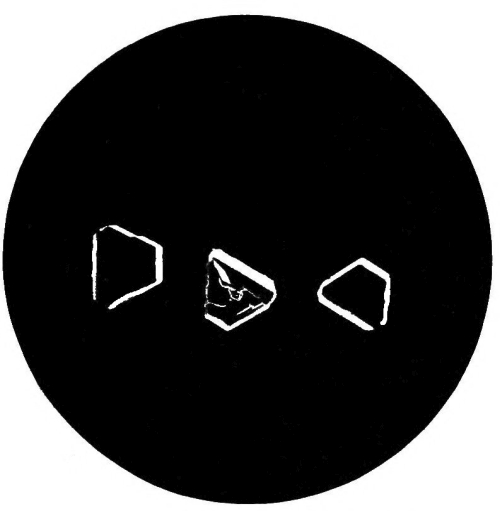
Fig. 33.—Photo-micrograph
of crystals
of tartarated antimony, × 50.
(R. J. M. Buchanan.)
Fig. 33.—Photo-micrograph of tartarated antimony crystals, × 50.
(R. J. M. Buchanan.)
Chronic.—The symptoms which mark the chronic form of poisoning differ chiefly in being less intense and less rapid than in the acute. Chronic poisoning by small repeated doses is that form of poisoning which appears most in vogue of late years—as certain diseases, enteritis, etc., can be simulated by the administration of repeated small doses. The unfortunate victim complains of constant nausea and [Pg 294] retching, with great depression. Food is objected to, as it only increases the vomiting. The matters vomited are at first merely mucus, but after a time they become mixed with bile. Each time the poison is repeated, the symptoms become aggravated. Emaciation gradually sets in, and the person dies from complete exhaustion, or from the effects of a larger dose than usual. Chronic poisoning has given rise to several errors in diagnosis, and the histories of recorded cases should put medical men on their guard. In all doubtful cases examine the urine.
Chronic.—The symptoms that characterize chronic poisoning are mainly less severe and develop more slowly than those of acute poisoning. Chronic poisoning from small, repeated doses has become more common in recent years, as it can mimic certain diseases like enteritis. The unfortunate victim experiences constant nausea and retching, along with significant depression. Food is typically rejected, as it only worsens the vomiting. At first, the vomit consists mainly of mucus, but over time it starts to include bile. Each time the poison is administered, the symptoms get worse. Gradual weight loss occurs, and the individual may die from total exhaustion or from receiving a larger dose than usual. Chronic poisoning has led to several diagnostic errors, and the documented cases should alert medical professionals. In any uncertain cases, examine the urine.
Post-mortem Appearances.—The mucous membrane of the throat, gullet, and stomach is inflamed, and in some places softened and corroded. Aphthous-looking spots are not infrequently found on the mucous membrane of the stomach, and these may also be observed on the throat and on the small intestines. The liver has been found in some cases of chronic poisoning, where the fatal termination has been for some time retarded, enlarged, and its structure so soft as to be easily broken down. Fatty degeneration of the internal organs has been found after protracted fatal administration of the drug. It is stated that in Brunswick the fatty livers of the geese are produced by the judicious administration of antimony. The appearances above detailed may be more or less absent or present, according to the time that may have elapsed from the swallowing of the poison to the time at which death has occurred.
Post-mortem Appearances.—The mucous membrane of the throat, esophagus, and stomach is inflamed, and in some areas, it is softened and damaged. Aphthous-looking spots are often found on the mucous membrane of the stomach, and these can also appear on the throat and small intestines. In some cases of chronic poisoning, where death has been delayed for some time, the liver is enlarged and its structure so soft that it can be easily broken down. Fatty degeneration of the internal organs has been observed following prolonged fatal doses of the drug. It is reported that in Brunswick, the fatty livers of geese are produced by carefully administering antimony. The conditions described above may vary in presence or absence, depending on the time that has passed from the ingestion of the poison to the point of death.
At the post-mortem examination on the body of Mr. Bravo, poisoned with tartar emetic in 1876, the mucous surfaces of the stomach and duodenum were found pale and yellowish. Ulcers were present in the cæcum, and the rest of the large intestine blood-stained, but not ulcerated. Stevenson records the post-mortem appearances of the bodies of three women poisoned by tartar emetic (B. M. J., 1903, vol. i. p. 873). They are of peculiar interest in reference to the preservative action of antimony upon the bodies of those poisoned by it. In the case of M. E. Marsh, upon whose body the examination was made eight days after death, there was no odour of putrefaction, the bowels were in a condition of acute catarrh and streaky congestion without ulceration.
At the post-mortem examination of Mr. Bravo, who was poisoned with tartar emetic in 1876, the mucous surfaces of the stomach and duodenum were found to be pale and yellowish. There were ulcers in the cæcum, and the rest of the large intestine was blood-stained, but not ulcerated. Stevenson documents the post-mortem findings of three women who were poisoned by tartar emetic (B. M. J., 1903, vol. i. p. 873). These cases are particularly interesting regarding the preservative effect of antimony on the bodies of those poisoned by it. In the case of M. E. Marsh, whose body was examined eight days after death, there was no smell of decay, and the intestines were in a state of acute catarrh and streaky congestion without ulceration.
The body of Bessie Taylor had been buried for twenty-one months; ordinary putrefactive changes were absent, and with the exception of the integuments, it was in a remarkably good state of preservation. The alimentary canal showed acute non-ulcerative gastro-enteritis, the stomach and duodenum were of a cinnabar-red colour, the jejunum also in patches. The ileum was covered with orange-red mucus in its lower portion, and there was an orange-coloured patch twelve inches above the ileo-cæcal valve. The patch on analysis was proved to contain antimonious sulphide. Similar orange patches were present in the colon and upper portion of the rectum. There were no ulcerations, but the mucous membrane of the intestines was of a dull cinnabar-red colour.
The body of Bessie Taylor had been buried for twenty-one months; typical decomposition was absent, and aside from the skin, it was in surprisingly good condition. The digestive tract showed acute non-ulcerative gastroenteritis, with the stomach and duodenum having a bright red color, and the jejunum also displaying patches of the same hue. The ileum was coated with orange-red mucus in its lower section, and there was an orange patch twelve inches above the ileocecal valve. Analysis revealed that the patch contained antimonious sulfide. Similar orange patches were found in the colon and upper rectum. There were no ulcers, but the mucous membrane of the intestines was a dull red color.
Elimination of Antimony from the System
Getting Rid of Antimony from the Body
Antimony, taken in a large dose, or in small doses frequently repeated, appears to be rapidly absorbed, and then eliminated from the system by the kidneys. Dating from the time at which the poison was swallowed, it will be found in the organs of the body in the following order:—
Antimony, when taken in a large dose, or in smaller doses taken repeatedly, seems to be quickly absorbed and then removed from the body by the kidneys. From the moment the poison is ingested, it can be found in the body's organs in the following order:—
1. Stomach and bowels, but slightly in the liver.
1. Stomach and intestines, but a little in the liver.
2. Absent from the stomach, but present in the liver, spleen, and kidneys—traces in the blood.
2. Not in the stomach, but found in the liver, spleen, and kidneys—traces in the blood.
3. Present in the fat and bones, with traces in the liver, fæces, and urine.
3. Present in the fat and bones, with traces in the liver, feces, and urine.
4. The period required for its complete elimination from the vital organs varies from fifteen to thirty days.
4. It takes about fifteen to thirty days for it to be completely eliminated from the vital organs.
In other words, the presence of antimony in the stomach and intestines points to the recent administration of the poison; and its absence from those organs, and presence in the others above mentioned, to a more remote period of administration. It has been suggested that in some cases the poison may be eliminated by the mucous membrane of the stomach. This assumption has been proved to be correct, for it has been shown that antimony may be found in the stomach after the inhalation of antimoniuretted hydrogen.
In other words, finding antimony in the stomach and intestines indicates that the poison was ingested recently, while its absence from those organs and presence in the others mentioned suggests a longer period since it was taken. Some have proposed that in certain cases, the poison can be removed by the mucous membrane of the stomach. This idea has been validated, as it has been demonstrated that antimony can still be present in the stomach after inhaling antimoniuretted hydrogen.
Fatal Dose.—It is impossible to state with certainty the exact amount of antimony—tartar emetic—which may prove fatal, as recoveries have taken place even after an ounce had been taken. Large doses are uncertain in their effects, as the severe vomiting which they produce generally helps to get rid of the poison. In small doses, death may result from the depressing action which it exerts over the heart.
Fatal Dose.—It's impossible to determine with certainty the exact amount of antimony—tartar emetic—that could be fatal, as people have recovered even after taking an ounce. Large doses have unpredictable effects, as the intense vomiting they cause usually helps eliminate the poison. In smaller doses, death can occur due to its depressing effect on the heart.
Fatal Period.—From a few hours to several weeks, and even months.
Fatal Period.—From a few hours to several weeks, and even months.
Treatment.—Promote vomiting by the administration of warm water, or warm greasy water, or the stomach may be washed out with a syphon tube, unless the chloride of antimony is the poison, and then give tannic acid in drachm doses in warm water, or any vegetable infusion containing tannin—viz., tea, oak bark, or cinchona bark. Demulcent drinks may be administered, and warmth applied. Opium may be given to relieve pain, and stimulants for the depression.
Treatment.—Induce vomiting by giving warm water or warm oily water, or the stomach can be flushed with a siphon tube, unless the poison is chloride of antimony, in which case administer tannic acid in drachm doses in warm water, or any herbal infusion containing tannin—like tea, oak bark, or cinchona bark. You can give soothing drinks and apply warmth. Opium may be used to ease pain, and stimulants for the low energy.
The Detection of Antimony
Antimony Detection
Prepare the solutions of the liver and other solid organs, and also the contents of the stomach, as described under the detection of arsenic, using tartaric acid instead of hydrochloric acid. Through a portion of one of the solutions, obtained by filtration or dialysis, pass a current of sulphuretted hydrogen, which will produce, if antimony be present, an orange-coloured precipitate of the sulphide of antimony. The precipitated sulphide is dissolved by hot hydrochloric acid with the evolution of sulphuretted hydrogen; and if the resulting solution [Pg 296] be poured into water, a white precipitate is formed of oxychloride of antimony, soluble in tartaric acid. Chloride of bismuth is precipitated when poured into water, but the precipitate is turned black by sulphide of ammonium, the antimonial orange-red; the precipitate of bismuth is not soluble in tartaric acid, the antimonial is soluble.
Prepare solutions from the liver and other solid organs, as well as the contents of the stomach, following the detection method for arsenic, but use tartaric acid instead of hydrochloric acid. Pass a stream of hydrogen sulfide through a portion of one of the filtered or dialyzed solutions. If antimony is present, this will create an orange-colored precipitate of antimony sulfide. The sulfide can be dissolved in hot hydrochloric acid, releasing hydrogen sulfide gas. If you then pour the resulting solution into water, a white precipitate of antimony oxychloride will form, which is soluble in tartaric acid. When chloride of bismuth is poured into water, it precipitates, but this precipitate turns black with ammonium sulfide, while the antimony precipitate remains orange-red. The bismuth precipitate is not soluble in tartaric acid, but the antimony one is.
Marsh‘s and Reinsch‘s processes may also be used for the detection of antimony. The former is, however, open to the objection that antimony, when present in any quantity, rapidly precipitates on the zinc in the form of a flocculent black deposit, while the issuing gas is found to contain only traces of the metal.
Marsh's and Reinsch's methods can also be used to detect antimony. However, the first method has the drawback that when antimony is present in any amount, it quickly forms a flaky black deposit on the zinc, while the gas that comes off contains only small amounts of the metal.
Reinsch‘s process is, however, very delicate, and its application is in every respect similar to that in use for the detection of arsenic. The acid liquid should, however, be boiled down to a small bulk with the copper, before a conclusion is drawn as to the entire absence of the metal.
Reinsch's process is very sensitive, and its application is similar in every way to the method used for detecting arsenic. However, the acid solution should be boiled down to a small volume with the copper before concluding that the metal is completely absent.
Table giving the Characteristic Reactions
of Antimonial and Arsenical Deposits
on Copper.
Table showing the Characteristic Reactions
of Antimonial and Arsenical Deposits
on Copper.
| Antimony. | Arsenic. | |
|---|---|---|
| The colour of the | Lustrous, with a violet hue. | Dark steel-grey colour, |
| deposit on copper | and lustrous. | |
| by Reinsch‘s | ||
| process is— | ||
| The coated copper | No effect, or only a | Well-marked sublimate |
| heated in the end | trifling white sublimate, | of octahedral crystals; |
| of a small tube. | non-crystalline, | is readily volatile. |
| non-volatile. If the | ||
| sublimate be dissolved | ||
| in solution of tartaric | ||
| acid and sulphuretted | ||
| hydrogen passed | ||
| through the solution, | ||
| the orange antimonious | ||
| sulphide is thrown down. |
It may be noted that mercury likewise yields a deposit on copper with Reinsch‘s process; but the coating is in this case either of a grey colour or white, and silvery on the application of friction. When the coated copper is heated in a glass tube, there is a sublimate of metallic mercury readily aggregating into globules on being rubbed with a glass rod. If the deposit is trifling in quantity, a magnifying-glass should be used to identify the metallic globules. This test at once distinguishes a deposit on copper due to mercury from that produced under similar conditions by arsenic or antimony.
It’s worth noting that mercury also leaves a deposit on copper using Reinsch’s process; however, the coating is either gray or white, and turns silvery when rubbed. When the coated copper is heated in a glass tube, metallic mercury sublimes and easily aggregates into tiny droplets when rubbed with a glass rod. If the deposit is small, a magnifying glass should be used to see the metallic droplets. This test quickly distinguishes a deposit on copper caused by mercury from one created under similar conditions by arsenic or antimony.
Recapitulation of the
Leading Facts
with regard to Poisoning
with Antimony.
Summary of the Key Facts
About Poisoning
with Antimony.
MERCURY
Metallic mercury possesses no toxicological interest, as it appears to be almost inert, even in very large doses. If applied to the skin in a finely-divided state, as in mercurial ointment, or internally, as blue pill, its toxic effects may be produced. The vapour given off from the metal is highly poisonous, producing salivation, emaciation, and death. A singular accident of poisoning by mercurial vapour occurred on board H.M.S. Triumph in 1810, owing to the bursting of bladders containing large quantities of the metal; in three weeks 200 men were affected with salivation, etc., nearly all the cattle on board died, as well as the mice, a dog, and a canary-bird.
Metallic mercury isn’t considered toxic, as it seems to be almost harmless, even in large amounts. However, when it’s applied to the skin in a finely ground form, like in mercurial ointment, or taken internally, as with blue pill, it can be toxic. The vapor released from the metal is very dangerous, causing excessive saliva production, weight loss, and even death. A notable incident of mercury vapor poisoning happened on H.M.S. Triumph in 1810 when bladders containing large amounts of the metal burst; in just three weeks, 200 men showed symptoms like excessive salivation, and nearly all the cattle on board died, along with a dog, a canary, and some mice.
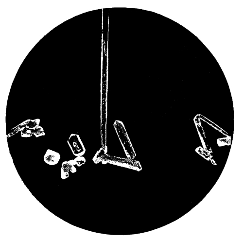
Fig. 34.—Photo-micrograph
of crystals of corrosive sublimate, × 50.
(R. J. M. Buchanan.)
Fig. 34.—Photo-micrograph of crystals of corrosive sublimate, × 50.
(R. J. M. Buchanan.)
Corrosive Sublimate
Corrosive Sublimate
This is the most important of the preparations of mercury. It occurs either in crystalline masses of prismatic crystals or as a white powder. It is now known among chemists as the perchloride, though it is frequently spoken of as the bichloride, chloride, and oxymuriate of mercury. It has a powerful metallic and styptic taste, and is soluble in about sixteen parts of cold water and three of boiling water. Alcohol and ether readily dissolve it, the latter having the power of abstracting it from its solution in water. This property of ether is of importance as a means of separating corrosive sublimate from its solution in other liquids. It is important to remember that corrosive sublimate is soluble in alcohol (R. v. Walsh). The liquor hydrargyri perchloridi of the Pharmacopœia contains half a grain [Pg 299] of the salt to a fluid ounce of water. Half a grain of the muriate of ammonia is added to increase the solubility of the mercurial salt. Applied externally to the unbroken skin, corrosive sublimate has caused death in several cases, the symptoms being almost identical with those which follow the entrance of the poison into the stomach. Toxic symptoms have followed intra-uterine or vaginal injections of solution of perchloride of mercury, also when it has been used to wash out abscess cavities. Acute poisoning, in some cases fatal, has resulted from the external application of corrosive sublimate to ulcers and tumours.
This is the most important preparation of mercury. It can appear as crystalline masses of prismatic crystals or as a white powder. Chemists now refer to it as perchloride, although it’s often called bichloride, chloride, or oxymuriate of mercury. It has a strong metallic and styptic taste and is soluble in about sixteen parts of cold water and three parts of boiling water. Alcohol and ether dissolve it easily, with ether being especially effective at pulling it out of its water solution. This characteristic of ether is significant for separating corrosive sublimate from its solution in other liquids. It's crucial to note that corrosive sublimate is also soluble in alcohol (R. v. Walsh). The liquor hydrargyri perchloridi from the Pharmacopœia contains half a grain of the salt per fluid ounce of water. Half a grain of ammonium chloride is added to enhance the solubility of the mercurial salt. When applied externally to intact skin, corrosive sublimate has caused death in several instances, with symptoms nearly identical to those that occur when the poison enters the stomach. Toxic symptoms have also followed intra-uterine or vaginal injections of perchloride of mercury solution, as well as when used to cleanse abscess cavities. Acute poisoning, sometimes fatal, has resulted from the external application of corrosive sublimate to ulcers and tumors.
Symptoms of Poisoning by
Corrosive Sublimate
Symptoms of Corrosive Sublimate Poisoning
Acute.—The symptoms come on almost immediately the poison is swallowed. A strong metallic coppery taste in the mouth is experienced, and a choking sensation in the throat. Pain of a burning character is felt, extending from the mouth to the stomach, followed by nausea and vomiting of stringent mucus, more or less tinged with blood, and violent purging, the evacuations being also mixed with blood and mucus. The pulse is feeble, quick, and irregular; the countenance flushed or pale, and the tongue white and shrivelled. This appearance of the tongue is not present in all cases. Broncho pneumonia may occur. The skin is cold and clammy, and the functions of the kidneys are altered and albuminuria present, there being in many cases complete suppression of urine. As is the case with other irritant poisons, the symptoms and effects produced admit of considerable variation. Thus, there may be no pain in the stomach, and no purging. Salivation is present in some cases, but chiefly in those in whom the fatal termination is somewhat prolonged. This sign is not infrequently absent. Poisoning with corrosive sublimate differs from arsenical poisoning in the following particulars:—Corrosive sublimate has a distinct metallic taste, arsenic is almost tasteless; the symptoms in the former supervene immediately the poison is swallowed, in the latter there is a short delay. The discharges in corrosive sublimate are more frequently bloody than in arsenic poisoning.
Acute.—The symptoms appear almost right after the poison is swallowed. A strong metallic, coppery taste is felt in the mouth, along with a choking sensation in the throat. A burning pain extends from the mouth to the stomach, followed by nausea and vomiting of thick mucus, which is sometimes mixed with blood, along with severe diarrhea that also contains blood and mucus. The pulse is weak, rapid, and irregular; the face may be flushed or pale, and the tongue is white and shriveled. This appearance of the tongue doesn’t occur in every case. Bronchopneumonia may develop. The skin feels cold and clammy, and kidney function is altered, often showing albuminuria, with many cases experiencing complete suppression of urine. As with other irritant poisons, the symptoms and effects can vary significantly. For instance, there may be no stomach pain or diarrhea. Salivation occurs in some cases, but mainly in those where the fatal outcome is somewhat delayed. This sign is often absent. Poisoning from corrosive sublimate is different from arsenic poisoning in several ways: corrosive sublimate has a distinct metallic taste, while arsenic is nearly tasteless; symptoms from the former appear immediately after swallowing, while there’s a brief delay with the latter. Discharges from corrosive sublimate are more likely to be bloody than those from arsenic poisoning.
Chronic.—The symptoms present in this form of poisoning are modified by the size of the dose, and the interval allowed to elapse between each dose. Nausea, followed by occasional vomiting, and pains in the stomach, are complained of by the patient. There is general constitutional disturbance, with anæmia and cachexia, and consequent mental depression. Salivation, as might be expected, is a more prominent symptom than in acute poisoning; but the salivation may be intermittent—that is, it may cease and then reappear, even after the lapse of months, without an additional dose of mercury having been given in the interval. Salivation may also come on in the course of certain diseases, attacking the salivary glands, and it may also be produced by other causes—pregnancy, etc. The glands of the mouth become swollen and painful, the gums tender, and the teeth loosened fall out of the mouth. Periostitis of the jaw may occur. The breath has [Pg 300] a peculiar, offensive smell, the bowels are irritable, and diarrhœa is not infrequently present. It must be borne in mind that in certain diseases—granular disease of the kidney—the smallest dose of any mercurial preparation may produce profuse ptyalism. And the toxicologist must be careful not to mistake the affection known as cancrum oris, or “the canker,” most common in delicate, ill-fed children and adults, for the effects of mercury. The nervous system is more or less affected, neuralgic pains and mercurial tremors being present in many cases. The tremors commence in the tongue and face and spread to the arms and legs—they are similar to those of paralysis agitans; at first they are invoked by exertion, finally they become continuous; they cease during sleep. Paralysis may also occur, especially in those exposed to the vapour of mercury. Habit appears to exert some influence on the action of corrosive sublimate, if we may accept the story of the old man of Constantinople, who for thirty years took large doses till his daily allowance was a drachm, and then died at the respectable age of one hundred years.
Chronic.—The symptoms of this type of poisoning vary depending on the dose size and the time between doses. Patients often report nausea, occasional vomiting, and stomach pains. There is a general disruption of health, along with anemia and severe weakness, leading to mental depression. Salivation is a more noticeable symptom compared to acute poisoning; however, it can be intermittent—it may stop and then start again, even after months without an additional mercury dose. Salivation can also occur during certain illnesses, affecting the salivary glands, and can be caused by other factors, like pregnancy. The mouth glands become swollen and painful, the gums are tender, and loose teeth may fall out. There may be inflammation of the jawbone. The breath has a distinctive, foul odor, the bowels are overly sensitive, and diarrhea is often present. It's important to remember that in certain conditions—like kidney disease—even the smallest dose of mercury can cause excessive salivation. A toxicologist should be careful not to confuse the condition known as cancrum oris, or “the canker,” which is most common in frail, undernourished children and adults, with the effects of mercury. The nervous system can also be affected, with neuralgic pain and mercury-induced tremors appearing in many patients. These tremors begin in the tongue and face and spread to the arms and legs, resembling those seen in Parkinson’s disease; initially, they are triggered by movement but eventually become constant, stopping only during sleep. Paralysis can also occur, especially in those exposed to mercury vapor. Habit seems to play a role in how corrosive sublimate acts, as suggested by the story of an old man from Constantinople who took large doses for thirty years until his daily amount reached a drachm, and then lived to the respectable age of one hundred years.
Post-mortem Appearances.—The morbid appearances are chiefly confined, as is the case with arsenic, to the stomach and bowels; but the corrosive action of the mercurial sublimate is more marked. Inflammation more or less intense is always present in the stomach, the mucous membrane of which may be found of a slate-grey colour, corroded, and so soft as to scarcely admit of the removal of the organ without tearing it. The cæcum and rectum are also sometimes found inflamed, and the mucous membrane softened. Perforation of the stomach is very rare, only one case having been recorded in which this was present. The mouth, throat, and gullet may also present signs of the action of the poison similar to those just described as seen in the stomach. Patches of pneumonia are present in the lungs of some cases, and the kidneys show coagulative and desquamative nephritis.
Post-mortem Appearances.—The signs of illness are mainly found, as in the case of arsenic, in the stomach and intestines; however, the corrosive effects of mercurial sublimate are more pronounced. There is always some level of inflammation in the stomach, which may appear slate-grey, corroded, and so fragile that it can barely be removed without tearing. The cecum and rectum are sometimes also inflamed, with softened mucous membranes. Stomach perforation is very rare, with only one documented case. The mouth, throat, and esophagus may also show signs of the poison’s effects similar to those seen in the stomach. Some cases may have patches of pneumonia in the lungs, and the kidneys show signs of coagulative and desquamative nephritis.
Fatal Dose.—The smallest dose was two grains in the case of a child, but the exact amount to cause death in an adult has not been accurately determined. Recovery has taken place after one hundred grains has been taken.
Fatal Dose.—The smallest dose was two grains for a child, but the exact amount needed to cause death in an adult hasn't been clearly defined. Recovery has occurred after taking one hundred grains.
Fatal Period.—From half an hour to five days. No exact time can be stated. In one case death took place on the twelfth day after swallowing seventy grains of the perchloride.
Fatal Period.—From half an hour to five days. There's no specific time that can be given. In one instance, death occurred on the twelfth day after ingesting seventy grains of the perchloride.
Treatment.—Vomiting, if present, must be encouraged; if absent, it must be produced by emetics—zinc sulphate or cupric sulphate, followed by copious draughts of warm water. The hypodermic injection of ¹/₂₀ to ⅒ grain of apomorphine may also be used to produce vomiting. Albumen, the white of egg, or vegetable gluten produced from flour by washing it in a muslin bag, should be given. The albuminate of mercury so formed should be quickly removed by an emetic or lavage by means of a soft tube and funnel in mild cases, as it is soluble in excess of albumen, and may be digested or absorbed. The rapid removal of the poison from the stomach, however, is the end to which all our exertions must tend. The forcible use of the stomach pump should not be employed [Pg 301] if it can possibly be avoided, as it may greatly injure the softened mucous membrane of the gullet and stomach. Opium may be given to relieve pain, and opium enemata to relieve purging and tenesmus.
Treatment.—If vomiting is present, it should be encouraged; if not, it must be induced using emetics like zinc sulfate or copper sulfate, followed by large amounts of warm water. A hypodermic injection of 1/20 to 1/10 grain of apomorphine can also be used to induce vomiting. Albumen, which is egg white, or vegetable gluten obtained from flour by washing it in a muslin bag should be administered. The albuminate of mercury that forms should be quickly eliminated using an emetic or gastric lavage with a soft tube and funnel in mild cases, as it dissolves in excess albumen and may be digested or absorbed. The main goal of our efforts must be to remove the poison from the stomach as quickly as possible. The forcible use of a stomach pump should be avoided whenever possible, as it can severely damage the softened mucous membrane of the esophagus and stomach. Opium may be given to relieve pain, and opium enemas can help alleviate diarrhea and tenesmus.
Calomel
Calomel
Calomel, or the subchloride of mercury, is not used as a poison. In large doses it may act as an irritant poison, and death has not infrequently occurred even from comparatively small doses. Profuse salivation and gangrene of the mouth have resulted from its use, and cases are recorded of death resulting from these. In many cases idiosyncrasy appears to modify, more or less, the action of this preparation of mercury. The poisonous effect of calomel has been attributed to—(1) Adulteration with corrosive sublimate. (2) Conversion of the calomel into corrosive sublimate by the action of the hydrochloric acid of the gastric juice.
Calomel, or mercury(I) chloride, isn't used as a poison. In large amounts, it can act as an irritant poison, and deaths have often occurred even from relatively small doses. Excessive salivation and oral gangrene have resulted from its use, and there are recorded cases of death from these effects. In many instances, individual reactions seem to alter the effects of this mercury compound. The toxic effects of calomel have been linked to: (1) Adulteration with corrosive sublimate. (2) The transformation of calomel into corrosive sublimate by the hydrochloric acid in gastric juice.
N.B.—The free acid of the gastric juice is probably in too small a quantity to materially alter the composition of the calomel.
N.B.—The free acid in gastric juice is likely present in such small amounts that it doesn't significantly change the composition of the calomel.
Ammonio-Chloride of Mercury
Mercury Ammonium Chloride
White precipitate may, if taken in large doses, produce alarming effects, but it cannot be regarded as an active poison. Pavy‘s experiments on rabbits indicate that it is a more powerful poison than was formerly thought to be the case. Its action is that of an irritant, accompanied with, in some cases, severe salivation.
White precipitate can cause concerning effects if taken in large doses, but it shouldn't be considered a strong poison. Pavy's experiments on rabbits suggest that it is a more potent toxin than previously believed. Its effect is that of an irritant, often leading to severe salivation in some cases.
Red Precipitate
Red Precipitate
Red oxide of mercury possesses poisonous properties, but it is seldom employed as a poison. The symptoms most frequently present are vomiting, coldness of the surface of the body, stupor, pain in the abdomen, and cramps of the muscles of the lower extremities. The vomited matters are generally mixed with some of the red oxide.
Red oxide of mercury is toxic, but it's rarely used as a poison. The most common symptoms are vomiting, cold skin, lethargy, abdominal pain, and cramps in the leg muscles. The vomit usually contains some of the red oxide.
Cinnabar. Vermilion
Cinnabar. Vermilion
A compound of sulphur and mercury in the form of a dark red crystalline mass is known as cinnabar; and to the same substance reduced to a fine powder the name vermilion has been given. It is used as a red pigment. It can scarcely be considered as a poison, Orfila asserting that it is not poisonous. The vapour of this substance appears, however, to be capable of producing severe symptoms, and in one case, profuse salivation resulted from the application of the vapour to the body.
A compound of sulfur and mercury that appears as a dark red crystalline mass is called cinnabar; when it's ground into a fine powder, it’s referred to as vermilion. This substance is used as a red pigment. It’s not really viewed as a poison, with Orfila claiming that it isn’t toxic. However, the vapor from this substance can lead to serious symptoms, and in one instance, excessive salivation occurred when the vapor was applied to the body.
Cyanide of Mercury
Mercury Cyanide
This substance, though an active poison little inferior to corrosive sublimate, is seldom used as such, probably from its being better known to chemists than to the general public. It differs from corrosive [Pg 302] sublimate in having no local corrosive action. It has been supposed, but proof is wanting, that its injurious effects are due to its decomposition by the acids of the stomach and the formation of prussic acid. Death has occurred in nine days from a dose of ten grains. It acts as an irritant. The sulphocyanide of mercury is used in the manufacture of the toy known as Pharaoh‘s serpents.
This substance, while a potent poison almost as dangerous as corrosive sublimate, is rarely used in that way, likely because it's more familiar to chemists than the general public. Unlike corrosive sublimate, it doesn't cause local corrosion. It has been suggested, though there’s no solid proof, that its harmful effects result from its breakdown by stomach acids that create prussic acid. Death can occur within nine days from a ten-grain dose. It works as an irritant. Mercury thiocyanate is used in making the toy known as Pharaoh’s serpents.
Turbith Mineral
Turbith Mineral
A powerful irritant poison, but seldom used. A drachm has caused death in a boy sixteen years of age. Coldness of the surface, burning pain in the stomach and bowels, with other symptoms of irritant poisoning, were present. After death, the mucous membranes of the throat, stomach, and bowels were found considerably inflamed.
A strong irritant poison, but rarely used. A drachm has caused the death of a sixteen-year-old boy. He experienced coldness on the skin, burning pain in his stomach and intestines, along with other signs of irritant poisoning. After death, the mucous membranes in his throat, stomach, and intestines were found to be significantly inflamed.
Nitrates of Mercury
Mercury Nitrates
These substances—the nitrate and sub-nitrate—are used in the arts for various purposes. They act as powerful irritant poisons, with symptoms and post-mortem appearances not unlike those before described when speaking of corrosive sublimate and other irritants.
These substances—the nitrate and sub-nitrate—are used in the arts for various purposes. They act as strong irritant poisons, with symptoms and post-mortem appearances similar to those previously described when discussing corrosive sublimate and other irritants.
Chemical Analysis:
Chemical Analysis:
Table showing the Reaction of Mercuric and
Mercurous Salts with Reagents
Table showing the Reaction of Mercuric and
Mercurous Salts with Reagents
Detection of Mercury in the Tissues and
in the Contents of the Stomach.
Finding Mercury in the Tissues and
in the Stomach Contents.
Mercury is particularly liable to be absorbed by the tissues; it also readily combines with various organic substances, gelatine, albumin, &c.
Mercury tends to be absorbed by tissues easily; it also readily combines with different organic substances, like gelatin, albumin, etc.

Fig. 35.—Photo-micrograph
of globules of
mercury obtained by Reinsch‘s process, × 50.
(R. J. M. Buchanan.)
Fig. 35.—Photomicrograph of mercury globules obtained by Reinsch's process, × 50.
(R. J. M. Buchanan.)
A.—If the contents of the stomach are under examination, they should be diluted with distilled water, filtered, and the residue pressed and reserved for further examination.
A.—If the stomach contents are being examined, they should be diluted with distilled water, filtered, and the residue pressed and saved for further analysis.
The liquid thus obtained may be concentrated, and, while still warm, slightly acidified with hydrochloric acid. A slip of zinc foil, with a piece of gold foil twisted round it, is then introduced. If mercury be present, the gold will, sooner or later, lose its yellow colour, and its surface become white and silvery, while the zinc is wholly or partially dissolved. The gold being removed, separated from the zinc, washed first with water and then with ether, is divided into two equal parts. One half may be heated in a reduction tube, when it will yield a sublimate of metallic mercury, identified by the spherical form of the globules under a magnifying-glass, and their metallic reflection and complete opacity. The other half of the gold may be treated with nitric acid and heated, which will dissolve off the mercury. The resulting solution, after expelling the excess of acid by evaporation, will give a scarlet precipitate with iodide of potassium soluble in excess; and, with protochloride of tin, a black precipitate of metallic mercury.
The liquid obtained can be concentrated and, while still warm, lightly acidified with hydrochloric acid. A strip of zinc foil, with a piece of gold foil wrapped around it, is then added. If mercury is present, the gold will eventually lose its yellow color, turning white and silvery, while the zinc is entirely or partially dissolved. After removing the gold and separating it from the zinc, it is washed first with water and then with ether, and divided into two equal parts. One half can be heated in a reduction tube, yielding a sublimate of metallic mercury, identifiable by the spherical shape of the globules under a magnifying glass, along with their metallic shine and complete opacity. The other half of the gold can be treated with nitric acid and heated, which will dissolve the mercury. The resulting solution, after removing the excess acid through evaporation, will produce a scarlet precipitate with iodide of potassium that is soluble in excess; and, with protochloride of tin, a black precipitate of metallic mercury.
B.—For the detection of mercury in the insoluble form, the [Pg 304] residue from A is dried; or, if the tissues are under examination, they should be finely divided, and freed from superfluous moisture. In either case, the substance is boiled in moderately strong nitric or hydrochloric acid (about one part of acid to four of water). After digestion for some time, the liquid is filtered, concentrated, and tested as in A. When there is reason to infer the presence of corrosive sublimate in considerable quantity in an organic liquid, advantage may be taken of the solubility of the salt in ether, and the power possessed by this liquid of abstracting it from its aqueous solutions. The liquid is agitated with an equal volume of ether, the ethereal solution poured off and allowed to spontaneously evaporate, when the corrosive sublimate will be left in white silky prisms, yielding all the characteristic reactions of the salt. In obscure cases of salivation, the saliva should be examined as follows: Take about two drachms of the saliva, acidulate with pure hydrochloric acid, and immerse in the mixture a very small piece of copper gauze attached to a platinum wire, and set aside in a warm place for some hours. If mercury be present, the copper will be covered with a white coating; this should be washed and heated in a reduction tube, when globules of mercury will be formed, and examined with a lens.
B.—To detect mercury in its insoluble form, the [Pg 304] residue from A should be dried; if examining tissues, they need to be finely chopped and moisture should be removed. In either case, the substance is boiled in moderately strong nitric or hydrochloric acid (about one part acid to four parts water). After digestion for a while, the liquid is filtered, concentrated, and tested as in A. If there's a reason to suspect a significant amount of corrosive sublimate in an organic liquid, take advantage of the salt's solubility in ether and its ability to extract it from aqueous solutions. Mix the liquid with an equal volume of ether, then pour off the ethereal solution and let it evaporate on its own; the corrosive sublimate will remain as white silky crystals, showing all the characteristic reactions of the salt. In unclear cases of salivation, saliva should be examined as follows: Take about two drachms of saliva, add pure hydrochloric acid, and immerse a small piece of copper gauze attached to platinum wire in the mixture, then place it in a warm area for several hours. If mercury is present, the copper will have a white coating; wash this off and heat it in a reduction tube, where it will produce mercury globules that can be examined with a lens.
C.—Mercury in solution may be detected by Reinsch‘s process. On boiling with pure copper foil after acidifying the solution with hydrochloric acid, the mercury is deposited on the copper in the metallic state, as a whitish silvery film, which can be polished to a silvery mirror surface. On washing the film in water, ether, and absolute alcohol and allowing it to dry, then subliming it in a reduction tube by the aid of heat, the mercury deposits on the cool part of the tube in the form of minute globules, which may be easily recognised through the microscope. If a small crystal of iodine be now placed in the tube and gently warmed so as to volatilise it, but not the mercury, and the tube be left to stand for a short time, the iodine combines with the mercury to form the scarlet iodide, the colour of which renders it easy of recognition.
C.—You can detect mercury in a solution using Reinsch's process. When you boil it with pure copper foil after adding hydrochloric acid to the solution, mercury gets deposited on the copper as a whitish-silvery film that can be polished to a shiny mirror finish. If you wash this film with water, ether, and absolute alcohol and let it dry, then heat it in a reduction tube, mercury will deposit as tiny globules on the cooler part of the tube, which can be easily seen under a microscope. If you then place a small crystal of iodine in the tube and gently warm it to vaporize the iodine without vaporizing the mercury, and let the tube sit for a bit, the iodine will react with the mercury to create scarlet iodide, which is easy to recognize due to its color.
According to Bonnewyn, the presence of an extremely small quantity of corrosive sublimate (¹/₅₀₀₀₀) in calomel may be detected by immersing a clean knife blade, moistened with alcohol or ether, in the suspected calomel. A black spot is formed on the steel very difficult of removal. No spot is formed when the calomel is pure.
According to Bonnewyn, you can detect the presence of a tiny amount of corrosive sublimate (¹/₅₀₀₀₀) in calomel by dipping a clean knife blade, moistened with alcohol or ether, into the suspected calomel. A black spot appears on the steel that is very hard to remove. If the calomel is pure, no spot will form.
LEAD
Metallic lead is not poisonous; but it appears probable that when it is acted upon by the acids of the intestinal secretions, it may become so changed as to produce unpleasant symptoms. Any salt of lead is poisonous when in a condition to be absorbed into the system.
Metallic lead isn't toxic, but it seems likely that when it interacts with the acids in intestinal secretions, it may change in a way that causes unpleasant symptoms. Any lead salt is toxic when it's in a form that can be absorbed into the body.
Sugar of Lead
Lead Sugar
Acetate of Lead. Subacetate. Goulard‘s Extract
Lead Acetate. Subacetate. Goulard's Extract
General Character.—The acetate of lead, better known as sugar of lead, is not unlike loaf-sugar in its general appearance. It is usually met with in the form of solid crystalline masses of a white or brownish-white colour. To the taste it is sweet, a metallic astringent taste being left in the mouth. Acetate of lead is soluble in water and in alcohol. The subacetate is a more active poison than the neutral acetate. Sugar of lead is popularly considered as an active poison, but this does not appear to be the case. Sir R. Christison gave eighteen grains daily in divided doses for eight or ten days with no other unpleasant symptoms than slight colicky pains in the abdomen. Lead is probably eliminated from the system by the urine, and also by the milk; but there is reason to believe that when once deposited in the body, some considerable time is required for its complete elimination. Dr. Wilson is of opinion that in chronic lead poisoning the lead is more largely deposited in the spleen than in any other organ of the body. This organ should therefore always be carefully examined in suspected cases of poisoning by this metal.
General Character.—Lead acetate, commonly known as sugar of lead, resembles loaf sugar in its overall appearance. It usually occurs in solid crystalline forms that are white or brownish-white. It has a sweet taste, leaving a metallic and astringent sensation in the mouth. Lead acetate is soluble in water and alcohol. Subacetate is a more potent poison than neutral acetate. Sugar of lead is often thought to be a strong poison, but this may not be accurate. Sir R. Christison administered eighteen grains daily in divided doses for eight to ten days without any other unpleasant effects besides slight abdominal cramps. Lead is likely expelled from the body through urine and milk; however, it seems that once deposited in the body, it takes a significant amount of time for complete removal. Dr. Wilson believes that in cases of chronic lead poisoning, lead accumulates more in the spleen than in any other organ. Therefore, this organ should always be thoroughly examined in suspected cases of lead poisoning.
Goulard‘s Extract is a solution of the subacetate of lead. It may be of a reddish colour, from the employment of common vinegar in the place of pure acetic acid in the manufacture.
Goulard's Extract is a solution of lead subacetate. It can have a reddish color due to using regular vinegar instead of pure acetic acid during its production.
Goulard‘s Lotion is the extract diluted with water.
Goulard's Lotion is the extract mixed with water.
White Lead
White Lead
White lead, carbonate of lead, ceruse, or kremser white, is used as a pigment. It is generally in the form of white, heavy chalky masses, insoluble in water, and, when taken in large doses, poisonous. It is this substance which, in the majority of cases, causes chronic lead poisoning, or painter‘s colic.
White lead, lead carbonate, ceruse, or kremser white, is used as a pigment. It typically appears as white, heavy chalky lumps that don't dissolve in water, and can be toxic when consumed in large quantities. This substance is what mostly leads to chronic lead poisoning, or painter’s colic.
The chloride and nitrate, the oxides, litharge and red lead, are all poisonous; but the sulphate, due probably to its insolubility, appears to be inert.
The chloride and nitrate, the oxides, litharge and red lead, are all toxic; however, the sulphate, likely because it doesn't dissolve, seems to be inactive.
Lead poisoning may result from—
Lead poisoning can result from—
1. Constant contact with lead and its salts in manufactories.
1. Ongoing exposure to lead and its compounds in factories.
2. Its use in the arts and as a pigment. The injurious effects of this substance are strikingly seen among painters, the makers of glazed cards, and the workmen engaged in preparing Brussels lace—this material being whitened by beating white lead into it. All thus employed are liable to suffer more or less from chronic poisoning.
2. Its use in the arts and as a pigment. The harmful effects of this substance are clearly seen among painters, card makers, and workers involved in making Brussels lace—this material is whitened by hammering white lead into it. Everyone who works with it is at risk of suffering from chronic poisoning to some degree.
3. Its application to the surface of the body in the form of ointment, plasters, cosmetics, and hair-dyes.
3. Its use on the skin as ointments, patches, cosmetics, and hair dyes.
4. Drinking water impregnated with lead, from being stored in leaden cisterns or conveyed in leaden pipes.
4. Drinking water contaminated with lead, due to being stored in lead cisterns or transported through lead pipes.
“The action of water upon lead is much modified by the presence of saline substances. It is increased by chlorides and nitrates, and diminished by carbonates, sulphates, and phosphates, and especially by carbonate of lime, which, held in solution by excess of carbonic acid, [Pg 306] is a frequent ingredient of spring and river water. But water highly charged with carbonic acid may become dangerously impregnated with lead, in the absence of any protecting salt, in consequence of its solvent power over carbonate of lead. In general, water which is not discoloured by sulphuretted hydrogen may be considered as free from lead; but there are few waters which have passed through leaden pipes, or have been retained in leaden cisterns, in which a minute analysis will not detect a trace of the metal; and were it not for the great convenience of lead, iron pipes and slate cisterns would, from a sanitary point of view, be in all cases preferable.
The way water reacts with lead changes significantly with the presence of salt substances. Chlorides and nitrates enhance this reaction, while carbonates, sulfates, and phosphates reduce it, especially carbonate of lime, which is often found in spring and river water, dissolved in excess carbonic acid. However, water that contains a lot of carbonic acid can become dangerously contaminated with lead if there are no protective salts, due to its ability to dissolve lead carbonate. Generally, water that isn’t discolored by hydrogen sulfide can be considered free of lead; yet, few waters that have flowed through lead pipes or been stored in lead cisterns won't show at least a trace of the metal upon careful analysis. If it weren’t for the convenience of lead, iron pipes and slate cisterns would be the better sanitary option in all cases. [Pg 306]
“Another case of contamination by lead may arise from electric action, as where iron, copper, or tin is in contact with or soldered into lead; and in these cases, owing to the action of alkaline bases as well as of acids upon the lead, danger may occur when it is thrown into an electro-negative as well as into an electro-positive state.
“Another instance of lead contamination can happen due to electrical activity, such as when iron, copper, or tin is touching or soldered to lead. In these situations, because of the effect of alkaline substances as well as acids on the lead, there can be risks when it's put into both an electro-negative and an electro-positive state.”
“Cisterns are sometimes corroded and their bottoms are perforated by pieces of mortar having dropped into them, the lime of which has caused the oxidation of the metal and a solution of the oxide.”
“Cisterns can sometimes get corroded, and their bottoms can have holes caused by chunks of mortar falling in. The lime from the mortar leads to the oxidation of the metal and dissolves the oxide.”
5. Lead may also find its way into the system by means of the food. Farinaceous foods, chocolate, and tea may become contaminated if lead wrappers be used; and confectionery from the use of lead chromate as a colouring agent. The use of leaden vessels in the manufacture of cider is attended with danger, and also the keeping of pickles in glazed earthenware jars. The celebrated “Devonshire Colic” was the result of cider-making in leaden vats. Beer may be contaminated with lead if allowed to stand in leaden pipes overnight. Rum has been known to have been dangerously impregnated with lead, leaden worms having been used attached to the stills. Many tobacconists are in the habit of using lead foil to wrap up their tobacco and snuff; this practice has resulted in several cases of chronic lead poisoning. Soda and Seltzer waters may contain lead when kept in syphons with leaden caps or valves.
5. Lead can also get into the system through food. Starchy foods, chocolate, and tea can become contaminated if lead wrappers are used; and candy can be affected by lead chromate as a coloring agent. Using lead vessels in cider production is risky, as is storing pickles in glazed earthenware jars. The infamous “Devonshire Colic” resulted from cider made in lead vats. Beer can become contaminated with lead if it sits in lead pipes overnight. Rum has been known to dangerously contain lead due to lead worms being used on the stills. Many tobacco shops often wrap their tobacco and snuff in lead foil, which has led to several cases of chronic lead poisoning. Soda and seltzer waters can contain lead when stored in siphons with lead caps or valves.
6. Acute and subacute cases of poisoning occur from the taking of lead in the form of diachylon pills to procure abortion.
6. Acute and subacute cases of poisoning happen from taking lead in the form of diachylon pills to induce abortion.
Symptoms of Poisoning by Lead
Symptoms of Lead Poisoning
Acute.—A metallic taste in the mouth, accompanied with dryness in the throat and intense thirst, is experienced by the patient soon after the poison is swallowed. In some cases, however, two or more hours may elapse before the effects of the poison begin to show themselves. Vomiting may or may not be present. Twisting colicky pains are felt in the abdomen, relieved in some cases by pressure. The paroxysms of pain may be separated by intervals of ease. The bowels are, as a rule, obstinately confined, and the fæces are of a dark colour, from the formation of the sulphuret of lead. The skin is cold, the pulse quick and weak, and there is considerable prostration of strength. In some cases the patient suffers from cramps of the calves of the legs, and sometimes, in protracted cases, paralysis of one or more of the extremities may supervene. The effect on the nervous [Pg 307] system, headache, temporary blindness, is marked by giddiness, and stupor, in some cases delirium even resembling acute mania, terminating in coma, or convulsions and death. Albuminuria may occur.
Acute.—The patient experiences a metallic taste in the mouth, along with a dry throat and intense thirst, soon after swallowing the poison. In some cases, though, two or more hours might pass before the poison's effects start to appear. Vomiting may or may not occur. Twisting, cramping pain is felt in the abdomen, which may be relieved by pressure in some instances. The pain episodes may be separated by periods of relief. Generally, the bowels are persistently constipated, and the stool is a dark color due to the formation of lead sulfide. The skin is cold, the pulse rapid and weak, and there is significant weakness. In some cases, the patient may experience calf cramps, and in prolonged cases, paralysis of one or more limbs might develop. The impact on the nervous [Pg 307] system includes headaches and temporary blindness, accompanied by dizziness and stupor; in some instances, delirium may even resemble acute mania, leading to coma, convulsions, and death. Albuminuria may occur.
Chronic.—This form of poisoning generally occurs among painters, manufacturers of white lead, pewterers, and others. The early symptoms are those of ordinary colic, only more severe. The patient generally complains, in the first instance, of feeling unwell, and of general debility. He then suffers from pain of a twisting, grinding nature, felt in the region of the navel, the abdomen being retracted and hard. The bowels are obstinately confined. The appetite becomes capricious, and may be entirely lost. The mouth is parched, the breath fœtid, the countenance sallow, the skin dry, and general emaciation sets in. A nasty sweetish metallic taste in the mouth is present in most cases. Not infrequently the subjects of lead poisoning experience a peculiar form of paralysis of the upper extremities, well known as “dropped hand.” It appears that this condition is the result of paralysis of the extensor muscles of the wrist, the long supinator escaping. In other cases the muscles of the shoulder girdle or pelvic girdle and legs may be affected. Acute general paralysis has occurred in workers with lead, and may prove fatal. Optic neuritis and optic atrophy occur in some cases. Persistent headache may be present in others. The muscles undergo a form of fatty degeneration. The lead appears to act primarily on the muscles, then on the nerves, and lastly on the nerve centres. The absorption of lead causes marked anæmia with degeneration of the red corpuscles, which show marked punctate basophilia. One other symptom of importance has yet to be noticed. The gums, at their margins where they join the teeth, present a well-marked blue line, absent where a tooth has been removed. This is not present in all cases, but it should be looked for. Chronic lead absorption produces cardiovascular changes with arterio-sclerosis, valvular degeneration, cardiac hypertrophy, and chronic renal changes with albuminuria. Closely associated with these conditions are saturnine arthritis and gout. The effects upon the vessels may predispose to cerebral hæmorrhage or lead encephalopathy. Cicconardi suggests as a method of diagnosis in lead poisoning, where the cause of the colic is uncertain, to paint the skin with a 6 per cent. solution of sodium sulphite. If lead be the cause the painted part will become darkened in colour.
Chronic.—This type of poisoning usually happens to painters, manufacturers of white lead, pewter workers, and others. The initial symptoms are similar to regular colic, but more intense. The patient typically starts by feeling unwell and generally weak. Then they experience a twisting, grinding pain in the belly area, with a retracted and hard abdomen. The bowels become stubbornly constipated. Their appetite may turn erratic, and they might completely lose it. The mouth feels dry, the breath is foul, the face looks pale, the skin is dry, and overall weight loss begins. Many people suffering from lead poisoning experience a strange sweet metallic taste in their mouth. It's also common for those affected to have a specific type of paralysis in their arms, known as “dropped hand.” This condition results from paralysis of the wrist extensors, while the long supinator muscle remains functional. In some cases, the muscles in the shoulder or pelvic areas and legs might also be impacted. Severe general paralysis can occur in lead workers and may be fatal. Optic neuritis and optic atrophy can happen in some cases. Persistent headaches may affect others. The muscles can undergo a kind of fatty degeneration. Lead appears to primarily impact the muscles, then the nerves, and finally the nerve centers. Lead absorption leads to significant anemia with degradation of the red blood cells, which show noticeable punctate basophilia. Another crucial symptom to note is that the gums, where they meet the teeth, show a distinct blue line, absent where a tooth has been taken out. This isn’t present in every case, but it should be checked for. Chronic lead absorption results in cardiovascular changes such as arteriosclerosis, valve degeneration, cardiac hypertrophy, and chronic kidney changes with albuminuria. Close to these conditions are saturnine arthritis and gout. The effects on the blood vessels may increase the risk of cerebral hemorrhage or lead encephalopathy. Cicconardi suggests a diagnostic method for lead poisoning, where the cause of colic is unclear, by applying a 6 percent sodium sulfite solution to the skin. If lead is the cause, the painted area will darken in color.
N.B.—The symptoms produced by white lead—carbonate of lead—are those of colica pictonum, or painter‘s colic, described under the head of Chronic Lead Poisoning.
N.B.—The symptoms caused by white lead—lead carbonate—are similar to those of colica pictonum or painter's colic, which are discussed in the section on Chronic Lead Poisoning.
Post-mortem Appearances.—In acute poisoning the mucous membrane of the stomach and intestines is inflamed, and is in some cases covered by layers of white or whitish-yellow mucus, more or less impregnated with the salt of lead swallowed. Corrosion of the mucous membrane may occur if the dose be large, and this condition is more frequently present when the neutral salt is taken.
Post-mortem Appearances.—In cases of acute poisoning, the lining of the stomach and intestines becomes inflamed and may be covered with layers of white or yellowish-white mucus, which is partially saturated with the lead salt that was ingested. If the dose is high, the mucous membrane can be damaged, and this situation is more commonly seen when the neutral salt is consumed.
Fatal Dose.—Sugar of lead is not an active poison, recovery having taken place after one ounce had been swallowed.
Fatal Dose.—Lead sugar isn't a strong poison; there have been recoveries even after swallowing one ounce.
Fatal Period.—Uncertain.
Critical Time.—Unclear.
Treatment.—The stomach should be emptied by means of the pump or syphon tube, followed by the free administration of the sulphates of soda and magnesia. The carbonates should not be given, the carbonate of lead being poisonous. Vomiting should be promoted, and a powerful cathartic administered. Albumen and milk should also be given, as these precipitate the oxide. In the chronic form of poisoning, the iodide of potash and aperients, notably the sulphate of magnesia, should be administered. Dixon Mann does not consider that iodide of potash is of any value as an eliminator of lead, as the latter forms a stable compound with the tissues. Sulphur baths are also useful in removing the lead from the system. Lately the galvanic bath has been tried with great success. By way of prophylaxis, it has been recommended that all those engaged in lead manufactories, or who are obliged to handle this metal frequently, should partake largely of lemonade made with sulphuric acid, should not take their meals in the factories, or without well washing the hands.
Treatment.—The stomach should be emptied using a pump or siphon tube, followed by the administration of sulfate of soda and magnesia. Carbonates should be avoided, as lead carbonate is toxic. Vomiting should be encouraged, and a strong laxative should be given. Albumen and milk should also be provided, as these help eliminate the oxide. In cases of chronic poisoning, potassium iodide and laxatives, particularly magnesium sulfate, should be administered. Dixon Mann believes that potassium iodide is not effective in removing lead since it forms a stable compound with body tissues. Sulfur baths are also helpful for removing lead from the body. Recently, the galvanic bath has been tested with great success. As a form of prophylaxis, it is recommended that anyone working in lead factories or frequently handling the metal should drink plenty of lemonade made with sulfuric acid, avoid eating meals in the factories, and wash their hands thoroughly.
Chemical Analysis.—When the solid acetate is heated on platinum foil, it melts, then solidifies, becomes dark in colour, and gives off fumes of acetic acid.
Chemical Analysis.—When the solid acetate is heated on platinum foil, it melts, then solidifies, turns dark in color, and releases fumes of acetic acid.
The following are the liquid tests for lead in solution:
The following are the tests for lead in a liquid solution:
1. Dilute sulphuric acid gives a white precipitate of the sulphate, which is insoluble in nitric, but soluble in hydrochloric acid, in excess of caustic potash solution, and in ammonium acetate solution.
1. Diluted sulfuric acid produces a white precipitate of sulfate, which doesn’t dissolve in nitric acid but does dissolve in hydrochloric acid, in excess caustic potash solution, and in ammonium acetate solution.
2. Solution of potassium iodide gives a yellow precipitate, soluble in boiling water and caustic potash solution.
2. A solution of potassium iodide produces a yellow precipitate that dissolves in boiling water and caustic potash solution.
3. Sulphuretted hydrogen, or ammonium sulphide, gives a black precipitate.
3. Hydrogen sulfide, or ammonium sulfide, forms a black solid.
4. Potassium bichromate a yellow precipitate.
4. Potassium bichromate is a yellow solid that forms as a precipitate.
Detection of Lead in Organic Mixtures
Detection of Lead in Organic Mixtures
The contents of the stomach or vomited matters must be diluted with water and filtered. The residue left on the filter, washed with distilled water, should be set aside for further examination; the filtrate and washings acidified with nitric acid. A current of sulphuretted hydrogen passed through the solution will then throw down the whole of the lead, should any of that metal be present, in the form of a brownish-black sulphide, which may be collected on a small filter and dried. The sulphide, boiled with dilute nitric acid, is partly converted into insoluble sulphate, and in part dissolved as nitrate. The carefully neutralised solution may be either tested at once or carefully concentrated. In either case, the production of a bright yellow precipitate, with a solution of bichromate of potash, and a similar one with a solution of iodide of potassium, may be taken as [Pg 309] conclusive of the presence of lead. The portion of lead deposited as sulphate will be found to be soluble in a solution of pure potash, the resulting liquid giving a brown-black precipitate on the addition of sulphide of ammonium.
The contents of the stomach or any vomited matter must be diluted with water and filtered. The residue left on the filter, washed with distilled water, should be set aside for further examination; the filtrate and washings should be acidified with nitric acid. A stream of hydrogen sulfide passed through the solution will then precipitate all the lead, if any is present, as a brownish-black sulfide, which can be collected on a small filter and dried. The sulfide, when boiled with dilute nitric acid, is partly converted into insoluble sulfate and partly dissolved as nitrate. The carefully neutralized solution can be tested immediately or concentrated carefully. In either case, the appearance of a bright yellow precipitate with a solution of potassium bichromate and a similar one with a solution of potassium iodide can be taken as [Pg 309] conclusive evidence of the presence of lead. The lead deposited as sulfate will be soluble in a solution of pure potash, and the resulting liquid will produce a brown-black precipitate when ammonium sulfide is added.
The insoluble residue left on the filter should be incinerated in a porcelain crucible, either with or without nitric acid, care being taken not to raise the temperature more than is necessary to produce the desired effect: the carbonised mass boiled with dilute nitric acid evaporated to dryness, extracted with distilled water, and then filtered, the filtrate tested as before mentioned. It is often useful, as a preliminary test for the presence of lead in a soluble form, to dip a piece of bibulous paper into the clear liquid obtained by submitting the contents of the stomach or vomited matters to filtration, and then exposing the paper to the action of a current of sulphuretted hydrogen. If lead be present, blackening of the paper will take place.
The leftover solid on the filter should be burned in a porcelain crucible, either with or without nitric acid, making sure not to heat it more than necessary to get the desired result: the carbonized mass should be boiled with dilute nitric acid, evaporated to dryness, extracted with distilled water, and then filtered, with the filtrate tested as previously mentioned. It can be helpful, as a preliminary test for detecting lead in a soluble form, to dip a piece of absorbent paper into the clear liquid obtained by filtering the contents of the stomach or vomited material, and then expose the paper to a current of hydrogen sulfide. If lead is present, the paper will turn black.
To detect the lead in the urine and fæces, Dixon Mann advised the urine to be evaporated to the consistency of gruel, and the fæces to be mixed with distilled water to a similar consistence; the organic matters are then to be destroyed with hydrochloric acid and chlorate of potash with the aid of heat, and the solution filtered. The filtrate is then placed in a cell with a parchment bottom, and this into another cell containing distilled water acidulated with sulphuric acid. Two pieces of platinum foil are now placed in the inner and outer cells, separated by the parchment; that in the inner cell is connected with the cathode, that in the outer with the anode of four Grove cells, and the current closed for several hours. The lead, if present, is deposited on the platinum connected with the cathode.
To detect lead in urine and feces, Dixon Mann recommended evaporating the urine until it reaches a thick, porridge-like consistency, and mixing the feces with distilled water to a similar thickness. The organic materials are then destroyed using hydrochloric acid and potassium chlorate with heat, and the solution is filtered. The filtered solution is placed in a container with a parchment bottom, which is then set into another container filled with distilled water mixed with sulfuric acid. Two pieces of platinum foil are then placed in the inner and outer containers, separated by the parchment; the foil in the inner container is connected to the cathode, while the outer one is connected to the anode of four Grove cells, and the current is allowed to flow for several hours. If lead is present, it will be deposited on the platinum connected to the cathode.
The magnesium method (Marsden & Abram) is useful. A strip of pure magnesium is placed in the urine, to which has previously been added ammonium oxalate, about 1 gramme to 150 c.c. Lead, if present, is deposited on the magnesium in about half an hour, but it may take some hours. The strip is washed in distilled water and dried—warmed with a crystal of iodine the yellow iodide will form; or the deposit may be dissolved in HNO₃ and tested in the usual way.
The magnesium method (Marsden & Abram) is effective. A strip of pure magnesium is placed in urine that has previously had about 1 gram of ammonium oxalate added to 150 c.c. If lead is present, it will deposit on the magnesium in about half an hour, but it could take several hours. The strip is washed in distilled water and dried—when warmed with a crystal of iodine, the yellow iodide will form; alternatively, the deposit can be dissolved in HNO₃ and tested in the usual manner.
Goadby recommends inoculating the urine with bacillus coli, which during its growth produces H₂S; this precipitates any lead as sulphide. After filtering, the filtrate is dissolved in a small quantity of 10 per cent. nitric acid and tested by the usual methods.
Goadby suggests treating the urine with bacillus coli, which, when it grows, produces H₂S; this causes any lead to settle as sulphide. After filtering, the liquid is mixed with a small amount of 10 percent nitric acid and tested using the standard methods.
Recapitulation of the Leading Facts with
regard to Poisoning by Lead
Summary of Important Facts about Lead Poisoning
COPPER
Metallic copper, like metallic lead, is not poisonous, but its oxides are; it should, therefore, not be swallowed, as it is rapidly acted on by the intestinal secretions and poisonous compounds formed. An alloy of copper is used for ornamenting ginger-bread, &c. All the salts of copper are poisonous. The most important are, however, the sulphate, blue-stone, or blue vitriol, and the subacetate or verdigris.
Metallic copper, like metallic lead, isn’t toxic, but its oxides are. So, it shouldn’t be ingested, as it quickly reacts with the intestinal secretions and produces harmful compounds. An alloy of copper is used for decorating gingerbread, etc. All copper salts are toxic. The most significant ones are the sulphate, blue-stone, or blue vitriol, and the subacetate or verdigris.
Copper is eliminated to a slight extent by the urine. It has been found in the stomach, liver, and intestines eight months after its administration had been discontinued. It has also been detected more readily in the bronchial secretion than in the urine.
Copper is removed to a small degree through urine. It has been discovered in the stomach, liver, and intestines eight months after it was last given. It has also been found more easily in bronchial secretions than in urine.
Symptoms of Poisoning by Copper
Symptoms of Copper Poisoning
Acute.—The primary action of the sulphate of copper in from five- to fifteen-grain doses is that of a quick emetic; in larger doses, a powerful irritant; but when absorbed, it appears to act chiefly on the brain and nervous system. Its irritant action is marked by nausea, vomiting, griping pain in the belly, which is greatly distended, and increased flow of saliva. The vomited matters are of a bluish or greenish colour, and the discharges from the bowels greenish and containing blood. The vomited matters become blue on the addition of ammonia. The above-mentioned symptoms usually follow immediately after the poison is swallowed, and rapidly increase in severity. After a time, the remote effects supervene, marked by headache, giddiness, laboured breathing, quick irregular pulse, coma or convulsions, paralysis, and death. [Pg 311]
Acute.—The main effect of copper sulfate in doses ranging from five to fifteen grains acts as a rapid emetic. In larger doses, it becomes a strong irritant, but once absorbed, it mainly affects the brain and nervous system. Its irritating effects include nausea, vomiting, and severe abdominal cramps, with a noticeably distended belly and an increased flow of saliva. The vomit appears bluish or greenish, and bowel movements are greenish and may contain blood. When ammonia is added to the vomit, it turns blue. These symptoms typically occur right after ingestion of the poison and quickly worsen. After a while, delayed effects develop, characterized by headaches, dizziness, difficulty breathing, a rapid and irregular pulse, coma or convulsions, paralysis, and death. [Pg 311]
In poisoning by this substance, the convulsions are most violent, and wild incoherent delirium not infrequent.
In cases of poisoning by this substance, the convulsions are extremely severe, and episodes of wild, nonsensical delirium are common.
The subacetate of copper or verdigris produces symptoms not unlike those just described. Jaundice and suppression of urine may result when either this or the sulphate is taken.
The subacetate of copper, or verdigris, causes symptoms that are quite similar to those already mentioned. Jaundice and decreased urine output can occur when either this substance or the sulfate is ingested.
Chronic.—Constant and troublesome irritation of the stomach and bowels; vomiting and purging, attended with considerable straining at stool; loss of appetite, loss of power, and general emaciation set in. The patient is subject to frequent trembling of the limbs, which may end in paralysis. The mouth is unpleasant, and a coppery, metallic taste is experienced. Cramps or colicky pains in the belly are not infrequently present. Jaundice is sometimes present. The vomited matters are greenish; but the practitioner must not be led away, and thus mistake the colour of the vomited matters which occur in some morbid states of the bile, for the result of poisoning by a salt of copper. A form of chronic poisoning affecting workers in this metal has been described by some French pathologists as “copper-colic.” A cachectic condition of the system, accompanied with one or more of the symptoms already detailed, marks this form of poisoning. A purple line along the margins of the gums is present in some cases.
Chronic.—Ongoing and annoying irritation of the stomach and intestines; vomiting and diarrhea, accompanied by significant straining when trying to have a bowel movement; loss of appetite, weakness, and overall weight loss occur. The patient often experiences frequent shaking in the limbs, which can lead to paralysis. The mouth feels unpleasant, and there's a metallic, coppery taste. Cramps or colicky pain in the abdomen are often present. Jaundice may also occur. The vomited material is greenish; however, the healthcare provider must not be misled by the color of the vomit seen in certain bile-related conditions, mistaking it for symptoms of copper poisoning. Some French pathologists have identified a type of chronic poisoning affecting workers with copper as “copper-colic.” This form of poisoning is marked by a weakened state of the body, coupled with one or more of the symptoms mentioned earlier. A purple line along the edges of the gums is seen in some cases.
Copper poisoning may result from—
Copper poisoning can result from—
1. Its introduction into the system by using, for culinary purposes, copper vessels not properly tinned. An interesting account of poisoning from this source may be found in the second volume of the Medical Observations and Inquiries by a Society of Physicians in London, published 1764. The cases there recorded occurred on board ship, with most alarming symptoms.
1. Its introduction into the system by using copper pots that weren't properly tinned for cooking. An interesting account of poisoning from this source can be found in the second volume of the Medical Observations and Inquiries by a Society of Physicians in London, published in 1764. The cases documented there happened on board a ship, with very alarming symptoms.
2. By constant application of the metal to the surface of the body, necessitated by certain processes in its manufacture and in its application for industrial purposes. M. Michel Levy, however, says in his work, Traité d‘Hygiène, Publique et Privée, that workmen in copper may pass green-coloured urine and yet be as robust and as long-lived as other workmen.
2. By constantly applying the metal to the surface of the body, which is required by certain processes in its production and its use for industrial purposes. M. Michel Levy, however, mentions in his book, Traité d‘Hygiène, Publique et Privée, that workers handling copper may have green-colored urine and still be just as strong and live as long as other workers.
3. The use of certain preparations of this metal as pigment.
3. The use of specific formulations of this metal as a pigment.
4. The use of German silver—an alloy of copper, zinc, and nickel—may be rendered dangerous by the action of acid food upon the compound.
4. The use of German silver—an alloy made of copper, zinc, and nickel—can become hazardous due to the reaction of acidic foods with the material.
5. The use of a salt of copper to give a green fresh colour to certain tinned vegetables and fruits, peas, &c., now introduced into this country from France.
5. The use of a copper salt to provide a fresh green color to certain canned vegetables and fruits, like peas, etc., now being introduced into this country from France.
Post-mortem Appearances.—The mucous membrane of the stomach is inflamed, the inflammation extending sometimes into the gullet. The intestines may be found perforated. The lining membrane of the whole alimentary canal presents a deep green colour, distinguished from that the result of a morbid condition of the bile by being turned blue on the addition of ammonia.
Post-mortem Appearances.—The stomach's mucous membrane is inflamed, and this inflammation can sometimes extend into the esophagus. The intestines may show signs of perforation. The lining of the entire digestive tract exhibits a deep green color, which can be differentiated from the green caused by a disease related to bile, as it turns blue when ammonia is added.
Fatal Dose.—Nothing certain is known as to the exact quantity that may prove fatal, as the evidence of the poisonous action of copper [Pg 312] is somewhat contradictory. It appears to be more dangerous in small doses than in large ones. Half an ounce of verdigris or subacetate has proved fatal to an adult.
Fatal Period.—The shortest time on record is four hours.
Fatal Period.—The shortest time on record is four hours.
Treatment.—Induce vomiting, and assist the emetic action of the copper salts by the free use of warm water, milk, or any demulcent drink. The stomach tube may be used if vomiting does not occur. As an antidote, large quantities of albumen and iron filings have been given, of which the former appears to be most efficacious.
Treatment.—Induce vomiting and help the emetic effect of the copper salts by giving plenty of warm water, milk, or any soothing drink. If vomiting doesn’t happen, you can use a stomach tube. As an antidote, large amounts of egg whites and iron filings have been administered, with the former seeming to be the most effective.
Chemical Analysis.—The following are the liquid tests for copper in solution:—
Chemical Analysis.—Here are the liquid tests for copper in solution:—
1. Ammonia gives a bluish-white precipitate soluble in excess, forming a blue solution.
1. Ammonia produces a bluish-white precipitate that dissolves with more added, creating a blue solution.
2. Sulphuretted hydrogen and ammonium sulphide give a chocolate-coloured precipitate.
2. Hydrogen sulfide and ammonium sulfide produce a chocolate-colored precipitate.
3. Ferrocyanide of potassium gives a port-wine colour, or reddish-brown precipitate.
3. Potassium ferrocyanide produces a port-wine color or reddish-brown precipitate.
4. If a bright steel needle be introduced into an acid solution of copper, the metal is deposited on the needle.
4. If you put a shiny steel needle into an acid solution of copper, the metal will stick to the needle.
5. If a piece of zinc bound with platinum wire be placed in a solution of a copper salt, the metal is deposited on the platinum; it is turned violet on exposure to the vapour from sulphuric acid mixed with potassium bromide.
5. If you put a piece of zinc wrapped in platinum wire into a solution of copper salt, the metal will stick to the platinum; it turns violet when exposed to the vapor from a mixture of sulfuric acid and potassium bromide.
Detection of Copper in Organic Liquids
Detecting Copper in Organic Liquids
A.—The finely-divided tissue, or the contents of the stomach, diluted with water, are thrown on a filter, and the insoluble portion set aside for further treatment. (See B.)
A.—The finely ground tissue, or the stomach contents, mixed with water, are poured through a filter, and the insoluble part is set aside for further processing. (See B.)
The filtrate and washings may now be concentrated, acidified with sulphuric acid, and a polished needle inserted in the liquid; and should no immediate deposition of metallic copper occur, it may be allowed to remain for several hours. The colour of the metallic deposit is highly characteristic of copper. As a corroborative proof, the concentrated liquid may be placed in a platinum capsule with some fragments of zinc, when the copper will be deposited on the platinum capsule at the parts in contact with the zinc; the liquid poured off, and the excess of zinc adhering to the platinum removed by dilute hydrochloric acid. The copper may now be dissolved off the platinum by nitric acid, the excess of acid driven off by heat, and the solution subjected to the wet tests given above.
The filtrate and washings can now be concentrated and acidified with sulfuric acid, and a polished needle should be inserted into the liquid. If metallic copper does not immediately deposit, it can be left for several hours. The color of the metallic deposit is a clear indicator of copper. For additional proof, the concentrated liquid can be placed in a platinum capsule with some zinc fragments, which will cause copper to deposit on the parts of the platinum that are in contact with the zinc. The liquid should then be poured off, and any excess zinc sticking to the platinum can be removed using dilute hydrochloric acid. The copper can now be dissolved off the platinum with nitric acid, the excess acid should be evaporated off with heat, and the solution should be subjected to the wet tests mentioned above.
B.—The insoluble portion from A is incinerated in a porcelain crucible. The ash thus obtained is digested in hydrochloric acid with the aid of heat, and evaporated nearly to dryness. The residue, dissolved in distilled water, may be tested as under ‘A.’
B.—The insoluble part from A is burned in a porcelain crucible. The ash that results is mixed with hydrochloric acid while applying heat and then evaporated down to almost dry. The remaining residue, dissolved in distilled water, can be tested as described in ‘A.’
ZINC
The sulphate and the chloride of zinc are alone important. Poisoning by the chloride of zinc has been described (p. 265). [Pg 313]
The sulfate and chloride of zinc are the only ones that matter. Poisoning from zinc chloride has been reported (p. 265). [Pg 313]
Sulphate of Zinc
Zinc Sulfate
White vitriol or white copperas.
White vitriol or white copperas.
Symptoms.—The sulphate of zinc acts as a pure irritant. Violent vomiting, accompanied with pain in the abdomen, and purging, are the symptoms which first make their appearance. These may be followed by symptoms which betoken collapse, viz. coldness of the limbs, paleness of the face, irregular pulse, and fainting.
Symptoms.—Zinc sulfate acts as a strong irritant. Severe vomiting, along with abdominal pain and diarrhea, are the initial symptoms that show up. These may be followed by signs of collapse, such as cold limbs, pale face, irregular pulse, and fainting.
Post-mortem Appearances.—Presence of inflammatory action.
Post-mortem Appearances.—Inflammation present.
Fatal Dose.—Uncertain.
Deadly Dose.—Uncertain.
Fatal Period.—Death has occurred in four hours.
Critical Time.—Death has happened in four hours.
Chemical Analysis.—Distinguished from oxalic acid by remaining fixed when heated on platinum foil.
Chemical Analysis.—Different from oxalic acid because it stays solid when heated on platinum foil.
In Solution:
In Solution:
1. Ammonia gives a white precipitate soluble in excess.
1. Ammonia creates a white solid that dissolves in larger amounts.
2. Ferrocyanide of Potassium, a white precipitate.
Potassium Ferrocyanide, a white powder.
3. Sulphuretted Hydrogen or Ammonium Sulphide, a milky-white precipitate in a neutral pure solution.
3. Hydrogen Sulfide or Ammonium Sulfide, a milky-white precipitate in a neutral pure solution.
4. Nitrate of Baryta, a white precipitate showing the presence of sulphuric acid.
4. Barium Nitrate, a white solid indicating the presence of sulfuric acid.
5. Caustic potash or soda, a white precipitate soluble in excess.
5. Caustic potash or soda, a white solid that dissolves in excess.
In Organic Mixtures pass sulphuretted hydrogen, collect the sulphuret, and decompose it with boiling hydrochloric acid, then test for zinc.
In Organic Blends pass hydrogen sulfide, collect the sulfide, and break it down with boiling hydrochloric acid, then check for zinc.
Treatment.—Tea, coffee, milk, warm water, albumen, and in some cases enemata of gruel and other emollients.
Treatment.—Tea, coffee, milk, warm water, egg white, and in some cases enemas of porridge and other soothing remedies.
IRON
The preparations of iron which are of importance are the sulphate and the muriate.
The important forms of iron are sulfate and muriate.
Sulphate of Iron
Iron Sulfate
Copperas or Green Vitriol—has been administered as a poison, but more frequently to procure abortion. An ounce has been taken with no other serious effect than the production of violent pain, purging, and vomiting. Constant application of this substance to the body has produced vomiting, pains in the belly and limbs. These symptoms disappear on treatment.
Copperas or Green Vitriol has been used as a poison, but more often to induce abortion. An ounce has been consumed with no serious effects other than causing intense pain, diarrhea, and vomiting. Continuous use of this substance on the body has led to vomiting and pain in the abdomen and limbs. These symptoms go away with treatment.
Chemical Analysis.—(1) Hydrosulphuret of ammonia gives a black precipitate. (2) Ferrocyanide of potassium added to it, in solution, gives rise to a greenish-blue precipitate, becoming dark blue on exposure. (3) Chloride of barium will point to the nature of the acid present.
Chemical Analysis.—(1) Ammonium hydrosulfide produces a black precipitate. (2) When potassium ferrocyanide is added in solution, it results in a greenish-blue precipitate that turns dark blue upon exposure. (3) Barium chloride will indicate the type of acid present.
Muriate of Iron
Iron Chloride
Better known as the Tincture of Sesquichloride of Iron, or the Tinctura Ferri Perchloridi.—The tincture acts as a corrosive and irritant poison, death having followed in five weeks after an ounce and a half had been swallowed. It is sometimes used as an abortifacient. Recovery has, however, taken place after three ounces had [Pg 314] been swallowed. The symptoms present in most cases observed were those of a corrosive and irritant.
Better known as the Tincture of Sesquichloride of Iron, or the Tinctura Ferri Perchloridi.—The tincture acts as a corrosive and irritant poison, with death occurring five weeks after taking an ounce and a half. It is sometimes used as an abortifacient. However, recovery has happened after swallowing three ounces. The symptoms observed in most cases were those of corrosive and irritant poisoning. [Pg 314]
Chemical Analysis.—(1) The addition of nitrate of silver, causing a white precipitate insoluble in nitric acid, points to the presence of chlorine. (2) The peroxide of iron, indicated by the formation of Prussian blue on adding a solution of the ferrocyanide of potassium.
Chemical Analysis.—(1) Adding silver nitrate creates a white precipitate that doesn't dissolve in nitric acid, indicating the presence of chlorine. (2) The presence of iron oxide is shown by the formation of Prussian blue when a solution of potassium ferrocyanide is added.
BISMUTH
The preparations of this metal act as irritant poisons, death having occurred from a dose of two drachms of the sub-nitrate. Dr. Trail (Outlines of Medical Jurisprudence, p. 116) mentions the ease of a patient of his who took six drachms in three days in divided doses. The symptoms were vomiting, extreme pain in the abdomen and throat, a weak, feeble pulse, and much anxiety about the præcordia. Recovery took place. A case of severe vomiting during pregnancy, ending fatally, was mistaken for arsenic poisoning. The error arose from mistaking a greyish powder on the walls of the stomach for arsenic. It turned out on further inquiry that it was bismuth, given medicinally to prevent the vomiting. Bismuth carbonate mixed with gruel or bread and milk is given in large quantities for the purposes of radiography of the alimentary canal without any untoward effects.
The preparations of this metal act as irritating poisons, with death occurring after a dose of two drachms of the sub-nitrate. Dr. Trail (Outlines of Medical Jurisprudence, p. 116) mentions a patient of his who took six drachms over three days in divided doses. The symptoms included vomiting, severe pain in the abdomen and throat, a weak, feeble pulse, and significant anxiety about the chest area. Recovery happened. In another case, severe vomiting during pregnancy, which ended fatally, was mistaken for arsenic poisoning. The error occurred because a greyish powder found on the walls of the stomach was mistaken for arsenic. Further investigation revealed that it was bismuth, given medically to prevent the vomiting. Bismuth carbonate mixed with gruel or bread and milk is administered in large quantities for radiography of the digestive tract without any adverse effects.
POTASSIUM
Nitrate of Potash
Potassium Nitrate
This substance is well known as nitre, saltpetre, and sal prunella. In large doses it acts as an irritant, and cases are recorded in which it has been used to poison children. In one case, the presence of crystals of the salt in some of the dried vomited matter on the child‘s shoe, led to an explanation of the cause of death.
This substance is commonly known as nitre, saltpetre, and sal prunella. In large doses, it acts as an irritant, and there are reports of it being used to poison children. In one instance, the presence of salt crystals in some dried vomit on the child's shoe helped explain the cause of death.
Symptoms.—Those of a pure irritant, to which death must be referred, and not to any constitutional action of the drug. The nervous symptoms, which are sometimes very marked, are, as is well known, common to the action of many pure irritants. In some cases there is suppression of urine.
Symptoms.—These are specific to a pure irritant, leading to death directly from it and not due to any overall effect of the drug. The nervous symptoms, which can be quite pronounced, are, as is widely recognized, typical of the effects of many pure irritants. In some instances, there is a reduction in urine output.
Post-mortem Appearances.—Those produced by irritants generally.
Post-mortem Appearances.—Those caused by irritants in general.
Chemical Analysis.—Separate the poison by dialysis, evaporate, and test the crystals as directed under nitric acid.
Chemical Analysis.—Separate the poison using dialysis, evaporate, and test the crystals as instructed with nitric acid.
Fatal Dose.—About an ounce.
Deadly Dose.—About an ounce.
Fatal Period.—Two hours.
Critical Time.—Two hours.
Treatment.—The same as for other irritants; demulcent drinks. Promote vomiting.
Treatment.—The same as for other irritants; soothing drinks. Induce vomiting.
Sulphate of Potash
Potassium Sulfate
Sal Polychrist, Sal de Duobus, or sulphate of potash, acts as an irritant poison, being largely used in France as an abortive. The symptoms and the post-mortem appearances are much [Pg 315] the same as those produced by the nitrate. A like treatment may also be adopted. In the detection of this substance, the nitrate of baryta will point to the acid present, and bichloride of platinum to the presence of potash.
Sal Polychrist, Sal de Duobus, or potash sulfate, acts as an irritant poison and is widely used in France as an abortifacient. The symptoms and the post-mortem findings are very similar to those caused by the nitrate. The same treatment can be applied. To detect this substance, barium nitrate will indicate the presence of the acid, and platinum dichloride will indicate the presence of potash.
Chlorate of Potash
Potassium Chlorate
This salt acts as a poison when taken in large doses, producing symptoms which might be mistaken for poisoning with arseniuretted hydrogen, or for such diseases as hæmoglobinuric fever.
This salt becomes toxic when consumed in large amounts, causing symptoms that could be confused with poisoning from arseniuretted hydrogen or conditions like hemoglobinuric fever.
Symptoms.—These comprise pain in the stomach and bowels with vomiting, collapse and stupor, cyanosis, jaundice; diminution of the urine, which contains hæmoglobin, casts, and albumen. It is a question whether the jaundice is hæmatogenous or hepatogenous. Chlorate of potash destroys the red corpuscles, the hæmoglobin is dissolved out and is set free in the liquor sanguinis.
Symptoms.—These include stomach and bowel pain with vomiting, collapse, and stupor, along with a bluish discoloration of the skin, jaundice; decreased urine output, which contains hemoglobin, casts, and albumin. There is some debate about whether the jaundice is caused by blood or liver issues. Potassium chlorate destroys red blood cells, causing hemoglobin to be released into the bloodstream.
Fatal Dose.—45-50 grains proved fatal to a child three years old. For an adult 390 grains to an ounce and a half.
Fatal Dose.—45-50 grains were fatal for a three-year-old child. For an adult, 390 grains to an ounce and a half.
Fatal Period.—From five hours to several days.
Critical Time Frame.—From five hours to several days.
Treatment.—The stomach should be washed out through the syphon tube. Venæsection may be useful, with subsequent transfusion of fresh blood. The nephritis and other symptoms must be treated generally.
Treatment.—The stomach should be cleared out using a siphon tube. Bloodletting might help, followed by a transfusion of fresh blood. The nephritis and other symptoms need to be treated as well.
Post-mortem Appearances.—The mucous membrane of the stomach may be inflamed and submucous hæmorrhages may be found. The blood is chocolate in colour and gives the spectrum of methæmoglobin. The spleen is enlarged and chocolate-coloured, and the kidneys acutely inflamed.
Post-mortem Appearances.—The stomach's mucous membrane might be inflamed, and there could be submucosal hemorrhages present. The blood appears chocolate-colored and shows the spectrum of methemoglobin. The spleen is enlarged and also chocolate-colored, and the kidneys are acutely inflamed.
Chemical Analysis.—Chlorate of potash may be separated from organic material by dialysis.
Chemical Analysis.—Chlorate of potash can be separated from organic material through dialysis.
1. If to a solution of the salt a few drops of indigo sulphate be added, and then a few drops of strong sulphuric acid, the indigo-blue is bleached.
1. If you add a few drops of indigo sulfate to a solution of the salt, and then a few drops of strong sulfuric acid, the indigo blue gets bleached.
2. If a small crystal of the chlorate be heated in a test tube with a drop of strong sulphuric acid, it explodes with detonation.
2. If you heat a small crystal of chlorate in a test tube with a drop of strong sulfuric acid, it will explode with a bang.
BARIUM
The chloride, nitrate, and carbonate of barium are all irritant poisons. But besides their irritant action, the salts of barium also appear to act on the nervous system and the heart, arresting its action in systole. The symptoms, post-mortem appearances, and treatment are the same as for the other irritant poisons. Sulphate of magnesia, or other soluble sulphate, should be given to form an insoluble sulphate of baryta. Sulphate of barium is also used like bismuth carbonate for radiography.
The chloride, nitrate, and carbonate of barium are all irritating poisons. In addition to their irritating effects, barium salts also seem to affect the nervous system and the heart, stopping its function during contraction. The symptoms, post-mortem findings, and treatment are the same as those for other irritant poisons. Magnesium sulfate, or another soluble sulfate, should be given to create an insoluble barium sulfate. Barium sulfate is also used like bismuth carbonate for X-ray imaging.
Chemical Analysis.—Sulphuric acid or alkaline sulphate gives a white precipitate with solution of chloride of baryta, insoluble in nitric acid. The salts impart to flame a greenish-yellow colour. The chlorine is detected by nitrate of silver. Dissolve the carbonate in hydrochloric acid, and test as above. [Pg 316]
Chemical Analysis.—Sulfuric acid or alkaline sulfate produces a white precipitate with barium chloride solution, which is not soluble in nitric acid. The salts give a greenish-yellow color to the flame. Chlorine can be identified using silver nitrate. Dissolve the carbonate in hydrochloric acid and test as described above. [Pg 316]
CHROMIUM
Two compounds of this metal are largely used in the arts for dyeing purposes—the neutral chromate and the acid bichromate of potash. The bichromate of potash is a powerful poison, and death may occur from its direct action on the nervous system, without the development of any of the signs of irritation; in other cases, however, well-marked irritant symptoms have been present. Applied externally, it produces deep fistulous sores, especially on the mucous membrane of the septum of the nose, in the workmen who are engaged in its manufacture. These sores are prevented to some extent by taking snuff. Dyers not infrequently suffer severely on their arms when using it in the course of their trade. Death has resulted in four hours after its administration.
Two compounds of this metal are commonly used in the arts for dyeing—the neutral chromate and the acid bichromate of potash. The bichromate of potash is a strong poison, and death can occur from its direct effect on the nervous system without showing any signs of irritation; in other cases, however, clear irritant symptoms have been observed. When applied to the skin, it causes deep sores, particularly on the mucous membrane of the nasal septum, in workers involved in its production. These sores can be somewhat prevented by using snuff. Dyers often experience severe pain in their arms when using it during their work. Death has occurred within four hours after exposure.
Chemical Analysis.—A solution of the bichromate of potash, added to a solution of acetate of lead, gives a yellow precipitate; with nitrate of silver, a red. The salt boiled with hydrochloric or sulphuric acid and alcohol, gives a green liquid.
Chemical Analysis.—A solution of potassium bichromate, mixed with a solution of lead acetate, produces a yellow precipitate; when combined with silver nitrate, it creates a red one. The salt boiled with hydrochloric or sulfuric acid and alcohol results in a green liquid.
Treatment.—Emetics, magnesia, chalk, demulcent drinks, &c.
Treatment.—Induce vomiting, use magnesium, chalk, soothing drinks, etc.
Mode of Action.—The general effects produced by the somewhat large class of vegetable irritants are—
Mode of Action.—The general effects produced by the relatively large group of plant irritants are—
1. Severe abdominal pain, accompanied with vomiting and purging.
1. Intense stomach pain, along with vomiting and diarrhea.
2. Absence in most cases of any cerebral or nervous symptoms.
2. Usually, there are no signs of any brain or nervous system issues.
3. The irritant properties appear to reside in an acrid oil or resin. In colchicum, stavesacre, and some others, the presence of an alkaloid may account for their active properties.
3. The irritating effects seem to come from a bitter oil or resin. In colchicum, stavesacre, and a few others, the presence of an alkaloid might explain their active properties.
4. In medicinal doses, the vegetable irritants act as safe purgatives.
4. In medicinal amounts, plant irritants work as safe laxatives.
5. The post-mortem appearances found in the alimentary canal betoken inflammation, the result of irritation.
5. The post-mortem appearances seen in the digestive tract indicate inflammation caused by irritation.
6. Applied externally, they produce inflammation, pustular eruptions, and sometimes unhealthy callous sores.
6. When applied to the skin, they can cause inflammation, pimples, and sometimes unhealthy thick patches.
SAVIN
The leaves and tops of this plant, Juniperus Sabina (N.O. Coniferæ), yield an acrid volatile oil, to the presence of which the poisonous properties are due. The oil is colourless or pale yellow, with a peculiar terebinthinate odour. It is used in medicine both internally and externally, and is supposed to possess emmenagogue properties. The dried powder is less active than the fresh tops. Savin is seldom used as a poison, more frequently to procure abortion. Its use for this purpose is mentioned in the old ballad of “Marie Hamilton”:
The leaves and tops of this plant, Juniperus Sabina (N.O. Coniferæ), produce a sharp, volatile oil, which is responsible for its toxic properties. The oil is colorless or pale yellow and has a unique turpentine-like smell. It's used in medicine both internally and externally and is believed to have properties that stimulate menstruation. The dried powder is less potent than the fresh tops. Savin is rarely used as a poison, but it's more commonly used to induce abortion. Its use for this purpose is mentioned in the old ballad of “Marie Hamilton”:
- “The King has gane to the Abbey garden,
- And pu‘d the savin tree,
- To scale the babe from Marie‘s heart;
- But the thing it wadna be.”
Symptoms.—Those of irritant poisoning. Violent pain in the abdomen, followed by vomiting, and in some cases salivation and strangury. Purging is not always present. When taken to procure abortion, death often takes place before the object for which it was taken is attained.
Symptoms.—Those of irritant poisoning. Severe abdominal pain, followed by vomiting, and in some cases excessive drooling and painful urination. Diarrhea isn't always present. When used to induce an abortion, death often occurs before the intended outcome is achieved.
Chemical Analysis.—When an infusion or decoction of the leaves has been taken, chemical analysis is of no assistance. The oil may be separated from the contents of the stomach by subjecting them to distillation, and then shaking the distillate with ether, when the oil is dissolved out. On the evaporation of the ether, the oil is left for examination. When the powder is taken the contents of the stomach are not unlike green pea-soup. If a small portion of the green liquid be taken, and diluted with water, the green chlorophyll, being insoluble, will sink; but if the colour be due to bile, the liquid will remain of a uniform green colour. If a portion of the green matter be collected, dried, and then rubbed in a mortar, the characteristic odour of savin will be given off. The microscope may detect bits of the twigs.
Chemical Analysis.—After consuming an infusion or decoction of the leaves, chemical analysis is not helpful. The oil can be separated from the stomach contents through distillation, followed by shaking the distillate with ether, which dissolves the oil. Once the ether evaporates, the oil is left for examination. When powdered, the stomach contents resemble green pea soup. If a small amount of the green liquid is taken and diluted with water, the insoluble green chlorophyll will settle; however, if the color comes from bile, the liquid will stay a consistent green. If some of the green matter is collected, dried, and ground in a mortar, it will emit the distinct smell of savin. A microscope can reveal bits of the twigs.
The oil, on the addition of strong sulphuric acid, gives a brown colour. On diluting the coloured liquid with water, a dense white precipitate forms.
The oil, when mixed with strong sulfuric acid, turns brown. When you dilute the colored liquid with water, a thick white precipitate forms.
CROTON OIL
The oil expressed from the seeds of Croton tiglium (N.O. Euphorbiaceæ).
The oil extracted from the seeds of Croton tiglium (N.O. Euphorbiaceæ).
The seeds, when taken, produce violent pains in the stomach and purging. Pereira has described the case of a man who suffered severely from inhaling the dust of the seeds. The dose of the oil is from half a minim to a minim. Dr. Trail mentions the case of a delicate lady patient who took three drops for a dose without inconvenience. Dr. Adam records a case (Edinburgh Medical Journal, 1856) of a man who, in mistake, drank three drachms of a liniment containing about fifty drops of croton oil. After the most alarming symptoms, the patient ultimately recovered. Two drachms and a half have caused death (Journal de Clinic Médicale, 1839, p. 509). The poisonous properties depend upon the presence of a fatty acid.
The seeds, when ingested, cause intense stomach pain and lead to diarrhea. Pereira described a case of a man who suffered greatly from inhaling the dust from the seeds. The dosage of the oil ranges from half a drop to a drop. Dr. Trail notes the case of a sensitive female patient who took three drops without any problems. Dr. Adam reports a case (Edinburgh Medical Journal, 1856) of a man who accidentally drank three drachms of a liniment that contained about fifty drops of croton oil. After experiencing serious symptoms, the patient eventually recovered. Two and a half drachms have resulted in death (Journal de Clinic Médicale, 1839, p. 509). The toxic effects are due to the presence of a fatty acid.
A medical friend informed Husband that in Shetland six drops in as many colocynth pills have, in cases there, only produced “a comfortable ‘aisement’ of the bowels.” This is attributed to the dura ilia, resulting from a constant fish diet.
A doctor friend told Husband that in Shetland, six drops in as many colocynth pills there have only provided “a comfortable 'aisement' of the bowels.” This is blamed on the dura ilia, which comes from a steady fish diet.
Symptoms.—Pain in the abdomen, vomiting, and purging, followed by exhaustion and collapse. In some cases, when the dose is large, the pain is hot and burning, and may be felt from the mouth downward.
Symptoms.—Abdominal pain, vomiting, and diarrhea, followed by fatigue and fainting. In some instances, when the dose is high, the pain is intense and burning, and can be felt from the mouth downwards.
Chemical Analysis.—Separate the oil from the contents of the stomach by means of ether, and then drive off the ether by means of heat. The oil then warmed with nitric acid becomes of a brown colour, and nitrous acid vapours are given off.
Chemical Analysis.—Separate the oil from the contents of the stomach using ether, and then evaporate the ether using heat. The oil, when warmed with nitric acid, turns brown, and nitrous acid vapors are released.
COLCHICUM
In June 1875 an epidemic of gastric irritation among the inhabitants of Rione Boego was traced to the use of the milk of goats which had accidentally eaten the leaves of colchicum.
In June 1875, an outbreak of stomach issues among the people of Rione Boego was linked to the consumption of milk from goats that had accidentally eaten colchicum leaves.
Symptoms.—Colchicum, in medicinal doses, increases the activity of the liver, and bile is freely secreted. The action of the kidneys and of the skin is also increased. The heart is more or less affected, and its frequency diminished. In large doses, all the symptoms of irritant poisoning are present, and in some cases have been likened to those observed in Asiatic cholera.
Symptoms.—When taken in medicinal doses, colchicum boosts liver activity, leading to increased bile secretion. It also stimulates the kidneys and skin. The heart experiences some impact, often slowing down. In larger doses, symptoms resembling those of irritant poisoning occur, and in some instances, they have been compared to those seen in Asiatic cholera.
Post-mortem Appearances.—Death may result from its use without leaving any morbid appearances. In other cases, however, the usual signs of inflammation were present. Casper describes the colour and condition of the blood in those poisoned by colchicum as dark cherry-red, with the consistency of treacle. A marked congestion of the vena cava may also be present.
Post-mortem Appearances.—Death can occur from its use without showing any abnormal signs. In other situations, though, the usual signs of inflammation were evident. Casper describes the color and condition of the blood in those poisoned by colchicum as a dark cherry-red, with a syrupy consistency. A significant congestion of the vena cava may also be present.
Chemical Analysis.—Colchicine, obtained by Stas process, added to concentrated nitric acid, becomes of a violet colour, changing to blue and brown. The violet solution changes to yellow on dilution with water, then to red on adding caustic soda. Tincture of iodine precipitates colchicine of a kermes brown colour, platinum bichloride yellow, and tannic acid white, the precipitate being soluble in alcohol, acetic acid, and alkaline carbonates. Strong sulphuric acid gives a yellow colour with colchicine, which changes to green, violet, and reddish-brown on the addition of nitric acid.
Chemical Analysis.—Colchicine, obtained using the Stas process, when added to concentrated nitric acid, turns a violet color, which then changes to blue and brown. The violet solution turns yellow when diluted with water and then red when caustic soda is added. Tincture of iodine precipitates colchicine as a kermes brown color, platinum bichloride produces yellow, and tannic acid creates a white precipitate, which is soluble in alcohol, acetic acid, and alkaline carbonates. Strong sulfuric acid gives a yellow color with colchicine, which changes to green, violet, and reddish-brown upon the addition of nitric acid.
Fatal Dose.—One ounce of the tincture.
Deadly Dose.—One ounce of the tincture.
Treatment.—Stimulants and opium should be given to counteract its depressing effects. Tannin is said to be an antidote.
Treatment.—Stimulants and opium should be administered to counteract its depressing effects. Tannin is believed to be an antidote.
ERGOT
Like savin, ergot is more frequently used to procure abortion than as a poison. When taken in a large dose it causes vomiting, purging, intense thirst, hurried breathing, and irregularity of the heart‘s action. Ergot appears to act powerfully on non-striated muscular fibre wherever it exists in the body; hence the vessels contract powerfully, and the peristaltic action of the intestinal canal is greatly increased. On the pregnant uterus its action is uncertain, as it does not appear to have any marked power in inducing labour, but on the parturient uterus its effects are most marked. A case is recorded in the Lancet (vol. ii. 1882) in which ergot had been taken for some time to procure abortion, but this end not being accomplished, the patient took “two hands full” of the powdered ergot to expedite matters, which caused the following fatal symptoms: There was some amount of jaundice, and the expression of the face was anxious. Occasionally fits of stupor occurred, and the general condition of the patient was maudlin, but there was no smell of alcohol in the breath; but during the course of the case, which ended fatally, a distinct etherish smell could be perceived. The pulse was so quick that it could not be counted, and it had also a peculiar jerky feeling under the finger. Attempts were made [Pg 320] to induce labour by passing a bougie-a-boule, but the patient died collapsed before delivery could be effected.
Like savin, ergot is used more often to induce abortion than as a poison. When taken in large doses, it causes vomiting, diarrhea, intense thirst, rapid breathing, and irregular heartbeats. Ergot seems to act powerfully on smooth muscle wherever it is found in the body; because of this, blood vessels contract strongly, and the movement of the intestines is significantly increased. Its effects on a pregnant uterus are unpredictable, as it doesn't clearly induce labor, but on a uterus that is already in labor, its effects are very noticeable. A case is recorded in the Lancet (vol. ii. 1882) where ergot was taken for a while to try to induce abortion, but when that didn't work, the patient consumed “two handfuls” of powdered ergot to speed things up, resulting in the following fatal symptoms: there was some jaundice, and the patient’s face showed signs of anxiety. Occasionally, she experienced bouts of stupor, and her overall condition was tearful, but there was no smell of alcohol on her breath; however, during the course of her case, which ended fatally, a distinct ether-like smell was noticeable. Her pulse was so rapid it couldn't be counted, and it also had a strange, jerky feeling under the finger. Attempts were made [Pg 320] to induce labor by passing a bougie-a-boule, but the patient collapsed and died before delivery could occur.
Where the drug has been taken for some time in the form of rye-bread made from the diseased grain, the symptoms in some cases are referable to the nervous system; in others, the blood appears to undergo certain changes; and hæmorrhages into the internal organs, as in the case just mentioned, have been frequently noticed. Gangrene of one or more of the extremities has also been known to occur. To chronic poisoning by this drug the term Ergotism has been applied, and may occur under two forms—the spasmodic and the gangrenous; the former marked by convulsions, giddiness, delirium, dimness of vision, and tetanic spasms; the latter, as a rule, by dry gangrene of the nose or extremities.
When the drug has been used for some time in the form of rye bread made from infected grain, the symptoms in some cases relate to the nervous system; in others, the blood seems to undergo certain changes; and internal bleeding, like in the case just mentioned, has often been observed. Gangrene of one or more of the limbs has also been known to happen. The term Ergotism has been applied to chronic poisoning by this drug and can occur in two forms—the spasmodic and the gangrenous; the former is characterized by convulsions, dizziness, delirium, blurred vision, and muscular spasms; the latter typically involves dry gangrene of the nose or extremities.
Chemical Analysis.—Ergot has a peculiar, slightly fishy odour, which is increased by rubbing up the powder with liquor potassæ and heating the mixture. At the same time it turns a reddish colour. The production of this odour, and the appearance under the microscope, are the only tests yet known for this substance in powder. From organic mixture it may be extracted with hot alcohol acidulated with sulphuric acid. The solution is red in colour, and shows two bands in the spectrum, one in the green, and a second, broader and more marked, in the blue.
Chemical Analysis.—Ergot has a distinctive, slightly fishy smell that gets stronger when you grind the powder with potassium hydroxide and heat the mixture. At the same time, it changes to a reddish color. The release of this smell and its appearance under a microscope are the only tests currently available for this powdered substance. It can be extracted from an organic mixture using hot alcohol mixed with sulfuric acid. The solution appears red and displays two bands in the spectrum: one in the green and another, broader and more pronounced, in the blue.
Treatment.—Wash out the stomach, and give inhalations of amyl nitrite.
Treatment.—Perform a stomach washout and provide inhalations of amyl nitrite.
BLACK HELLEBORE
This plant, Helleborus niger—Black Hellebore—(N.O. Ranunculaceæ), known as the Christmas rose, is the melampodium of the old pharmacopœias. All parts of the plant are poisonous.
This plant, Helleborus niger—Black Hellebore—(N.O. Ranunculaceæ), commonly called the Christmas rose, is the melampodium of old pharmacopoeias. All parts of the plant are toxic.
Symptoms.—Purging, vomiting, pain in the bowels, and cold sweats. Death is generally preceded by convulsions and insensibility.
Symptoms.—Purging, vomiting, abdominal pain, and cold sweats. Death is usually preceded by convulsions and unconsciousness.
Post-mortem Appearances.—Those common to the action of other irritants.
Post-mortem Appearances.—Those typical of the effects of other irritants.
WHITE HELLEBORE
White Hellebore, Veratrum album (N.O. Melanthaceæ), acts very much in the same manner as the black hellebore, but is more powerful. The powder causes violent sneezing. The alkaloid Veratria appears to be the active principle. The symptoms and post-mortem appearances are analogous to those produced by black hellebore.
White Hellebore, Veratrum album (N.O. Melanthaceæ), works similarly to black hellebore but is more potent. The powder induces intense sneezing. The alkaloid Veratria seems to be the active ingredient. The symptoms and post-mortem findings are similar to those caused by black hellebore.
GAMBOGE
Gamboge is the gum resin of Garcinia Morella. It is an active ingredient in certain quack “vegetable pills.” One drachm has caused death by its irritant action. Owing to the imperfect pulverisation of gamboge in quack pills, they have caused violent irritation of the bowels, straining at stool, and prolapsus uteri, due to the irritating action of small pieces of this substance. [Pg 321]
Gamboge is the resin from Garcinia Morella. It’s an active ingredient in some questionable “vegetable pills.” Just one drachm can be fatal because of its irritating effects. Due to the poor grinding of gamboge in these pills, they can cause severe irritation in the intestines, leading to straining during bowel movements and uterine prolapse from the irritating action of small bits of this substance. [Pg 321]
JALAP
Jalap, the powder obtained from the tubers of Exogonium Purga. The active properties of the drug reside in a resin. It is a drastic purgative; twelve grains have killed a dog.
Jalap is the powder made from the tubers of Exogonium Purga. The drug's active properties come from a resin. It is a strong purgative; twelve grains have been fatal to a dog.
SCAMMONY
Scammony is obtained from the dry root of Convolvulus Scammonia. Like the last mentioned, it is a powerful purgative, and may cause death if given in large doses to debilitated individuals.
Scammony is obtained from the dry root of Convolvulus Scammonia. Like the one previously mentioned, it is a strong laxative and can be fatal if given in large amounts to weak individuals.
CASTOR-OIL
The oil expressed, with or without the aid of heat, from the seeds of Ricinus communis. A girl, eighteen years of age, died in Liverpool in 1837 from eating a few of the castor-oil seeds.
The oil extracted, with or without heat, from the seeds of Ricinus communis. A girl, 18 years old, died in Liverpool in 1837 after eating a few castor-oil seeds.
ARUM MACULATUM
Cuckoo-pint, Wake-robin, or Lords and Ladies, is one of the most acrid of indigenous vegetables. The active property of the plant appears to be lost by drying, and by distillation in water. Children have been poisoned by its leaves.
Cuckoo-pint, Wake-robin, or Lords and Ladies, is one of the most pungent native plants. The active component of the plant seems to be destroyed by drying and boiling in water. Its leaves have caused poisoning in children.
YEW
The twigs and fruit of Taxus baccata act as irritant poisons, producing also symptoms which point to cerebro-spinal mischief. A case is recorded of poisoning by yew leaves, in which only five grains of the leaves were found in the stomach; yet death took place within an hour from the time the symptoms commenced (British Medical Journal, 1876, vol. ii. p. 392). In the above-mentioned case, vomiting and other signs of gastric irritation were absent. The chief symptoms present were—pallor of the face, faintness, an almost imperceptible pulse, facial convulsions, foaming at the mouth, stertorous breathing, loss of consciousness, ending in death. The symptoms are due to an alkaloid toxin. Several children have died after eating the fruit. Post-mortem signs of irritation of the alimentary canal.
The twigs and fruit of Taxus baccata are irritant poisons, causing symptoms that indicate issues with the cerebrospinal system. There’s a recorded case of poisoning from yew leaves where only five grains were found in the stomach, yet death occurred within an hour after the symptoms started (British Medical Journal, 1876, vol. ii. p. 392). In that case, vomiting and other signs of stomach irritation were not present. The main symptoms observed included a pale face, faintness, a barely detectable pulse, facial spasms, foaming at the mouth, labored breathing, loss of consciousness, and eventual death. These symptoms result from an alkaloid toxin. Several children have died after consuming the fruit. Post-mortem signs showed irritation in the digestive tract.
LABURNUM
Cytisus Laburnum, or common Laburnum, the seeds, bark, and wood of which are poisonous. They contain a narcotico-acrid, crystallisable alkaloid—Cytisine—producing vomiting, foaming at the mouth, convulsions, and insensibility. Recovery took place in two cases mentioned by Trail, from the use of emetics and ammonia.
Cytisus Laburnum, also known as common Laburnum, has seeds, bark, and wood that are toxic. They contain a narcotic, bitter, crystallizable alkaloid—Cytisine—which can cause vomiting, foaming at the mouth, convulsions, and loss of consciousness. Recovery occurred in two cases noted by Trail, through the use of emetics and ammonia.
FOOL‘S PARSLEY
Æthusa Cynapium has been mistaken for parsley. Nausea, vomiting, giddiness, and severe abdominal pains are among the most common symptoms of poisoning by this plant. [Pg 322]
Æthusa Cynapium is often confused with parsley. Nausea, vomiting, dizziness, and intense abdominal pain are some of the most common symptoms of poisoning from this plant. [Pg 322]
BRYONY
Two plants are included under this name, Bryonia dioica, white bryony (N.O. Cucurbitaceæ), the only indigenous cucurbitaceous plant, and the Tamus communis, black bryony (N.O. Dioscoreaceæ). Both the bryonia dioica and the tamus communis possess active irritant properties. They are of importance from the fact of their growing wild, and the possibility of the fruit being eaten by children.
Two plants are included under this name, Bryonia dioica, white bryony (N.O. Cucurbitaceæ), which is the only native cucurbit plant, and Tamus communis, black bryony (N.O. Dioscoreaceæ). Both Bryonia dioica and Tamus communis have active irritating properties. They are significant because they grow wild and there’s a risk of children eating the fruit.
ELATERIUM
Elaterium, the inspissated juice of Ecballium officinarum, or Squirting Cucumber. It is a powerful drastic purgative, one grain having given rise to alarming symptoms in man.
Elaterium, the concentrated juice of Ecballium officinarum, or Squirting Cucumber. It is a strong laxative, with one grain causing serious symptoms in humans.
ANIMAL IRRITANTS
ANIMAL TRIGGERS
CANTHARIDES
Cantharides—Cantharis vesicatoria (N.O. Coleoptera)—is seldom given as a poison, but is most frequently employed to procure abortion, or for its supposed aphrodisiac properties.
Cantharides—Cantharis vesicatoria (N.O. Coleoptera)—is rarely used as a poison, but is more often used to induce abortion or for its believed aphrodisiac effects.
Cantharides is a pure irritant. Applied externally, it produces vesication; and if absorbed, strangury.
Cantharides is a strong irritant. When applied to the skin, it causes blistering; and if absorbed, it leads to painful urination.
Cantharidine—the active principle of Cantharides—is insoluble in water and bisulphide of carbon. It is but slightly soluble in alcohol, but it is dissolved by chloroform, ether, and some oils. Four parts of cantharidine have been procured from a thousand parts of the flies.
Cantharidin—the active ingredient in Cantharides—is not soluble in water and carbon bisulfide. It's barely soluble in alcohol, but it dissolves in chloroform, ether, and some oils. Four parts of cantharidine can be obtained from a thousand parts of the flies.
Symptoms.—An acrid taste is first experienced in the mouth, followed by a burning heat in the throat, stomach, and abdomen. There is constant vomiting of bloody mucus, and the stools also contain blood. The patient complains of intense thirst, pains in the loins, and an incessant desire to void urine, which is frequently mixed with blood. Salivation in some cases is a prominent symptom. Strangury may result from the external application of cantharides as a blister, &c. Priapism is often obstinate and painful, and the fatal termination is generally ushered in by violent convulsions and delirium. In pregnant women, abortion may take place as a result of the general irritation and disturbance of the system, there being no proof that the uterus is particularly affected by the drug. The vomited matters may contain shining green particles, the presence of which indicates the nature of the poison taken. The invasion of the symptoms may in some cases be retarded.
Symptoms.—An acrid taste is first felt in the mouth, followed by a burning sensation in the throat, stomach, and abdomen. There is persistent vomiting of bloody mucus, and the stools also contain blood. The patient reports extreme thirst, pain in the lower back, and an ongoing urge to urinate, which is often mixed with blood. Excessive salivation can be a prominent symptom in some cases. Painful and frequent urination may result from applying cantharides as a blister, etc. Priapism is often stubborn and painful, and death usually occurs after severe convulsions and delirium. In pregnant women, miscarriage may happen due to overall irritation and disruption of the system, with no evidence that the uterus is specifically affected by the drug. The vomited materials may have shiny green particles, indicating the type of poison ingested. The onset of symptoms may, in some cases, be delayed.
Post-mortem Appearances.—Those of powerful irritation. The mucous membrane of the whole alimentary canal, from the mouth to the rectum, has been found in a state of acute inflammation. The uterus, kidneys, and internal organs of generation share also in the general irritation, ulceration of the bladder having been met with in some cases. Portions of the wings and elytra are sometimes found adhering to the coats of the stomach. [Pg 323]
Post-mortem Appearances.—Those caused by severe irritation. The mucous membrane throughout the entire digestive tract, from the mouth to the rectum, has been observed to be in a state of acute inflammation. The uterus, kidneys, and internal reproductive organs are also affected by this overall irritation, with cases of bladder ulceration reported. Sometimes, parts of the wings and elytra are found stuck to the stomach walls. [Pg 323]
Fatal Dose.—One ounce of the tincture has caused death in fourteen days. This is perhaps the smallest fatal dose on record. Six ounces have been stated to have produced no dangerous symptoms. The worthlessness of the preparation may account for this result.
Fatal Dose.—One ounce of the tincture has led to death in fourteen days. This might be the smallest fatal dose on record. Six ounces have reportedly caused no serious symptoms. The ineffectiveness of the preparation might explain this outcome.
Treatment.—Vomiting should be promoted and warm mucilaginous drinks given. If vomiting be absent, emetics should be administered. Oil should not be given, as it dissolves out the active principle. Opium may be given with advantage.
Treatment.—Encourage vomiting and provide warm, soothing drinks. If there's no vomiting, administer emetics. Avoid giving oil, as it washes out the active ingredient. Opium can be beneficial.
Chemical Analysis.—The contents of the stomach should be concentrated and then treated with chloroform, filtered, and the filtrate allowed to spontaneously evaporate. A portion of the residue should then be placed on the skin, and the presence or absence of vesication noticed. Examined under the microscope, portions of the wing-cases may be detected. No change of colour is produced in cantharidine by the action of sulphuric or nitric acid, thus distinguishing this substance from any of the vegetable alkaloids.
Chemical Analysis.—The contents of the stomach should be concentrated and then treated with chloroform, filtered, and the filtrate allowed to evaporate naturally. A small amount of the residue should then be applied to the skin, and the appearance or absence of blistering noted. When examined under a microscope, parts of the wing cases may be found. Cantharidine does not change color when exposed to sulfuric or nitric acid, which helps to distinguish it from any plant alkaloids.
PUTREFACTIVE OR BACTERIAL
ALKALOIDS
The processes by which complex and highly organised substances are broken up into their primary elements are largely synthetical. The putrefactive processes brought about by the action of bacteria result in the formation of special products, some of which combine with certain mineral and vegetable acids to form definite chemical salts; in this respect they correspond with inorganic and organic bases. These products are called ptomaines, a name suggested by an Italian toxicologist, Selmi, and it is derived from the Greek word πτῶμα, a cadaver or corpse.
The ways in which complex and highly organized substances break down into their basic elements are mostly synthetic. The decay processes caused by bacteria lead to the creation of specific products, some of which combine with certain mineral and plant acids to form distinct chemical salts; in this way, they correspond to inorganic and organic bases. These products are known as ptomaines, a term introduced by an Italian toxicologist, Selmi, and it comes from the Greek word πτῶμα, meaning cadaver or corpse.
On account of their basic properties, resembling the vegetable alkaloids, they are called putrefactive or bacterial alkaloids. They have been called animal alkaloids, but some ptomaines may be produced by the action of bacteria upon vegetable proteids; so this term is not strictly applicable, and should be restricted to those basic bodies or “leucomaines” that result from metabolism of the tissues in the animal body.
Due to their basic properties, which are similar to vegetable alkaloids, they are referred to as putrefactive or bacterial alkaloids. Although they have also been called animal alkaloids, some ptomaines can be produced by bacteria acting on plant proteins; therefore, this term is not completely accurate and should be limited to those basic substances or "leucomaines" that come from tissue metabolism within the animal body.
The essential element of their basic nature is nitrogen, and in this they resemble the vegetable alkaloids. Some contain oxygen, like the fixed alkaloids, while others do not, like the volatile alkaloids nicotine and conine. The kind of ptomaine formed depends upon the nature of the bacterium, the material upon which, and the conditions under which, it grows; the amount of oxygen present; the temperature and the period of growth. All ptomaines are not necessarily poisonous. Albumin is the origin from which all alkaloids, vegetable or animal, are derived. The following is a list of the principal ptomaines: [Pg 324]
The fundamental element of their basic structure is nitrogen, which is similar to vegetable alkaloids. Some have oxygen, like the fixed alkaloids, while others don’t, like the volatile alkaloids nicotine and conine. The type of ptomaine produced depends on the specific bacterium, the material it grows on, and the conditions in which it develops; this includes the amount of oxygen available, the temperature, and the growth duration. Not all ptomaines are necessarily toxic. Albumin is the source from which all alkaloids, whether plant or animal, are created. Here’s a list of the main ptomaines: [Pg 324]
Methylamine, CH₃NH₂.—Found in herring brine and decomposing fish—non-poisonous.
Methylamine, CH₃NH₂.—Found in herring brine and decaying fish—safe to consume.
Dimethylamine, (CH₃)₂NH₂.—From putrefying gelatine, yeast, fish, and sausage—non-poisonous.
Dimethylamine, (CH₃)₂NH₂.—Derived from rotting gelatin, yeast, fish, and sausage—non-toxic.
Trimethylamine, (CH₃)₃N.—Various decomposing animal and vegetable tissues, ergot—poisonous in large quantities.
Trimethylamine, (CH₃)₃N.—Different decomposing animal and plant tissues, ergot—dangerous in large amounts.
Ethylamine, C₂H₅NH₂.—Beet-sugar, wheat-flour—non-poisonous.
Ethylamine, C₂H₅NH₂.—Beet sugar, wheat flour—non-toxic.
Diethylamine, (C₂H₅)₂NH₂.—Putrid fish and sausage—non-poisonous.
Diethylamine (C₂H₅)₂NH₂ — Rancid fish and sausage — non-toxic.
Triethylamine, (C₂H₅)₃N.—Putrid fish and sausage—non-poisonous.
Triethylamine, (C₂H₅)₃N. —Rotten fish and sausage—non-toxic.
Propylamine, C₃H₇NH₂.—From cultures of bacteria of fæces—non-poisonous.
Propylamine, C₃H₇NH₂.—Derived from bacterial cultures found in feces—non-toxic.
Butylamine, C₄H₁₁N.—From cod-liver oil. Diaphoretic and diuretic—in large doses causes vomiting and stupor.
Butylamine, C₄H₁₁N.—Derived from cod-liver oil. It induces sweating and urination; in high doses, it can lead to vomiting and drowsiness.
Iso-amylamine, (CH₃)₂·CH·CH₂CH₂NH₂.—Decomposing yeast and cod-liver oil—active poison, causes convulsions and death.
Iso-amylamine, (CH₃)₂·CH·CH₂CH₂NH₂.—Decomposing yeast and cod-liver oil—this is a potent poison that can lead to convulsions and death.
Caproylamine, C₆H₁₅N.—Called septicin by Hager.
Caproylamine, C₆H₁₅N.—Also known as septicin by Hager.
Collodine, C₈H₁₁N.—The first ptomaine obtained in a chemically pure condition—from putrid horse flesh, pancreas, gelatine, and mackerel.
Collodine, C₈H₁₁N.—The first ptomaine acquired in a chemically pure form—from decayed horse meat, pancreas, gelatin, and mackerel.
Hydrocollodine, C₈H₁₃N.—Putrefying horse flesh and mackerel—highly poisonous.
Hydrocollodine, C₈H₁₃N.—Rotting horse meat and mackerel—extremely toxic.
Parvoline, C₉H₁₃N.—Putrid horse flesh and mackerel.
Parvoline, C₉H₁₃N.—Rotten horse meat and mackerel.
Unnamed base, C₁₀H₁₅N.—From decomposing fibrin and jelly-fish. Like curare in its action.
Unnamed base, C₁₀H₁₅N.—Derived from decomposing fibrin and jellyfish. Its effects are similar to those of curare.
Putrescine, C₄H₁₂N₁₂.—From human corpses—feebly poisonous.
Putrescine, C₄H₁₂N₁₂.—From human bodies—slightly toxic.
Cadaverine, C₅H₁₆N₂.—From human corpses—causes suppuration.
Cadaverine, C₅H₁₆N₂.—From human bodies—causes pus formation.
Neuridine, C₅H₁₄N₂.—Common product of putrefaction—quite inert.
Neuridine, C₅H₁₄N₂.—Common product of decay—fairly inert.
Neurine, C₅H₁₃NO.—From human corpses, intensely poisonous—resembles muscarin in its action.
Neurine, C₅H₁₃NO.—Extracted from human bodies, highly toxic—has effects similar to muscarin.
Choline, C₅H₁₅NO₂.—From putrefying animal and vegetable substances—feebly poisonous; by giving up one molecule of water it changes to neurine—this may be brought about by bacteria or chemical agencies.
Choline, C₅H₁₅NO₂.—Derived from decomposing animal and plant materials—it is slightly toxic; by losing one molecule of water, it transforms into neurine—this can occur due to bacteria or chemical processes.
Muscarine, C₅H₁₃NO₂.—From putrid fish and horse flesh. The active principle of poisonous mushroom.
Muscarine, C₅H₁₃NO₂.—Derived from decaying fish and horse meat. The main active component in toxic mushrooms.
Gadinine, C₇H₁₆NO₂.—From putrefying codfish, haddock, and gelatine, in pure cultures of proteus vulgaris—poisonous in large quantities.
Gadinine, C₇H₁₆NO₂.—Derived from decaying codfish, haddock, and gelatin, in pure cultures of proteus vulgaris—it is toxic in large amounts.
A Base (?), C₇H₁₇NO₂.—From decomposing horse flesh—its action is like curare: causes loss of temperature, rigors, convulsions, and general paralysis: the heart stops in diastole.
A Base (?), C₇H₁₇NO₂.—Derived from decomposing horse meat—its effects resemble those of curare: it leads to a drop in body temperature, chills, convulsions, and overall paralysis: the heart stops during diastole.
Mydaleine.—Composition not determined—from human corpses—actively poisonous.
Mydaleine.—Composition not established—from human bodies—highly toxic.
Even after prolonged periods and with access of air, any putrefactive alkaloids which may form do so in very small quantities, and they are very unstable. In their chemical reactions they respond to many of the group-tests used for alkaloids, but they differ in their reaction to the special tests used for vegetable alkaloids. There is no test that will differentiate between putrefactive and vegetable alkaloids, as a class; at the same time no putrefactive alkaloid will give the same chemical reactions, and have the same physiological properties, as any one of the vegetable alkaloids.
Even after long periods and with exposure to air, any putrefactive alkaloids that may form do so in very small amounts, and they are highly unstable. In their chemical reactions, they respond to many of the group tests used for alkaloids, but they react differently to the specific tests for plant alkaloids. There is no test that can distinguish between putrefactive and plant alkaloids as a whole; however, no putrefactive alkaloid will produce the same chemical reactions or have the same physiological properties as any of the plant alkaloids.
Neurine was first obtained by Liebreich by boiling protagon with concentrated baryta. Since then it has been extracted from putrefying animal tissues. The free base is strongly alkaline, and gives a white cloud with the vapour of hydrochloric acid. It is intensely poisonous, resembling muscarine in its action. Very small quantities cause complete paralysis in frogs. Respiration ceases first, and the heart beats become more and more feeble, until it stops in diastole. If atropine be now injected the heart begins to beat again. [Pg 325]
Neurine was first obtained by Liebreich by boiling protagon with concentrated baryta. Since then, it has been extracted from rotting animal tissues. The free base is extremely alkaline and creates a white cloud with hydrochloric acid vapor. It is highly poisonous, acting similarly to muscarine. Even very small amounts can cause complete paralysis in frogs. Respiration stops first, and the heartbeat becomes weaker and weaker until it stops in diastole. If atropine is injected at this point, the heart starts beating again. [Pg 325]
As a defence set up in cases of poisoning, when one or other of the rarer alkaloids has been used, it has been suggested that the poison discovered in the body of the deceased was due to the processes of putrefaction of the tissues themselves. In view of this it is important to know the toxic power of such putrefactive alkaloids as may be found in the human cadaver.
As a defense in poisoning cases where one of the rarer alkaloids has been used, it has been suggested that the poison found in the body of the deceased resulted from the natural decay of the tissues. Given this, it's crucial to understand the toxic effects of any putrefactive alkaloids that might be present in a human body after death.
Two only of these are actively poisonous—neurine and mydaleine; others are toxic in so small a degree that large amounts would be required to produce lethal effects, far more in proportion to the body weight than any vegetable alkaloid for which it may be alleged they have been mistaken.
Two of these are actually poisonous—neurine and mydaleine; the others are toxic only to a small extent, meaning you'd need to consume a lot of them to cause lethal effects, far more relative to body weight than any plant alkaloid they might be confused with.
Neurine does not appear before the fifth or sixth day after death, mydaleine not until the seventh day, and only in traces; it does not appear in amount sufficient for quantitative analysis until the end of the second or third week.
Neurine doesn't show up until the fifth or sixth day after death, mydaleine appears only by the seventh day, and just in small amounts; it doesn't reach levels suitable for quantitative analysis until the end of the second or third week.
At the period after death when a medico-legal analysis has generally to be made, choline is the only alkaloid present, and it is but feebly poisonous.
At the time after death when a medical-legal examination usually has to be done, choline is the only alkaloid found, and it is only slightly toxic.
In rabbits neurine causes marked salivation and increased flow of secretion from the eyes and nose. The heart beats more quickly at first, but gradually slows down and stops in diastole. There is increased peristalsis of the intestines with profuse diarrhœa. There is narrowing of the pupil both after injection or local installation. Clonic spasms and violent convulsions occur, and are followed by paralysis first of the hind then of the fore legs, ending in death. The symptoms are prevented or relieved by atropine.
In rabbits, neurine causes significant salivation and an increased flow of secretions from the eyes and nose. The heart initially beats faster, but gradually slows down and stops during diastole. There is increased intestinal peristalsis accompanied by severe diarrhea. The pupils narrow both after injection and local application. Clonic spasms and violent convulsions occur, followed by paralysis starting in the hind legs and then moving to the front legs, ultimately leading to death. The symptoms can be prevented or alleviated by atropine.
If atropine be injected first the poisonous effects of the neurine do not show themselves.
If atropine is injected first, the harmful effects of the neurine don't appear.
Mydaleine was discovered by Brieger in putrefying cadaveric organs. Small doses injected into guinea-pigs cause profuse lachrymation and coryza.
Mydaleine was found by Brieger in decaying body organs. Small amounts injected into guinea pigs lead to excessive tearing and a runny nose.
The pupils dilate and then become motionless. The temperature rises from 1° to 2° Centigrade. There is somnolence at this stage, with increased intestinal peristalsis. The pulse and respirations are quickened; later these with the temperature return to the normal, and the animal recovers. Large doses cause death with the heart in diastole and the intestines contracted.
The pupils widen and then stay still. The temperature goes up by 1° to 2° Celsius. At this point, there is drowsiness, along with increased movement in the intestines. The heart rate and breathing speed up; later, these along with the temperature go back to normal, and the animal recovers. Large doses can lead to death with the heart in a relaxed state and the intestines tight.
Clonic spasms and stupor precede death.
Clonic spasms and unconsciousness happen before death.
The Extraction of putrefactive alkaloids from organic matters may be carried out by the process for alkaloid extraction (vide p. 335 et seq.).
The extraction of decaying alkaloids from organic materials may be done using the process for alkaloid extraction (vide p. 335 et seq.).
Amongst the attempts made to distinguish the putrefactive from vegetable alkaloids by chemical reactions one method was based on the rapid reduction of potassium ferricyanide to the ferrocyanide. After converting the alkaloid to a sulphate, a solution of it is mixed with a drop of potassium ferricyanide and a drop of ferric chloride added: the deep blue colour of Prussian blue is produced if reduction to the ferrocyanide has taken place. However, certain vegetable alkaloids, viz. morphine, aconitine, eserine, and hyoscyamine act rapidly as reducing agents upon the ferricyanide. Emetine, igasurine, nicotine, [Pg 326] colchicine act less rapidly. Brieger considers that when the reaction occurs with putrefactive alkaloids it is due to impurities present in them. Brouardel and Boutmy have suggested making use of the action of alkaloids upon photographic silver bromide paper as a means of distinction. The paper is written upon with a solution of the alkaloid and kept light free for half an hour; it is then fixed in a solution of sodium hyposulphite and washed in water. The putrefactive alkaloids are said to reduce and blacken the silver compound, while the vegetable alkaloids do not. Neither of these processes is to be relied upon for medico-legal purposes.
Among the methods used to differentiate putrefactive alkaloids from vegetable alkaloids through chemical reactions, one approach involves the quick reduction of potassium ferricyanide to ferrocyanide. After converting the alkaloid into a sulfate, a solution of it is mixed with a drop of potassium ferricyanide, and then a drop of ferric chloride is added: if reduction to ferrocyanide occurs, a deep blue color called Prussian blue is produced. However, some vegetable alkaloids, such as morphine, aconitine, eserine, and hyoscyamine, quickly act as reducing agents on the ferricyanide. Emetine, igasurine, and nicotine, as well as colchicine, react more slowly. Brieger believes that any reactions seen with putrefactive alkaloids are due to impurities within them. Brouardel and Boutmy have suggested using the interaction of alkaloids with photographic silver bromide paper as a distinguishing method. The paper is written on with a solution of the alkaloid and kept in the dark for half an hour; it is then fixed in a sodium hyposulphite solution and washed with water. It is claimed that putrefactive alkaloids reduce and darken the silver compound, while vegetable alkaloids do not. Neither of these methods can be fully trusted for medico-legal purposes.
LEUCOMAINES OR
ANIMAL ALKALOIDS
Leucomaines or animal alkaloids are basic substances which originate from the metabolic processes taking place in the animal body. They closely resemble the vegetable alkaloids, and some are found in plants as well as animals. It is probable that some of them may have originated primarily from the putrefactive processes in the intestines and been absorbed into the system. The following is a list of the principal leucomaines resulting from the metabolism of the tissues of the animal body:
Leucomaines or animal alkaloids are basic substances that come from the metabolic processes occurring in the animal body. They closely resemble plant alkaloids, with some found in both plants and animals. It's likely that some of them originated from the decay processes in the intestines and were absorbed into the body. Here is a list of the main leucomaines that result from the metabolism of animal body tissues:
Adenin, C₅H₅N₅.—From thymus gland, from all tissues animal or vegetable which are rich in nucleinic acid—poisonous in large doses.
Adenine, C₅H₅N₅.—Found in the thymus gland and in all animal or plant tissues that are high in nucleic acid—hazardous in large amounts.
Sarkine or hypoxanthine, C₅H₄N₄O.—From urine and flesh—causes increased reflex excitability and convulsive seizures.
Sarkine or hypoxanthine, C₅H₄N₄O.—Derived from urine and meat—leads to heightened reflex sensitivity and can trigger convulsive seizures.
Guanine, C₅H₅N₅O.—From flesh and guano—it is inert.
Guanine, C₅H₅N₅O.—Derived from meat and bird droppings—it is inactive.
Xanthine, C₅H₅N₄O₂.—From flesh and urine—acts as a muscle stimulant.
Xanthine, C₅H₅N₄O₂.—Derived from flesh and urine—it stimulates muscles.
Heteroxanthine, C₆H₆N₄O₃.—From urine.
Heteroxanthine, C₆H₆N₄O₃.—From urine.
Methylxanthine, C₆H₆N₄O₂.—From urine.
Methylxanthine, C₆H₆N₄O₂.—From urine.
Paraxanthine, C₇H₈N₄O₂.—From urine—destroys spontaneous muscular action, lessens reflex excitability.
Paraxanthine, C₇H₈N₄O₂.—Derived from urine—it stops spontaneous muscle movements and reduces reflex sensitivity.
Carnine, C₇H₈N₄O₃.—From fresh meat.
Carnine, C₇H₈N₄O₃.—From fresh meat.
Gerotine, C₅H₁₄N₂.—From liver and kidneys, an isomer of cadaverine—exerts a paralysing action upon the nerve centres and cardiac ganglia.
Gerotine, C₅H₁₄N₂.—Derived from the liver and kidneys, it's an isomer of cadaverine—has a paralyzing effect on the nerve centers and cardiac ganglia.
Spermin, C₂H₅N.—From semen, testicles, ovaries, breast, thyroid, pancreas, and spleen, normal bone marrow. Poehl states that it has a tonic effect on the nervous system.
Spermin, C₂H₅N.—Derived from semen, testicles, ovaries, breast, thyroid, pancreas, and spleen, as well as normal bone marrow. Poehl notes that it has a stimulating effect on the nervous system.
Creatinine, C₄H₇N₃O.—From urine.
Creatinine, C₄H₇N₃O.—From urine.
Crusocreatinine, C₅H₈N₄O.—From fresh meat.
Creatinine, C₅H₈N₄O.—From fresh meat.
Xanthocreatinine, C₅H₁₀N₄O.—From fresh meat—causes depression, fatigue, somnolence, defæcation, and vomiting.
Xanthocreatinine, C₅H₁₀N₄O.—From fresh meat—causes depression, fatigue, drowsiness, bowel movements, and vomiting.
Betaine, C₅H₁₁NO₂.—From urine.
Betaine, C₅H₁₁NO₂.—From urine.
Mytilotoxine, C₆H₁₅NO₂.—From poisonous mussels.
Mytilotoxin, C₆H₁₅NO₂.—From poisonous mussels.
The Relation of Leucomaines
to Disease
The Connection of Leucomaines
to Disease
It will be necessary in considering the relation of leucomaines to disease to give the term a wider significance than that relative to the chemistry of these bodies. Autogenous diseases may be looked upon as having their origin in altered metabolism of the tissue cells, apart from the introduction of foreign cells or poisons. “It is certainly true that if we should drink only chemically pure water, take only that [Pg 327] food which is free from all adulteration and infection, and breathe the purest air free from all organic matter living and dead, yet our excretions would contain poisons. It is true that the excretions of all living things, plants, and animals contain substances which are poisonous to the organisms excreting them” (Vaughan). Bouchard estimates that the amount of a certain poison formed in the intestines of a healthy man in twenty-four hours, if absorbed, would prove fatal. Unless free elimination takes place, elevation of temperature may follow.
It’s important to expand the meaning of leucomaines in relation to disease beyond just their chemistry. Autogenous diseases can be seen as originating from changes in the metabolism of tissue cells, independent of any foreign cells or toxins. “It’s definitely true that if we only drink chemically pure water, eat food that is completely unadulterated and uninfected, and breathe the cleanest air free from all organic matter, both living and dead, our waste would still contain poisons. All living things, including plants and animals, excrete substances that are toxic to themselves” (Vaughan). Bouchard estimates that the amount of a certain toxin produced in the intestines of a healthy person within twenty-four hours, if absorbed, would be deadly. If waste isn't eliminated properly, it can lead to a rise in body temperature.
The products of imperfect digestion, if absorbed, may give rise to serious disturbances. Hildebrandt has shown by his experiments that subcutaneous injection of pepsin into dogs is followed by elevation of body temperature, which he calls “ferment fever.” The fever reaches a maximum within a few hours and may last several days. Rigors are frequent. The animals suffer from trembling in the limbs, uncertainty of gait, vomiting, dyspnœa, and coma followed by death. On post-mortem examination there are found degeneration of the heart, muscles, liver, and kidneys, abundant hæmorrhages into the intestine, Peyer‘s patches, the mesenteric glands, and occasionally into the lungs. The blood is at first lessened in coagulability, afterwards increased, and thrombi formed which have been found in the lungs and kidneys.
The products of poor digestion, if absorbed, can cause serious health issues. Hildebrandt demonstrated through his experiments that injecting pepsin under the skin of dogs leads to a rise in body temperature, which he refers to as “ferment fever.” The fever peaks within a few hours and can last for several days. Chills are common. The animals experience trembling in their limbs, unsteady walking, vomiting, difficulty breathing, and can go into a coma followed by death. During the post-mortem examination, there are signs of degeneration in the heart, muscles, liver, and kidneys, along with extensive hemorrhaging in the intestines, Peyer‘s patches, mesenteric glands, and sometimes even in the lungs. Initially, the blood has reduced ability to clot, but later it becomes more prone to clotting, forming blood clots that have been found in the lungs and kidneys.
Excessive formation of these poisonous substances within the body or insufficient elimination of them produces serious disturbances. Fatigue fever is an example. A considerable rise of temperature may follow excessive and prolonged exercise, the appetite is impaired, and insomnia is present from excitation of the brain and the senses being rendered more acute. There may be rigors simulating malaria. This fatigue fever occurs particularly amongst recruits in armies subjected to prolonged marching. From his observations of this disease in the Italian army, Mosso states that it is due to the absorption of poisonous substances into the blood from the tissues, which, if injected into the circulation of healthy animals, produces symptoms of exhaustion. The fever of prostration or exhaustion is similar but less in degree, it is more likely to be produced by prolonged exertion with insufficient food, it may resemble typhus fever, delirium may be present, and loss of muscular control over the bowels; death may result. In non-fatal cases weeks may elapse before recovery takes place.
Excessive buildup of these toxic substances in the body or inadequate removal of them causes serious issues. Fatigue fever is one example. A significant temperature increase may occur after excessive and extended exercise, appetite can decrease, and insomnia may arise due to brain stimulation and heightened senses. There might be chills similar to malaria. This fatigue fever often happens among recruits in armies subjected to long marches. Based on his observations of this condition in the Italian army, Mosso indicates that it is caused by the absorption of toxic substances from the tissues into the bloodstream, which, if injected into healthy animals, leads to symptoms of fatigue. The fever of exhaustion is similar but less severe; it is more likely triggered by prolonged exertion without enough food, may resemble typhus fever, could involve delirium, and may result in loss of muscular control over the bowels; death may occur. In non-fatal cases, recovery can take weeks.
Rachford has pointed out that an excess of paraxanthine in the blood is followed by migraine, and it may give rise to epileptic seizures, gastric neurosis, and asthma; and by injecting paraxanthine into the blood of mice and rats he has produced symptoms of certain forms of epilepsy, and others similar to the nervous symptoms of chronic lead poisoning.
Rachford has highlighted that having too much paraxanthine in the blood can lead to migraines, and it may also cause epileptic seizures, gastric issues, and asthma. He has injected paraxanthine into the blood of mice and rats, which resulted in symptoms of certain types of epilepsy, as well as symptoms resembling the nervous effects of chronic lead poisoning.
Instances have occurred from time to time of serious illness attacking individuals either separately or collectively shortly after the ingestion of food. The food may be rendered poisonous in the following ways:
Instances have happened occasionally where people, either alone or in groups, experience severe illness shortly after eating. The food can become toxic in the following ways:
1. A poisonous substance may have been added to it, intentionally or accidentally.
1. A toxic substance might have been added to it, either on purpose or by mistake.
2. Grain may become infected with poisonous fungi, e.g. ergot.
2. Grain can get infected with harmful fungi, like ergot.
3. Plants or animals may feed upon materials harmless to them, but which render them poisonous to man—birds that have fed on mountain laurel are said to have proved poisonous to man.
3. Plants or animals might eat substances that are harmless to them but make them toxic to humans—birds that have eaten mountain laurel are reported to be poisonous to people.
4. During periods of physiological activity of certain of their glands, the flesh of some animals becomes poisonous to man; some fish, for example, are poisonous during the spawning season.
4. During times when certain glands in some animals are active, their flesh can become toxic to humans; for instance, some fish are poisonous during their spawning season.
5. Food may carry infection by contamination with germs, e.g. typhoid bacilli in milk.
5. Food can spread infection through contamination with germs, e.g. typhoid bacteria in milk.
6. The animal may suffer from a specific disease, and it may be transmitted to man, e.g. tuberculosis.
6. The animal might have a specific disease that can be transmitted to humans, e.g. tuberculosis.
7. Foods may be contaminated with bacteria which produce poisons either before or after the food has been eaten.
7. Foods can be contaminated with bacteria that produce toxins either before or after the food is consumed.
8. The food may be infected with parasites or their ova, and which develop in the individual who partakes of it, e.g. trichiniasis.
8. The food might be contaminated with parasites or their eggs, which can develop in the person who eats it, e.g. trichinosis.
In cases in which the poison has been added or preformed, the symptoms of poisoning come on almost immediately or within a short space of time; there may, however, in the latter, be a delay in the appearance of the symptoms in instances where the bacterial poison is formed subsequent to the ingestion of the food. This delay is bordering on the nature of a true infection. In those cases when the bacteria have been present in the animal before, or develop in it subsequent to its death, and which develop in the person who eats it as food, symptoms may not come on for some time; the condition is a true infection, and there may be an incubation period over six or seven days.
In cases where the poison has been added or formed beforehand, the symptoms of poisoning appear almost immediately or within a short time. However, in some cases, there might be a delay in the onset of symptoms if the bacterial poison forms after the food has been eaten. This delay resembles a true infection. In situations where the bacteria were already present in the animal beforehand or develop after its death, and then infect the person who consumes it, symptoms may take a while to show up. This condition is a true infection, and there can be an incubation period of six or seven days or more.
Meat Poisoning
(Kreotoxismus)
Apart from those cases of poisoning following the ingestion of food to which poison has been added, or from meat affected by parasitic disease, there have occurred outbreaks of serious illness following the partaking of meat. Vaughan, in the Twentieth Century Practice of [Pg 329] Medicine, vol. xiii. p. 20, holds that “there can scarcely be any difference of opinion on the following points: (1) With fresh food to act upon and with normal gastric juice to act, the process of peptic digestion proceeds without the formation of any harmful substance. (2) With putrid food, containing poisons to start with, the most active digestion does not guarantee the destruction of those poisons. (3) With even the best of food, peptic digestion may proceed so slowly and imperfectly that during the process poisons may be formed by bacterial agencies.” During the process of decomposition of meat and other albuminous foods by bacterial agency, certain poisonous substances are formed prior to the production of the ptomaines or bacterial alkaloids. These are known as toxalbumoses and enzymes; they are unstable bodies, they cannot be obtained in a crystalline form, and their composition is not fully understood. They give certain reactions with a few group reagents, but they are recognised only by their effects upon living animals. As decomposition advances the more stable alkaloids are formed, but those which are poisonous, like the toxalbumoses, are readily converted by further processes of putrefaction or by chemical means into innocuous bodies. Toxins is the general term used in toxicology for these poisonous substances formed from animal tissues.
Aside from cases of poisoning that result from adding poison to food or from meat infected by parasites, there have been outbreaks of serious illness after eating meat. Vaughan, in the Twentieth Century Practice of [Pg 329] Medicine, vol. xiii. p. 20, states that “there’s hardly any disagreement on the following points: (1) With fresh food and normal gastric juice, the process of peptic digestion happens without creating any harmful substances. (2) When dealing with spoiled food that already contains poisons, even the most vigorous digestion doesn’t ensure those poisons are destroyed. (3) Even with the best food, peptic digestion can be so slow and incomplete that poisons may form through bacterial action during the process.” As meat and other protein-rich foods decompose due to bacteria, certain poisonous substances are created before the formation of ptomaines or bacterial alkaloids. These are called toxalbumoses and enzymes; they are unstable, cannot be crystallized, and their makeup isn’t fully understood. They react in specific ways with a small number of group reagents, but they can only be identified by their effects on living animals. As decomposition continues, more stable alkaloids are created; however, those that are toxic, like the toxalbumoses, can easily be transformed by further decomposition or chemical processes into harmless substances. Toxins is the general term in toxicology for these poisonous substances derived from animal tissues.
It is not necessary that complete putrefaction should have taken place for meat to prove poisonous. In fact many of the severest cases are those in which it has not fully putrefied. The most poisonous toxins are present during the early stages of decomposition, and the changes are not recognisable by the senses—smell or taste—which would ensure the rejection of the meat as food.
It isn't required for meat to be fully rotten to be harmful. In fact, some of the worst cases occur when it hasn't completely decayed. The most dangerous toxins are found during the early stages of spoilage, and the changes aren't noticeable by smell or taste, which would otherwise lead to avoiding the meat as food.
The poisonous effects are rarely due to the ingestion of bacterial products alone; those cases in which no bacteriological investigation of the food has been made cannot be taken into consideration. The toxalbumoses are destroyed by a few minutes‘ exposure to a temperature at boiling-point, 212° F. (Durham, B. M. J., 1898, vol. ii. p. 797).
The harmful effects are rarely caused by just swallowing bacterial products alone; cases where no microbiological testing of the food has been done can't be considered. Toxalbumoses are destroyed after just a few minutes at boiling point, 212° F. (Durham, B. M. J., 1898, vol. ii. p. 797).
In reference to the toxic action of the alkaloids, these have been noted only from the results following subcutaneous injection; their effects when taken per orem have not been established by experiment. In all instances where the necessary bacteriological investigation has been properly carried out a true infection has been proved to have taken place.
In relation to the harmful effects of the alkaloids, these have only been observed from the results after subcutaneous injection; their effects when taken orally haven't been tested experimentally. In every case where the required bacteriological investigation has been conducted properly, a genuine infection has been shown to occur.
In cases of meat poisoning the principal bacteria concerned are not the ordinary putrefactive organisms. The Bacillus enteritidis of Gärtner, which has been found associated with twelve epidemics, and the Bacillus botulinus of Ermengem are the most important causative agents.
In cases of meat poisoning, the main bacteria involved are not the usual spoilage organisms. The Bacillus enteritidis of Gärtner, linked to twelve outbreaks, and the Bacillus botulinus of Ermengem are the most significant causes.
The Bacillus enteritidis is killed by proper cooking. It is destroyed in one minute at a temperature of 180° F. At 41° F. it will not grow, but, in meat kept at 68° F. for seventy-two hours, it flourishes abundantly. Freezing will not kill it. In meat which has been infected with the bacilli post-mortem they do not penetrate the meat more [Pg 330] than 1 cm. in ten days. Roasting or boiling will sterilise it. In those instances in which poisoning has taken place after cooking, the bacilli have either been present in the meat beforehand, and the temperature has not been sufficiently high or the cooking sufficiently prolonged, to ensure their destruction in the deepest portions; or the meat after cooking has become contaminated, and been insufficiently warmed up again after keeping it for a day or so. Exposure to sewer gas will not affect meat and contaminate it with the Bacillus enteritidis. The chief symptoms due to the Bacillus enteritidis are vomiting and diarrhœa, herpes labialis, rashes on the skin followed by desquamation in about fourteen days, jaundice, and great thirst. The onset is sudden, with nausea, headache, pains in the back and limbs, rigors, fever lasting a few days, general weakness, and, in cases which recover, convalescence extending over a period of from three to six weeks.
Bacillus enteritidis is killed by proper cooking. It is destroyed in one minute at a temperature of 180°F. At 41°F, it won't grow, but in meat kept at 68°F for seventy-two hours, it thrives abundantly. Freezing won’t kill it. In meat infected with the bacilli post-mortem, they don’t penetrate more than 1 cm in ten days. Roasting or boiling will sterilize it. In cases where poisoning occurs after cooking, the bacilli were either present in the meat beforehand, and the temperature wasn't high enough or the cooking time wasn't long enough to kill them in the deepest parts; or the meat became contaminated after cooking and wasn't properly reheated after being stored for a day or so. Exposure to sewer gas won't affect meat or contaminate it with Bacillus enteritidis. The main symptoms from Bacillus enteritidis are vomiting and diarrhea, cold sores, skin rashes followed by peeling in about fourteen days, jaundice, and extreme thirst. The onset is sudden, with nausea, headaches, back and limb pain, chills, fever lasting a few days, general weakness, and in cases that recover, convalescence may last from three to six weeks.
The symptoms of botulismus, due to the Bacillus botulinus of Ermengem, and associated with sausage poisoning, are, as a rule, dryness of the mouth, constriction of the fauces, nausea, vomiting, purgation, vertigo, dilatation of the pupils, with dimness of vision and diplopia, and a sense of suffocation. Marked muscular weakness and nervous prostration are prominent symptoms. In fatal cases there is weakness of the pulse and cyanosis, with coldness of the surface and perspiration. The temperature is raised at first and may reach 103° F., but ultimately falls below normal. Delirium comes on late, followed by coma and death.
The symptoms of botulism, caused by the Bacillus botulinus from Ermengem and linked to sausage poisoning, generally include a dry mouth, tightness in the throat, nausea, vomiting, diarrhea, dizziness, dilated pupils, blurred vision and double vision, along with a feeling of suffocation. Significant muscle weakness and extreme fatigue are key symptoms. In fatal cases, there is a weak pulse, blue discoloration of the skin, cold skin, and sweating. Initially, the temperature rises and can reach 103° F., but eventually drops below normal. Delirium occurs late, followed by coma and death.
In dangerous cases obstinate constipation may follow after a few hours of watery stools.
In serious situations, stubborn constipation can occur after a few hours of watery diarrhea.
On post-mortem examination of the bodies in fatal cases the following appearances have been noted: a white, dried, parchment condition of the mouth, fauces, throat, and gullet; hyperæmia of the mucous membrane of the stomach and intestines with submucous extravasations of blood. The abdominal and thoracic viscera have been found engorged with blood, with enlargement of the spleen; the former are due to failure of the heart, and cannot be regarded as characteristic of sausage poisoning. Some stress has been laid on the observation that putrefaction is unusually delayed, but Müller has shown that no reliance can be placed upon it; he says that in forty-eight autopsies it has been noted that in eleven of them putrefaction had developed rapidly.
On post-mortem examination of the bodies in fatal cases, the following findings have been observed: a white, dried, parchment-like condition of the mouth, throat, and esophagus; increased blood flow in the mucous membranes of the stomach and intestines, along with blood collecting beneath the mucous layer. The organs in the abdomen and chest have been found swollen with blood, and the spleen is enlarged; these conditions are due to heart failure, and they cannot be considered characteristic of sausage poisoning. Some emphasis has been placed on the observation that decomposition is unusually slow, but Müller has demonstrated that this observation cannot be trusted; he notes that in forty-eight autopsies, it was observed that in eleven of those cases, decomposition had progressed quickly.
The symptoms of meat poisoning are grouped by Dixon Mann into two divisions: (1) those due to a true infection, (2) those due to simple poisoning.
The symptoms of meat poisoning are categorized by Dixon Mann into two divisions: (1) those caused by a true infection, (2) those resulting from simple poisoning.
In (1) the symptoms are those of an infectious disease—they include headache, anorexia, rigors, constipation followed by diarrhœa, pains in the back and limbs, photophobia, delirium, skin eruptions, meteorism, and enlargement of the spleen. The post-mortem appearances greatly resemble those of enterica—infiltration, ulceration, and sloughing of Peyer‘s patches; hæmorrhage into the bowels, enlargement of the spleen, with possibly some pus depots. [Pg 331]
In (1) the symptoms are those of an infectious disease—they include headache, loss of appetite, chills, constipation followed by diarrhea, pain in the back and limbs, sensitivity to light, confusion, skin rashes, bloating, and an enlarged spleen. The post-mortem findings closely resemble those of enterica—infiltration, ulceration, and sloughing of Peyer’s patches; bleeding into the intestines, enlarged spleen, possibly with some pus accumulations. [Pg 331]
In (2) the symptoms are those of acute gastro-enteritis—violent vomiting, purging, prostration, cramps in the legs, and collapse; the temperature is generally subnormal, but may be elevated. The post-mortem appearances are those produced by gastro-enteritis, with hæmorrhages into the intestinal mucous membrane; the spleen is frequently enlarged, and Peyer‘s patches may be infiltrated.
In (2), the symptoms are those of acute gastroenteritis—severe vomiting, diarrhea, extreme weakness, leg cramps, and collapse; the temperature is usually lower than normal but can be high. The post-mortem appearances are those caused by gastroenteritis, with bleeding into the intestinal lining; the spleen is often enlarged, and Peyer’s patches may show signs of infiltration.
Meat poisoning has usually been most frequently associated with the ingestion of pork, veal, beef, meat pies, potted meat, tinned meat, sausages, and brawn. The more finely divided the meat, the more easily and completely it may become infected and poisonous. Cases of poisoning from the ingestion of canned meats are not uncommon. In some instances they may be due to metallic poisoning, in the great majority they are due to putrefactive changes having taken place in the meat. Ungefug reports a case confirmed by the celebrated chemist Heinrich Rose, in which sulphate of zinc had been used as a preservative instead of saltpetre. In some the canning may have been imperfect, and putrefaction taken place before reaching the consumer; in others decomposition may have begun after opening the can. The meat may have been taken from diseased animals, or decomposed prior to canning.
Meat poisoning is usually most commonly linked to eating pork, veal, beef, meat pies, potted meat, canned meat, sausages, and brawn. The finer the meat is ground, the more easily and completely it can become infected and toxic. Cases of poisoning from canned meats are not rare. Sometimes, this can be due to metal poisoning, but in most cases, it results from spoilage that has occurred in the meat. Ungefug reports a case confirmed by the noted chemist Heinrich Rose, where zinc sulfate was used as a preservative instead of saltpeter. In some cases, the canning process may not have been done properly, leading to spoilage before it reached the consumer; in others, decomposition might have started after opening the can. The meat may have been sourced from sick animals or may have spoiled before being canned.
Poisoning by tinned provisions with the metal used for tinning is more likely to occur with fruits than meat. The malic acid of the juice probably dissolves the solder and forms a malate of tin. Cherries, apples, pineapples, and tomatoes are the most likely to do this.
Poisoning from canned goods due to the metal used for canning is more likely to happen with fruits than with meat. The malic acid in the juice probably breaks down the solder and creates a tin malate. Cherries, apples, pineapples, and tomatoes are the most likely culprits.
In 1890 Luff investigated four cases of tin poisoning due to the consumption of tinned cherries. Some of the material left was analysed, and the juice contained malate of tin in solution equivalent to two grains of the higher oxide of tin per fluid ounce. It was estimated that the symptoms were produced by doses of two to four grains of malate of tin. Two of the patients nearly died from the diarrhœa and collapse.
In 1890, Luff looked into four cases of tin poisoning caused by eating canned cherries. Some of the leftover material was analyzed, and the juice contained tin malate in a solution equivalent to two grains of tin oxide per fluid ounce. It was estimated that the symptoms were triggered by doses of two to four grains of tin malate. Two of the patients almost died from severe diarrhea and collapse.
Sulphate of copper is used to give a full green colour to peas, olives, and pickles, or it may contaminate preserved fruits if they be left in copper vessels. The copper combines with the phyllocyanic acid of the chlorophyll, and although insoluble in the surrounding liquor, is set free and absorbed by the process of digestion.
Copper sulfate is used to give a rich green color to peas, olives, and pickles, but it can contaminate preserved fruits if they're left in copper containers. The copper combines with the phyllocyanic acid in chlorophyll, and even though it's insoluble in the surrounding liquid, it's released and absorbed during digestion.
Fish Poisoning
(Ichthyotoxismus)
Fish may cause poisoning in two ways: in one the poison is a physiological product of certain glands of the animal, and is quite independent of bacteria; the other is due to the poisonous products of bacterial growth. The fish that are inherently poisonous as a rule occupy tropical waters: several of them exist in Japanese waters. Mackerel, carp, barbel, and herrings may become poisonous at times; some of these, especially mackerel, may rapidly become unfit for food [Pg 332] after they are dead. Caviare and the roe of herrings have caused poisoning. Shell-fish, especially mussels, also may prove poisonous.
Fish can cause poisoning in two ways: one is that the poison is a natural product of specific glands in the animal and is completely independent of bacteria; the other is caused by harmful products from bacterial growth. Fish that are naturally poisonous usually live in tropical waters, with several found in Japanese waters. Mackerel, carp, barbel, and herring can occasionally become poisonous; some of these, particularly mackerel, can quickly become unsafe to eat after they die. Caviar and herring roe have been linked to poisoning. Shellfish, especially mussels, can also be toxic. [Pg 332]
The symptoms of fish and shell-fish poisoning are variable. In some cases disturbance of the nervous system predominates, with delirium, convulsions, and paralysis. There may be dryness and constriction of the throat, dyspnœa, disturbed vision, vertigo, jerky speech or aphonia, rapid pulse, loss of co-ordination, numbness, formication, coldness of the limbs, dilated pupils, paralysis, and collapse, followed by death in a few hours. Other cases exhibit symptoms of severe gastro-intestinal irritation, with nausea, vomiting, pain, tenesmus, mucous and bloody stools; in the most dangerous cases the bowels are constipated. Cases exhibiting the nervous type of symptoms resemble poisoning by atropine, and an alkaloid—ptomatropine—is regarded as the cause. It has never been obtained in the pure state, and nothing is known of its composition. It must not be mistaken, in toxicological examination, for atropine; its presence can only be recognised by its action on the pupil.
The symptoms of fish and shellfish poisoning can vary. In some cases, nervous system issues are the main concern, leading to delirium, convulsions, and paralysis. There might be dryness and tightness in the throat, shortness of breath, blurred vision, dizziness, slurred speech or loss of voice, a fast heartbeat, lack of coordination, numbness, tingling sensations, cold limbs, dilated pupils, paralysis, and collapse, which can result in death within a few hours. Other cases show signs of severe gastrointestinal irritation, including nausea, vomiting, pain, a constant urge to defecate, and stools that contain mucus and blood; in the most severe cases, the bowels may be constipated. Cases that display nervous system symptoms resemble atropine poisoning, and a related alkaloid called ptomatropine is believed to be the cause. It has never been isolated in its pure form, and nothing is known about its makeup. It should not be confused with atropine in toxicology testing; the presence of ptomatropine can only be identified by its effects on the pupil.
Many cases of fish poisoning are accompanied by erythema, urticaria, and severe itching of the skin. In probably all cases there is an elevation of the body temperature.
Many instances of fish poisoning come with redness, hives, and intense skin itching. In almost all cases, there is a rise in body temperature.
Tinned fish has caused poisoning on many occasions. In one case of tinned salmon poisoning, which proved fatal, parts of the stomach and intestines were almost gangrenous from the intensity of the inflammation.
Tinned fish has led to poisoning multiple times. In one instance of tinned salmon poisoning, which was deadly, parts of the stomach and intestines were nearly gangrenous due to the severity of the inflammation.
Stevenson (Brit. Med. Journ., 1892) records a case of sardine poisoning which proved fatal, and in which the tissues post-mortem were found to be emphysematous. He extracted an alkaloid from some of the sardines, and the stomach contents; it was highly toxic and proved fatal to rats.
Stevenson (Brit. Med. Journ., 1892) reports a case of sardine poisoning that was fatal, where the tissues found during the autopsy were discovered to be emphysematous. He extracted an alkaloid from some of the sardines and the stomach contents; it was highly toxic and lethal to rats.
It is most probable in poisoning by tinned fish that the contents of the tins have become contaminated with bacteria before being sealed up.
It’s most likely that poisoning from canned fish happens because the contents were contaminated with bacteria before they were sealed.
Shell-fish may become contaminated with bacteria and cause true infections in people who eat them. Typhoid fever has been carried in this way by oysters, and probably cockles. The fish may develop toxins and prove poisonous, and as an example of this mussels produce a powerful toxin—mytilotoxine—while they are alive, which gives rise to a serious illness termed mytilotoxismus. There are three quite different classes of symptoms induced by poisonous mussels. In one the symptoms are principally those of acute gastro-enteritis; in another skin eruptions are the principal feature; and the third is known as mytilotoxismus paralyticus, in which there is great disturbance of the cerebro-spinal nervous system, with paralysis. The two former groups of symptoms are due to putrefactive processes in the mussels, but the third or paralytic group is due to the alkaloid mytilotoxine, which is not a product of putrefaction, as it is not found in mussels that have been allowed to decay.
Shellfish can get contaminated with bacteria and lead to actual infections in people who consume them. Typhoid fever has been spread this way by oysters and probably cockles. Fish can develop toxins and become poisonous; for example, mussels produce a strong toxin—mytilotoxinemytilotoxine, which does not come from decay, as it is not found in mussels that have begun to rot.
Poisoning by Milk and
Milk Products
The term milk poisoning or galactotoxismus is used here to indicate the results following the drinking of milk infected with saprophytic toxicogenic bacteria, and which are mainly responsible for the high mortality from “summer diarrhœas” of artificially-fed infants. One of the products of these bacterial infections of milk is the alkaloid tyrotoxicon. It has been isolated by Vaughan from cheese, and has also been found in ice-cream, frozen custards, and cream puffs. Vaughan, however, asserts that it is not the one most frequently present, nor is it the most actively poisonous. There are others which he considers are poisonous albumins (Vaughan, Twentieth Cent. Pract. Med., vol. xiii.).
The term milk poisoning or galactotoxismus is used here to describe the effects that occur after drinking milk contaminated with saprophytic toxic bacteria, which are primarily responsible for the high mortality rates from “summer diarrheas” in artificially-fed infants. One of the byproducts of these bacterial infections in milk is the alkaloid tyrotoxicon. Vaughan isolated it from cheese and it has also been found in ice cream, frozen custards, and cream puffs. However, Vaughan claims that it is not the most common one present, nor is it the most toxic. He identifies other substances that he believes are toxic proteins (Vaughan, Twentieth Cent. Pract. Med., vol. xiii.).
The symptoms of poisoning by tyrotoxicon are mainly those of acute gastro-enteritis, and comprise constriction of the fauces, nausea and vomiting, sharp griping intestinal pains, headache, thoracic oppression, chilliness, dizziness, and purging. In severe forms exhaustion, subnormal temperature, coma, collapse, and death may follow.
The signs of tyrotoxicon poisoning are primarily those of acute gastroenteritis and include a tight feeling in the throat, nausea and vomiting, sharp stomach cramps, headache, chest tightness, chills, dizziness, and diarrhea. In severe cases, symptoms can progress to exhaustion, low body temperature, coma, collapse, and potentially death.
TRICHINIASIS
This disease is due to the introduction of the Trichina spiralis into the human body. The encysted worm is found embedded in the fibres of all the striped muscles of the trunk and limbs, and even in the heart, where it appears in the form of white ovoid bodies or capsules, the capsules being sometimes calcareous. The worm passes the greater part of its existence in the chrysalis state in the muscular system of one animal, and only reaches its mature condition in the stomach of another. Virchow and Zenker assert that the trichina not only frequently presents itself in the human organism, but that this organism is most favourable for its full development. Once in the stomach, the period of incubation is about three to eight days, and then propagation rapidly begins and continues, so that Dr. Kellen estimates that in about seven days after the ingestion of half a pound of meat the stomach and intestines may contain thirty millions of the worms. The worms when introduced into the stomach leave their capsules, become free, produce young, and these leave the stomach through its coats for the muscles, where they become encysted. The trichina is most frequently found in pork, seldom in sheep, horses, or oxen—the last being the most free.
This disease is caused by the introduction of Trichina spiralis into the human body. The encysted worms are found embedded in the fibers of all the striped muscles in the trunk and limbs, and even in the heart, where they show up as white oval bodies or capsules, which can sometimes be calcareous. The worm spends most of its life in the larval stage within the muscular system of one animal and only reaches maturity in the stomach of another. Virchow and Zenker state that trichina not only often appears in humans, but that the human body is particularly favorable for its complete development. Once in the stomach, the incubation period is about three to eight days, after which reproduction quickly begins and continues. Dr. Kellen estimates that within about seven days of consuming half a pound of meat, the stomach and intestines can contain thirty million of the worms. Once the worms enter the stomach, they leave their capsules, become free, reproduce, and then exit the stomach through its walls to enter the muscles, where they encyst. The trichina is most commonly found in pork, but rarely in sheep, horses, or cattle—the latter being the least affected.
Symptoms.—Intestinal irritation, loss of appetite, sickness, malaise, general weakness of the limbs, and diarrhœa. The eyelids swell as well as the joints, the skin is bathed in cold, clammy sweat, and a low form of fever sets in. Death may be due to peritonitis, paralysis of the muscles—the result of their destruction—or to irritative fever. During the perforation of the coats of the stomach and bowels by the worms, the mucous membrane becomes inflamed, pus is formed on the surface, and the stools become bloody. [Pg 334]
Symptoms.—Intestinal irritation, loss of appetite, nausea, fatigue, overall weakness in the limbs, and diarrhea. The eyelids and joints swell, the skin becomes cold and sweaty, and a mild fever develops. Death can occur from peritonitis, muscle paralysis—caused by their destruction—or from irritative fever. When the worms perforate the stomach and intestinal walls, the mucous membrane gets inflamed, pus forms on the surface, and the stools turn bloody. [Pg 334]
TOXICOHÆMIC,
SNAKE POISONS, ETC.
Under this head may be classed all those effects produced by the sting or bite of various insects and reptiles, and also by the bite of the mad dog and wolf.
Under this category, we can include all the effects caused by the sting or bite of different insects and reptiles, as well as by the bite of a rabid dog and wolf.
No medico-legal question is likely to be raised on this subject, at least in this country, where, with the exception of the common viper or adder, all our reptiles are harmless enough.
No medical-legal issues are likely to come up on this subject, at least in this country, where, except for the common viper or adder, all our reptiles are pretty harmless.
Vegetable alkaloids may be classified in three groups: (1) derivatives of pyridine, e.g. atropine, conine; (2) derivatives of quinoline, e.g. cinchonine, narcotine; (3) substituted amines and amides. The majority of the vegetable alkaloids belong to the first two groups. They are for the most part solid, crystalline, and colourless; a few, such as conine, nicotine, and pilocarpin, are liquid. They combine with acids to form salts, and the salts are more soluble in water than the free alkaloid. Alkaloids possess certain properties in common, amongst which is that of being precipitated from their solutions by certain reagents, which are called alkaloidal grouping reagents. Some are precipitated by all the group reagents, others only by a few.
Vegetable alkaloids can be classified into three groups: (1) derivatives of pyridine, like atropine and conine; (2) derivatives of quinoline, such as cinchonine and narcotine; (3) substituted amines and amides. Most vegetable alkaloids fall into the first two groups. They are typically solid, crystalline, and colorless; a few, like conine, nicotine, and pilocarpin, are liquid. They react with acids to form salts, which are more soluble in water than the free alkaloid. Alkaloids share certain common properties, one of which is that they can be precipitated from their solutions by specific reagents known as alkaloidal grouping reagents. Some alkaloids are precipitated by all of the group reagents, while others are only affected by a few.
Alkaloidal Group Reagents
1. Iodine dissolved in solution of Potassium Iodide—Wagner‘s reagent.—Gives a reddish-brown precipitate with most alkaloids.
1. Iodine dissolved in potassium iodide solution—Wagner's reagent.—Produces a reddish-brown precipitate with most alkaloids.
2. Phosphomolybdic Acid—Sonnenschein‘s reagent.—Made by dissolving phosphomolybdate of soda in water containing one-tenth its volume of strong nitric acid. It gives a yellow precipitate with most of the alkaloids; it also precipitates ammonium salts and ammonia derivatives, also salts of lead, silver, and mercury unless there be sufficient acid to keep them in solution.
2. Phosphomolybdic Acid—Sonnenschein's reagent.—Created by dissolving sodium phosphomolybdate in water mixed with one-tenth of its volume of strong nitric acid. It produces a yellow precipitate with most alkaloids; it also precipitates ammonium salts and ammonia derivatives, as well as salts of lead, silver, and mercury, unless there is enough acid present to keep them dissolved.
3. Potassio-mercuric Iodide—Mayer‘s reagent.—Made by adding a solution of potassium iodide to one of mercuric chloride until the red precipitate first formed be just dissolved. This solution precipitates most of the alkaloids. The solution to be tested must contain acetic acid.
3. Potassium Mercuric Iodide—Mayer's Reagent.—This is made by adding a solution of potassium iodide to a solution of mercuric chloride until the red precipitate that initially forms is just dissolved. This solution can precipitate most alkaloids. The solution being tested must contain acetic acid.
4. Phosphotungstic Acid—Scheibler‘s reagent.—This acts in a manner very similar to phosphomolybdic acid.
4. Phosphotungstic Acid—Scheibler's reagent.—This works in a way that's very similar to phosphomolybdic acid.
Methods for detecting
Vegetable Alkaloids
There are several methods recommended for the isolation and detection of the vegetable alkaloids, and their separation from the contents of the stomach or from the membranes and tissues of the body. The process, however, most generally pursued is that of Stas, which may be briefly described as follows:
There are several methods suggested for isolating and detecting vegetable alkaloids, as well as separating them from the stomach contents or from body membranes and tissues. However, the most commonly used process is the one developed by Stas, which can be briefly described as follows:
(b) If the membranes or organs have to be examined, they are finely divided, treated with absolute alcohol, squeezed, and again treated with fresh alcohol as in (a).
(b) If the membranes or organs need to be examined, they are finely cut, treated with pure alcohol, squeezed, and then treated again with fresh alcohol as in (a).
In either case, the mixture, when quite cold, is filtered, and the alcoholic solution is concentrated by evaporation, either in vacuo or in a current of air not exceeding 95° F. or 35° C.
In either case, the mixture, when fully chilled, is filtered, and the alcoholic solution is concentrated by evaporation, either in vacuo or in a current of air not exceeding 95° F. or 35° C.
The liquid residue is now passed through a moistened filter, which separates the fat and other insoluble matters. The filtrate is evaporated to dryness over sulphuric acid or in vacuo, and the acid residue of this evaporation dissolved in the smallest possible quantity of distilled water. The acid liquid is then gradually neutralised with the bicarbonate of potash or soda until effervescence ceases, and afterwards shaken in a flask with four or five times its bulk of pure ether, and allowed to settle. When the ether has become quite clear, a small portion of it is decanted into a small glass capsule, and allowed to spontaneously evaporate in a dry place. If during evaporation streaks of liquid appear on the side of the capsule, running together at the bottom, a liquid volatile alkaloid is probably present. If none of these manifestations occur, the alkaloid is in all probability solid and non-volatile.
The liquid residue is now filtered through a damp filter, which separates the fat and other insoluble materials. The filtered solution is evaporated until dry over sulfuric acid or in a vacuum, and the remaining acid from this evaporation is dissolved in the smallest possible amount of distilled water. The acidic solution is then gradually neutralized with potassium bicarbonate or sodium bicarbonate until bubbling stops, and then shaken in a flask with four or five times its volume of pure ether, letting it settle. Once the ether is completely clear, a small amount is poured into a small glass capsule and left to evaporate on its own in a dry place. If streaks of liquid appear on the sides of the capsule, coming together at the bottom during evaporation, a volatile alkaloid is likely present. If none of these signs happen, the alkaloid is probably solid and non-volatile.
| The Alkaloid is Volatile. |
The Alkaloid is Non-Volatile. |
|---|---|
| To the original mixture in a flask | To the original mixture in a |
| add a moderate quantity of a strong | flask add strong caustic potash or |
| solution of caustic potash or soda, | soda solution, and agitate with |
| mixed with ether; agitate, and allow | successive portions of pure ether |
| the mixture to settle. Pour off the | allowing it to completely settle |
| ethereal solution, and re-shake | each time. The ethereal solutions, |
| residue with a fresh quantity of | being mixed, are evaporated, leaving |
| ether; decant, and mix both solutions. | the alkaloid in an impure state. |
| The ethereal solution is now shaken | To purify it, the solid residue left |
| with a mixture of four parts of water | on evaporation is treated with a |
| and one of sulphuric acid, which | small quantity of dilute sulphuric |
| withdraws the alkaloid from its | acid, which dissolves the alkaloid, |
| solution, leaving any fatty matter | leaving any fatty impurities behind. |
| dissolved in the ether. The acid | The acid liquid is evaporated to |
| solution is now mixed with strong | three-quarters of its bulk over |
| potash or soda solution in excess,[20] | strong sulphuric acid, and then a |
| agitated with ether, the ether poured | saturated solution of carbonate of |
| off, and then evaporated at as low | potash or soda added. The absolute |
| a temperature as possible,[21] | alcohol will then dissolve out the |
| leaving the pure alkaloid with all its | pure alkaloid, giving it, on in the |
| characteristic chemical and | crystalline form, and in evaporation, |
| physical properties. | a state to show its characteristic |
| reactions. |
If morphine has to be sought for, the liquid should be shaken with ether immediately after being neutralised with carbonate of sodium, and [Pg 337] the ether poured off as quickly as possible; for if the alkaloid have time to separate in the crystalline form, scarcely any of it is dissolved by the ether (Otto).
If you need to find morphine, the liquid should be shaken with ether right after it's neutralized with sodium carbonate, and then the ether should be poured off as quickly as possible. If the alkaloid has time to crystallize, hardly any of it will dissolve in the ether (Otto).
The method of Stas is based upon the fact that the salts of the alkaloids, as a class, are soluble in water and alcohol, but are insoluble in ether; and that these salts when in solution are readily decomposed by the mineral alkalies with the elimination of the alkaloids, which, in their free and uncombined state, are more or less readily soluble in ether.
The Stas method relies on the fact that the salts of alkaloids, as a group, dissolve in water and alcohol but do not dissolve in ether. When these salts are in solution, they can be easily broken down by mineral alkalis, releasing the alkaloids, which, in their free and uncombined form, are somewhat soluble in ether.
Otto‘s Method.—Otto‘s modification of Stas‘s process is simpler, and at the same time equally accurate. Instead of numerous treatments and evaporations which have to be gone through in the original process, Otto converts the alkaloid into a salt, such as the sulphate, by the addition of acid, and after solution in a small quantity of water, agitates with successive quantities of ether, which remove all foreign fatty matters, leaving the solution of the alkaloid comparatively pure, and from which the alkaloid may be obtained in a state of great purity, by first rendering the solution alkaline and then using ether to dissolve the alkaloid.
Otto's Approach.—Otto's modification of Stas's process is simpler and just as accurate. Instead of going through numerous treatments and evaporations in the original process, Otto turns the alkaloid into a salt, like the sulfate, by adding acid. After dissolving it in a small amount of water, he mixes it with successive amounts of ether, which removes all the unwanted fatty substances, leaving the alkaloid solution relatively pure. The alkaloid can then be extracted in a very pure form by first making the solution alkaline and then using ether to dissolve the alkaloid.
R. Wagner‘s Method.—The presence of alkaloids in organic liquids—strychnia in beer, for example—may, according to R. Wagner (Zeitschr. Anal. Chem., vol. iv. p. 387), be detected by mixing the liquid, diluted with two vols. water (½ to 1 litre), with about 5 c.c. of a solution of iodine in potassium iodide (12.7 grains iodine to the litre) and a few drops of sulphuric acid. The precipitate separated from the supernatant liquid is dissolved in a dilute solution of sodium hyposulphite, and again precipitated by means of the iodine solution. If this new precipitate be now dissolved in aqueous sulphurous acid, the solution will leave, on evaporation, the pure sulphate of the base.
R. Wagner's Approach.—The presence of alkaloids in organic liquids—like strychnine in beer, for instance—can be detected, according to R. Wagner (Zeitschr. Anal. Chem., vol. iv. p. 387), by mixing the liquid, diluted with two volumes of water (½ to 1 liter), with about 5 c.c. of a solution of iodine in potassium iodide (12.7 grains of iodine to the liter) and a few drops of sulfuric acid. The precipitate that forms from the supernatant liquid is then dissolved in a dilute solution of sodium hyposulfite and precipitated again using the iodine solution. If this new precipitate is then dissolved in aqueous sulfurous acid, the solution will leave behind the pure sulfate of the base when evaporated.
Dragendorff‘s Method.—This is intended for the purpose of separating alkaloids from each other when more than one are in aqueous solution, by using different solvents in sequence. Some solvents take up certain alkaloids to the exclusion of others. The process consists of extracting the aqueous acid solution of the alkaloids successively with petroleum spirit, benzene, chloroform, and amyl-alcohol, then alkalising it and repeating with the same solvents.
Dragendorff's Method.—This method is used to separate alkaloids from one another when multiple alkaloids are dissolved in water, by using a sequence of different solvents. Some solvents selectively dissolve certain alkaloids while leaving others behind. The process involves repeatedly extracting the aqueous acidic solution of the alkaloids with petroleum spirit, benzene, chloroform, and amyl alcohol, then adding a base and repeating the extraction with the same solvents.
1. From the acid solution benzene removes caffeine, colchicine, santonin, digitalin, cantharidin. Chloroform removes papaverine, colchicine, narceine, picrotoxin.
1. From the acid solution, benzene takes out caffeine, colchicine, santonin, digitalin, and cantharidin. Chloroform extracts papaverine, colchicine, narceine, and picrotoxin.
2. From the alkaline solution petroleum ether removes strychnine, brucine, aconitine, veratrine, conine, nicotine, lobeline, emetine, and aniline. Benzene removes atropine, hyoscyamine, physostigmine, codeine, narcotine, and further quantities of strychnine, brucine, aconitine, veratrine, and emetine. Chloroform removes morphine, narceine, papaverine, strychnine, and brucine. Amyl-alcohol removes morphine, solanine, and narceine.
2. From the alkaline solution, petroleum ether extracts strychnine, brucine, aconitine, veratrine, conine, nicotine, lobeline, emetine, and aniline. Benzene extracts atropine, hyoscyamine, physostigmine, codeine, narcotine, and additional amounts of strychnine, brucine, aconitine, veratrine, and emetine. Chloroform extracts morphine, narceine, papaverine, strychnine, and brucine. Amyl-alcohol extracts morphine, solanine, and narceine.
The Stas process cannot be recommended for the detection of opium in organic liquids, for two reasons. Firstly, that it altogether fails to indicate the presence of meconic [Pg 338] acid; and, secondly, because morphine is almost insoluble in ether. Dragendorff recommends the use of benzole for separating the alkaloids, but in this substance morphia is nearly insoluble. It is, however, applicable to strychnine, aconitine, conine, and atropine; but for the two last, on account of their volatility, ether is preferable.
The Stas process isn't a good option for detecting opium in organic liquids for two main reasons. First, it completely misses the presence of meconic [Pg 338] acid. Second, morphine is almost insoluble in ether. Dragendorff suggests using benzole to separate the alkaloids, but morphine is nearly insoluble in that substance as well. However, it works for strychnine, aconitine, conine, and atropine; but for the last two, ether is a better choice due to their volatility.
Rodger‘s and Girdwood‘s Method.—Extraction with dilute hydrochloric acid and the use of chloroform instead of ether. Chloroform is a much better solvent of most alkaloids than ether. Particularly useful for the isolation of strychnine and for most alkaloids, but there is a little danger of hydrolysis of the alkaloid in the use of a mineral acid, e.g. hyoscine.
Rodger's and Girdwood's Approach.—Extraction with diluted hydrochloric acid and the use of chloroform instead of ether. Chloroform is a much better solvent for most alkaloids than ether. It's especially useful for isolating strychnine and many other alkaloids, but there is some risk of hydrolysis of the alkaloid when using a mineral acid, e.g. hyoscine.
Stevenson‘s Modification of the Otto-Stas Process.—The material to be examined, if solid, is finely divided, and digested for twenty-four hours with twice its weight of rectified spirit at 35° C.; if fluid, with twice its volume. The clear liquid is decanted and the residue again digested with fresh spirit; this is again decanted, and mixed with the first alcoholic solution. The residue is now digested with spirit faintly acidified with acetic acid; this is decanted, and the residue digested with two or three lots of unacidified alcohol. The alcoholic extracts obtained before acidification are mixed together and rapidly raised to 70° C. for a moment or two. They are quickly cooled and filtered, and the filter washed with spirit. The acidified extract and those after it are mixed and treated in the same way. The extracts are then separately evaporated at a temperature not above 35° C. to the consistency of a syrup, the excess of acid being neutralised with soda; these are extracted with absolute alcohol, and the extracts evaporated to a syrup as before. The syrupy extracts are now diluted with a small quantity of water, filtered, the filters washed with water, and the filtrates mixed. The liquid will contain the whole of the alkaloids, and will be free from albuminoids, which have been coagulated while the extracts were at 70° C. The liquid containing the alkaloids is extracted several times with washed ether, which removes fatty acids or oils, but does not remove alkaloidal salts. The ether should be washed with water to which a few drops of sulphuric acid has been added, and the water kept: this has to be done because some alkaloidal salts are slightly soluble in ether. The acid liquid and the acidified aqueous washings of the ether are mixed together, rendered alkaline with sodium carbonate, and exhausted firstly with a mixture of one volume of chloroform to three of ether, and lastly three or four times with ether alone.
Stevenson's Update to the Otto-Stas Process.—If the material to be tested is solid, it should be finely ground and soaked for twenty-four hours in twice its weight of purified alcohol at 35° C.; if it is liquid, use twice its volume. The clear liquid is poured off, and the leftover material is soaked again in fresh alcohol; this is also poured off and combined with the first alcoholic solution. The leftover material is then soaked in slightly acidic alcohol, and this is poured off, with the residue being soaked in two or three batches of unacidified alcohol. The alcoholic extracts collected before acidifying are combined and quickly heated to 70° C. for a brief moment. They are then quickly cooled and filtered, with the filter being rinsed with alcohol. The acidic extract and those obtained afterward are mixed and treated in the same way. The extracts are then evaporated separately at a temperature not exceeding 35° C. until they reach a syrup-like consistency, and any excess acid is neutralized with soda; these are then extracted with absolute alcohol, and the extracts are evaporated to syrup, as before. The syrupy extracts are now diluted with a small amount of water, filtered, and the filters are rinsed with water, with the filtrates combined. The liquid will contain all the alkaloids and will be free from albuminoids, which have coagulated at 70° C. The liquid with the alkaloids is extracted several times with washed ether, which removes fatty acids or oils but does not affect alkaloidal salts. The ether should be washed with water containing a few drops of sulfuric acid, and that water should be set aside; this is necessary because some alkaloidal salts are slightly soluble in ether. The acidic liquid and the acidified washings from the ether are combined, made alkaline with sodium carbonate, and extracted first with a mixture of one part chloroform to three parts ether, and finally three or four times with ether alone.
The alkalisation with sodium carbonate liberates the alkaloids from their salts, and these are soluble in the chloroform-ether and ether. These ethereal extracts are then washed with water acidified with sulphuric acid, and water alone, and the washings mixed. The water acidulated with sulphuric acid converts them into sulphates, which are insoluble in the ether and chloroform, and are removed by the acidified water, while impurities are left behind. The mixed aqueous and acid extracts are again washed with ether, the ether removed, and the liquid re-alkalised with sodium carbonate and then re-extracted with chloroform-ether and ether. [Pg 339]
The process of alkalizing with sodium carbonate releases the alkaloids from their salts, making them soluble in a chloroform-ether mix and ether. These ether extracts are then rinsed with water that's been acidified with sulfuric acid, and also with plain water, mixing the washings together. The water with sulfuric acid turns them into sulfates, which don't dissolve in ether or chloroform and are removed by the acidified water, leaving impurities behind. The combined aqueous and acid extracts are washed again with ether, the ether is separated out, and the liquid is re-alkalized with sodium carbonate before being re-extracted with chloroform-ether and ether. [Pg 339]
The ethereal solutions are removed and are washed with water slightly alkalised with sodium carbonate. The ethereal solution is filtered through a dry filter, the filtrate evaporated to dryness first at 35° C. then at 100° C., and cooled over sulphuric acid. The residue is weighed and represents the weight of the alkaloids. A test quantity should be evaporated to see if there be any oily odorous residue, i.e. a volatile alkaloid, nicotine or conine. If so, the chloroform and ether extracts should be mixed with a little pure ether and strong hydrochloric acid; the alkaloids are thus changed into non-volatile hydrochlorides, which are left behind after evaporation of the chloroform and ether. Any alkaloid found should be converted into the hydrochloride, dissolved, and tested by special tests. Morphine cannot be extracted except in very minute amounts by this method. To obtain it, the first alkaline solution from which the other alkaloids have been removed should be extracted with acetic ether and ether, in which morphine is soluble.
The ethereal solutions are removed and washed with water that has been slightly alkalized with sodium carbonate. The ethereal solution is filtered through a dry filter, and the filtrate is evaporated to dryness first at 35°C, then at 100°C, and cooled over sulfuric acid. The residue is weighed, which represents the weight of the alkaloids. A test quantity should be evaporated to check for any oily odorous residue, meaning a volatile alkaloid like nicotine or conine. If present, the chloroform and ether extracts should be mixed with a little pure ether and strong hydrochloric acid; this converts the alkaloids into non-volatile hydrochlorides, which remain after the chloroform and ether are evaporated. Any alkaloid found should be converted into the hydrochloride, dissolved, and tested using specific methods. Morphine cannot be extracted except in very small amounts through this method. To obtain it, the first alkaline solution, from which the other alkaloids have been removed, should be extracted with acetic ether and ether, where morphine is soluble.

Fig. 36.—Photo-micrograph
of crystals
of hydrochloride of morphine, × 50.
(R. J. M. Buchanan.)
Fig. 36.—Microscopic photo of hydrochlori de of morphine crystals, × 50.
(R. J. M. Buchanan.)
Taylor‘s method for the extraction of morphine may be briefly described as follows:
Taylor's method for extracting morphine can be briefly described as follows:
The liquid—porter, &c.—to be examined is acidified with acetic acid; or, if a solid organ is to be tested, it must be cut into thin slices and placed in distilled water acidified in a similar way. In either case the liquid is digested for one or two hours at a gentle heat, and filtered. Acetate of lead is now added to the filtrate until no further precipitation occurs; the liquid is then boiled and filtered. The meconic acid remains on the filter as meconate of lead, while the filtrate contains the morphine as acetate. The liquid is freed from excess of lead by passing through it a current of sulphuretted hydrogen, filtered to remove the precipitated sulphide of lead, and the [Pg 340] resulting liquid evaporated to an extract on a water bath, and treated with alcohol. The alcoholic solution on evaporation gives acetate of morphine, which may then be tested.
The liquid—porter, etc.—that needs to be examined is acidified with acetic acid; or, if a solid organ is to be tested, it should be cut into thin slices and placed in distilled water that is also acidified in the same way. In either case, the liquid is heated gently for one or two hours and then filtered. Acetate of lead is added to the filtered liquid until no more precipitation occurs; then the liquid is boiled and filtered again. The meconic acid stays on the filter as meconate of lead, while the filtered liquid contains the morphine as acetate. The liquid is cleared of excess lead by bubbling it with hydrogen sulfide, then filtered to remove the precipitated lead sulfide, and the resulting liquid is evaporated to yield an extract on a water bath, which is then treated with alcohol. Evaporating the alcoholic solution produces acetate of morphine, which can then be tested.

Fig. 37.—Photo-micrograph
of meconic acid
crystallised from aqueous solution, × 50.
(R. J. M. Buchanan.)
Fig. 37.—Photo-micrograph of meconic acid
crystallized from water, × 50.
(R. J. M. Buchanan.)

Fig. 38.—Photo-micrograph
of meconic acid
crystallised from an alcoholic solution, × 50.
(R. J. M. Buchanan.)
Fig. 38.—Photo-micrograph of meconic acid
crystallized from an alcoholic solution, × 50.
(R. J. M. Buchanan.)
The meconate of lead which remains on the filter is decomposed by [Pg 341] treating it with dilute sulphuric acid, and gently boiling the mixture. The filtered liquid should be neutralised before the tests for the presence of meconic acid are applied.
The lead meconate left on the filter is broken down by treating it with dilute sulfuric acid and gently boiling the mixture. The filtered liquid should be neutralized before testing for the presence of meconic acid.
The reactions of both morphine and meconic acid are best seen from the following Table:—
The reactions of both morphine and meconic acid are best shown in the following table:—
| Morphine—Solid | |
|---|---|
| Treated with strong nitric acid. | Dissolves with effervescence and |
| the production of ruddy fumes, | |
| forming a rich orange-coloured | |
| solution not changed by the | |
| addition of stannous chloride. | |
| Mixed with a little iodic acid | A blue colour, due to the |
| and starch paste. | liberation of iodine. |
| Dissolved in cold strong | Bright-green colour. |
| sulphuric acid, and a drop of | |
| strong solution of bichromate | |
| of potash added. | |
| Rubbed with sulphomolybdic acid | A violet colour changing to |
| (Frohde‘s reagent). | green, and then sapphire-blue. |
| Morphine and Meconic Acid in Solution | ||
|---|---|---|
| Morphine. | Meconic Acid. | |
| Tested with litmus paper. | Slightly alkaline. | Very distinctly acid. |
| A little perchloride | An inky-blue colour, | Deep red colour, not |
| of iron, rendered | destroyed and changed | easily destroyed by |
| of nearly neutral | to orange-red by | a solution of |
| as possible. | nitric acid. | corrosive sublimate or |
| dilute mineral acids. | ||
The characteristic tests for morphine are its reactions with nitric acid, iodic acid and starch, and perchloride of iron. The reaction with the perchloride of iron is also characteristic of meconic acid. This last-mentioned test is a very conclusive one for meconic acid, when certain precautions are taken; for the property of striking a deep red with a persalt of iron is shared equally by sulphocyanides and alkaline acetates. The colour produced by sulphocyanic acid is instantly bleached on the addition of corrosive sublimate. The question thus lies between acetic and meconic acid. To distinguish the one from the other, the solution to be tested should be boiled for a short time after the addition of a few drops of sulphuric acid. Any acetate present is decomposed and the acetic acid is expelled by the boiling; so that if, after allowing the solution to cool, it still gives the red colour with perchloride of iron, the reaction may be taken as conclusive of meconic acid. By these means morphine and meconic acid may be detected in porter and other liquids. [Pg 342]
The key tests for morphine involve its reactions with nitric acid, iodic acid, starch, and iron(III) chloride. The reaction with iron(III) chloride is also indicative of meconic acid. This specific test is quite definitive for meconic acid when certain precautions are observed, since the ability to produce a deep red color with iron(III) salts is also a trait of thiocyanates and alkaline acetates. The color generated by thiocyanic acid is instantly bleached when corrosive sublimate is added. Hence, the distinction lies between acetic acid and meconic acid. To differentiate them, the solution being tested should be boiled for a short period after adding a few drops of sulfuric acid. Any acetate present will decompose, and the acetic acid will evaporate during boiling; therefore, if the solution still produces a red color with iron(III) chloride after cooling, this result can be regarded as conclusive for meconic acid. Using these methods, morphine and meconic acid can be identified in porter and other beverages. [Pg 342]
Table showing the Characters and
Tests of the Following Poisons
Table showing the Characters and
Tests of the Following Poisons
| Morphine. | Strychnine. |
|---|---|
| 1. Crystallises in colourless | 1. Crystallises in white |
| transparent prisms, belonging | four-sided prisms, terminated |
| to the trimetric system. | by four-sided pyramids. |
| 2. Sulphuric acid and | 2. Treated with cold sulphuric |
| bichromate of potash give | acid, no reaction; on |
| a bright-green coloration. | the addition of a crystal of |
| potassium bichromate, an | |
| intense purple colour is | |
| produced, becoming | |
| crimson and then light red | |
| 3. Strong colourless nitric acid, | 3. Strong nitric acid usually |
| added freely to a cold | produces a yellow or |
| solution, produces a deep | yellow-brown colour. |
| orange-red coloration, not | |
| changed by stannous chloride. | |
| Brucine. | Narcotine. |
| 1. Crystallises in oblique rhomboidal | 1. Crystallises in right rhombic |
| prisms, sometimes agglomerated | prisms, or in needles |
| mushroom-like heads. | grouped in bundles. |
| 2. Sulphuric acid gives a | 2. Sulphuric acid a bright |
| rich rose-pink tint; on the | sulphur-yellow colour, |
| addition of potassium | potassium bichromate added a |
| bichromate, none of the | green colour as with morphine, |
| reactions of strychnine | but slower in production. |
| are observed. | |
| 3. Strong nitric acid produces a | 3. Strong nitric acid forms |
| blood-red colour, changed | a colourless fluid, becoming |
| after warming and diluting | yellow on heating. |
| with distilled water to purple | |
| by stannous chloride; | |
| ammonium sulphide gives | |
| a similar but less marked | |
| reaction. Excess of stannous | |
| chloride discharges the | |
| blood-red colour in the cold. |
Sleep-inducing
OPIUM
Opium is the inspissated juice of the Papaver somniferum, the garden or opium poppy. The plant is a native of Egypt and Syria, cultivated in England.
Opium is the concentrated sap of the Papaver somniferum, the garden or opium poppy. The plant originates from Egypt and Syria and is grown in England.
Opium is sometimes taken in its crude state as a poison, but more frequently one of its preparations is thus employed—notably the tincture, better known as laudanum.
Opium is sometimes used in its raw form as a poison, but more often, one of its preparations is used—especially the tincture, commonly known as laudanum.
The poisonous properties of this drug reside in an alkaloid, morphine—in combination with an acid, meconic acid. The several varieties of opium vary considerably in the quantity of morphine which they contain, the amount varying from 2 to 9 per cent.
The harmful effects of this drug come from an alkaloid, morphine, combined with an acid called meconic acid. Different types of opium have a significant range in the amount of morphine they contain, with levels varying from 2 to 9 percent.
Opium, or its alkaloid, morphine, forms an important ingredient in Dalby‘s Carminative, Winslow‘s Soothing Syrup, Godfrey‘s Cordial, Chlorodyne, Nepenthe, &c.
Opium, or its alkaloid morphine, is a key ingredient in Dalby’s Carminative, Winslow’s Soothing Syrup, Godfrey’s Cordial, Chlorodyne, Nepenthe, etc.
Of all forms of poisoning, that by opium and its preparations is the most frequent; and it is stated that three-fourths of all the deaths from opium occur among children under five years of age.
Of all types of poisoning, opium and its products are the most common; it's reported that three-quarters of all deaths from opium happen among children under five years old.
Symptoms.—The rapidity with which the symptoms of poisoning by opium make their appearance will depend upon the form in which the poison is taken—solution, of course, increasing the activity of the drug. In most cases, an interval of from half an hour to an hour elapses after the poison has been swallowed before any evil effects become apparent. Christison, however, mentions a case in which stupor did not show itself for eighteen hours. During the first stage of poisoning by opium, the patient may become slightly excited; this state is, however, soon followed by giddiness and drowsiness. The eyes are kept open with difficulty. Stupor and insensibility now supervene, from which he may, in most cases, be temporarily aroused by a loud noise or a smart blow. As the case progresses, coma and stertorous breathing occur, and it becomes almost impossible to rouse him at all. The pulse, at first small, quick, and irregular, becomes slow and full as the coma increases. The breathing, hurried in the early stages, is now slow and stertorous. The pupils are contracted in the early stages, and may be in the later stages dilated; the former condition is most frequently present, together with insensibility to light. The pupils may be contracted in cases of hæmorrhage into the pons Varolii, and [Pg 344] this disease has been mistaken for opium poisoning. In uræmic coma, coming on in the course of Bright‘s disease, the pupils may also be contracted; the nature of the case will be explained by the history and presence of dropsy. All the secretions, except that of the skin, are suspended, and the bowels are usually obstinately confined. The breath may be impregnated with the odour of opium. Certain anomalies in the symptoms may occur; thus, there may be vomiting and purging, convulsions (the last most frequent in children), delirium, tetanic spasms, one pupil dilated and the other contracted, paralysis, and anæsthesia. It must be borne in mind that remissions sometimes occur in the symptoms, the patient dying after an attempt at recovery.
Symptoms.—The speed at which symptoms of opium poisoning appear depends on how the poison is taken—liquid forms obviously increase the drug's effects. In most cases, there’s a delay of about half an hour to an hour after swallowing the poison before any harmful effects show up. Christison notes a case where stupor didn’t happen until eighteen hours later. During the initial stage of opium poisoning, the patient might feel slightly energetic; however, this is quickly followed by dizziness and drowsiness. The eyes are hard to keep open. Then stupor and unresponsiveness set in, from which the patient can usually be momentarily roused by a loud noise or a sharp blow. As the situation worsens, coma and labored breathing occur, making it nearly impossible to wake them up. The pulse, initially small, rapid, and irregular, becomes slow and strong as the coma deepens. Breathing, which was quick in the early stages, is now slow and labored. The pupils are constricted at first and may be dilated later on; usually, they are constricted along with a lack of response to light. In cases of hæmorrhage into the pons Varolii, the pupils may also be constricted, and this condition has been confused with opium poisoning. In uræmic coma, which occurs during Bright's disease, the pupils may similarly be constricted; the case will be clarified by the patient's history and the presence of swelling. All secretions, except for sweat, are halted, and constipation is usually severe. The breath may smell like opium. Certain unusual symptoms can also arise; for example, vomiting and diarrhea, convulsions (these are most common in children), delirium, muscle spasms, one pupil dilated and the other constricted, paralysis, and loss of sensation. It's important to remember that symptoms can sometimes lessen, with the patient eventually dying after showing signs of recovery.
A question of some importance may arise as to the amount of volition and power of locomotion which may exist for some time after a poisonous dose has been taken. Death may be due to causes other than the effect of poison. It must, at least, be admitted as possible, that a person, after swallowing a quantity of opium sufficient to cause death, may yet be able to walk and move about for one or two hours.
A significant question may come up regarding how much control and mobility a person can have for some time after taking a lethal dose of poison. Death might result from factors other than the poison itself. It's at least possible to acknowledge that someone who has ingested a fatal amount of opium could still walk and move around for one or two hours afterward.
Opium-eating.—If opium be taken for some time in small doses, the system becomes tolerant of it, so that a dose which would be poisonous to most people only produces a slight and pleasurable excitement. De Quincey was in the habit of taking daily nine ounces of laudanum. The habitual opium-eater generally suffers from disorders of the digestive organs, dyspepsia and its train of unpleasant symptoms; the body becomes thin, the countenance attenuated, the eyes sunken and glassy, the gait halting, and the body bent. The craving for the drug, which becomes greater and greater, is only temporarily satisfied by larger and larger doses. The opium-eater seldom attains a great age, usually dying before forty. This is perhaps a somewhat exaggerated picture of the ill effects of opium-eating. Christison, after quoting the results of his observations in twenty-five cases of confirmed opium-eaters, concludes as follows: “These facts tend on the whole rather to show that the practice of eating opium is not so injurious, and an opium-eater‘s life is not uninsurable, as is commonly thought, and that an insured person, who did not make known his habit, could scarcely be considered guilty of concealment to the effect of voiding his insurance. But I am far from thinking (as several represent who have quoted this work) that what has now been stated can with justice be held to establish such important inferences; for there is an obvious reason why, in an inquiry of this kind, those instances chiefly should come under notice where the constitution has escaped injury—cases fatal in early life being more apt to be lost sight of, or more likely to be concealed.”
Opium use.—If someone uses opium regularly in small doses, their body builds up a tolerance, so a dose that would be toxic for most people only causes a mild and enjoyable high. De Quincey used to take nine ounces of laudanum every day. Regular opium users typically experience digestive issues, stomach problems, and a range of unpleasant symptoms; they become thin, with sunken and glassy eyes, a shaky walk, and a hunched posture. The desire for the drug grows stronger and is only temporarily satisfied with larger doses. Opium users rarely live long, often dying before they turn forty. This might be an exaggerated portrayal of the negative effects of opium use. Christison, after reviewing twenty-five cases of chronic opium users, concludes: “These facts overall suggest that the habit of using opium is not as harmful and that an opium user’s life is not uninsurable, as is usually believed, and that a person with insurance who didn’t disclose their habit could hardly be considered guilty of concealment in a way that would void their insurance. However, I don’t believe (as several who have cited this work suggest) that what has been presented here can justifiably support such significant conclusions; there is a clear reason why, in investigations like this, we mostly notice cases where individuals remain unharmed—instances that end in early death are more likely to be overlooked or hidden.”
Effects of External Application.—The application of opium to the surface of the body is not usually attended with dangerous symptoms; but, in a few cases, due probably to some idiosyncrasy, alarming effects, or even death, have resulted from the external application of the drug. Orfila has tried to show that opium is readily absorbed by the coats of the rectum, and that it acts more rapidly than [Pg 345] when taken into the stomach. This statement does not appear to be correct, for the dose administered by enema is usually twice that given by the mouth.
Effects of External Application.—Applying opium to the skin typically doesn’t cause dangerous symptoms; however, in a few cases, likely due to individual sensitivity, there have been serious effects or even death from using the drug externally. Orfila has attempted to demonstrate that opium is easily absorbed through the rectal lining and that it acts faster than when ingested. This claim seems inaccurate, as the dose given by enema is usually double that of the oral dose. [Pg 345]
Post-mortem Appearances.—As might be expected, the appearances found after death are not very characteristic. The vessels of the brain are congested, and serous effusions in the ventricles or between the membranes are not uncommon. Engorgement of the lungs is most frequently present in those cases in which convulsions have occurred. The stomach is in most cases found quite healthy. The bladder may be full of urine, due probably to the person being unable to empty it from loss of consciousness.
Post-mortem Appearances.—As expected, the appearances observed after death are not very distinctive. The brain's blood vessels are congested, and fluid build-up in the ventricles or between the membranes is common. Lung congestion is most often seen in cases where convulsions have occurred. The stomach is usually found to be quite healthy. The bladder may be full of urine, likely because the person was unable to empty it due to loss of consciousness.
Fatal Period.—From three-quarters of an hour and upwards.
Fatal Period.—From 45 minutes and longer.
Fatal Dose.—Four grains is about the smallest fatal dose of opium in an adult; but cases of recovery, where an ounce or more of laudanum has been taken, are not very rare. Children are very susceptible to opium. The smallest dose of morphine that has proved fatal to an infant is one-twelfth of a grain of the hydrochloride. Half a grain of the acetate has proved fatal to an adult, one grain of morphine or its salts has proved fatal on several occasions. With prompt treatment recovery has taken place after much larger doses, even as much as seventy-five grains.
Fatal Dose.—Four grains is roughly the smallest lethal dose of opium for an adult; however, there have been instances where individuals have recovered after taking an ounce or more of laudanum. Children are particularly sensitive to opium. The smallest dose of morphine that has been fatal to an infant is one-twelfth of a grain of the hydrochloride. Half a grain of the acetate has been fatal to an adult, and one grain of morphine or its salts has been fatal on several occasions. With prompt treatment, recovery has been possible even after much larger doses, including as much as seventy-five grains.
Chemical Analysis and Tests.—These have been described on p. 339 et seq.
Chemical Analysis and Tests.—These have been described on p. 339 et seq.
Treatment.—The stomach pump should be used without delay, and the stomach thoroughly washed out. The washing water should contain about ten to fifteen grains of permanganate of potash to the pint, and the washing repeated at short intervals, as the permanganate destroys the morphine. If the stomach tube be not at hand, the patient should be made to drink the permanganate solution if possible. This treatment should be carried out even when morphine has been administered hypodermically, as it is excreted by the stomach. Emetics should also be given if the patient can swallow; if unable to do so, a hypodermic of ⅒ grain of apomorphine may be given. The administration of strong coffee or tea, the application of ammonia to the nostrils, flagellation of the soles of the feet, and keeping the patient constantly walking about (a procedure of doubtful value) are among the measures usually adopted by way of treatment. Galvanism and artificial inflation of the lungs have done good service even in the most hopeless cases. The student is referred to some important cases recorded by Dr. Burgess and others in the Medical Press and Circular, vol. i. p. 369, for the year 1892. Dr. Burgess strongly recommends prolonged artificial respiration, the interrupted current, and the administration of stimulants, externally, internally, and hypodermically. Dr. Finny is of opinion that, while opium may be useful in cases of atropine poisoning, atropine is of little use in opium poisoning; in this opinion Dr. Burgess concurred. The state of the respiration is a better test than the condition of the pupil when atropine is used as an antidote. If the administration of atropine does not quicken the respiration it should be discontinued, and other methods tried. Vinegar should not be given, [Pg 346] as it dissolves the morphine and renders it more easy of absorption. Death is rare in those cases in which proper remedies have been resorted to before the stage of stupor has commenced.
Treatment.—The stomach pump should be used immediately, and the stomach should be thoroughly washed out. The washing water should contain about ten to fifteen grains of potassium permanganate per pint, and the washing should be repeated at short intervals, as the permanganate neutralizes the morphine. If a stomach tube isn't available, the patient should be encouraged to drink the permanganate solution if possible. This treatment should continue even if morphine has been given via injection, as it's also excreted by the stomach. Emetics should be given if the patient can swallow; if not, a hypodermic injection of ⅒ grain of apomorphine may be used. Strong coffee or tea, applying ammonia to the nostrils, tapping the soles of the feet, and keeping the patient walking around (although this has questionable effectiveness) are all common treatment measures. Galvanism and artificially inflating the lungs have been helpful even in the most serious cases. Students are referred to key cases recorded by Dr. Burgess and others in the Medical Press and Circular, vol. i. p. 369, from the year 1892. Dr. Burgess strongly recommends prolonged artificial respiration, using an interrupted current, and giving stimulants both externally and internally, including injections. Dr. Finny believes that while opium might be effective in cases of atropine poisoning, atropine is not particularly useful for opium poisoning; Dr. Burgess agrees with this view. The state of respiration is a better indicator than pupil reaction when using atropine as an antidote. If administering atropine does not improve respiration, it should be stopped, and other methods should be attempted. Vinegar should not be given, [Pg 346] as it dissolves the morphine and makes it easier to absorb. Death is rare in cases where proper treatment has begun before the onset of stupor.
Synopsis of the Effects of Opium
upon the System
Synopsis of the Effects of Opium
on the Body
1. The Mental Faculties.—The first effect noticed when opium is taken in small doses is a primary exaltation of the mental faculties; the imagination is rendered brilliant, and the passions exalted; after a time drowsiness supervenes, followed by deep sleep. A dose of thirty drops of the tincture caused in one experimenter an exhilaration of the mental faculties, and an aptitude for study; the subsequent drowsiness being removed by a dose of a hundred drops or more, when the greatest mental excitement was the result.
1. The Mental Faculties.—The first noticeable effect when taking small doses of opium is an immediate boost in mental abilities; the imagination becomes vivid, and emotions are heightened. After a while, drowsiness sets in, followed by deep sleep. In one experiment, a dose of thirty drops of tincture led to heightened mental abilities and an eagerness to study; the following drowsiness was eased by taking a hundred drops or more, resulting in the highest level of mental excitement.
2. The Respiration.—The frequency of the respiration is diminished, and the oxidation of the blood impaired.
2. The Respiration.—The breathing rate is reduced, and the blood's ability to oxidize is affected.
3. The Pulse.—The first effect on the circulatory system is that of a stimulant, and then sedative. By the administration of repeated small doses, the force of the circulation may be maintained for some time.
3. The Pulse.—The initial effect on the circulatory system is that of a stimulant, followed by a sedative effect. By giving repeated small doses, the strength of the circulation can be sustained for a while.
4. The Eyes and Countenance.—The pupils, when the patient is powerfully under the influence of opium, are contracted even to a point. Dilatation, has, however, been noticed in some cases, especially when death approaches. In apoplexy of the pons Varolii, the pupils are contracted. The countenance is placid, pale, and ghastly; the eyes heavy, and the lips livid.
4. The Eyes and Face.—The pupils, when the person is heavily affected by opium, can contract to a pinpoint. However, dilation has been observed in some cases, especially as death nears. In cases of stroke affecting the pons Varolii, the pupils are contracted. The face appears calm, pale, and ghostly; the eyes are heavy, and the lips are bluish.
5. The Cutaneous System.—The skin, although cold, is not infrequently bathed in profuse perspiration.
5. The Cutaneous System.—The skin, even though it feels cold, is often covered in excessive sweat.
6. The Alimentary Canal.—Sometimes there is vomiting and even purging; but, as a rule, the secretions along the whole alimentary canal are diminished, and constipation is the result. According to Dr. Walter Smith, of Dublin, morphine is mainly excreted into the stomach and bowels, and so cast out in the fæces. Very little goes out in the urine.
6. The Alimentary Canal.—Sometimes there is vomiting and even diarrhea; however, generally, the secretions throughout the entire digestive system are reduced, leading to constipation. According to Dr. Walter Smith from Dublin, morphine is primarily eliminated into the stomach and intestines, and is thus expelled in the feces. Very little is excreted in the urine.
7. The Average Commencement of Symptoms.—Much depends upon the size and form of the dose. In most cases the first appearance of the symptoms is seldom delayed beyond an hour after the poison is taken.
7. The Average Commencement of Symptoms.—A lot depends on the size and shape of the dose. In most cases, the first signs of symptoms usually show up within an hour after the poison is ingested.
Table showing some of the Symptoms and Effects
of Opium and Belladonna
Table displaying some of the Symptoms and Effects
of Opium and Belladonna
| Opium. | Belladonna. |
|---|---|
| 1. Slight excitement, coma, | 1. Active, busy delirium preceding |
| lethargy, and no return of the | the coma, followed by delirium, |
| excitement should the patient | if recovery takes place. |
| recover. | |
| 2. Coma is of shorter duration than | 2. Coma is of longer duration than |
| in poisoning by belladonna. | in poisoning by opium. |
| 3. Pupils contracted. | 3. Pupils dilated. |
| 4. Local application to the eye | 4. Dropped into the eye, the pupils |
| does not affect the pupil. | are dilated. |
| 5. Bowels as a rule confined. | 5. Bowels not affected. |
| 6. Acts powerfully on children. | 6. Well borne by children. |
Table showing the Points
of Distinction between
Apoplexy and Narcotic Poisoning
Table displaying the Differences between
Stroke and Drug Overdose
Table showing the Condition of the Pupils in—
Table showing the Status of the Students in—
| Ordinary sleep | The eyes turned upwards; pupils contracted. |
| Chloroform narcosis | When the liquid is taken, coma; pupils |
| dilated; eyes suffused or glistening, and | |
| turned upwards. When the vapour is | |
| inhaled, pupils first contracted; when coma | |
| supervenes, dilated. | |
| Apoplexy | Pupils dilated; insensible to light. Sometimes |
| unequal. Apoplexy of pons Varolii, | |
| pupils contracted. | |
| Alcoholic coma | The pupils dilated or variable, and not affected |
| by a bright light placed before them. | |
| Poisoning by opium | Contracted in some cases to a pin‘s head; as |
| death approaches, the pupils dilate. | |
| Carbolic acid | Contracted and insensible to light. |
| Calabar bean | Powerful contraction of the pupils. |
| Hyoscyamus or atropine | Dilatation of the pupils. |
| Strychnine | In some cases the pupils, during the |
| paroxysms, are dilated, and contracted | |
| during the intermissions. | |
| Aconite | Sometimes contracted; but in 17 out of 20 |
| cases recorded by Dr. Tucker, dilatation | |
| was present. |
Under this head will be noticed those poisons whose action on the animal economy is characterised by delirium, illusion of the senses, and marked dilatation of the pupil. In some cases there is considerable irritation of the digestive organs, accompanied with a difficulty in passing water, sometimes ending in complete suppression of urine. The mydriatic alkaloids atropine, hyoscine, hyoscyamine, daturine, duboisine, scopolamine, are practically identical in chemical composition and action, and produce similar symptoms.
Under this topic, we will discuss those poisons that affect the body by causing delirium, sensory illusions, and noticeable dilatation of the pupils. In some cases, there is significant irritation of the digestive system, along with difficulty urinating, which can sometimes lead to complete urinary retention. The mydriatic alkaloids atropine, hyoscine, hyoscyamine, daturine, duboisine, and scopolamine are chemically very similar and have the same effects, leading to comparable symptoms.
The following are among the most important poisons of this group:—
The following are some of the most important poisons in this group:—
- 1. Belladonna.
- 2. Hyoscyamus.
- 3. Stramonium.
- 4. Solanum Dulcamara.
- 5. Solanum Nigrum.
- 6. Solanum Tuberosum.
Those of less importance are Œnanthe crocata or Dropwort, Camphor, Salicylic Acid, and Yew—the last already described among the Vegetable Irritants.
Those of lesser importance are Œnanthe crocata or Dropwort, Camphor, Salicylic Acid, and Yew—the last already mentioned among the Vegetable Irritants.
BELLADONNA
Symptoms.—Taken internally or applied externally, belladonna, Atropa Belladonna (N. O. Solanacea), or its alkaloid atropine, causes dryness of the mouth and throat, with intense thirst. Nausea and vomiting are present in most cases, accompanied with giddiness, double or indistinct vision, active delirium, convulsions, ending in stupor and coma. In the majority of cases an erythematous rash appears on the skin, with elevation of temperature resembling scarlet fever. A very marked characteristic of poisoning by solanaceous plants is dilatation of the pupil, the iris in some cases being reduced to a mere line round the pupil. The symptoms in some cases which have been recorded are almost identical with those of delirium tremens. In other instances there has been little or no delirium, the patient at once passing into fatal lethargy. Alarming symptoms have followed from drinking a decoction of belladonna leaves, which were mistaken and supplied for those of the ash. Accidental poisoning has also frequently occurred among children from their eating the ripe berries of the belladonna plant. Slight symptoms of poisoning are sometimes met with from the use of belladonna plasters to remove the [Pg 350] milk from the breasts of women delivered of still-born children, or in cases where the child has died soon after birth. In these cases the patients complain of intense dryness of the mouth, dimness of vision, and itching of the skin. The removal of the plasters will at once arrest the unpleasant symptoms.
Symptoms.—Whether taken internally or applied externally, belladonna, Atropa Belladonna (N. O. Solanacea), or its alkaloid atropine causes dryness in the mouth and throat, accompanied by severe thirst. Nausea and vomiting are common, often accompanied by dizziness, blurred or double vision, intense delirium, seizures, and can lead to stupor and coma. In most cases, a red rash appears on the skin, with a temperature rise that mimics scarlet fever. A very notable sign of poisoning from solanaceous plants is dilatation of the pupil, with the iris in some cases reduced to just a thin ring around the pupil. Some reported symptoms are almost identical to those of delirium tremens. In other cases, there may be little or no delirium, with the patient quickly slipping into fatal lethargy. Serious symptoms have resulted from drinking a decoction of belladonna leaves, which were mistakenly provided instead of ash leaves. Accidental poisoning has also happened frequently among children who eat the ripe berries of the belladonna plant. Mild poisoning symptoms can sometimes occur from using belladonna plasters to help remove milk from the breasts of women who have delivered stillborn children or when the baby has died shortly after birth. In these situations, patients report severe dryness of the mouth, blurred vision, and itchy skin. Removing the plasters will immediately stop the unpleasant symptoms.
In the Gazette des Hôpitaux, July 1859, a case is recorded of poisoning by the outward application of belladonna in the form of the following liniment: Camphorated oil of henbane, ten ounces; extract of belladonna, four scruples. The patient was seriously ill for some days, but ultimately recovered.
In the Gazette des Hôpitaux, July 1859, there’s a report of poisoning from using belladonna externally in the following liniment: ten ounces of camphorated oil of henbane and four scruples of belladonna extract. The patient was very sick for several days but eventually got better.
Poisoning has also resulted from the use of a solution of atropine (four grains to one ounce) dropped into the eye in the treatment of iritis. (See British Medical Journal, 1876, vol. i.)
Poisoning has also occurred from using a solution of atropine (four grains to one ounce) dropped into the eye to treat iritis. (See British Medical Journal, 1876, vol. i.)
Post-mortem Appearances.—Congestion of the vessels of the brain, sometimes with fluid blood, at other times with thick black blood. The stomach may or may not be congested; but in cases where the ripe berries have been taken, the mucous lining may be seen deeply dyed by the juice of the berries. The pupils are usually found dilated.
Post-mortem Appearances.—Swelling of the blood vessels in the brain, occasionally with fluid blood, other times with thick, dark blood. The stomach might be swollen or not; however, in cases where ripe berries have been consumed, the mucous lining may show a deep color from the berry juice. The pupils are typically dilated.
Fatal Dose.—One teaspoonful of belladonna liniment and one drachm of tincture have proved fatal. Recovery has taken place after half an ounce of liniment and extract. Children are less affected than adults. Of atropine half a grain has proved fatal. Recovery has taken place after five grains of the sulphate.
Fatal Dose.—One teaspoon of belladonna liniment and one dram of tincture have been deadly. People have recovered after taking half an ounce of liniment and extract. Children are less affected than adults. A half grain of atropine has been fatal. Recovery has occurred after taking five grains of the sulfate.
Fatal Period.—Twelve hours to several days.
Fatal Period.—Twelve hours to a few days.
Chemical Analysis.—From organic mixtures the alkaloid may be obtained by Stas‘s process, and treated according to Vitali with a little fuming nitric acid, and then dried in a water bath: when cold, it must be moistened with a drop of potassæ dissolved in absolute alcohol. A violet colour changing to red is produced, the violet being characteristic, as strychnia when treated as above gives a red colour. The physiological action on the pupil must also be noted. When the berries are taken, the mucous membrane of the stomach may be found dyed of a purple colour, turned green by alkalies and red by acids. Fragments of the berries may also be found in the stomach.
Chemical Analysis.—From organic mixtures, the alkaloid can be extracted using Stas's process and then treated according to Vitali with a small amount of fuming nitric acid, and subsequently dried in a water bath. Once cool, it should be moistened with a drop of potassium dissolved in pure alcohol. A violet color that changes to red is produced, with the violet being distinctive, as strychnine treated in the same manner yields a red color. It's also important to note the physiological effect on the pupil. When the berries are ingested, the mucous membrane of the stomach may appear purple, turning green with alkaline substances and red with acids. Fragments of the berries may also be present in the stomach.
To a small quantity of solid atropine add a drop or two of strong sulphuric acid, then a crystal of sodium nitrite; a yellow colour is produced, which alcoholic solution of potash changes to reddish-violet and then pale rose.
To a small amount of solid atropine, add a drop or two of strong sulfuric acid, then a crystal of sodium nitrite; a yellow color forms, which alcoholic potassium hydroxide then changes to reddish-violet and finally pale rose.
Free atropine gives a red colour with phenolpthalein; the colour is discharged with alcohol, but reappears on evaporating it.
Free atropine turns red with phenolphthalein; the color fades with alcohol, but comes back when the alcohol evaporates.
Gerrard‘s Test.—Mercuric chloride dissolved in alcohol gives a red colour.
Gerrard's Test.—Mercuric chloride mixed with alcohol produces a red color.
Blyth‘s Test.—To the solid alkaloid add strong solution of baryta, evaporate, to dryness, and heat the residue, when the smell of hawthorn blossom is given off.
Blyth's Test.—Add a strong barium solution to the solid alkaloid, evaporate it until dry, and then heat the leftover material. You will notice the scent of hawthorn blossoms.
Wormley‘s Test.—An alcoholic solution of bromine gives a crystalline yellow precipitate.
Wormley’s Test.—An alcoholic solution of bromine produces a crystalline yellow precipitate.
N.B.—Belladonna has been stated to act in antagonism to opium, and its administration recommended in poisoning by that drug.
N.B.—It has been said that Belladonna works against opium, and its use is suggested in cases of poisoning by that drug.
HYOSCYAMUS
Hyoscyamus, Hyoscyamus niger, or henbane (N.O. Solanaceæ), alkaloids hyoscine, hyoscyamine, taken in large doses, produces symptoms not unlike those due to belladonna. There is the same affection of sight—double vision; the same dilatation of the pupils, delirium, confusion of thought, insensibility, and coma. Hyoscine has a hypnotic effect in comparison to the deliriant action of belladonna. But its action varies very much on different people. A form of mania, with wild hallucinations, has sometimes been observed to follow the administration of this drug.
Hyoscyamus, Hyoscyamus niger, or henbane (N.O. Solanaceæ), contains alkaloids like hyoscine and hyoscyamine, which can cause symptoms similar to those of belladonna when taken in large doses. These symptoms include issues with vision—specifically double vision; expanded pupils, delirium, confusion, lack of responsiveness, and coma. Hyoscine has a sedative effect, unlike the delirious effects of belladonna. However, its effects can vary significantly between individuals. There have been instances of a form of mania, accompanied by severe hallucinations, occurring after taking this drug.
The peculiar property of henbane is marked by its tendency to produce a general paralysis of the nervous system. The root has been eaten by mistake for parsnips, when all the foregoing symptoms were present. The seeds are more poisonous than the roots, the leaves being the least poisonous part of the plant.
The strange characteristic of henbane is its ability to cause overall paralysis of the nervous system. The root has been mistakenly consumed as parsnips, leading to the symptoms mentioned earlier. The seeds are more toxic than the roots, while the leaves are the least toxic part of the plant.
Post-mortem Appearances.—The morbid appearances are not unlike those which result from poisoning with belladonna.
Post-mortem Appearances.—The unusual signs are similar to those caused by poisoning with belladonna.
Fatal Dose.—Nothing certain can be stated as to the amount required to cause death. Alarming symptoms are said to have followed the administration of ten minims of the tincture, repeated every six hours. Twenty of the seeds have caused active delirium. Idiosyncrasy may have something to do with this result. Half a drachm of the tincture is often given to an adult, and repeated every four hours, without any unpleasant result.
Fatal Dose.—Nothing certain can be said about the amount needed to cause death. Disturbing symptoms have been reported after taking ten minims of the tincture, repeated every six hours. Twenty of the seeds have led to severe delirium. Individual differences in response may play a role in this outcome. Half a drachm of the tincture is often given to an adult and repeated every four hours, without any negative effects.
Treatment.—As for belladonna, emetics and purgatives, to expel the poison from the system.
Treatment.—For belladonna, use emetics and laxatives to get the poison out of the system.
STRAMONIUM
The Thorn Apple, Datura Stramonium (N.O. Solanaceæ), possesses powerful poisonous properties. These are marked by the production of giddiness, impairment of vision, and syncope. Furious delirium is not infrequent; and in one case where this state was present there was loss of speech. The face is usually flushed, the eyes glistening and restless, and the pupils dilated; in short, the countenance is that of one intoxicated. Taken together, the symptoms are not unlike those produced by belladonna.
The Thorn Apple, Datura Stramonium (N.O. Solanaceæ), has strong toxic effects. These include dizziness, blurred vision, and fainting. Severe delirium isn’t uncommon; in one case, the person even lost their ability to speak. The face often appears flushed, the eyes shiny and restless, and the pupils are dilated; overall, the person's expression looks intoxicated. Taken as a whole, the symptoms are similar to those caused by belladonna.
Poisoning by stramonium seeds is a favourite mode of procedure among the Hindoos; but as the poison is most frequently given to facilitate robbery, death seldom results from its use. In India, the seeds are mixed with the boiled rice so commonly eaten there, and as they closely resemble the seeds of the common capsicum, the dangerous nature of the [Pg 352] mixture is not readily detected. The seeds of the datura can be distinguished by the taste, which is slightly bitter, whereas that of the capsicum is hot and pungent. The outward application of the leaves may give rise to all the appearances of poisoning.
Poisoning with stramonium seeds is a common method among the Hindus, but since the poison is often used to aid in robbery, it rarely causes death. In India, the seeds are mixed with the boiled rice that's commonly eaten, and because they look a lot like the seeds of regular capsicum, the dangerous nature of the mix is not easily noticed. The seeds of datura can be recognized by their slightly bitter taste, while capsicum has a hot and spicy flavor. Applying the leaves externally may create all the signs of poisoning.
The active principle of stramonium is the alkaloid Daturine, which crystallises in colourless quadrangular prisms, with a bitter acrid taste. It resembles atropine and hyoscyamine in chemical properties.
The active ingredient in stramonium is the alkaloid Daturine, which crystallizes in colorless, four-sided prisms and has a bitter, sharp taste. It has similar chemical properties to atropine and hyoscyamine.
Post-mortem Appearances.—Congestion of the vessels of the brain and its membranes, with some slight gastric irritation.
Post-mortem Appearances.—Swelling of the blood vessels in the brain and its membranes, along with some mild stomach irritation.
Treatment.—As for belladonna, emetics and purgatives, to get rid of the portions of the plant swallowed.
Treatment.—For belladonna, use emetics and laxatives to eliminate the parts of the plant that were ingested.
Some other solanaceous plants—Solanum Dulcamara, Bittersweet or Woody Nightshade, Solanum nigrum, or Garden Nightshade, and the Solanum tuberosum, or Potato—possess poisonous properties. They, like the other members of the order to which they belong, give rise to symptoms characterised by giddiness, dimness of sight, trembling of the limbs, and delirium. The water in which the potato has been boiled is sometimes used by the vulgar as an application to favus of the scalp.
Some other nightshade plants—Solanum Dulcamara, known as Bittersweet or Woody Nightshade, Solanum nigrum, or Garden Nightshade, and Solanum tuberosum, or Potato—contain toxic properties. They, like other members of their family, can cause symptoms such as dizziness, blurred vision, shaking limbs, and confusion. The water that the potatoes are boiled in is sometimes used by common people as a treatment for scalp ringworm.
The active principle of these plants resides in an alkaloid, Solanine, which is not a very powerful poison. A rabbit has been killed in a few hours by two grains of the sulphate of solanine.
The active ingredient in these plants is an alkaloid, Solanine, which isn't a very strong poison. A rabbit has died in just a few hours from two grains of solanine sulfate.
ŒNANTHE CROCATA
Hemlock-Dropwort, or Dead-tongue, is a poisonous indigenous, umbelliferous plant.
Hemlock-Dropwort, or Dead-tongue, is a poisonous native plant in the parsley family.
Accidental poisoning by this plant has occurred, the root having been mistaken for parsnip. The symptoms in one of the cases which have been recorded were those of delirium tremens; in another, which terminated fatally, vomiting of blood was followed by convulsions. First contraction and then dilatation of the pupil, spasmodic respiration, and an almost imperceptible pulse were the effects noticed. Death may take place in a few hours.
Accidental poisoning from this plant has happened because the root was confused with parsnip. In one reported case, the symptoms resembled delirium tremens; in another case, which ended fatally, blood vomiting was followed by convulsions. The observed effects included initial pupil constriction followed by dilation, spasmodic breathing, and a nearly imperceptible pulse. Death can occur within a few hours.
Post-mortem Appearances.—Congestion of the vessels of the brain, and gastric irritation. The face has sometimes a bloated expression, and blood may escape from the ears and mouth.
Post-mortem Appearances.—Swelling of the blood vessels in the brain and irritation of the stomach. The face may appear puffy, and blood can come from the ears and mouth.
Treatment.—Purgatives and emetics, to evacuate the bowel and stomach, and thus get rid of the poison.
Treatment.—Laxatives and vomits to clear out the bowel and stomach, removing the poison.
CAMPHOR
Camphor is a concrete vegetable oil obtained from Camphora officinarum (N.O. Lauraceæ). Its employment for the purpose of homicide is rare, but several cases of accidental poisoning from the use of the homœopathic solution have been recorded (British Medical Journal, 1873, vol. ii. p. 617).
Camphor is a solid plant oil extracted from Camphora officinarum (N.O. Lauraceæ). It's not commonly used for murder, but there have been several documented cases of accidental poisoning from the use of the homeopathic solution (British Medical Journal, 1873, vol. ii. p. 617).
The symptoms are—languor, giddiness, delirium, foaming at the mouth, [Pg 353] vomiting of blood-tinged fluid, convulsions, gastric irritation, and great abdominal pain. In one case—that of a young lady aged twenty, who took twenty-five drops of “Epps‘ Concentrated Solution of Camphor” for a sore throat—all the above-mentioned symptoms were present; she was also unconscious for several hours, and partially paralysed for several days—perfect recovery from the nervous symptoms not taking place for more than six months.
The symptoms include fatigue, dizziness, confusion, foaming at the mouth, [Pg 353] vomiting blood-tinged fluid, seizures, stomach irritation, and intense abdominal pain. In one case—of a twenty-year-old woman who took twenty-five drops of “Epps' Concentrated Solution of Camphor” for a sore throat—she exhibited all the symptoms listed above; she was also unconscious for several hours and partially paralyzed for several days, with full recovery from the nervous symptoms taking more than six months.
The homœopathic solution (Rubini‘s) is stronger than that of the British Pharmacopœia in the proportion of 7.2 to 1. For its detection in organic fluids, it may be removed by chloroform; and from fixed oils, by distillation. Water precipitates it from its alcoholic solution.
The homeopathic solution (Rubini's) is stronger than that of the British Pharmacopoeia in the ratio of 7.2 to 1. To detect it in organic fluids, it can be removed using chloroform, and from fixed oils, by distillation. Water will precipitate it from its alcoholic solution.
Post-mortem Appearances.—Those produced by irritants.
Post-mortem Appearances.—Those caused by irritants.
Treatment.—Purgation and emetics, to empty the bowel and stomach.
Treatment.—Laxatives and vomiting aids, to clear out the intestines and stomach.
SALICYLIC ACID
This substance, prepared by acting on a mixture of carbolic acid and sodium with carbonic acid at a moderate heat, is used largely for acute rheumatism. In some cases premonitory symptoms of poisoning have demanded a cessation in the administration of the drug. The most usual of these are noises in the ears, difficulty of hearing, amblyopia, delirium, and profuse perspiration. There may be hæmorrhages from the mucous membranes, and into the retina. When the drug is discontinued the symptoms pass off. The symptoms are held by some observers to be due to the artificial and not the natural acid.
This substance, made by reacting a mixture of carbolic acid and sodium with carbonic acid at a moderate temperature, is commonly used for acute rheumatism. In some cases, early signs of poisoning have required stopping the drug's use. The most common symptoms include ringing in the ears, trouble hearing, blurred vision, delirium, and excessive sweating. There may also be bleeding from the mucous membranes and into the retina. When the drug is stopped, the symptoms go away. Some observers believe these symptoms are caused by the synthetic acid rather than the natural one.
The poisons grouped under this head are characterised by causing delirium, followed by narcotism. Recovery is not infrequently slow, the system suffering more or less severely from the effects of the poison.
The poisons classified under this category are known to cause delirium, followed by drowsiness. Recovery is often slow, with the body experiencing varying levels of damage due to the poison's effects.
In the case of alcohol, loss of appetite, accompanied with considerable gastric irritation, are among the after-effects of the poison.
In the case of alcohol, loss of appetite, along with significant stomach irritation, are some of the after-effects of the toxin.
The chief of this group are—Alcohol, Cocculus Indicus, Poisonous Fungi, Nitro-Benzene.
The main substances in this group are—Alcohol, Cocculus Indicus, Poisonous Fungi, Nitro-Benzene.
Others of less importance will be briefly considered.
Others of lesser importance will be discussed briefly.
ALCOHOL
It will be necessary to consider poisoning by this substance under two forms—acute and chronic. So many anomalies present themselves that it is difficult to give a clear outline of the symptoms.
It’s important to look at poisoning from this substance in two ways—acute and chronic. There are so many irregularities that it’s hard to provide a clear outline of the symptoms.
Acute.—In most cases the symptoms come on within a few minutes after the poison is swallowed. Giddiness, confusion of ideas, and a difficulty in walking straight are among the first effects produced, these being followed by stupor and coma. Nausea and vomiting are the early signs of recovery. In some cases there may be no premonitory symptoms, sudden and complete stupor supervening some time after a large dose of alcohol has been taken.
Acute.—In most cases, the symptoms appear within a few minutes after the poison is ingested. Dizziness, confusion, and trouble walking straight are among the first effects. These are followed by unconsciousness and coma. Nausea and vomiting are early signs of recovery. In some cases, there may be no warning signs, with sudden and complete unconsciousness occurring some time after a large dose of alcohol has been consumed.
The patient not infrequently recovers from the first symptoms. A relapse takes place; he becomes insensible, and dies convulsed. The countenance wears a vacant expression, the face flushed and bloated, the lips livid, and the pupils dilated and insensible to light. The pupils may be contracted, but dilate on irritating the skin by a pin-prick or pinch. The sensibility of the pupil to the action of light should be regarded as a favourable symptom. The rapidity with which alcohol acts is not so great as to prevent the individual from walking some distance and performing certain acts of volition. The rapidity with which the symptoms show themselves will depend upon the previous habits of the individual, and the strength and quantity of the alcohol taken. Alcohol, when diluted, induces a preliminary stage of excitement, followed by stupor; but when concentrated, stupor may come on almost immediately after the spirit is swallowed.
The patient often recovers from the initial symptoms. However, a relapse occurs; they become unconscious and die convulsing. Their face has a blank look, appears flushed and swollen, the lips are bluish, and the pupils are dilated and unresponsive to light. The pupils might be constricted, but they will dilate if the skin is irritated with a pin-prick or pinch. The responsiveness of the pupil to light should be seen as a positive sign. The speed at which alcohol takes effect isn’t so fast that the person can't walk a short distance or carry out some conscious actions. The quickness with which symptoms appear will depend on the person's previous habits and the strength and amount of alcohol consumed. When diluted, alcohol leads to an initial stage of excitement followed by stupor; however, when concentrated, stupor can set in almost immediately after the drink is swallowed.
The vapour of alcohol may act as a poison, giving rise to the symptoms above mentioned.
The vapor from alcohol can be toxic, causing the symptoms mentioned above.
Congestion of the lungs or brain, or both together, is in most cases the cause of death in acute poisoning by alcohol.
Congestion in the lungs or brain, or both, is usually the cause of death in cases of acute alcohol poisoning.
Chronic.—The habitual dram-drinker suffers from many diseases. [Pg 355] The appetite becomes impaired; there is considerable irritation of the stomach and bowels, marked by vomiting and purging. Then follows a long list of organic diseases. The structure of the liver becomes changed; it may increase in size, become lighter in colour and cirrhotic, being then known as “hobnailed” or dram-drinker‘s liver. Jaundice and dropsy may be present as the result of this altered condition of the gland. The kidneys also suffer from granular degeneration. Then follow a long series of nervous complaints: congestion of the brain, paralysis, delirium tremens, and insanity. Sudden death by coma not infrequently ends the career of the drunkard.
Chronic.—The regular drinker experiences many health issues. [Pg 355] Their appetite gets worse; there’s significant irritation of the stomach and intestines, leading to vomiting and diarrhea. This is followed by a long list of organ diseases. The liver changes in structure; it might enlarge, become lighter in color, and develop cirrhosis, commonly referred to as “hobnailed” or drunkard’s liver. Jaundice and swelling can occur due to this altered state of the gland. The kidneys also suffer from granular degeneration. This leads to a series of nervous system issues: brain congestion, paralysis, delirium tremens, and insanity. Sudden death from coma often ends the life of the alcoholic.
Delirium tremens is one of the most common results of the habit of drinking; and this affection, it is stated, may be induced by the sudden discontinuance of alcohol in those who are habitually given to its use.
Delirium tremens is one of the most common outcomes of drinking habits; it's said that this condition can be triggered by suddenly stopping alcohol intake for those who regularly consume it.
Post-mortem Appearances.—The stomach may present the usual signs of inflammation, due to the irritant action of alcohol. The colour of the mucous membrane of the stomach may be bright red, dark red, brown, or quite pale. The brain and its membranes are sometimes congested, and the intracranial vessels gorged with blood. The odour of alcohol may be present in the contents of the stomach; and alcohol may, in some cases, be detected in the lungs, brain, and other organs of the body. The lungs are not infrequently found congested, and the right cavities of the heart full of dark-coloured blood. Casper examined a case in which the cavities of the heart were empty. The blood is remarkably fluid, and of a dark colour. “Lymphatic exudation between the cerebral meninges, so that the pia mater upon the cerebral hemispheres is seen here and there whitish as if varnished, is not a result of death from drinking, but is the result of the chronic irritation of the brain by habitual drunkenness, and is therefore a very common appearance in the bodies of all drunkards, from whatever cause they have died.” One other condition occurring in those dying from the effects of alcohol, is the remarkably long continued presence of the rigor mortis, and perfect freedom from putrefaction, even up to the ninth day, in an atmosphere by no means unfavourable to early decomposition. A condition of the skin known as cutis anserina, or “goose skin,” was present in some of the cases examined by Casper.
Post-mortem Appearances.—The stomach may show the typical signs of inflammation due to the irritating effects of alcohol. The color of the stomach’s mucous membrane can be bright red, dark red, brown, or very pale. The brain and its coverings are sometimes congested, with the blood vessels inside the skull filled with blood. The smell of alcohol might be found in the stomach contents, and in some instances, alcohol can be detected in the lungs, brain, and other organs. The lungs are often congested, and the right chambers of the heart are filled with dark blood. Casper examined a case where the heart's chambers were empty. The blood is notably thin and dark. "Lymphatic exudation between the brain's membranes, causing the pia mater on the brain's hemispheres to appear whitish, as if varnished, is not a result of death from drinking, but rather from the chronic irritation of the brain caused by habitual drunkenness, making it a very common finding in the bodies of all alcoholics, regardless of the cause of death." Another condition seen in those who die from alcohol effects is the unusually prolonged presence of rigor mortis, and a complete lack of decomposition, even up to the ninth day, in an environment that is by no means favorable for early decay. A skin condition known as cutis anserina, or “goose skin,” was observed in some of the cases Casper examined.
Absorption and Elimination.—From experiments on animals, it has been shown that alcohol is rapidly absorbed, and then eliminated from the system, and that all traces of alcohol may disappear in a few hours, and yet death be the result of its action. Alcohol is supposed to be decomposed in the body, but the exact changes it undergoes do not appear to be very clearly made out.
Absorption and Elimination.—Studies on animals have demonstrated that alcohol is quickly absorbed and then eliminated from the body, with all traces of it potentially disappearing within a few hours, yet it can still lead to death due to its effects. Alcohol is thought to break down in the body, but the specific changes it goes through are not fully understood.
Fatal Period.—Death has occurred in a few minutes after a large dose of alcohol had been swallowed. The average fatal period is about twenty-four hours. Death may also be an indirect result of the action of alcohol on the system.
Fatal Period.—Death can happen within minutes after consuming a large amount of alcohol. The typical fatal period is around twenty-four hours. Death can also result indirectly from alcohol's effects on the body.
Table showing the Points of Distinction between Concussion
of the Brain, Alcoholic Poisoning, and
Poisoning by Opium.
Table showing the Differences between Concussion
of the Brain, Alcohol Poisoning, and
Opium Poisoning.
| Concussion of the Brain. | Alcoholic Poisoning. | Poisoning by Opium. |
|---|---|---|
| 1. Marks of violence on | 1. The absence of | 1. Same as under |
| the head. | marks of violence, | alcohol. |
| unless the person has | ||
| fallen on the ground. | ||
| The history of the | ||
| case will help in | ||
| forming an opinion. | ||
| 2. Stupor comes on | 2. Excitement | 2. The symptoms |
| suddenly. | previous to the | slow in appearing; |
| stupor, which comes | drowsiness, stupor, | |
| on suddenly. | lethargy. Muscles | |
| relaxed, and | ||
| locomotion impossible. | ||
| The patient may be | ||
| roused by a sharp | ||
| question. | ||
| 3. Face pale and cold; | 3. Face flushed; and | 3. The face pale, |
| the pupils sluggish and | pupils generally | pupils contracted. |
| insensible to light | dilated. | |
| sometimes dilated. | ||
| 4. Remissions are | 4. Partial recovery | 4. Remissions are, as |
| rare, the patient | may take place, | a rule, rare in this |
| recovering slowly, | followed by death | form of poisoning. |
| and with some | after the lapse | |
| confusion of ideas. | of some hours. | |
| 5. Absence of the | 5. Presence of the | 5. Odour of opium |
| odour of alcohol in | odour of alcohol in | in the breath. |
| breath; if present, | the breath. | |
| it is probably due | ||
| to the treatment of | ||
| bystanders. |
Chemical Analysis.—Tests for Alcohol:
Chemical Analysis.—Alcohol Tests:
1. Characteristic smell.
Distinctive scent.
2. It dissolves camphor.
It dissolves camphor.
3. Treated with dilute sulphuric acid and a strong solution of bichromate of potash, the green oxide of chromium is set free, and the vapour of aldehyde may be detected by the smell.
3. When treated with diluted sulfuric acid and a strong solution of potassium bichromate, the green chromium oxide is released, and the vapor of aldehyde can be detected by its smell.
4. Burnt under the mouth of a test tube, moistened with solution of baryta or lime-water, a deposit is formed in the tube of carbonate of baryta or lime.
4. When heated at the mouth of a test tube, and moistened with a solution of baryta or lime water, a deposit of carbonate of baryta or lime forms in the tube.
5. If a few drops of a solution of iodine in iodide of potassium be added to alcohol, and then sufficient caustic potash be added to decolourise it, a crystalline precipitate of iodoform with its characteristic odour will be formed.
5. If you add a few drops of a solution of iodine in potassium iodide to alcohol, and then enough caustic potash to decolorize it, a crystalline precipitate of iodoform with its distinct smell will form.
6. If copper turnings be added to a solution containing alcohol, then some strong nitric and sulphuric acid, and the mixture warmed, the odour of sweet spirit of nitre will be given off.
6. If you add copper shavings to a solution that contains alcohol, then some concentrated nitric and sulfuric acid, and warm the mixture, it will release the smell of sweet spirit of nitre.
7. On warming with sodium or lead acetate and sulphuric acid the odour of acetic ether is evolved.
7. When heated with sodium or lead acetate and sulfuric acid, the smell of acetic ether is released.
[Pg 357] Alcohol in the Contents of the Stomach or in the Tissues.—The contents of the stomach, or the tissues bruised and macerated in distilled water, should be carefully distilled in a water bath. It will be necessary to neutralise the liquid prior to distillation. The distillate should be mixed with chloride of calcium or anhydrous sulphate of copper, and re-distilled. The liquid thus obtained is shaken with dry carbonate of potash, and allowed to settle. The alcohol rises to the top of the mixture, whence it may be removed by the aid of a pipette, and tested as before mentioned.
[Pg 357] Alcohol in the Contents of the Stomach or in the Tissues.—The contents of the stomach, or the tissues that have been crushed and soaked in distilled water, should be carefully distilled in a water bath. It's necessary to neutralize the liquid before distilling. The distillate should be mixed with calcium chloride or anhydrous copper sulfate, and then re-distilled. The liquid obtained is then shaken with dry potassium carbonate and allowed to settle. The alcohol will float to the top of the mixture, from which it can be extracted using a pipette and tested as mentioned earlier.
Treatment.—Immediate use of the stomach pump and emetics; to empty the stomach a hypodermic injection of apomorphine may be given. Affusion of cold water to the head, or the injection of cold water into the ears, may be tried. The administration of ammonia, and the employment of galvanism, have been of service in some cases.
Treatment.—Immediately use a stomach pump and induce vomiting; to empty the stomach, a hypodermic injection of apomorphine can be given. Pouring cold water on the head or injecting cold water into the ears may be attempted. The use of ammonia and the application of galvanism have been helpful in some cases.
COCAINE
Cocaine is an alkaloid obtained from the Erythroxylon Coca. It produces a paralysing effect upon the endings of sensory nerves, and is used as a local anæsthetic. When absorbed into the blood it paralyses the vagus and causes increased rapidity of the pulse. Applied to the eye it causes dilatation of the pupil. It first has a stimulating action on the centres of the brain and spinal cord, finally paralysing them. It produces death by paralysis of respiration, according to Mosso, by causing tetanus of the respiratory muscles.
Cocaine is an alkaloid derived from the Erythroxylon Coca. It causes a numbing effect on sensory nerve endings and is used as a local anesthetic. When it enters the bloodstream, it affects the vagus nerve and increases the heart rate. When applied to the eye, it dilates the pupil. Initially, it stimulates the brain and spinal cord, but ultimately leads to paralysis. According to Mosso, it can cause death by paralyzing respiration, leading to spasms of the respiratory muscles.
Symptoms.—The symptoms produced are pallor, cyanosis, faintness, and cold sweats, pain in the precordial region, rapid pulse, intermittent heart beat, laboured respiration. The pupils are dilated. Speech becomes incoherent, there may be trismus of the jaws, the ideas are confused, and there may be delirium. Tetanic spasms of muscles may occur, and convulsions, also loss of consciousness.
Symptoms.—The symptoms include pale skin, bluish tint to the skin, lightheadedness, and cold sweats, along with chest pain, a fast heartbeat, irregular heartbeat, and difficulty breathing. The pupils are dilated. Speech may become unclear, there might be jaw stiffness, thoughts can become confused, and delirium may occur. Muscle spasms can happen, as well as convulsions and loss of consciousness.
Chronic poisoning, following the cocaine habit, produces a long series of symptoms which are manifestations of mental and physical degeneration, which in extreme cases may pass on to insanity, with hallucinations and delusions.
Chronic poisoning from cocaine use leads to a range of symptoms that show both mental and physical decline, which in severe cases can result in insanity, accompanied by hallucinations and delusions.
Fatal Dose.—Half a grain injected into the gum of an adult has caused alarming symptoms, and two-thirds of a grain has caused death. Recovery has taken place after forty-three grains were taken by the mouth.
Fatal Dose.—Half a grain injected into the gum of an adult has led to serious symptoms, and two-thirds of a grain has resulted in death. Recovery has occurred after forty-three grains were ingested orally.
Fatal Period.—Death has occurred in twenty minutes after three and a half grains by hypodermic injection.
Fatal Period.—Death occurred twenty minutes after administering three and a half grains through a hypodermic injection.
Chemical Analysis.—The alkaloid may be separated from the stomach contents or viscera by the usual procedure for extraction of alkaloids.
Chemical Analysis.—The alkaloid can be separated from the stomach contents or internal organs using the standard method for extracting alkaloids.
1. On the addition of strong nitric acid and evaporating to dryness, the residue when treated with alcoholic solution of potash gives off an odour like peppermint or meadow-sweet.
1. When you add strong nitric acid and evaporate it until dry, the leftover substance, when mixed with an alcoholic solution of potash, releases a smell similar to peppermint or meadow-sweet.
2. Goeldner‘s Test.—Strong sulphuric acid and resorcin when mixed with cocaine gives a blue colour, changing to rose-pink on [Pg 358] addition of caustic potash. Goeldner considers this a reaction peculiar to cocaine.
2. Goeldner's Test.—When you mix strong sulfuric acid and resorcin with cocaine, it produces a blue color that changes to rose-pink when you add caustic potash. Goeldner believes this reaction is unique to cocaine.
3. Metzer‘s Test.—If a few drops of a 5 per cent. solution of chromic acid in water be added to a solution of cocaine hydrochloride, each drop gives a yellow precipitate which redissolves. The addition of strong hydrochloric acid produces a yellow precipitate of chromate of cocaine. Metzer considers this reaction peculiar to cocaine.
3. Metzer's Test.—If you add a few drops of a 5 percent solution of chromic acid in water to a solution of cocaine hydrochloride, each drop will create a yellow precipitate that redissolves. Adding strong hydrochloric acid results in a yellow precipitate of chromate of cocaine. Metzer believes this reaction is unique to cocaine.
4. When applied to the tongue or lips a feeling of numbness is produced; it is rendered more effectual if a solution of sodium bicarbonate be first applied to the mucous membrane.
4. When applied to the tongue or lips, it creates a feeling of numbness; it becomes more effective if a sodium bicarbonate solution is applied to the mucous membrane first.
Treatment.—Wash out the stomach and encourage vomiting. Stimulants and ammonia should be given freely, and if convulsions occur chloroform should be inhaled. Tannic acid or gallic acid in thirty-grain doses have been recommended, also iodine one grain with potassium iodide ten grains, in a wine-glassful of water between the stomach-washing or emesis. Oxygen inhalations and artificial respiration may be resorted to in failure of the respiration.
Treatment.—Flush the stomach and induce vomiting. Give stimulants and ammonia generously, and if there are convulsions, inhalation of chloroform is advised. Tannic acid or gallic acid in thirty-grain doses has been suggested, along with one grain of iodine and ten grains of potassium iodide, dissolved in a wine glass of water between the stomach wash or vomiting. If breathing fails, inhaling oxygen and performing artificial respiration may be necessary.
COCCULUS INDICUS
The fruit of Cocculus Indicus, Anamirta paniculata (N.O. Menispermaceæ), is poisonous, and is frequently used by poachers to capture fish. The berries are ground to powder, mixed with bread, and then thrown into the water. When taken by the fish, they become stupefied, float to the surface, and are then taken.
The fruit of Cocculus Indicus, Anamirta paniculata (N.O. Menispermaceæ), is toxic and is often used by poachers to catch fish. The berries are ground into a powder, mixed with bread, and then tossed into the water. When the fish consume it, they become disoriented, float to the surface, and are then collected.
The poisonous properties are due to a crystalline alkaloid, Picrotoxin. Fraudulent publicans have used this drug for the adulteration of beer. The strength of the beer is first reduced by the addition of salt and water, and then the cocculus indicus is added, to give to it an intoxicating property. The effect produced on the unfortunate customers is a strong desire to sleep, with more or less wakefulness. Loss of voluntary power is present, but consciousness is not lost, the sufferer lying in a state bordering on nightmare. Cocculus is not used in medicine or the arts, and yet a large quantity is imported, and mysteriously disappears in this country.
The toxic effects come from a crystalline alkaloid, Picrotoxin. Dishonest pub owners have used this drug to adulterate beer. First, they weaken the beer by adding salt and water, then they mix in cocculus indicus to make it intoxicating. This results in unfortunate customers feeling an overwhelming urge to sleep, though they remain somewhat aware. They experience a loss of control, but their consciousness stays intact, leaving them in a state that feels like a nightmare. Cocculus isn't used in medicine or in the arts, yet a significant amount is imported and mysteriously vanishes in this country.
Symptoms.—The symptoms which have been noticed in poisoning by this substance are—nausea, vomiting, severe abdominal pains, stupor, and intoxication. Two deaths at least have been reported as resulting from it. In the case of R. v. Cluderay, “the defendant administered to a child two cocculus indicus berries, entire in the pod, with intent to murder the child.” The kernel is a poison; the pod is not, and will not dissolve in the stomach; and they were therefore harmless. This was held to be administering poison with intent to murder, within the section of the Statute.
Symptoms.—The symptoms that have been observed in poisoning by this substance include nausea, vomiting, severe abdominal pain, stupor, and intoxication. At least two deaths have been reported as a result of it. In the case of R. v. Cluderay, “the defendant gave a child two whole cocculus indicus berries, still in their pod, with the intent to murder the child.” The seed is a poison; the pod is not and won't dissolve in the stomach; thus, they were harmless. This was determined to be administering poison with intent to murder, as outlined in the relevant section of the Statute.
Picrotoxin, the alkaloid, is in fine white crystals, intensely bitter to the taste, soluble in boiling water, slightly so in cold. Alcohol and ether readily dissolve it. Strong nitric acid dissolves it, [Pg 359] without change of colour; and sulphuric acid produces an orange-yellow colour, changed to pale yellow by dilution. In organic liquids it might be mistaken for sugar, or vice versa, as it precipitates the oxide of copper when boiled with the sulphate of copper and potash. In examining beer supposed to be adulterated with picrotoxin, the beer should be acidulated with hydrochloric acid, and then shaken up with ether. On spontaneous evaporation of the ether, the picrotoxin is left in crystals.
Picrotoxin is an alkaloid that appears as fine white crystals. It has an intense bitter taste, is soluble in boiling water, and only slightly soluble in cold water. It dissolves easily in alcohol and ether. Strong nitric acid dissolves it without changing its color, while sulfuric acid turns it orange-yellow, which becomes pale yellow when diluted. In organic liquids, it can be confused with sugar because it causes the copper oxide to precipitate when boiled with copper sulfate and potash. When testing beer suspected of being contaminated with picrotoxin, the beer should first be made acidic with hydrochloric acid and then mixed with ether. As the ether evaporates, picrotoxin will be left behind in crystal form.
Treatment.—Stomach pump, emetics, apomorphine subcutaneously; then chloral and the bromide of potassium. Chloroform may be inhaled. Paraldehyde is said to be a specific antidote.
Treatment.—Use a stomach pump, induce vomiting, administer apomorphine subcutaneously; then give chloral and potassium bromide. Chloroform can be inhaled. Paraldehyde is claimed to be a specific antidote.
LOLIUM TEMULENTUM
The seeds of Lolium temulentum, or common darnel, are poisonous. Cases of poisoning have occurred from these seeds being accidentally ground with wheat or rye, and then made into bread.
The seeds of Lolium temulentum, or common darnel, are toxic. There have been instances of poisoning from these seeds being unintentionally ground with wheat or rye and then made into bread.
Symptoms.—Gastric irritation, nausea, and vomiting followed by giddiness, deafness, loss of vision, and, in some cases, delirium. Not infrequently the symptoms resemble those produced by ergot. No death has been recorded as resulting from the use of these seeds. Three ounces of paste made from darnel flour, given to a dog, did not cause death.
Symptoms.—Stomach irritation, nausea, and vomiting, followed by dizziness, hearing loss, vision problems, and sometimes, confusion. Often, the symptoms are similar to those caused by ergot. No deaths have been reported from using these seeds. Giving a dog three ounces of paste made from darnel flour did not result in death.
POISONOUS FUNGI
Accidental poisoning by mushrooms is by no means rare. The Agaricus campestris, and a few others, are edible; but it is a fact worthy of notice that the poisonous properties of mushrooms are modified by climate and the seasons of the year at which they are collected. Idiosyncrasy may have something to do with the injurious effects produced on some persons by the fungi.
Accidental poisoning from mushrooms is not uncommon. The Agaricus campestris and a few others are safe to eat; however, it’s important to note that the toxic effects of mushrooms can change based on the climate and the time of year they are picked. Individual reactions may also play a role in the harmful effects some people experience from eating fungi.
The Agaricus campestris, or common mushroom of this country, is sometimes poisonous; and in some countries—Italy and Hungary—it is usually avoided. In Russia and in France certain fungi are eaten which are regarded as poisonous by us.
The Agaricus campestris, or common mushroom of this country, is sometimes toxic; and in some countries—Italy and Hungary—it is generally avoided. In Russia and France, certain mushrooms are eaten that we consider poisonous.
Bentley gives, in his Botany, the following table, by which edible and poisonous mushrooms may be known:
Bentley provides the following table in his Botany, which can help identify edible and poisonous mushrooms:
| Edible. | Poisonous. |
|---|---|
| 1. Grow solitary in dry airy | 1. Grow in clusters in woods |
| places. | and dark damp places. |
| 2. Generally white or brownish. | 2. Usually with bright colours. |
| 3. Have a compact, brittle | 3. The flesh tough, soft, and |
| flesh. | watery. |
| 4. Do not change colour by the | 4. Acquire a brown, green, or |
| action of the air when cut. | blue tint when cut and |
| exposed to the air. | |
| 5. Juice watery. | 5. Juice often milky. |
| 6. Odour agreeable. | 6. Odour commonly powerful |
| and disagreeable. | |
| 7. Taste not bitter, acrid, salt, | 7. Have an acrid, astringent, |
| or astringent. | acid, salt, or bitter taste. |
[Pg 360] Symptoms.—Two sets of symptoms may follow the use of mushrooms as food—those of irritant and those of narcotic poisoning. In the latter class, giddiness, double vision, and even delirium, have been present. Nausea, vomiting, purging, and convulsions characterise those of the former class. In some cases the individual has presented all the appearances of intoxication.
[Pg 360] Symptoms.—There are two types of symptoms that can occur after eating mushrooms—those from irritants and those from narcotic poisoning. In the case of narcotic poisoning, symptoms can include dizziness, double vision, and even delirium. The irritant symptoms are marked by nausea, vomiting, diarrhea, and convulsions. In some instances, the person may show all signs of being intoxicated.
Post-mortem Appearances.—These will depend to a great extent upon the character of the symptoms prior to death. If signs of irritation have been present, inflammation of the stomach and bowels will most probably be found; but if, on the other hand, narcotic symptoms were predominant, congestion of the vessels of the brain will most likely be present. Arsenic and other poisons have been mixed with mushrooms with intent to kill; the probability of this occurring should be borne in mind, and a rigid examination of the contents of the stomach made in all doubtful cases.
Post-mortem Appearances.—These will largely depend on the nature of the symptoms before death. If there were signs of irritation, inflammation of the stomach and intestines will likely be found; however, if narcotic symptoms were more prominent, congestion of the brain's blood vessels is probably present. Arsenic and other poisons have been combined with mushrooms with the intent to kill; this possibility should be kept in mind, and a thorough examination of the stomach's contents should be conducted in all uncertain cases.
Treatment.—Castor-oil and emetics, atropine hypodermically.
Treatment.—Castor oil and emetics, atropine injection.
NITROBENZENE, OR
ESSENCE OF MIRBANE
This substance, prepared by acting on benzene by nitric acid, is largely used for flavouring sweets, &c. Nitrobenzene is a heavy, yellow, oily substance with a strong odour of bitter-almond oil, from which, however, it differs by undergoing no change of colour when agitated with strong sulphuric acid. The natural oil acquires a fine crimson colour when treated with strong sulphuric acid.
This substance, made by treating benzene with nitric acid, is commonly used for flavoring sweets, etc. Nitrobenzene is a dense, yellow, oily liquid with a strong smell similar to bitter almond oil, but it doesn't change color when mixed with strong sulfuric acid. The natural oil, on the other hand, turns a deep crimson when exposed to strong sulfuric acid.
Symptoms.—These may not make their appearance for three or four hours after the poison is swallowed or inhaled. The vapour is more powerful than the liquid. In some cases which have been described, the patient has complained of feeling drunk, with pain in the head, giddiness, faintness, distorted vision, drowsiness, ending in coma and death. The face is flushed, the jaws sometimes spasmodically closed, and the lips livid. Vomiting then supervenes, the vomited matters having the odour of bitter almonds. Symptoms not unlike those produced by prussic acid or the essential oil of bitter almonds have been noticed in one or two cases; but, as a rule, the insensibility is not immediate, as in prussic acid poisoning, and in this fact lies the distinction between the two substances. Rapidly fatal cases might be mistaken for apoplexy, but the odour betrays the cause of death.
Symptoms.—These may not appear for three or four hours after the poison is swallowed or inhaled. The vapor is more potent than the liquid. In some described cases, the patient has reported feeling drunk, along with headaches, dizziness, faintness, blurred vision, drowsiness, leading to coma and death. The face is flushed, the jaws may be spasmodically clenched, and the lips become blue. Vomiting then occurs, with the vomit having the smell of bitter almonds. Symptoms similar to those caused by prussic acid or the essential oil of bitter almonds have been observed in one or two cases; however, typically, the unconsciousness does not happen immediately, unlike prussic acid poisoning, which distinguishes the two substances. Rapidly fatal cases might be mistaken for apoplexy, but the odor reveals the cause of death.
Post-mortem Appearances.—Nothing very characteristic is found after death due to this poison. The blood is sometimes black and fluid and gives the spectrum of acid hæmatin, the lungs congested, and the liver of a purple colour. The blood, contents of the stomach, and even the tissues, may smell strongly of this substance.
Post-mortem Appearances.—Nothing particularly distinctive is observed after death from this poison. The blood can sometimes be dark and liquid, showing the spectrum of acid hematin; the lungs may be congested, and the liver appears purple. The blood, stomach contents, and even the tissues may have a strong odor of this substance.
Chemical Analysis.—Nitrobenzene may be separated by distilling the organic mixture with sulphuric acid, when the distillate will contain the poison if present. It is converted into aniline by heating it with acetic acid and iron filings. (See test for aniline, infra.) On account of its odour, the only substance with which it can be confounded is the essential oil of bitter almonds, which owes its poisonous properties to the prussic acid it contains. [Pg 361]
Chemical Analysis.—You can separate nitrobenzene by distilling the organic mixture with sulfuric acid, and the distillate will contain the poison if it’s there. It turns into aniline when heated with acetic acid and iron filings. (See test for aniline, infra.) Because of its smell, the only substance it could be confused with is the essential oil of bitter almonds, which is toxic due to the prussic acid it contains. [Pg 361]
The following Table may assist in its Detection.
The table below may assist in identifying it.
| Nitrobenzene. | Oil of Bitter Almonds. | |
|---|---|---|
| Strong sulphuric acid. | No change of colour. | A rich crimson colour. |
| Proto-sulphate and the | No blue colour. | Prussian blue. |
| persulphate of iron, | ||
| liquor potassæ, and | ||
| hydrochloric acid. | ||
| Solution of sulphate | Insoluble. | Soluble. |
| of soda. |
Treatment.—Stomach pump, emetics, stimulants, cold douche, artificial respiration.
Treatment.—Stomach pump, vomiting aids, stimulants, cold shower, artificial respiration.
DINITROBENZENE
This substance is a solid of a yellow colour, and is used in the manufacture of roburite, bellite, and sicherite, explosives used in coal mines for blasting. Poisoning by it occurs amongst the workmen who come in contact with it in factories where it is used, by inhaling either the vapour or fine particles, and by handling it may become absorbed through the skin.
This substance is a yellow solid, and it's used in the production of roburite, bellite, and sicherite, explosives utilized in coal mines for blasting. Workers who come into contact with it in factories where it's used can experience poisoning by inhaling the vapor or fine particles, and it can also be absorbed through the skin when handled.
Symptoms.—In acute cases these are similar to poisoning by nitrobenzene. In chronic poisoning there is a marked and peculiar pallor of the face, with a livid blue colour of the ears, lips, fingers, and toes. Nausea and vomiting occur, with weakness, giddiness, and staggering. Amblyopia is a common symptom, with concentric contraction of the visual field and central scotoma. The blood resembles that of pernicious anæmia, and the urine is brown or blackish, due to some pigments of the aromatic series.
Symptoms.—In acute cases, these are similar to poisoning from nitrobenzene. In chronic poisoning, there is a noticeable and distinctive pale complexion, with a bluish color in the ears, lips, fingers, and toes. Nausea and vomiting can occur, along with weakness, dizziness, and unsteadiness. Amblyopia is a common symptom, characterized by a narrowing of the visual field and central blind spots. The blood appears similar to that seen in pernicious anemia, and the urine is brown or blackish, due to certain pigments from the aromatic series.
Post-mortem Appearances.—The blood has been found chocolate-coloured, and ecchymoses have been noted in mucous membranes.
Post-mortem Appearances.—The blood has been found to be chocolate-colored, and bruising has been observed in the mucous membranes.
Treatment.—As for nitrobenzene.
Treatment.—For nitrobenzene.
ANILINE
Aniline is a colourless oily liquid gradually changing to brown on exposure to air. The various aniline dyes are obtained by oxidation of aniline. Aniline is produced by reduction of nitrobenzene. It is slightly soluble in water, freely so in alcohol or ether. It can be absorbed through the unbroken skin as well as by the lungs and mucous membranes. It is used in the manufacture of marking inks. It has very toxic properties.
Aniline is a colorless oily liquid that slowly turns brown when exposed to air. The different aniline dyes are made by oxidizing aniline. Aniline is created by reducing nitrobenzene. It is slightly soluble in water and easily soluble in alcohol or ether. It can be absorbed through intact skin, as well as through the lungs and mucous membranes. It is used in making marking inks and has highly toxic properties.
Symptoms.—The symptoms come on rapidly—nausea and vomiting, with giddiness and drowsiness; the lips, face, ears, fingers, toes, conjunctivæ, and mucous membranes become cyanotic. The respirations are slow and laboured. The pulse may be full and slow, or small and [Pg 362] irregular. The body surface is cold, the pupils react sluggishly to light. The blood is chocolate-coloured, and is said to give the spectrum of methæmoglobin. The blue colour is held to be due to pigment changes, and not to true cyanosis. Convulsions and coma may come on in fatal cases.
Symptoms.—The symptoms appear quickly—nausea and vomiting, along with dizziness and drowsiness; the lips, face, ears, fingers, toes, conjunctivae, and mucous membranes turn blue. Breathing is slow and difficult. The pulse can be strong and slow, or weak and irregular. The skin feels cold, and the pupils respond slowly to light. The blood is chocolate-colored and is said to show the spectrum of methemoglobin. The blue coloration is believed to be due to pigment changes rather than true cyanosis. In severe cases, seizures and coma may occur.
Buchanan met with a case of aniline poisoning in a man who by mistake swallowed about half an ounce of marking ink. Vomiting came on early, with giddiness and staggering gait. The body became changed in colour very rapidly—the colour being between a slate and leaden hue. The eyeballs were of the same colour but of a lighter shade, the mouth and tongue exhibited the colour most markedly. The temperature was subnormal, the pulse quick and feeble, and the breathing occasionally interrupted with sighing respirations. The blood failed to give the spectrum of methæmoglobin. The symptoms passed off within twenty-four hours. During the illness, the man passed several green-coloured motions. The vomit was of a purplish-black colour—from the marking ink—and on analysis gave the reactions of aniline. Some of the ink was procured, and on being analysed was found to consist of hydrochloride of aniline and chloride of copper. The treatment consisted of stomach lavage and inhalations of oxygen, which gave the patient much relief.
Buchanan dealt with a case of aniline poisoning in a man who accidentally swallowed about half an ounce of marking ink. He started vomiting early, experienced dizziness, and had a staggering walk. His skin changed color quickly, taking on shades between slate and lead. His eyeballs were the same color but lighter, and his mouth and tongue showed the color most clearly. His temperature was below normal, his pulse was fast and weak, and his breathing was sometimes interrupted with sighing breaths. The blood didn’t show the spectrum of methaemoglobin. The symptoms went away within twenty-four hours. During his illness, the man had several green-colored bowel movements. The vomit was a purplish-black color from the marking ink and, upon analysis, showed reactions consistent with aniline. Some of the ink was obtained, and an analysis revealed it was made of aniline hydrochloride and copper chloride. The treatment involved stomach washing and oxygen inhalation, which provided significant relief to the patient.
Cases have been recorded of aniline poisoning in infants from absorption of the material from linen napkins, which were stamped with marking ink. Buchanan has seen lividity arise from the dry hydrochloride of aniline having been carried in a paper parcel in the waistcoat pocket for two or three days.
Cases have been reported of aniline poisoning in infants caused by absorbing the material from linen napkins that were marked with ink. Buchanan has observed lividity resulting from carrying the dry hydrochloride of aniline in a paper bag in the pocket of a waistcoat for two or three days.
Post-mortem Appearances.—None characteristic.
Post-mortem Appearances.—No distinct features.
Fatal dose.—Six drachms have proved fatal, probably less might do so.
Fatal dose.—Six drachms have been lethal; even a smaller amount could be dangerous.
Chemical Analysis.—Aniline may be separated from organic matter by alkalising and distilling the mixture.
Chemical Analysis.—Aniline can be separated from organic matter by making it alkaline and distilling the mixture.
1. If chloride of lime (bleaching powder) be added slowly to an aqueous solution of aniline, a deep purple colour is produced, which changes to brownish-red.
1. If you slowly add bleaching powder (chloride of lime) to a water solution of aniline, it creates a deep purple color that changes to brownish-red.
2. If strong sulphuric acid be added to aniline in a porcelain capsule it forms a dirty-white mass; on adding water and then potassium bichromate a bronze-green colour is produced, which changes rapidly to blue and then black.
2. If you add strong sulfuric acid to aniline in a porcelain dish, it creates a dirty-white substance; when you then add water followed by potassium bichromate, a bronze-green color appears, which quickly shifts to blue and then to black.
3. If aniline be dissolved in excess of aqueous solution of phenol, and bleaching powder dropped into the mixture, a yellow streak changing to blue follows each drop.
3. If you dissolve aniline in a large amount of water with phenol and then add bleaching powder to the mixture, a yellow streak that turns blue follows each drop.
4. Heated with corrosive sublimate a rich crimson colour is produced.
4. When heated with corrosive sublimate, a deep crimson color is produced.
5. If aniline be mixed with a little chloroform and alcoholic solution of potash and heated, the peculiar odour of phenyl-isocyanide is given off.
5. If you mix aniline with a bit of chloroform and an alcoholic solution of potash and heat it up, it releases the distinctive smell of phenyl-isocyanide.
Treatment.—As for nitrobenzene.
Treatment.—Regarding nitrobenzene.
Nitro-glycerine.—In liquid or vapour, violent headache and throbbing in the temples are produced by this substance, which is used in the treatment of angina pectoris.
Nitroglycerin.—In liquid or vapor form, this substance can cause severe headaches and pulsating pain in the temples. It is used to treat angina pectoris.
ACETANILIDE (ANTIFEBRIN),
PHENAZONUM (ANTIPYRIN),
AND PHENACETIN
These substances are used extensively as antipyretics. They have been known to cause poisoning when administered in large doses.
These substances are widely used as fever reducers. They are known to cause poisoning if taken in large amounts.
Symptoms.—The symptoms are principally those of depression, impairment of sight, vertigo, sleepiness, and unconsciousness; collapse, cyanosis, and loss of body temperature; the pulse and respiration are lowered. Antipyrin causes tumultuous action of the heart, and there may be erythematous or herpetic eruptions on the skin. Aniline derivatives, like sulphonal and other synthetic drugs, tend to destroy the red corpuscles of the blood, and decompose hæmaglobin, producing hæmatoporphyrin which appears in the urine.
Symptoms.—The symptoms mainly include depression, impaired vision, dizziness, drowsiness, and unconsciousness; collapse, bluish skin, and a drop in body temperature; the pulse and breathing rate decrease. Antipyrin causes chaotic heart activity, and there may be red or blister-like rashes on the skin. Aniline derivatives, such as sulfonal and other synthetic drugs, tend to damage the red blood cells and break down hemoglobin, producing hematoporphyrin, which shows up in the urine.
Chemical Analysis.—Antifebrin may be extracted from an acid solution by chloroform; for antipyrin the solution should be alkaline.
Chemical Analysis.—You can extract Antifebrin from an acidic solution using chloroform; for antipyrin, the solution needs to be alkaline.
Antifebrin gives the phenyl-isocyanide reaction on warming with alcoholic solution of potash and chloroform. Bichromate of potassium dissolved in strong sulphuric acid gives a red colour, changing to brown and dirty green; sodium nitrite and strong hydrochloric acid give a yellow colour, changing to green and blue; on evaporation the residue is orange, and turns red on addition of ammonia.
Antifebrin reacts with phenyl-isocyanide when heated with an alcoholic solution of potash and chloroform. Potassium bichromate dissolved in strong sulfuric acid produces a red color that shifts to brown and then to a dirty green; sodium nitrite combined with strong hydrochloric acid yields a yellow color that changes to green and blue; when evaporated, the residue is orange and turns red upon the addition of ammonia.
Antipyrin.—Heated with strong nitric acid and the liquid allowed to cool, a purple colour is produced; if water be added a violet precipitate is thrown down, and the filtered liquid will be purplish-red. Ferric chloride gives a blood-red colour, destroyed by a mineral acid. An aqueous solution of potassium nitrite and strong sulphuric acid gives a green colour.
Antipyrin.—When heated with strong nitric acid and allowed to cool, a purple color is produced; if water is added, a violet precipitate forms, and the filtered liquid will appear purplish-red. Ferric chloride produces a blood-red color, which is destroyed by a mineral acid. An aqueous solution of potassium nitrite mixed with strong sulfuric acid creates a green color.
Phenacetin is coloured yellow by nitric acid, the colour persisting when heated. It dissolves in sulphuric acid without change of colour. Boiled with hydrochloric acid, then diluted with water and chromic acid solution added, gives a deep red solution.
Phenacetin turns yellow when mixed with nitric acid, and the color remains even when heated. It dissolves in sulfuric acid without any color change. When boiled with hydrochloric acid, then diluted with water and added to a chromic acid solution, it produces a deep red solution.
Cardiac
Heart
DIGITALIS
The common foxglove, Digitalis purpurea, (N.O. Scrophulariaceæ), grows wild in the hedges in the South of England. All parts of the plant are poisonous, from the presence of a glucoside digitalin, and in addition it also contains the glucosides digitoxin, digitonin, and digitalein; according to Kopp, digitoxin is six to ten times more toxic than digitalin.
The common foxglove, Digitalis purpurea, (N.O. Scrophulariaceæ), grows wild in the hedges of Southern England. All parts of the plant are toxic due to the presence of a glucoside called digitalin, and it also contains the glucosides digitoxin, digitonin, and digitalein; according to Kopp, digitoxin is six to ten times more toxic than digitalin.
Symptoms.—Nausea, salivation, vomiting, purging, and severe abdominal pains are first noticed. The patient then complains of pain in the head, giddiness, and a gradual loss of sight. The eyes protrude, the pupils are dilated and insensible to light, and the sclerotics, according to Tardieu, are of a characteristic blue colour; the pulse weak, slow (forty in the minute) and jerky, sometimes intermittent. The surface of the body is cold, and bathed in perspiration. An aggravation in the symptoms takes place whenever the patient attempts to leave the recumbent position; hence, in all cases of poisoning, and in those where the therapeutical action of the drug is sought, the patient should be warned of the danger of leaving the recumbent posture. A marked depression in the action of the heart is a characteristic effect of this poison. The effect on the heart may be divided into three stages: (1) diminution in the frequency of the pulse, and rise of arterial pressure; (2) both of these become abnormally low; (3) frequency of pulse abnormally high, arterial pressure abnormally low. Convulsions have sometimes been noticed, and syncope and stupor are not uncommon.
Symptoms.—Nausea, drooling, vomiting, diarrhea, and severe abdominal pain are the first signs. The patient then reports headaches, dizziness, and a gradual loss of vision. The eyes bulge, the pupils are dilated and unresponsive to light, and the whites of the eyes, according to Tardieu, have a distinctive blue color; the pulse is weak, slow (forty beats per minute), and erratic, sometimes irregular. The body feels cold and is covered in sweat. Symptoms worsen when the patient tries to get up from lying down; therefore, in all poisoning cases, and when seeking the therapeutic effect of the drug, the patient should be warned about the risks of getting up. A significant drop in heart function is a notable effect of this poison. The heart's response can be divided into three stages: (1) decrease in pulse frequency and increase in blood pressure; (2) both become abnormally low; (3) pulse frequency becomes abnormally high while blood pressure becomes abnormally low. Convulsions have been observed at times, and fainting and lethargy are not uncommon.
Post-mortem Appearances.—Congestion of the brain and its membranes, and some inflammatory redness of the mucous membrane of the stomach. The blood is fluid.
Post-mortem Appearances.—Swelling of the brain and its coverings, along with some inflammation and redness of the stomach lining. The blood is liquid.
Fatal Dose.—Uncertain. Large doses of the infusion and tincture have been given without any untoward results. Thirty-eight grains of the powdered leaves, and nine drachms of the tincture, have proved fatal. One-quarter to half a grain of digitalin might prove fatal to an adult.
Fatal Dose.—Uncertain. Large doses of the infusion and tincture have been given without any negative effects. Thirty-eight grains of the powdered leaves and nine drachms of the tincture have been fatal. One-quarter to half a grain of digitalin could be fatal to an adult.
Fatal Period.—From three-quarters of an hour to twenty-four hours.
Critical Period.—From 45 minutes to 24 hours.
The following are the tests for digitalin:
The following are the tests for digitalin:
1. An almost amorphous, white, or fawn-coloured inodorous substance.
1. An almost shapeless, white or tan-colored odorless substance.
2. Almost insoluble in water.
2. Nearly insoluble in water.
3. Decomposes nitric acid, with the evolution of nitrous acid fumes. An orange-yellow-coloured solution is formed, which, in a few days, assumes a golden-yellow tint.
3. Breaks down nitric acid, releasing fumes of nitrous acid. An orange-yellow solution forms, which, after a few days, takes on a golden-yellow color.
4. Sulphuric acid dissolves it, changing it to a reddish-brown colour, changed to violet by bromine vapour.
4. Sulfuric acid dissolves it, turning it a reddish-brown color, which becomes violet with bromine vapor.
5. Hydrochloric acid with it at first forms a yellow solution, which, when heated, changes to a bright green colour.
5. Hydrochloric acid initially forms a yellow solution, which changes to a bright green color when heated.
The physiological test may be employed by injecting a solution of a carefully prepared extract of the contents of the stomach or vomited matters under the skin of a frog, dog, or rabbit.
The physiological test can be done by injecting a solution made from a carefully prepared extract of stomach contents or vomited material under the skin of a frog, dog, or rabbit.
Treatment.—Purgatives and emetics should be given, followed by infusions containing tannin, green tea, oak bark, galls, strong coffee, and other stimulants. The patient should be kept in the recumbent posture, and on no account allowed to sit up.
Treatment.—Laxatives and vomits should be administered, followed by infusions that include tannin, green tea, oak bark, galls, strong coffee, and other stimulants. The patient should remain lying down and must not be allowed to sit up.
TOBACCO
The consumption of tobacco, Nicotiana Tabacum (N.O. Solanaceæ), has greatly increased of late years. In some countries its use was prohibited by stringent laws. In Russia amputation of the nose was the punishment. Several Popes have excommunicated those who smoked in St. Peter‘s at Rome; and in some parts of Switzerland it was ranked on the tables next to adultery. Amurath IV made smoking tobacco a capital offence. Be this as it may, the moderate use of tobacco does not appear to lead to injurious results; and it is found that workmen engaged in the manufacture of tobacco do not suffer from any diseases other than those affecting the generality of mankind.
The use of tobacco, Nicotiana Tabacum (N.O. Solanaceæ), has significantly increased in recent years. In some countries, its use has been banned by strict laws. In Russia, cutting off a person's nose was the punishment. Several Popes have excommunicated those who smoked in St. Peter's in Rome; and in some areas of Switzerland, it was considered almost as serious as adultery. Amurath IV made smoking tobacco a capital offense. Regardless, moderate use of tobacco doesn't seem to cause harmful effects; and it's found that workers involved in tobacco production do not suffer from any diseases beyond those affecting the general population.
Nicotine—the alkaloid—is a colourless or slightly amber-coloured, oily, volatile liquid. It is to this principle that the poisonous activity of the drug is due. It differs from the other oily alkaloid, conine, in appearing of a green colour when a drop is placed on the surface of white enamelled glass—conine having a pink colour. They both leave a greasy stain on paper. Nicotine has been detected by Stas‘s process in the tongue, stomach, lungs, and liver. A ptomaine not unlike nicotine has been discovered.
Nicotine—the alkaloid—is a colorless or slightly amber, oily, volatile liquid. This is the substance responsible for the drug's poisonous effects. It differs from another oily alkaloid, conine, which appears green when a drop is placed on white enamelled glass—while conine has a pink color. Both leave a greasy mark on paper. Nicotine has been found in the tongue, stomach, lungs, and liver using Stas's method. A ptomaine similar to nicotine has also been discovered.
Symptoms.—Symptoms of poisoning by tobacco are by no means uniform, and have been variously described by observers. As a type of the effects produced, the following may be noticed as occurring to the tyro after his first or second “pipe”: The pulse is primarily quickened; then follow nausea and faintness, accompanied with an intense feeling of sinking. The face is blanched, the pulse slow; perspiration stands on the forehead, and ultimately he vomits, and then gradually recovers. Cold air blowing on the face, or sponging the face [Pg 366] with cold water, materially hastens a return to comfort. Sometimes, as in the case related by Dr. Marshall Hall of a man who smoked two “pipes,” nausea, vomiting, and syncope occurred, followed by stupor, stertorous breathing, general spasms, and insensibility of the pupil. After an interval of a few hours, the above symptoms again returned, but from which the patient ultimately recovered. Death has resulted as a sequence to excessive smoking. Gruelin records two cases—one from seventeen, the other from eighteen, pipes smoked at a sitting. The symptoms after taking nicotine are more acute, and are a burning acrid taste in the mouth and throat, nausea, vomiting, unconsciousness, shock, sighing respirations, delirium, convulsions; the pupils first contracted then dilated.
Symptoms.—Symptoms of tobacco poisoning are definitely not the same for everyone, and different observers have described them in various ways. As an example of the effects, here are some symptoms that may occur in a beginner after their first or second “pipe”: The pulse speeds up initially; then nausea and faintness follow, along with a strong sense of sinking. The face becomes pale, the pulse slows down; sweat appears on the forehead, and eventually, they vomit, after which they gradually feel better. Cold air on the face or sponging the face with cold water can significantly speed up their recovery. Sometimes, as in the case reported by Dr. Marshall Hall of a man who smoked two “pipes,” nausea, vomiting, and fainting occurred, which was followed by stupor, gasping breaths, general spasms, and unresponsive pupils. After a few hours, these symptoms returned, but the patient eventually recovered. Death has been reported as a result of excessive smoking. Gruelin mentioned two cases—one involving seventeen, the other eighteen, pipes smoked in one sitting. The symptoms after consuming nicotine are more intense, including a burning, acrid taste in the mouth and throat, nausea, vomiting, unconsciousness, shock, shallow breathing, delirium, convulsions; the pupils initially constrict, then dilate.
The filthy habit of snuff-taking has also been accredited with one or two deaths. Santeuil, the French poet, died in two days from the effects of snuff mixed with his wine as a practical joke.
The disgusting habit of taking snuff has also been linked to one or two deaths. Santeuil, the French poet, died two days after the effects of snuff mixed with his wine as a prank.
In animals, the symptoms are—nausea, vomiting, purging, convulsions, stupor, and death. The heart becomes paralysed. One drop of the empyreumatic oil on the tongue of a cat killed it in two minutes, the animal dying in convulsions.
In animals, the symptoms include nausea, vomiting, diarrhea, convulsions, lethargy, and death. The heart becomes paralyzed. One drop of the empyreumatic oil on a cat's tongue killed it in two minutes, with the animal dying in convulsions.
Post-mortem Appearances.—These are by no means uniform or characteristic. If much vomiting precedes death, the vessels of the brain may be engorged with blood. Inflammation of the stomach and intestines is also present in some cases. The odour of nicotine may be detected in the vomit or the stomach contents.
Post-mortem Appearances.—These are not uniform or consistent. If there is significant vomiting before death, the blood vessels in the brain may be swollen with blood. Inflammation of the stomach and intestines can also be observed in some cases. The smell of nicotine may be found in the vomit or the contents of the stomach.
Fatal Period.—The symptoms soon make their appearance, and death has occurred in three-quarters of an hour, or even less—in three minutes after taking the nicotine, in fifteen minutes after enema of tobacco.
Fatal Period.—The symptoms quickly show up, and death can happen in as little as thirty minutes, or even sooner—in just three minutes after taking nicotine, or within fifteen minutes after a tobacco enema.
Fatal Dose.—One to three drops of nicotine would probably kill an adult in a few minutes; an enema containing half a drachm of the leaves has proved fatal.
Fatal Dose.—One to three drops of nicotine would likely kill an adult in a few minutes; an enema containing half a drachm of the leaves has been fatal.
As an enema, tobacco should be used with extreme care.
As an enema, tobacco should be used very carefully.
Chemical Analysis.—Nicotine obtained by the usual process for alkaloid extraction, and mixed with water, may have the following tests applied after solution in dilute hydrochloric acid:
Chemical Analysis.—Nicotine extracted using the standard alkaloid extraction method and dissolved in water can undergo the following tests after being mixed with dilute hydrochloric acid:
1. Chloride of platinum gives an orange-yellow crystalline precipitate.
1. Platinum chloride produces an orange-yellow crystalline precipitate.
2. Corrosive sublimate, a white crystalline precipitate.
2. Corrosive sublimate, a white crystalline residue.
3. Arsenio-nitrate of silver, a yellow precipitate.
3. Arsenic nitrate of silver, a yellow powder.
4. Caustic potash added to the hydrochloride and warmed causes a strong odour of tobacco.
4. When caustic potash is added to the hydrochloride and warmed up, it produces a strong smell of tobacco.
5. Solution of iodine in ether added to ethereal solution of nicotine is followed by the production of long needle crystals after some hours.
5. When a solution of iodine in ether is added to an ether solution of nicotine, long needle-like crystals will form after a few hours.
Treatment.—Promote vomiting, wash out the stomach, cold water douches, and stimulants. Inject strychnine hypodermically.
Treatment.—Induce vomiting, cleanse the stomach, use cold water douches, and administer stimulants. Inject strychnine under the skin.
LOBELIA
Symptoms.—Nausea, vomiting, giddiness, cold clammy sweats, and great depression. The pulse becomes irregular, and very feeble. Taken together, the symptoms are not unlike those produced by tobacco.
Symptoms.—Nausea, vomiting, dizziness, cold sweaty skin, and deep sadness. The pulse becomes irregular and very weak. All these symptoms together are similar to those caused by tobacco.
Fatal Period.—One to two days, or more.
Fatal Period.—One to two days, or longer.
Fatal Dose.—One drachm of the powder.
Fatal Dose.—One teaspoon of the powder.
Chemical Analysis.—The alkaloid is fluid and may be extracted like nicotine; with it (1) strong sulphuric acid gives a red colour; (2) sulphomolybdic acid gives a violet colour.
Chemical Analysis.—The alkaloid is liquid and can be extracted like nicotine; with it (1) strong sulfuric acid produces a red color; (2) sulfomolybdic acid produces a violet color.
Treatment.—The same as recommended under tobacco. Stimulants should be given, ether hypodermically or alcohol per rectum.
Treatment.—The same as suggested for tobacco use. Stimulants should be administered, either through injections or alcohol per rectum.
VERATRINE
The alkaloid Veratrine is obtained from the dried fruit of Asagrœa officinalis (N.O. Melanthaceæ).
The alkaloid Veratrine is obtained from the dried fruit of Asagrœa officinalis (N.O. Melanthaceæ).
The alkaloid is in the form of a white amorphous powder, bitter and acrid to the taste. It acts as a powerful errhine, causing violent sneezing. Insoluble in water, it is readily dissolved by alcohol, ether, and chloroform. When gently heated on a plate with strong sulphuric acid, it first turns yellow, then crimson. Veratrine is entirely dissipated by heat.
The alkaloid appears as a white powder that has no specific shape, and it tastes bitter and harsh. It works as a strong irritant, leading to intense sneezing. It's not soluble in water but easily dissolves in alcohol, ether, and chloroform. When it’s gently heated on a plate with concentrated sulfuric acid, it initially changes to yellow and then turns crimson. Veratrine completely breaks down when heated.
Two grains of the alkaloid killed a cat in one minute; a dog being destroyed in two hours by a dose of three grains. The one-sixteenth of a grain (?) of veratrine in a pill caused alarming symptoms in an adult woman, for whom it was ordered by a medical man.
Two grains of the alkaloid killed a cat in one minute, while a dog was killed in two hours by a dose of three grains. One-sixteenth of a grain (?) of veratrine in a pill caused alarming symptoms in an adult woman, for whom it was prescribed by a doctor.
Symptoms.—Acrid burning sensation in the throat and down the œsophagus to the stomach, vomiting, great thirst, diarrhœa may occur with tenesmus. The pulse is feeble and respiration slow. The pupils may be dilated or contracted. Collapse and twitching of muscles, loss of consciousness and convulsions, or delirium and stupor may come on.
Symptoms.—A sharp burning feeling in the throat and down the esophagus to the stomach, vomiting, intense thirst, and diarrhea may happen along with a feeling of incomplete bowel movements. The pulse is weak and breathing is slow. The pupils may be either dilated or constricted. There could be a collapse and muscle twitching, loss of consciousness, convulsions, or confusion and drowsiness may occur.
Post-mortem Appearances.—Are the same as in poisoning by any of the vegetable irritants.
Post-mortem Appearances.—Are the same as in poisoning by any of the plant irritants.
Treatment.—Stomach pump, and emetics. Astringent infusions should be given, and alcohol and opium administered if the condition of the patient seems to require them.
Treatment.—Stomach pump and emetics. Astringent infusions should be given, and alcohol and opium administered if the patient's condition seems to require them.
Chemical Analysis.—Extract in the usual way for alkaloids.
Chemical Analysis.—Extract using the standard method for alkaloids.
1. Strong sulphuric acid produces a yellow colour, changing to red, produced rapidly if heated.
1. Strong sulfuric acid creates a yellow color, which quickly changes to red when heated.
2. Strong hydrochloric acid and heat produces a red colour.
2. Strong hydrochloric acid and heat create a red color.
3. Sulphomolybdic acid produces a reddish colour, changing to dirty brown, greenish, and finally blue.
3. Sulphomolybdic acid creates a reddish color that shifts to a dirty brown, then to greenish, and finally to blue.
These tests should be done with the solid veratrine.
These tests should be conducted using solid veratrine.
HYDROCYANIC ACID
Hydrocyanic acid is a compound of cyanogen and hydrogen. It was first obtained by Scheele in 1782, but it was not until 1815 that Gay-Lussac [Pg 368] pointed out its real nature. Anhydrous hydrocyanic acid may be obtained by passing over cyanide of mercury, gently heated, a stream of dry sulphuretted hydrogen. It is now made by mixing ferrocyanide of potassium with dilute sulphuric acid, and applying heat, when the acid is distilled over and collected in a cooled receiver.
Hydrocyanic acid is a compound made up of cyanogen and hydrogen. It was first created by Scheele in 1782, but it wasn’t until 1815 that Gay-Lussac [Pg 368] clarified its true nature. You can obtain anhydrous hydrocyanic acid by passing dry sulfurized hydrogen over gently heated mercury cyanide. Nowadays, it's produced by mixing potassium ferrocyanide with diluted sulfuric acid and applying heat, which causes the acid to distill and collect in a cooled receiver.
Dilute hydrocyanic acid, the only important form of the acid from a toxicological point of view, is a colourless, feebly acid liquid, with a peculiar odour, like that of bitter almonds or peach kernels (specific gravity, 0.997). The Pharmacopœial acid contains 2 per cent. of anhydrous acid; that of Scheele 5 per cent. According to Taylor, however, the percentage of the acid varies from 1.3 to 6.5. Taking into consideration the smallness of the dose, and the shortness of the time before death occurs, it is the most deadly of all known poisons. Prussic acid is not regarded as a cumulative poison—that is, it does not gradually accumulate in the body and then break out with dangerous or fatal violence.
Dilute hydrocyanic acid, the only significant form of the acid from a toxicology standpoint, is a colorless, slightly acidic liquid with a distinctive smell, similar to that of bitter almonds or peach pits (specific gravity, 0.997). The pharmacopoeial acid contains 2 percent anhydrous acid, while Scheele's version has 5 percent. According to Taylor, the acid's concentration can range from 1.3 to 6.5 percent. Considering the small amount needed and the quick onset of death, it is the deadliest poison known. Prussic acid is not considered a cumulative poison; it does not gradually build up in the body only to cause serious or fatal effects later on.
Symptoms.—These will be more or less modified by the quantity of the dose, and in some cases closely resemble an attack of epilepsy. In most cases, the symptoms of poisoning are seldom delayed beyond one or two minutes; and if the dose be large, the symptoms of poisoning may come on while the person is drinking. Giddiness, followed by almost complete insensibility, mark the accession of the symptoms. The eyes are fixed, staring, and glassy; the pupils are dilated, and insensible to light. The muscles of the extremities are relaxed, and the limbs flaccid. A white or bloody froth surrounds the mouth, and the jaws are fixed. The surface of the body is cold and clammy to the touch; the respiration is sometimes long-drawn and spasmodic; and the pulse so reduced as to be almost imperceptible. The breathing is sometimes stertorous in character. This is an important fact; for, in ignorance of the occasional presence of this symptom, it was argued that Walter Palmer, whose breathing was stertorous, died of apoplexy, and not from prussic acid as was alleged. When the dose is small (between twenty and thirty drops of the dilute acid), the patient complains of nausea, giddiness, and a feeling of constriction round the head. The mind is confused, the pulse hurried, and the breathing irregular. Salivation may also be present. Tetanic spasms and involuntary evacuations precede the fatal termination. When the dose is from ten to twenty drops, the patient complains of nausea, giddiness, and a feeling of impending suffocation. These symptoms under treatment may soon pass off, or leave the patient more or less confused and listless. In most cases, where the dose is very large, death takes place suddenly, without convulsions; but the period of death does not appear to be as short in man as in the lower animals.
Symptoms.—These will vary depending on the dose taken, and in some instances, they can closely mimic an epileptic seizure. Typically, the symptoms of poisoning appear within one or two minutes; if the dose is large, symptoms may begin while the person is still drinking. Dizziness, followed by nearly complete unconsciousness, signals the onset of symptoms. The eyes are fixed, staring, and glassy; the pupils are dilated and unresponsive to light. The limbs are relaxed and floppy. A white or bloody foam fills the mouth, and the jaw is locked. The skin feels cold and clammy to the touch; breathing can sometimes be prolonged and convulsive, and the pulse is so weak that it’s nearly undetectable. The breathing may also be stertorous. This is significant because, due to a lack of understanding of this symptom's occasional presence, it was concluded that Walter Palmer, whose breathing was stertorous, died from apoplexy and not from prussic acid as claimed. If the dose is small (between twenty and thirty drops of the dilute acid), the patient reports nausea, dizziness, and a sensation of tightness around the head. The mind becomes confused, the pulse quickens, and breathing is irregular. Excessive salivation may also occur. Tetanic spasms and involuntary evacuations may occur before death. When the dose is between ten and twenty drops, the patient experiences nausea, dizziness, and a feeling of impending suffocation. These symptoms can resolve with treatment or leave the patient feeling more or less confused and lethargic. In most cases involving a very large dose, death occurs suddenly without convulsions; however, the time until death does not seem to be as brief in humans as it is in lower animals.
External Application.—Applied to the unbroken skin, prussic acid does not appear to have caused any alarming symptoms; but it should be used with the utmost caution where the skin is at all abraded or ulcerated.
External Application.—When applied to unbroken skin, prussic acid doesn’t seem to cause any serious symptoms; however, it should be used very carefully if the skin is even slightly damaged or ulcerated.
Post-mortem Appearances.—In making an inspection, care should be [Pg 369] taken; for, if the dose be large, the vapour from the corpse on opening it has been known to produce giddiness and fainting. Externally, the skin is pale, livid, or of a violet colour. The hands are clenched, and the nails blue. The jaws are firmly set, and there is usually some froth around the mouth. The internal organs are greatly congested, and the venous system gorged with fluid dark-coloured blood. The stomach and intestines are sometimes inflamed, but in many cases they present no material alteration in colour.
Post-mortem Appearances.—When conducting an inspection, caution should be exercised; because, if the dose is large, the vapor from the body upon opening it can lead to dizziness and fainting. Externally, the skin appears pale, livid, or violet in color. The hands are clenched, and the nails are blue. The jaws are tightly closed, and there is usually some froth around the mouth. The internal organs are heavily congested, and the venous system is filled with dark-colored blood. The stomach and intestines may be inflamed, but in many cases, they show no significant change in color.
The appearances, when only a small dose has been taken, are not unlike those of asphyxia. The detection of the odour of hydrocyanic acid in the body is of importance; but this may be absent from the following causes:
The symptoms, when only a small amount has been taken, are similar to those of asphyxia. Detecting the smell of hydrocyanic acid in the body is significant; however, it may be absent for the following reasons:
1. Smallness of the quantity of the acid present.
1. The small amount of acid present.
2. Volatilisation from exposure of the corpse to the air.
2. Evaporation from the corpse being exposed to the air.
3. The smallness of the dose, and its absence the result of absorption and elimination, if death has not rapidly taken place.
3. The small size of the dose, and its absence due to absorption and elimination, if death hasn’t occurred quickly.
4. The amount of dilution of the poison.
4. The level of dilution of the poison.
5. Concealed by other odorous substances.
5. Hidden by other smelly substances.
In some cases, the smell may be detected in the stomach seven or eight days after death. The viscera should, in all cases of suspected poisoning, be placed in a glass-stoppered jar, and the stopper covered by bladder and tinfoil. Hydrocyanic acid is so volatile that, unless the greatest care be taken, all traces of it may vanish; and thus the guilty person may be allowed to escape.
In some cases, the smell can be noticed in the stomach seven or eight days after death. The organs should always be put in a glass-stoppered jar when poisoning is suspected, and the stopper should be covered with bladder and tinfoil. Hydrocyanic acid is so volatile that, unless extreme caution is taken, all traces of it can disappear, allowing the guilty person to slip away.
Fatal Period.—From a few seconds to as many minutes. Under active treatment, if a patient survive forty minutes, he will generally recover.
Critical Period.—Ranging from a few seconds to several minutes. With active treatment, if a patient survives for forty minutes, they will usually recover.
Fatal Dose.—Thirty minims of the dilute acid of the Pharmacopœia. This contains six-tenths of a grain of the anhydrous acid. Recovery has, however, taken place even after comparatively large doses. The strength and age of the individual, and also the emptiness or fulness of the stomach at the time the poison is swallowed, will materially affect the issue.
Fatal Dose.—Thirty minims of the diluted acid from the Pharmacopoeia. This contains 0.6 grains of the dry acid. However, recovery has occurred even after relatively large doses. The strength and age of the person, as well as whether their stomach is empty or full when the poison is ingested, will significantly influence the outcome.
Experiments on Animals
Animal Testing
Numerous experiments on animals have been made to ascertain the rapidity with which prussic acid kills. The late Sir R. Christison found that three drops projected into the eye acted on a cat in twenty seconds, and killed it in twenty more. The same quantity dropped on a fresh wound in the loins acted in forty-five, and proved fatal in one hundred and five seconds. In the cases where death did not occur so rapidly, there were regular fits of violent tetanus; but in the very rapid cases, the animals perished, just as the fit was ushered in, with retraction of the head. In rabbits opisthotonos, in cats emprosthotonos, were the chief tetanic symptoms.
Numerous experiments on animals have been conducted to determine how quickly prussic acid can kill. The late Sir R. Christison found that three drops applied to a cat's eye took effect in twenty seconds, leading to death in another twenty seconds. The same amount dropped on a fresh wound in the back acted within forty-five seconds and was fatal in one hundred and five seconds. In cases where death didn’t happen as quickly, there were regular episodes of severe muscle spasms; however, in the rapid cases, the animals died just as the spasms began, with their heads retracting. In rabbits, the main symptom was opisthotonos, while in cats, it was emprosthotonos.
In the experiments on animals certain effects were noticed, which are as follows:
In the experiments on animals, some effects were observed, which are as follows:
Expulsion of the Fæces and Urine.—In some cases only the fæces, in others the urine alone, was involuntarily expelled; and in some other cases neither the one nor the other was present.
Expulsion of the Fæces and Urine.—In some cases, only the feces were involuntarily expelled, in others, only the urine was. In some instances, neither was present.
The Shriek or Cry.—This cry, though a common, is by no means a constant symptom.
The Shriek or Cry.—This cry, while common, is not a constant symptom.
Convulsions.—These are sometimes present.
Seizures.—These are sometimes present.
Acts of Volition.—Only slight acts are possible; in the case of one of the dogs experimented on by Mr. Nunneley, the animal “went down, came up, and then went down again the whole flight of a steep, winding staircase.”
Acts of Volition.—Only minor actions are possible; in the case of one of the dogs tested by Mr. Nunneley, the animal “went down, came up, and then went down again the entire flight of a steep, winding staircase.”
The Post-mortem Appearances were not well marked in the animals subjected to experiment. In chronic cases, Mr. Nunneley states that both sides of the heart were distended with black blood. The pure acid is stated to completely destroy the irritability of the heart and voluntary muscles, galvanism producing no effect whatever. “In eight experiments on cats and rabbits with the pure acid, the heart contracted spontaneously, as well as under stimuli, for some time after death, except in the instance of the rabbit killed with twenty-five minims, and one of the cats killed by three drops applied to the tongue. In the last two the pulsation of the heart ceased with the short fit of tetanus which preceded death; and in the rabbit, whose chest was laid open instantly after death, the heart was gorged, and its irritability utterly extinct.”
The Post-mortem Appearances were not clearly observed in the animals used for the experiments. In chronic cases, Mr. Nunneley noted that both sides of the heart were swollen with dark blood. The pure acid is said to completely eliminate the heart's and voluntary muscles' responsiveness, with galvanism having no effect at all. “In eight experiments on cats and rabbits with the pure acid, the heart contracted on its own, as well as in response to stimuli, for some time after death, except in the case of the rabbit that was killed with twenty-five minims, and one of the cats that was killed with three drops on the tongue. In those last two, heart pulsation stopped with the brief fit of tetanus that preceded death; and in the rabbit, whose chest was opened immediately after death, the heart was engorged, and its responsiveness was completely gone.”
Detection of Hydrocyanic Acid
in Cases of Poisoning
Detection of Cyanide
in Cases of Poisoning
The “Vapour Tests” are those most readily applied to organic mixtures; but in some cases it may be necessary to make a distillation of the suspected substance, in order to isolate the poison.
The “Vapour Tests” are the easiest to apply to organic mixtures; however, in some cases, it may be necessary to distill the suspected substance to isolate the poison.
The first point to be noticed is, whether any odour of the acid can be perceived in the substance under examination. In any case, the contents of the stomach or finely-divided tissues should be mixed with water, and examined as to the reaction with test paper. If the mixture be found to be alkaline, it must be neutralised by the addition of tartaric acid; if, on the contrary, it be acid, carbonate of soda must be carefully added to neutralisation. A state of neutrality is always necessary previous to distillation, for the following reasons:
The first thing to note is whether any odor of the acid can be detected in the substance being examined. In any case, the contents of the stomach or finely divided tissues should be mixed with water and checked for their reaction with test paper. If the mixture is found to be alkaline, it should be neutralized by adding tartaric acid; if, on the other hand, it is acid, sodium carbonate should be carefully added for neutralization. Achieving a neutral state is always necessary before distillation for the following reasons:
An alkaline state of the liquid would, on the one hand, prevent, or, at all events, retard, the evolution of the hydrocyanic acid; whilst, on the other, the existence of any free acid would decompose any cyanide which might be present, and thus give rise to an evolution of hydrocyanic acid not existing as such in the mixture.
An alkaline state of the liquid would, on one hand, prevent or, at least, slow down the formation of hydrocyanic acid; while, on the other hand, if there is any free acid, it would break down any cyanide that might be present, leading to the release of hydrocyanic acid that isn't already in the mixture.
The organic mixture is then placed in a flask, and the contents distilled at as low a temperature as possible by the aid of a water bath.
The organic mixture is then put in a flask, and the contents are distilled at the lowest possible temperature using a water bath.
Should hydrocyanic acid be present, the distillate will yield all the characteristic reactions of the dilute acid.
Should hydrocyanic acid be present, the distillate will show all the typical reactions of the dilute acid.
1. Nitrate of silver will give a curdy-white precipitate, insoluble in cold but soluble in boiling nitric acid. A portion of the precipitate, on the addition of some liquor potassæ, sulphate of iron, ferric chloride and hydrochloric acid, forms Prussian blue. In this test, which may be taken as quite conclusive, the hydrochloric acid decomposes [Pg 371] the cyanide of silver; and on the addition of the sulphate of iron, Prussian blue is formed.
1. Silver nitrate will produce a curdy-white precipitate that's insoluble in cold water but can dissolve in boiling nitric acid. When you add some potassium hydroxide, iron sulfate, ferric chloride, and hydrochloric acid to a portion of the precipitate, it turns into Prussian blue. This test, which is considered definitive, shows that the hydrochloric acid breaks down the silver cyanide, and when the iron sulfate is added, Prussian blue is created.
2. If a portion of the dry precipitate formed by the nitrate of silver be heated in a test tube, cyanogen gas will be evolved, known by its characteristic odour of peach blossoms, and by its burning at the mouth of the tube with a rose-coloured flame.
2. If you heat some of the dry precipitate made from silver nitrate in a test tube, cyanogen gas will be released, recognizable by its distinct smell of peach blossoms and by the fact that it burns at the mouth of the tube with a pink flame.
3. To the solution containing hydrocyanic acid add a few drops of potassium nitrite, two or three drops of ferric chloride solution and dilute sulphuric acid until a yellow tint is obtained; heat to boiling, cool, precipitate excess of iron with ammonia, filter, and add one or two drops of a very dilute solution of colourless ammonium sulphide. A very minute quantity of hydrocyanic acid gives a violet-red colour, changing to blue, green, and finally yellow.
3. To the solution with hydrocyanic acid, add a few drops of potassium nitrite, two or three drops of ferric chloride solution, and dilute sulfuric acid until you see a yellow tint; heat it until boiling, then cool it down, remove the extra iron with ammonia, filter it, and add one or two drops of a very dilute solution of colorless ammonium sulfide. A tiny amount of hydrocyanic acid will produce a violet-red color that changes to blue, green, and finally yellow.
4. If a solution of starch be tinged blue with iodine, the colour will be discharged by a minute quantity of hydrocyanic acid.
4. If a starch solution turns blue with iodine, a tiny amount of hydrocyanic acid will remove the color.
Vapour Tests.—There are three tests for the presence of hydrocyanic acid when present in organic mixtures, which have the advantage of being applicable without the addition of anything extraneous to the mixture to be tested. They are all dependent on the volatile nature of hydrocyanic acid, and may be applied as follows, the suspected mixture being divided into three portions:
Vapor Tests.—There are three tests to check for hydrocyanic acid when it’s mixed with organic substances, which can be done without adding anything extra to the mixture. They all rely on the volatile nature of hydrocyanic acid and can be performed as follows, with the suspected mixture being divided into three parts:
1. Iron or Prussian Blue Test.—The liquid mixture to be tested is placed in a small beaker glass, and covered with a glass plate the centre of which is smeared with a mixture of potash and proto-sulphate of iron. The whole is now left undisturbed for some time. The glass is eventually removed, and the mixture of potash and iron treated with hydrochloric acid, which, should hydrocyanic acid be present, will cause the development of Prussian blue.
1. Iron or Prussian Blue Test.—The liquid mixture to be tested is put in a small beaker, and covered with a glass plate that has a mix of potash and iron(II) sulfate smeared in the center. The whole setup is then left undisturbed for a while. Later, the glass is taken off, and the potash and iron mixture is treated with hydrochloric acid, which will produce Prussian blue if hydrocyanic acid is present.
2. Sulphur Test, or Liebig‘s Test.—A second portion of the original mixture is placed in a beaker, and a watch-glass containing a few drops of bisulphide of ammonium is suspended over the liquid, the mouth of the beaker being closed. A short time is allowed to elapse; the watch-glass is then removed, and its contents evaporated to dryness at a low temperature. A blood-red colour is developed on the addition of a little perchloride of iron to the dry residue. This effect is due to the absorption of the hydrocyanic acid vapour by the bisulphide of ammonium—sulphocyanide of ammonium being formed, which, on the addition of perchloride of iron, gives the blood-red colour of the sulphocyanide of iron, which is bleached by corrosive sublimate.
2. Sulphur Test, or Liebig‘s Test.—A second sample of the original mixture is placed in a beaker, and a watch glass with a few drops of ammonium bisulfide is suspended above the liquid, while the beaker's opening is sealed. After a short time, the watch glass is removed, and its contents are evaporated to dryness at a low temperature. A deep red color appears upon adding a little ferric chloride to the dry residue. This reaction occurs because the ammonium bisulfide absorbs the hydrocyanic acid vapor, forming ammonium thiocyanate, which, when ferric chloride is added, creates the deep red color of ferric thiocyanate, which gets bleached by mercuric chloride.
3. Silver Test.—This is the most successful of the vapour tests, a single apple pip yielding all the reactions. If a watch-glass containing a few drops of nitrate of silver solution be suspended in a beaker (as in 2), the silver solution will become white and opaque, from the formation of cyanide of silver; examined under the microscope it is seen to consist of small prismatic crystals. The cyanide as formed, treated with hydrochloric acid, liquor potassæ, and sulphate of iron, will give Prussian blue.
3. Silver Test.—This is the most effective of the vapor tests, with just one apple seed producing all the reactions. If you hang a watch glass with a few drops of silver nitrate solution in a beaker (as in 2), the silver solution will turn white and cloudy due to the formation of silver cyanide; when looked at under a microscope, it appears to consist of small prism-shaped crystals. The cyanide formed, when treated with hydrochloric acid, potassium hydroxide, and iron sulfate, will produce Prussian blue.
The Quantitative Analysis.—Use the precipitate of cyanide of silver, 100 grains being equal to 20.33 of pure anhydrous acid.
The Quantitative Analysis.—Use the precipitate of silver cyanide, with 100 grains equal to 20.33 grams of pure anhydrous acid.
Ammonia.—The use of this substance was first advocated by Mr. John Murray of London, and is no doubt a valuable remedy if given early. Care should be taken that the mucous membrane of the air passages and alimentary canal be not inflamed by using too strong a solution.
Ammonia.—The use of this substance was first promoted by Mr. John Murray of London, and it is undoubtedly a valuable remedy if administered early. It is important to ensure that the mucous membrane of the air passages and digestive tract is not irritated by using a solution that is too strong.
Chlorine.—Recommended by Riauz in 1822. Water impregnated with the vapour of chlorine may be given internally, and the gas may be breathed under proper precautions.
Chlorine.—Recommended by Riauz in 1822. Water infused with chlorine vapor can be taken internally, and the gas can be inhaled with appropriate precautions.
Cold Affusion.—First proposed by Dr. Herbst of Göttingen. Its success is most to be looked for when it is employed before the convulsive stage of the poisoning is over. The cold water should be poured on the head and down the spine.
Cold Affusion.—First suggested by Dr. Herbst of Göttingen. Its effectiveness is most likely when used before the convulsive stage of poisoning has ended. Cold water should be poured over the head and down the spine.

Fig. 39.—Photo-micrograph
of crystals of cyanide
of silver obtained by the vapour test, × 50.
(R. J. M. Buchanan.)
Fig. 39.—Photo-micrograph of silver cyanide crystals obtained through the vapor test, × 50.
(R. J. M. Buchanan.)
Bleeding from the Jugular Vein.—In one case treated by Magendie, bleeding from the jugular vein was attended with success.
Bleeding from the Jugular Vein.—In one case treated by Magendie, bleeding from the jugular vein was successfully managed.
Chemical Antidotes.—The administration of a solution of carbonate of potash, followed by a solution of the mixed sulphates of iron, has been suggested. The formation of Prussian blue is the result. The only objection to this treatment is, that prussic acid is so rapidly absorbed that death may result from the already absorbed acid before the antidote can be given.
Chemical Antidotes.—It's been suggested to administer a solution of potassium carbonate, followed by a solution of mixed iron sulfates. This results in the formation of Prussian blue. The only concern with this treatment is that prussic acid is absorbed so quickly that death may occur from the acid already in the system before the antidote can be administered.
Atropine.—This should be given hypodermically.
Atropine.—This should be given subcutaneously.
Cobalt nitrate 0.5 to 1 per cent. solution has been advocated hypodermically.
Cobalt nitrate 0.5 to 1 percent solution has been recommended for hypodermic use.
Sodium thiosulphate, in 10 per cent. solution, repeated hypodermically.
Sodium thiosulphate, in a 10 percent solution, administered repeatedly through hypodermic injection.
Cyanide of Potassium
Potassium Cyanide
This substance is used largely by photographers and electro-platers. It acts as a poison in a similar manner to hydrocyanic acid, and the symptoms are the same. As a commercial preparation it frequently contains undecomposed potassium carbonate, and may exert a corrosive action on the mucous membranes of the mouth and stomach, leading to the production of blood-stained mucus in the stomach.
This substance is mainly used by photographers and electroplaters. It works as a poison similar to hydrocyanic acid, and the symptoms are the same. As a commercial product, it often contains undecomposed potassium carbonate, which can have a corrosive effect on the mucous membranes of the mouth and stomach, resulting in blood-stained mucus in the stomach.
Post-mortem Appearances.—These are the same as those described under hydrocyanic acid, with the addition of the corrosive effects.
Post-mortem Appearances.—These are the same as those described under hydrocyanic acid, along with the added corrosive effects.
Fatal Dose.—Five grains have proved fatal in a quarter of an hour; recovery has taken place after forty grains.
Deadly Dose.—Five grains have been lethal in fifteen minutes; recovery has occurred after forty grains.
Chemical Analysis.—Same as for hydrocyanic acid.
Chemical Analysis.—The same as for hydrocyanic acid.
Treatment.—As for hydrocyanic acid.
Treatment.—Regarding hydrocyanic acid.
PREPARATIONS CONTAINING
HYDROCYANIC ACID
Preparations with
Hydrocyanic Acid
The following plants contain prussic acid, and are therefore more or less poisonous in proportion to the quantity of the acid which they severally contain:
The following plants contain prussic acid and are thus more or less toxic depending on the amount of acid they each have:
- Nat. Ord. Rosaceae
- Amygdalus Communis.—The Almond and its varieties.
- Prunus Domestica.—The Plum and its varieties.
- Cerasus.—The Cherry and its varieties.
- Pyrus Aria, or White Bean Tree.—The seeds are poisonous.
- Nat. Ord. Euphorbiaceae
- Jatropha Manihot, or Bitter Cassava.
Bitter Almonds
Bitter Almonds
The essential oil of bitter almonds is very poisonous. “The oil does not, like common essential oils, exist ready formed in the almond, but it is only produced when the almond pulp comes in contact with water. It cannot be separated by any process whatever from the almond without the co-operation of water—neither, for example, by pressing out the fixed oil, nor by the action of ether, nor by the action of absolute alcohol. After the almond is exhausted by ether, the remaining pulp gives the essential oil as soon as it is moistened; but if it is also exhausted by alcohol, the essential oil is entirely lost. The reason is, that alcohol dissolves out a peculiar crystalline principle named Amygdalin, which, with the co-operation of water, forms the [Pg 374] essential oil by reacting on a variety of the albuminous principle in the almond, called Emulsin, or Synaptase.
The essential oil from bitter almonds is extremely toxic. “This oil isn’t naturally present in the almond like most essential oils; it only forms when the almond pulp comes into contact with water. It can’t be separated from the almond using any method that doesn’t involve water—this includes pressing out the fixed oil, using ether, or even using pure alcohol. Once the almond has been treated with ether, the leftover pulp will release the essential oil as soon as it gets wet; however, if it has also been treated with alcohol, the essential oil is completely lost. The reason for this is that alcohol extracts a specific crystalline compound called Amygdalin, which, when combined with water, reacts with a certain albuminous substance in the almond known as Emulsin or Synaptase to create the essential oil." [Pg 374]
The essential oil of bitter almonds may contain from 6.0 to 14.33 per cent. of hydrocyanic acid. Deaths from the incautious use of this oil for flavouring articles of confectionery are not infrequent. As the flavour is not in the least injured, it has been suggested to subject the oil to repeated distillation with caustic potassæ, by which means the oil is purified from prussic acid.
The essential oil of bitter almonds can contain between 6.0 to 14.33 percent of hydrocyanic acid. There are not infrequent cases of death from the careless use of this oil in flavoring candy. Since the flavor remains unaffected, it's been suggested to distill the oil multiple times with caustic potash to purify it from prussic acid.
Symptoms in Man.—Nausea, vomiting, and diarrhœa, due to gastric irritation, have occurred when the dose has been small, as is the case when confectionery owes its flavour to the use of the essential oil. Idiosyncrasy may have something to do with these effects, for cases are on record where a single almond has produced a state resembling intoxication, followed by an eruption not unlike urticaria or nettle-rash. Taken in large doses, the symptoms produced are identical with those described under poisoning by prussic acid. The breath is usually strongly impregnated with the odour of bitter almonds.
Symptoms in Man.—Nausea, vomiting, and diarrhea, caused by stomach irritation, can occur even with small amounts, like when candies get their flavor from using essential oil. Individual reactions might play a role in these effects, since there are reports of cases where just one almond caused a state similar to intoxication, followed by a rash resembling hives. When taken in large doses, the symptoms are the same as those seen in poisoning from prussic acid. The breath often has a strong smell of bitter almonds.
Symptoms in Animals.—Vomiting, trembling, weakness, paralysis, tetanic convulsions, and coma.
Symptoms in Animals.—Vomiting, shaking, weakness, paralysis, muscle spasms, and loss of consciousness.
Post-mortem Appearances.—These are identical with those seen in poisoning by the pure acid.
Post-mortem Appearances.—These are the same as those observed in poisoning by the pure acid.
Fatal Dose.—The essential oil is from four to eight times as strong as the acid of the Pharmacopœia. From twenty to thirty drops have proved fatal. Death may take place in half an hour or less.
Fatal Dose.—The essential oil is four to eight times stronger than the acid in the Pharmacopœia. Twenty to thirty drops have been lethal. Death can occur in thirty minutes or less.
Treatment.—The same as that recommended under prussic acid.
Treatment.—The same as what is recommended for prussic acid.
Cherry-Laurel
Cherry laurel
The cherry-laurel, Prunus Laurocerasus—the leaves of which have been used for flavouring custards, &c.—contains prussic acid, and is therefore poisonous.
The cherry-laurel, Prunus Laurocerasus—the leaves of which have been used to flavor custards, etc.—contains prussic acid and is therefore poisonous.
In the British Pharmacopœia there is an Aqua Laurocerasi—laurel water—prepared from the leaves. It contains 0.1 per cent. of hydrocyanic acid. It should be used with extreme caution, as the amount of hydrocyanic acid contained in the leaves is uncertain. Death has frequently resulted from its use. The most important case, however, is that of Sir T. Broughton. His mother, who gave him his usual draught on the morning of his death, observed that it had a strong smell of bitter almonds. Two minutes after he took it she observed a rattling or gurgling in his stomach; in ten minutes more he seemed inclined to doze; and five minutes afterwards she found him quite insensible, with the eyes fixed upwards, the teeth locked, froth running out of his mouth, and a great heaving at his stomach, and gurgling in his throat. He died within half an hour after swallowing the draught. No light was thrown on the case by the carelessly conducted post-mortem; but the suddenness of his death, the improbability of apoplexy occurring at so early an age, and the odour of bitter almonds observed by his mother, pointed out clearly enough the true cause of death.
In the British Pharmacopœia, there is an Aqua Laurocerasi—laurel water—made from the leaves. It contains 0.1 percent of hydrocyanic acid. It should be used very carefully since the amount of hydrocyanic acid in the leaves is unpredictable. Death has often resulted from its use. The most significant case is that of Sir T. Broughton. His mother, who gave him his usual drink on the morning of his death, noticed that it had a strong smell of bitter almonds. Two minutes after he took it, she heard a rattling or gurgling in his stomach; after ten more minutes, he seemed to be dozing off; and five minutes later, she found him completely unresponsive, with his eyes fixed upward, teeth clenched, froth coming from his mouth, and severe heaving in his stomach along with gurgling in his throat. He died within half an hour of drinking it. No clarity came from the poorly conducted post-mortem; however, the sudden nature of his death, the unlikelihood of a stroke at such a young age, and the bitter almond smell noted by his mother clearly indicated the true cause of death.
ACONITE
All parts of this plant, the Aconitum Napellus (N.O. Ranunculaceæ), are poisonous. The poisonous properties depend upon the presence of an alkaloid—aconitine—chiefly found in the root.
All parts of this plant, the Aconitum Napellus (N.O. Ranunculaceæ), are toxic. The toxic properties are due to an alkaloid—aconitine—mainly found in the root.
Poisoning by the alkaloid came before the public mind in the case of Dr. Lamson, executed for the murder of his brother-in-law. The symptoms noticed in that case were very much as detailed below. When any part of the plant is chewed, a sensation of tingling is experienced in the mouth, and burning in the throat. Many of the aconites are, however, inert. The root, having been taken by mistake for horse-radish, has led to several cases of accidental poisoning.
Poisoning from the alkaloid first caught public attention in the case of Dr. Lamson, who was executed for murdering his brother-in-law. The symptoms observed in that case were quite similar to those listed below. Chewing any part of the plant causes a tingling sensation in the mouth and a burning feeling in the throat. However, many types of aconite are harmless. There have been several accidental poisoning cases when the root was mistakenly taken for horse-radish.
| Aconite | Horse-Radish |
|---|---|
| General Characteristics.— | General Characteristics.— |
| Root conical; dark brown externally, | Root cylindrical, of nearly the same |
| and with numerous twisted rootlets; | thickness down its whole length. |
| internally, the colour is whitish. | Externally, buff-coloured; |
| internally, white. | |
| Taste.—Produces a tingling | Taste.—Sweet and pungent. |
| and numbing sensation in the mouth. |
Symptoms in Man.—The patient complains, within a short time after the poison is taken, of dryness of the throat, accompanied with tingling and numbness of the mouth and tongue. He then complains of nausea, vomiting, pain in the epigastrium, and distressing dyspnœa, of a sensation of formication or tingling, with numbness in his face and limbs, which appear to him heavy and enlarged. In attempting to walk he staggers, his limbs losing their power of supporting his body. He becomes giddy, his pupils dilated, and his sight and hearing imperfect; but he is seldom unconscious till near death. His pulse irregular, gradually becomes weaker, and at last almost imperceptible; his skin cold and clammy; his features pale and bloodless; and his mind clear: then suddenly he dies, in some cases from shock, in others from asphyxia; or he may die from syncope, especially after some exertion.
Symptoms in Man.—The patient complains shortly after taking the poison of a dry throat, along with a tingling and numb feeling in the mouth and tongue. He then experiences nausea, vomiting, pain in the upper abdomen, and troubling difficulty breathing, along with a sensation of tingling and numbness in his face and limbs, which feel heavy and swollen. When he tries to walk, he staggers, as his limbs lose the strength to support his body. He feels dizzy, with dilated pupils, and his sight and hearing become impaired; however, he is rarely unconscious until just before death. His pulse becomes irregular, gradually getting weaker, and eventually almost undetectable; his skin turns cold and clammy, and his features are pale and lifeless while his mind remains clear. Then suddenly, he dies, sometimes from shock, other times from asphyxia, or he may pass away from fainting, especially after some effort.
Symptoms in Animals.—Weakness of the limbs and staggering, the respiration slow and laboured, loss of sensation, paralysis, dimness of vision, increasing difficulty in breathing, convulsions, and death by asphyxia.
Symptoms in Animals.—Weakness in the limbs and stumbling, slow and labored breathing, loss of feeling, paralysis, blurred vision, increasing difficulty in breathing, convulsions, and death from asphyxia.
Delirium is present in some cases, and dilatation of the pupil has also been noticed. In a case recorded in the British Medical Journal, 1877, vol. i. p. 258, two ounces of the tincture of aconite were drunk in mistake for Succus Limonis; recovery took place, but not before alarming symptoms had taken place, and death at one time appeared imminent.
Delirium occurs in some cases, and pupil dilation has also been observed. In a case reported in the British Medical Journal, 1877, vol. i. p. 258, two ounces of aconite tincture were accidentally consumed instead of Succus Limonis; the patient recovered, but not before experiencing alarming symptoms, and at one point, death seemed imminent.
Fatal Period.—The symptoms may come on immediately, or may be delayed for an hour or two. In the case mentioned in the British Medical Journal the patient walked about five miles after swallowing two ounces of the tincture, which he drank at 11 o‘clock, returning home at 2.30 P.M. An excise officer, who died in about four hours, was able to walk from the Custom House over London Bridge. Death has taken place in so short a time as one hour and a quarter.
Fatal Period.—The symptoms might show up right away or could be delayed for an hour or two. In a case reported in the British Medical Journal, the patient walked about five miles after drinking two ounces of the tincture at 11 o'clock and returned home at 2:30 P.M. An excise officer, who died in about four hours, was able to walk from the Custom House over London Bridge. Death has occurred in as little as one hour and fifteen minutes.
Fatal Dose.—About two grains of the extract, and one drachm of the tincture. Much will depend upon the amount of the alkaloid present. One drachm of the scraped root is said to have proved fatal. One-fifteenth of a grain of aconitine has proved fatal.
Fatal Dose.—About two grains of the extract and one teaspoon of the tincture. A lot will depend on how much alkaloid is in it. One teaspoon of the scraped root is said to have been fatal. One-fifteenth of a grain of aconitine has been fatal.
Chemical Analysis and Tests.—The alkaloid must be isolated from the contents of the stomach by the process of Stas. The physiological test consists in placing a small portion of the extract, or the alkaloid so obtained, on the tongue or lip, and noting if tingling be produced. To the pure alkaloid, nitric acid added produces no change of colour. Officinal phosphoric acid added, and the mixture carefully evaporated, a violet colour is produced; this reaction is due to impurities in the aconitine.
Chemical Analysis and Tests.—The alkaloid needs to be isolated from the stomach contents using the Stas method. The physiological test involves placing a small amount of the extract or the isolated alkaloid on the tongue or lip and observing if it causes a tingling sensation. When pure alkaloid is mixed with nitric acid, there’s no change in color. Adding commercial phosphoric acid and carefully evaporating the mixture produces a violet color; this reaction is caused by impurities in the aconitine.
Kundrat‘s Test.—A solution of ammonium vanadate in strong sulphuric acid produces a coffee colour with aconitine.
Kundrat‘s Test.—A solution of ammonium vanadate in strong sulfuric acid creates a coffee color when mixed with aconitine.
Treatment.—Emetics, stomach lavage, castor-oil, and animal charcoal should be given. The administration of digitalis in aconite poisoning has been attended with good results. (See British Medical Journal, 11th December 1872.) The drug may be given hypodermically as an antidote. Stimulants will be required; and friction down the spine, together with galvanism and artificial respiration, may be tried.
Treatment.—Give emetics, perform stomach washing, use castor oil, and administer activated charcoal. Administering digitalis in cases of aconite poisoning has shown positive outcomes. (See British Medical Journal, 11th December 1872.) The medication can be given through a needle as an antidote. You'll need to provide stimulants; also, try rubbing down the spine, using electrical stimulation, and artificial respiration.
Synopsis of the Action of Aconite
Summary of the Effects of Aconite
1. On Nervous System.—Giddiness, numbness, and tingling in the limbs is a primary effect, followed by gradually increasing paralysis of the muscles, and insensibility of the surface of the body to pinching and pricking. Dr. Fleming asserts that it produces a powerful sedative effect on the nervous system. At any rate, it now seems to be proved that aconite paralyses the sensory nerves, commencing at their peripheral endings.
1. On Nervous System.—Dizziness, numbness, and tingling in the limbs are initial effects, followed by a gradual increase in muscle paralysis and a lack of sensitivity on the body's surface to pinching and pricking. Dr. Fleming claims that it has a strong sedative effect on the nervous system. In any case, it now appears that aconite paralyzes the sensory nerves, starting from their outer ends.
2. On Vascular System.—Extreme depression of the circulation is produced by doses large enough to cause death. The pulse may become imperceptible at the wrist. In medicinal doses, aconite lowers the heart‘s action; in poisonous doses, it causes fatal syncope.
2. On Vascular System.—Severe suppression of the circulation occurs with doses large enough to be lethal. The pulse may become undetectable at the wrist. In medicinal doses, aconite reduces the heart's activity; in toxic doses, it leads to fatal fainting.
3. On Digestive System.—Some have denied the irritant action of aconite on the alimentary canal, but Sir R. Christison states that he was deterred from the use of aconite “by two patients being attacked with severe vomiting, griping, and diarrhœa.”
3. On Digestive System.—Some people have disputed the irritating effect of aconite on the digestive tract, but Sir R. Christison mentioned that he was discouraged from using aconite “because two patients experienced severe vomiting, cramping, and diarrhea.”
The symptom most characteristic of these poisons is the marked anæsthesia which they produce when their vapours are inhaled. The hydrate of chloral, though placed under the above heading, is more closely allied in its action to opium than to ether or chloroform.
The most distinctive symptom of these poisons is the significant numbness they cause when their vapors are inhaled. While chloral hydrate is categorized under the same classification, its effects are more similar to those of opium than to ether or chloroform.
ETHER
Ether, when taken in its liquid form, produces symptoms and post-mortem appearances not unlike those caused by alcohol.
Ether, when consumed in its liquid form, causes symptoms and post-mortem effects that are similar to those caused by alcohol.
Fatal Dose.—No death having been recorded, the fatal dose of this substance is unknown.
Fatal Dose.—Since no deaths have been reported, the lethal dose of this substance remains unknown.
Ether Vapour.—The vapour of ether has caused death. Entering the blood through the lungs, it acts with great rapidity, a state of lethargy being quickly induced.
Ether Vapor.—Ether vapor has been responsible for fatalities. It enters the bloodstream through the lungs and acts very quickly, leading to a rapid onset of lethargy.
The early symptoms are noticed in a modification of respiration, the breathing becoming slow, prolonged, and stertorous. The face is pale, the lips bluish, and the surface of the body cold and exsanguine. The pulse, at first quickened, becomes slower, as the inhalation of the vapour is continued. The pupils are dilated, and the eyes glassy and fixed. The voluntary muscles of the body become flabby and relaxed, the patient still, however, having the power to move the limbs. The involuntary muscles are not affected; as an instance, the uterus contracts and expels its contents with ease. If the inhalation of the vapour be pushed too far, the pulse sinks, and coma ensues, from which the patient can only with difficulty be aroused; but if in an early stage the ether be discontinued, the patient quickly regains consciousness, due to the rapid elimination of the ether by the lungs. A marked peculiarity in this form of poisoning is the complete anæsthesia or paralysis of the nerves of sensation.
The early symptoms include changes in breathing, with the breath becoming slow, prolonged, and labored. The face looks pale, the lips have a bluish tint, and the skin feels cold and lacking color. The pulse, initially rapid, slows down as the person continues to inhale the vapor. The pupils are dilated, and the eyes appear glassy and fixed. The voluntary muscles of the body become loose and relaxed, although the patient can still move their limbs. The involuntary muscles remain unaffected; for example, the uterus can contract and expel its contents easily. If too much vapor is inhaled, the pulse drops, and the patient may enter a coma, from which they can be hard to rouse. However, if the ether is stopped early, the patient quickly regains consciousness due to the rapid removal of the ether by the lungs. A notable feature of this type of poisoning is the complete loss of sensation or paralysis of the sensory nerves.
Post-mortem Appearances.—These are chiefly found in the brain and lungs, which in most cases are greatly congested. The cavities of the heart have been found full of dark-coloured liquid blood. A marked effect noticed in poisoning by ether is the congestion of the vessels of the upper portion of the spinal cord. The liver, kidneys, and spleen are sometimes congested.
Post-mortem Appearances.—These are mainly seen in the brain and lungs, which are often significantly congested. The heart’s chambers have been observed filled with dark-colored liquid blood. A notable effect seen in ether poisoning is the congestion of the blood vessels in the upper part of the spinal cord. The liver, kidneys, and spleen can also be congested at times.
Tests:
Exams:
1. The vapour passed into a solution of bichromate of potash, and sulphuric acid added, gives the reactions of alcohol.
1. The vapor mixed with a solution of potassium bichromate, and when sulfuric acid is added, produces the reactions of alcohol.
2. The vapour burns with a smoky flame, depositing carbon on any cool surface placed above the flame.
2. The vapor burns with a smoky flame, leaving carbon on any cool surface placed above it.
3. It is but sparingly soluble in water, on which liquid it floats.
3. It is only slightly soluble in water, where it floats.
Treatment.—When the pulse becomes weak, and the breathing laboured and stertorous, the inhalation should be discontinued, and cold water dashed in the face—free ventilation being also allowed. Galvanism and artificial respiration should also be tried.
Treatment.—When the pulse is weak and breathing is difficult and noisy, stop the inhalation and splash cold water on the face—make sure there is plenty of fresh air. Try using electricity (galvanism) and artificial breathing as well.
CHLOROFORM
The effects produced by chloroform when swallowed are not unlike those occasioned by alcohol. Four ounces have been taken without causing death; it is, therefore, not an active poison in this form.
The effects of swallowing chloroform are similar to those caused by alcohol. Four ounces have been consumed without leading to death; therefore, it isn’t a potent poison in this form.
Chloroform Vapour.—The symptoms occasioned by chloroform when inhaled are not unlike those caused by ether, with this exception, that insensibility and general relaxation of the muscles are more rapidly produced.
Chloroform Vapor.—The symptoms caused by inhaling chloroform are similar to those from ether, with one exception: loss of sensation and overall muscle relaxation happen more quickly.
Symptoms.—The symptoms of poisoning when chloroform is taken by the mouth are similar to those following inhalation, with irritation of the mucous membrane of the stomach and intestines. Vomiting generally occurs, the person becomes unconscious and comatose, the face cyanosed and the skin moist. The pupils are dilated and insensitive to light. The breathing is slow and stertorous, the pulse small and feeble. Death is due to respiratory paralysis first, and paralysis of the heart in addition, or the latter, according to some observers, may be the primary cause. On recovery diarrhœa may follow, with occasionally enlargement of the liver and jaundice.
Symptoms.—The symptoms of poisoning from ingesting chloroform are similar to those that occur from inhalation, causing irritation of the stomach and intestinal lining. Vomiting usually happens, the person may lose consciousness and enter a coma, their face turns blue and their skin feels moist. The pupils become dilated and unresponsive to light. Breathing is slow and labored, and the pulse is weak and thready. Death is primarily due to respiratory failure, with heart failure also contributing; however, some observers suggest that heart failure may be the main cause. After recovery, diarrhea may occur, occasionally accompanied by liver enlargement and jaundice.
Post-mortem Appearances.—Congestion of the vessels of the brain, and also of the lungs, is generally found. The cavities of the heart are usually empty; but, in some cases, the right side of the heart is found distended with dark-coloured fluid blood. Congestion of the spleen, liver, and kidneys is not of infrequent occurrence.
Post-mortem Appearances.—It's common to see congestion in the blood vessels of the brain and the lungs. The chambers of the heart are usually empty; however, in some instances, the right side of the heart can be swollen with dark-colored fluid blood. Congestion in the spleen, liver, and kidneys is not uncommon.
Fatal Period and Dose.—In one or two cases where the vapour was inhaled, death took place in from one to two minutes. Thirty drops thus taken destroyed life in one minute, and even fifteen drops have proved speedily fatal. It has thus destroyed life in a smaller dose, and more rapidly, than any other known poison. When swallowed, one fluid drachm has proved fatal in a boy, about four fluid drachms in an adult. Recovery has taken place after four fluid ounces. Three hours is the shortest fatal period after swallowing chloroform.
Fatal Period and Dose.—In one or two cases where the vapor was inhaled, death occurred within one to two minutes. Thirty drops taken this way caused death in one minute, and even fifteen drops have been quickly fatal. It has thus taken life in a smaller dose and more rapidly than any other known poison. When swallowed, one fluid drachm has been fatal in a boy, about four fluid drachms in an adult. Recovery has happened after four fluid ounces. Three hours is the shortest fatal period after swallowing chloroform.
1. The substance to be examined should be placed in a flask, to which is adapted a glass tube bent at right angles. A piece of blue litmus paper, and another portion of paper moistened with iodide of potassium and starch paste, are inserted into the end of the glass tube. The flask and its contents should now be placed in a water bath heated to a temperature of 161° F. (72° C.), and a portion of the glass tube just past the bend heated to redness. Any chloroform vapour evolved from the contents of the flask is decomposed during its passage through the heated glass tube into free chlorine and hydrochloric acid, the presence of the former being indicated by the starch paper becoming blue; while at the same time the reddening of the litmus paper reveals the presence of the acid. As a further corroboration, the exit tube may be made to dip into nitrate of silver solution, when a precipitate of the curdy-white chloride of silver will take place, insoluble in nitric acid, but dissolving on the addition of ammonia. Every 100 parts of chloride of silver formed, equals 27.758 of chloroform. By this process chloroform has been detected four weeks after death in putrid organs.
1. The substance to be tested should be placed in a flask, which has a glass tube attached that is bent at right angles. A piece of blue litmus paper and another piece of paper dampened with potassium iodide and starch paste are inserted into the end of the glass tube. The flask and its contents should now be placed in a water bath heated to a temperature of 161° F. (72° C.), and a section of the glass tube just past the bend should be heated until red hot. Any chloroform vapor released from the flask's contents is broken down as it passes through the heated glass tube into free chlorine and hydrochloric acid, with the presence of chlorine indicated by the starch paper turning blue, while the reddening of the litmus paper shows the presence of the acid. As further confirmation, the exit tube can be dipped into a silver nitrate solution, causing a curdy-white precipitate of silver chloride to form, which is insoluble in nitric acid but dissolves with ammonia. Every 100 parts of silver chloride produced equates to 27.758 parts of chloroform. This method has successfully detected chloroform four weeks after death in decayed organs.
2. Chloroform may be separated from organic mixtures by distillation. If aniline and alcoholic solution of potash be added to chloroform and heated, the peculiar odour of phenyl-isocyanide is given off.
2. Chloroform can be separated from organic mixtures through distillation. If you add aniline and an alcoholic solution of potash to chloroform and heat it, you'll release the distinct smell of phenyl-isocyanide.
3. A solution of β-naphthol dissolved in caustic potash, when added to chloroform and heated, gives a blue colour.
3. A solution of β-naphthol mixed with caustic potash, when added to chloroform and heated, produces a blue color.
4. Chloroform reduces Fehling‘s solution.
Chloroform reduces Fehling's solution.
Treatment.—The same as recommended with regard to ether. M. Nelaton recommends inversion of the body, and ascribes the recovery of one patient to his suddenly lifting him up and throwing him over his shoulder with his head hanging down.
Treatment.—The same as recommended for ether. M. Nelaton suggests inverting the body and credits the recovery of one patient to his quickly lifting him up and tossing him over his shoulder with his head hanging down.
CARBON BISULPHIDE
This substance is largely used in certain industries, as it dissolves oils, fats, caoutchouc, gutta percha, &c. It is very inflammable, burns with a blue flame, evolving sulphur dioxide. The odour, when this substance is impure, is very disagreeable. If taken internally, it produces an intense burning sensation in the throat, headache, and giddiness. In chronic poisoning from the vapour in manufactories where it is used, there appear to be two stages—one of excitement, and one of depression. In the former, there are more or less persistent headache, irritability of temper, tinnitus aurium, and even mania; in the latter, anæsthesia of the skin, even affecting the mucous membranes, patients complaining that their tongues feel as if tied in a cloth. Paralysis of the limbs has been noted in prolonged cases of chronic poisoning. The post-mortem appearances do not differ [Pg 380] much from those found after death from the inhalation of chloroform. Carbon bisulphide may be separated from organic liquids by distillation, and detected by its odour, and by a black precipitate of sulphide of lead when heated with nitrate of lead and potash.
This substance is widely used in certain industries because it dissolves oils, fats, rubber, gutta-percha, and similar materials. It's highly flammable, burns with a blue flame, and produces sulfur dioxide. When this substance is impure, it has a very unpleasant odor. If ingested, it causes a strong burning sensation in the throat, headaches, and dizziness. In cases of chronic poisoning from the fumes in factories where it's used, there appear to be two stages—one of excitement and one of depression. In the excitement stage, individuals experience persistent headaches, irritability, ringing in the ears, and even mania; in the depression stage, there’s a loss of sensation in the skin, including the mucous membranes, with patients complaining that their tongues feel like they’re tied up. Paralysis of the limbs has been noted in long-term cases of chronic poisoning. The post-mortem findings are not much different from those observed after death from inhaling chloroform. Carbon disulfide can be separated from organic liquids through distillation and identified by its odor and the formation of a black precipitate of lead sulfide when heated with lead nitrate and potash. [Pg 380]
CHLORAL HYDRATE
This substance is prepared by acting on alcohol by chlorine. It is used extensively as a hypnotic, and, owing to its indiscriminate use, many fatal cases have been recorded. Care should be taken when large doses are given not to repeat them too quickly, as there appears to be a tendency to accumulation, and sudden and dangerous action of the drug.
This substance is made by treating alcohol with chlorine. It is widely used as a sedative, and because of its widespread use, many fatal cases have been reported. Caution is advised when administering large doses; they should not be given too quickly, as there seems to be a tendency for the drug to build up in the system, leading to sudden and dangerous effects.
Symptoms.—Chloral, in moderate doses, acts on the brain as a powerful hypnotic, the early symptoms being gradual drowsiness, followed by deep sleep. With a dose of about 30 grains, the patient can, however, by walking about, ward off sleep. In large doses the narcosis becomes completely uncontrollable, and the poison then acts as a depressant to the basal ganglia of the brain, and on the spinal cord; and, as a result, there is weakness of the heart‘s action, with ultimate diastolic arrest, slowing of the respiratory movements, and general muscular weakness, with some anæsthesia. Under these circumstances the patient has all the appearance of a drunken person, the face is flushed, and the deep sleep may pass imperceptibly into death without any marked change. In some cases delirium precedes the condition of sleep. The pulse in some cases is quickened, and the face flushed; but, in other cases, the pulse becomes slow and almost imperceptible, the heart being ultimately arrested in diastole. In these cases the face is pale, and the breathing performed at long intervals. The motor paralysis present, when a poisonous dose is taken, is due to the action of the drug on the spinal cord, and not on the nerves. During the sleep produced by chloral, the pupils are contracted, but dilate on the person awakening. In a case described by Dr. Levinstein, and reported in the Lancet, 21st February 1874, the patient took six drachms with intent to commit suicide. The face was at first flushed, the veins swollen, and the pulse 160 per minute; he then became livid, the pupils contracted, and at times the circulation appeared to be entirely arrested. The temperature varied from 32.9° C. to 38.7° C. (89.6° F. to 100.4° F.). This case recovered under treatment by the subcutaneous injections of strychnine (.03 to .04 grain), and the use of faradisation in thirty-two hours after the poison had been taken. Chronic chloral poisoning, “chloral-drinking,” has unfortunately become far too common of late years, in which the mental faculties suffer severely, so that in our asylums, cases of mania and melancholia are rightly (or wrongly) attributed to the habit. A peculiar eruption, not unlike that produced by shell-fish, and followed by desquamation, sometimes occurs when this substance has been given for some time in medicinal doses.
Symptoms.—Chloral, in moderate doses, acts on the brain as a strong sedative, with the early symptoms being gradual drowsiness followed by deep sleep. With a dose of about 30 grains, the patient can, however, prevent sleep by walking around. In larger doses, the sedation becomes completely uncontrollable, and the drug then depresses the basal ganglia of the brain and the spinal cord; as a result, there is weakness in heart function, ultimately leading to diastolic arrest, slowed breathing, and general muscle weakness, accompanied by some numbness. In these situations, the patient resembles someone who is intoxicated—there is facial redness, and deep sleep may seamlessly transition into death without any noticeable change. In some cases, delirium comes before the state of sleep. The pulse may be quickened with a flushed face, but in other cases, the pulse becomes slow and almost imperceptible, ultimately resulting in the heart being arrested in diastole. In these latter cases, the face is pale, and breathing occurs at long intervals. The motor paralysis present when a toxic dose is taken results from the drug's effect on the spinal cord rather than the nerves. During the sleep induced by chloral, the pupils constrict but dilate upon waking. In a case described by Dr. Levinstein and reported in the Lancet on February 21, 1874, the patient ingested six drachms with the intent to commit suicide. Initially, the face was flushed, the veins were swollen, and the pulse was 160 beats per minute; then he became livid, the pupils constricted, and at times, circulation seemed completely arrested. The temperature varied from 32.9° C. to 38.7° C. (89.6° F. to 100.4° F.). This case recovered with treatment involving subcutaneous injections of strychnine (.03 to .04 grain) and the use of faradization thirty-two hours after the poison was taken. Chronic chloral poisoning, or "chloral-drinking," has unfortunately become far too common in recent years, leading to severe impairment of mental faculties. This has resulted in cases of mania and melancholia in our asylums being rightly (or wrongly) attributed to the habit. A peculiar rash, similar to that caused by shellfish, and followed by peeling, sometimes occurs when this substance has been administered for a prolonged period in medicinal doses.
Post-mortem Appearances.—These are not unlike those of asphyxia, the vessels of the brain being engorged, and the ventricles [Pg 381] containing an abnormal quantity of fluid. The mucous membrane of the larynx may be injected, and in some cases œdematous. The right side of the heart is engorged and the left empty, together with congestion of the lungs. Chloral is very little decomposed into chloroform by the blood. W. H. Roberts has in several cases recovered it as chloral from the blood.
Post-mortem Appearances.—These are similar to those seen in asphyxia, with the blood vessels in the brain swollen and the ventricles [Pg 381] holding an unusual amount of fluid. The mucous membrane of the larynx may be swollen and, in some instances, may show signs of edema. The right side of the heart is swollen while the left is empty, along with congestion in the lungs. Chloral breaks down into chloroform very little in the bloodstream. W. H. Roberts has recovered chloral from the blood in several cases.
Fatal Dose.—The fatal dose cannot be accurately stated, but children, as in the case of belladonna, are said to bear the drug better than adults. A child a year old died in ten hours from a dose of three grains. Ten grains proved fatal to an old lady seventy years of age. Twenty grains has caused death in an adult in half an hour, and in one case thirty grains. As a rule, any quantity over two drachms may be considered a dangerous, if not a fatal dose, although recovery has been stated to have occurred after one ounce. Dr. Richardson considers 120 grains, distributed over twenty-four hours, as a safe dose for an adult. Death may take place suddenly, or after the lapse of several hours.
Fatal Dose.—The exact fatal dose can't be determined, but children, like with belladonna, tend to handle the drug better than adults. A one-year-old child died within ten hours after taking three grains. Ten grains caused the death of a seventy-year-old woman. Twenty grains can lead to death in an adult within half an hour, and there have been cases of death after thirty grains. Generally, any amount over two drachms is considered dangerous, if not fatal, although recovery has been reported after one ounce. Dr. Richardson views 120 grains, spread over twenty-four hours, as a safe dosage for an adult. Death can occur suddenly or after several hours.
Fatal Period.—From fifteen minutes, which is the shortest time on record, to thirty-nine and a half hours, which is the longest period recorded.
Fatal Period.—From fifteen minutes, the shortest time on record, to thirty-nine and a half hours, the longest period recorded.
Chemical Analysis.—Chloral may be extracted from the stomach contents by digestion with absolute alcohol acidified by sulphuric acid. The alcoholic extract is filtered and evaporated. The residue is treated with petroleum ether to remove fatty substances, and finally shaken with pure ether to remove the chloral. On evaporating the ether the chloral hydrate is left. From urine it may be extracted by first acidifying with sulphuric acid and then treating it with petroleum ether and ether. Chloral hydrate in solution gives the following reactions:—
Chemical Analysis.—Chloral can be extracted from stomach contents by digesting with absolute alcohol that has been acidified with sulfuric acid. The alcoholic extract is then filtered and evaporated. The residue is treated with petroleum ether to remove fatty substances, and finally it is shaken with pure ether to extract the chloral. When the ether is evaporated, chloral hydrate is left behind. From urine, it can be extracted by first acidifying with sulfuric acid, followed by treatment with petroleum ether and ether. Chloral hydrate in solution gives the following reactions:—
1. On agitation with solution of caustic potash, and gently warmed if necessary, chloroform is evolved, which can be detected by its odour. From a strong solution the chloroform may separate in the form of minute globules.
1. When caustic potash solution is agitated and gently warmed if needed, chloroform is released, which can be identified by its smell. In a strong solution, the chloroform may appear as tiny droplets.
2. If one drop of ammonium sulphide be added to a solution of chloral hydrate and gently heated, a peculiar opalescent milky reddish-yellow precipitate forms. This test is extremely delicate, and differentiates chloral hydrate from chloroform.
2. If you add a drop of ammonium sulfide to a solution of chloral hydrate and gently heat it, a unique milky reddish-yellow precipitate forms that looks opalescent. This test is very sensitive and distinguishes chloral hydrate from chloroform.
3. Alcoholic solution of potash and aniline when added to chloral hydrate solution, shaken up and warmed, produces the peculiar odour of phenyl-isocyanide. This is due to the formation of chloroform on the addition of the caustic potash.
3. An alcoholic solution of potash and aniline, when mixed with a chloral hydrate solution, shaken, and warmed, creates the distinct smell of phenyl-isocyanide. This happens because chloroform is formed when caustic potash is added.
4. If β-naphthol dissolved in caustic potash solution be added to a solution of chloral hydrate and the mixture warmed, a blue colour is produced.
4. If β-naphthol dissolved in caustic potash solution is added to a solution of chloral hydrate and the mixture is warmed, a blue color is produced.
5. Chloral hydrate reduces Fehling‘s solution.
5. Chloral hydrate reacts with Fehling's solution.
To separate chloroform in the stomach contents from chloral hydrate they should be acidified with tartaric acid and distilled, when the chloroform which was free in the stomach will pass over to the receiver. If the residue be now rendered alkaline with caustic potash and again distilled, any chloroform which then comes over must have been derived from chloral hydrate in the stomach contents. [Pg 382]
To separate chloroform in the stomach contents from chloral hydrate, you should acidify the mixture with tartaric acid and distill it. The free chloroform in the stomach will then pass into the receiver. If you make the residue alkaline with caustic potash and distill it again, any chloroform collected must have come from the chloral hydrate in the stomach contents. [Pg 382]
Treatment.—The treatment consists in washing out the stomach, the administration of emetics, or hypodermic injection of apomorphine, in the use of galvanism, friction, mustard-plasters to the calves of the legs, artificial respiration, and the hypodermic injection of a solution of nitrate of strychnia or injection of atropine. The warmth of the body must be carefully maintained in all cases by suitable external applications. Oxygen inhalations are said to be beneficial.
Treatment.—The treatment involves flushing out the stomach, giving emetics, or administering apomorphine through a hypodermic injection. It also includes using galvanism, friction, mustard plasters on the calves, artificial respiration, and injecting a solution of strychnine nitrate or atropine. It's important to keep the body's temperature stable with appropriate external applications. Inhaling oxygen is considered helpful.
SULPHONAL
Sulphonal is a product of the oxidation of mercaptol, obtained from acetone and mercaptan; it occurs in tasteless, odourless, colourless crystals or powder, was introduced into medical practice in 1888, and has since caused many accidental deaths. The habit of taking it may be acquired. It has a tendency to accumulative action when taken for some time without interruption. A serious feature in most of the cases of poisoning is that usually those taking it have been apparently benefited by the drug up to the time of the appearance of toxic symptoms.
Sulphonal is made from the oxidation of mercaptol, which comes from acetone and mercaptan. It appears as tasteless, odorless, colorless crystals or powder, was introduced into medical use in 1888, and has since led to many accidental deaths. People can develop a habit of taking it. It tends to build up in the body when used continuously for a while. A concerning aspect of most poisoning cases is that those who take it often seem to benefit from the drug until toxic symptoms show up.
Symptoms.—In medicinal doses (15-30 grains) it has an action similar to that of paraldehyde, it causes sleep with little depression of the circulation and respiration. Larger doses produce variable symptoms, mental confusion with nervous ataxic manifestations, stupor, and insensibility; less frequently excitement and convulsions. Skin eruptions may follow a large dose or from its long continued use. In severe cases there is marked cyanosis, feeble pulse, and stertorous and irregular breathing. Death usually results from failure of respiration, or the urine may be suppressed and fatal coma supervene. Albuminuria and hæmatoporphyrinuria are common features, especially in chronic cases. The deep pink colour of the urine was attributed to hæmatoporphyrin, and indicating extensive destruction of the red blood corpuscles. It is a grave symptom. That the colour of the urine is due to hæmatoporphyrin is questionable, for on removing the hæmatoporphyrin the colour remains the same. The cumulative action is due to slow elimination by the kidneys. A case published by Drs. Taylor and Sailer, and reported in the Lancet, February 1900, was that of a woman aged fifty-two years, unmarried, and presenting symptoms of hysteria. On inquiry it was discovered that she had been in the habit of taking sulphonal for some time, the dose being fifteen grains per diem. She was informed of the evil results likely to follow the continued use of the drug, and she discontinued its use for some weeks. After this she resumed taking the drug, and began speedily to exhibit all the symptoms of sulphonal poisoning: viz. mental confusion, marked insomnia, difficulty of speech, and a peculiar sighing dyspnœa. The urine was tinged a deep pink colour (hæmatoporphyrinuria), and a stiffness and paralysis of both legs soon developed. The control of the sphincters was lost. Cutaneous sensibility was not affected. The paralysis spread rapidly upwards, and death resulted from cardiac failure. Spectroscopic examination of both urine and blood disclosed the presence of [Pg 383] hæmatoporphyrin. At the necropsy the following conditions were revealed: fatty degeneration was present in the heart, liver, and kidneys. The spleen was filled with a greenish pigment, both free and within the lymphoid corpuscles, the fibrous trabeculæ were greatly increased in size, and the lymphoid follicles were extremely well developed. Excessive pigmentation was found also in the lymphatic glands of the body and in the lungs. No morbid changes could be found in the spinal cord or brain.
Symptoms.—In medicinal doses (15-30 grains), it acts similarly to paraldehyde, causing sleep with minimal impact on circulation and breathing. Larger doses lead to a range of symptoms, including mental confusion with nervous coordination issues, stupor, and loss of consciousness; less commonly, it may cause excitement and convulsions. Skin rashes can occur after high doses or prolonged use. In severe cases, there may be noticeable cyanosis, weak pulse, and noisy, irregular breathing. Death typically results from respiratory failure, or urine may be suppressed, leading to fatal coma. Albuminuria and hematochromogenuria are common, especially in chronic cases. The deep pink color of the urine is linked to hematochromogen, indicating extensive destruction of red blood cells, which is a serious symptom. However, it is debated whether the pink color is actually due to hematochromogen, as destroying the hematochromogen does not change the color. The accumulation of the substance is attributed to slow kidney elimination. A case reported by Drs. Taylor and Sailer in the Lancet, February 1900, involved a 52-year-old unmarried woman presenting symptoms of hysteria. Upon investigation, it was found that she had been regularly taking sulphonal at a dose of fifteen grains per day. She was warned about the potential harmful effects of continued use and stopped for several weeks. Afterward, she resumed taking the drug and quickly showed all the symptoms of sulphonal poisoning: mental confusion, significant insomnia, speech difficulties, and a peculiar sighing difficulty in breathing. Her urine was deeply pink (hematochromogenuria), and stiffness and paralysis of both legs developed. She lost control of her sphincters, but her skin sensation was intact. The paralysis rapidly progressed upward, resulting in death from cardiac failure. Spectroscopic analysis of both her urine and blood revealed the presence of hematochromogen. At the autopsy, the following conditions were found: fatty degeneration in the heart, liver, and kidneys. The spleen was filled with a greenish pigment, found both freely and within the lymphoid cells, and the fibrous structures were significantly enlarged, with well-developed lymphoid follicles. Excessive pigmentation was also observed in the body’s lymph nodes and lungs. No abnormal changes were found in the spinal cord or brain.
Fatal Dose.—This is very uncertain, depending upon idiosyncrasy. Thirty grains has caused death in a woman in forty hours, while a case is reported in the Lancet, January 1904, of recovery after the taking of 365 grains. In many cases death has followed the daily use of moderate doses (10-20 grains) for several months.
Fatal Dose.—This is very uncertain, as it depends on individual reaction. Thirty grains has caused death in a woman within forty hours, while there is a case reported in the Lancet, January 1904, of recovery after taking 365 grains. In many cases, death has followed the daily use of moderate doses (10-20 grains) for several months.
Fatal Period.—May occur in a few hours, or days, or after months; also after the use of the drug has been discontinued.
Fatal Period.—Can happen within a few hours, days, or even after months; it can also occur after stopping the use of the drug.
Chemical Analysis.—Sulphonal is very stable, and is unaffected by boiling alkalies or by concentrated nitric or sulphuric acids. From its solution in concentrated sulphuric acid it may be recovered by dilution. It is easily recovered from the body after death, as its stability prevents its decomposition. It is isolated from organic matter by treatment with alcohol, evaporation, and extraction of the residue with hot water, evaporation and final extraction with ether.
Chemical Analysis.—Sulphonal is very stable and is not affected by boiling alkaline solutions or by concentrated nitric or sulfuric acids. It can be retrieved from its solution in concentrated sulfuric acid through dilution. After death, it can be easily recovered from the body since its stability stops it from breaking down. It is separated from organic matter by treating it with alcohol, evaporating it, extracting the residue with hot water, evaporating again, and finally extracting with ether.
Tests.—1. Heated in a test tube with powdered charcoal, sulphonal forms mercaptan, acetic acid, formic acid, and sulphur dioxide. The offensive odour of mercaptan may be noted, and the vapours will change blue litmus paper. Sulphur dioxide may be shown by its bleaching action on a piece of filter paper moistened with blue starch iodide and suspended in the mouth of the tube.
Tests.—1. When heated in a test tube with powdered charcoal, sulphonal produces mercaptan, acetic acid, formic acid, and sulfur dioxide. You’ll notice the strong smell of mercaptan, and the vapors will turn blue litmus paper. Sulfur dioxide can be identified by its bleaching effect on a piece of filter paper that is dampened with blue starch iodide and held at the opening of the tube.
2. When melted with potassium cyanide, sulphonal develops a mercaptan odour, and potassium sulphocyanate is formed at the same time. A blood-red colour is therefore produced on the addition of ferric chloride to a solution of the residue in water. Great care must be exercised in the carrying out of this test, as it is somewhat dangerous to the experimenter.
2. When mixed with potassium cyanide, sulphonal gives off a mercaptan smell, and potassium sulphocyanate is created simultaneously. A blood-red color appears when ferric chloride is added to a solution of the leftover material in water. Extreme caution is necessary when performing this test, as it can be somewhat hazardous to the person conducting it.
Treatment.—The stomach should be washed out in order to remove any drug unabsorbed. Diuretics, purgatives, and general stimulants given, such as strychnine, hot coffee, &c.
Treatment.—The stomach should be cleaned out to get rid of any unabsorbed medication. Diuretics, laxatives, and general stimulants like strychnine and hot coffee should be administered.
TRIONAL AND TETRONAL
These compounds are very similar in both their chemical and physiological properties to sulphonal, but both have a bitter taste. Both have caused fatal poisoning, the symptoms being very similar to those caused by sulphonal.
These compounds are very similar in both their chemical and physiological properties to sulphonal, but both have a bitter taste. Both have caused fatal poisoning, with symptoms that are very similar to those caused by sulphonal.
VERONAL
Symptoms.—In moderate doses it produces sleep without subsequent depression, and does not affect temperature or respiration. In larger doses it may cause erythema and constipation, with alternating semi-coma and delirium. During the last few years fatal cases of poisoning have become rather frequent.
Symptoms.—In moderate doses, it causes sleep without subsequent drowsiness, and it doesn’t impact temperature or breathing. In larger doses, it may lead to skin redness and constipation, along with alternating states of semi-coma and delirium. In recent years, fatal poisoning cases have become increasingly common.
Details of a fatal case by Dr. Durrant reported in Taylor, p. 616, are, briefly: A man aged thirty-three, a heavy drinker, to whom veronal had been prescribed, in fifteen-grain doses, with chloral hydrate and bromide of ammonium, had gradually increased the dose from 15 to 30 grains, or even 60 grains. Five weeks before death he had taken 100 grains, and had recovered in three days. Nine hours after taking 120 grains of veronal with 20 grains of chloral and 40 grains of ammonium bromide he was found deeply comatose, with surface warm, respirations 32, pulse 102, pupils dilated, corneæ insensitive, reflexes absent. Next day not so deeply comatose, he could be made to wince by slapping the face, the pupils were still dilated, temperature 102° F., pulse 110, reflexes still absent. Later he was still constipated, and the breathing impeded by mucus. On the third day he spoke when roused; the temperature, bowels, and pupils the same. With slight remission he became worse, and on the fourth day, in a comatose condition, with muscular flaccidity and absence of reflexes, temperature 104° F., respirations over 40, and the pulse 150, death took place. Post-mortem.—Nothing was found except intense congestion of the lungs, liver, spleen, and brain, due, no doubt, to the asphyxial form of death. The mucous membrane of the alimentary canal was congested, but otherwise normal.
Details of a fatal case by Dr. Durrant reported in Taylor, p. 616, are, briefly: A 33-year-old man, a heavy drinker, to whom veronal had been prescribed in 15-grain doses along with chloral hydrate and ammonium bromide, gradually increased his dose from 15 to 30 grains, and even up to 60 grains. Five weeks before his death, he took 100 grains and recovered in three days. Nine hours after ingesting 120 grains of veronal accompanied by 20 grains of chloral and 40 grains of ammonium bromide, he was found in a deep coma, with warm skin, a respiratory rate of 32, a pulse of 102, dilated pupils, insensitivity in the corneas, and absent reflexes. The next day, he was less deeply comatose; he could wince when his face was slapped, his pupils remained dilated, his temperature was 102° F, his pulse was 110, and reflexes were still absent. Later, he was still constipated, and his breathing was hindered by mucus. On the third day, he spoke when awakened; his temperature, bowel condition, and pupil reflexes were the same. After a slight improvement, he worsened, and by the fourth day, he was in a comatose state with muscle flaccidity and absent reflexes. His temperature reached 104° F, his respiration exceeded 40, and his pulse was 150, leading to his death. Post-mortem.—Nothing was found except severe congestion of the lungs, liver, spleen, and brain, likely due to asphyxiation. The mucous membrane of the digestive tract was congested but otherwise normal.
Chemical Tests.—1. Heated dry with sodium carbonate, ammonia is evolved. If held in the mouth of the tube red litmus paper turns blue, turmeric paper brown.
Chemical Tests.—1. When heated dry with sodium carbonate, ammonia is released. If held at the opening of the tube, red litmus paper turns blue, and turmeric paper changes to brown.
2. A saturated solution acidified with nitric acid gives a white precipitate with Millon‘s reagent soluble in excess.
2. A saturated solution mixed with nitric acid produces a white precipitate with Millon’s reagent that dissolves when more is added.
Treatment.—As for sulphonal.
Treatment.—Regarding sulphonal.
CONIUM
The common or spotted hemlock, Conium maculatum (N. O. Umbelliferæ), is indigenous. It must be distinguished from the Myrrhis temulenta, another indigenous, umbelliferous plant, which has also a spotted stem, but which is covered with hairs—the stem of the hemlock being smooth. Several cases of poisoning have occurred, hemlock having been mistaken for parsley, fennel, asparagus, and parsnip. The leaves of the plant have a peculiar mousy odour, which is intensified when they are rubbed in a mortar with some caustic potash. The poisonous properties reside in an alkaloid, conine. The activity of the plant appears to depend upon the time of the year when it is gathered, being most powerful in May. The ready decomposition of the alkaloid by heat or age renders the extract of conium a very uncertain preparation, the conine being converted into an inert resinoid matter.
The common or spotted hemlock, Conium maculatum (N. O. Umbelliferæ), is native. It should be distinguished from Myrrhis temulenta, another native umbelliferous plant that also has a spotted stem but is covered in hairs—the hemlock's stem is smooth. There have been several cases of poisoning because hemlock has been confused with parsley, fennel, asparagus, and parsnip. The leaves of this plant have a distinctive mousy smell, which gets stronger when they are crushed in a mortar with some caustic potash. The toxic properties come from an alkaloid called conine. The potency of the plant seems to depend on the time of year it's collected, being most potent in May. The rapid breakdown of the alkaloid due to heat or age makes the extract of conium very unreliable, as conine is transformed into an inert resin-like substance.
Conine the alkaloid is a colourless volatile oil, lighter than water, with an odour of mice. It is strongly alkaline, soluble in diluted acid, but its salts have not yet been crystallised. It has been suggested that a ptomaine not unlike conine may be formed in the body by the combination of one molecule of butyric acid and one molecule of ammonia with separation of water, thus:
Conine is a colorless, volatile oil that's lighter than water and has a smell that's reminiscent of mice. It's strongly alkaline and dissolves in diluted acid, but its salts have not been crystallized yet. It has been proposed that a type of ptomaine similar to conine might be created in the body through the combination of one molecule of butyric acid and one molecule of ammonia, with water being released in the process, like this:
| Butyric Acid. | Ammonia. | Conine. | |||
| 2C₄H₈O₂ | + | NH₃ | - | 2H₂O = | C₈H₁₅N |
Conine is a deadly poison, killing all animals, death resulting from asphyxia. Neutralised with an acid, its activity is increased, and it becomes more soluble in water. Almost instant death resulted in a dog from injecting two grains of conine, neutralised with hydrochloric acid, into the femoral vein.
Conine is a lethal poison that kills all animals, causing death through asphyxiation. When neutralized with an acid, its potency increases, and it becomes more soluble in water. A dog almost died instantly from an injection of two grains of conine, neutralized with hydrochloric acid, into the femoral vein.
Symptoms in Man.—The symptoms in some cases resemble those of poisoning with opium; in others, the patient complains of dryness and constriction of the throat, and drowsiness. There is dilatation of the pupil, with closure of the eyes or ptosis, and loss of power in the muscles of the extremities, so that the patient falls on attempting to walk. The paralysis does not appear to be due to any direct influence upon the muscles, but upon the motor nerves, and especially on their extreme peripheral ends, and in this differs from Calabar bean, which acts on the spinal cord. Gradual loss of power in the respiratory [Pg 386] muscles is the cause of death. Giddiness, coma, and convulsions were the typical symptoms of two cases of accidental poisoning recorded by Dr. Watson.
Symptoms in Man.—In some cases, the symptoms are similar to those of opium poisoning; in other cases, the patient reports dryness and tightness in the throat, along with drowsiness. There is dilation of the pupils, with eyes that may be closed or droopy, and a loss of strength in the limb muscles, causing the patient to fall when trying to walk. The paralysis seems to result from a direct effect on the motor nerves, especially at their farthest points, which sets it apart from Calabar bean, that acts on the spinal cord. A gradual decline in the strength of the respiratory muscles leads to death. Dizziness, coma, and seizures were the typical symptoms observed in two cases of accidental poisoning noted by Dr. Watson.
Symptoms in Animals.—“Palsy, first of the voluntary muscles, next of the chest, lastly of the diaphragm—asphyxia, in short, from paralysis, without insensibility, and with slight occasional twitches only of the limbs; and the heart was always found contracting vigorously for a long time after death” (Christison).
Symptoms in Animals.—“Weakness in voluntary muscles, then in the chest, and finally in the diaphragm—breathing difficulties due to paralysis, but without loss of consciousness, and only occasional slight twitches in the limbs; the heart continued to contract strongly for a long time after death” (Christison).
Post-mortem Appearances.—Congestion of the vessels of the brain and lungs. The blood is very fluid, and of a dark colour, the fluidity due probably to the mode of death—slowly induced asphyxia. There may be some redness of the mucous membrane of the alimentary canal.
Post-mortem Appearances.—There is congestion in the blood vessels of the brain and lungs. The blood is very fluid and dark in color, likely due to the way death occurred—slowly induced asphyxia. There might be some redness in the mucous membrane of the digestive tract.
Fatal Period.—The symptoms may come on in from ten minutes to an hour, or more, after the poison has been taken. Death usually takes place in about four hours.
Fatal Period.—The symptoms can appear anywhere from ten minutes to an hour, or even longer, after the poison has been ingested. Death typically occurs within about four hours.
Fatal Dose.—Uncertain. Thirty grains of the extract carefully prepared killed a rabbit in five minutes. A single drop of conine dropped into the eye of a rabbit killed it in nine minutes.
Fatal Dose.—Uncertain. Thirty grains of the extract, when carefully prepared, killed a rabbit in five minutes. A single drop of conine placed in the eye of a rabbit killed it in nine minutes.
Chemical Analysis.—Conine may be extracted from organic mixture by the general process for extracting alkaloids. The following tests may then be applied:
Chemical Analysis.—Conine can be extracted from organic mixtures using the standard process for extracting alkaloids. The following tests can then be conducted:
1. The odour of conine, when diluted with water, resembles that of mice. Harley states that a mixture of caustic potash with organic substances may evolve a similar odour even when conine is absent.
1. The smell of conine, when mixed with water, is similar to that of mice. Harley mentions that a combination of caustic potash and organic materials can produce a similar smell even when conine isn't present.
2. On warming conine with sulphuric acid and potassium bichromate butyric acid is produced, and can be recognised by its peculiar odour.
2. When conine is warmed with sulfuric acid and potassium bichromate, it produces butyric acid, which can be identified by its distinctive smell.
3. If conine be added to a solution of alloxan a reddish-purple colour is produced in a few minutes, and white needle-shaped crystals form on standing. These crystals if dissolved in caustic potash solution produce a purple colour, and the odour of conine is given off.
3. If you add conine to a solution of alloxan, a reddish-purple color appears in a few minutes, and white needle-like crystals form as it sits. When these crystals are dissolved in a caustic potash solution, they produce a purple color, and the smell of conine is released.
Treatment.—Emetics, stomach pump, castor-oil, followed by ammonia and other diffusible stimulants. Artificial respiration should be resorted to and kept up for a long time.
Treatment.—Use emetics, a stomach pump, castor oil, followed by ammonia and other stimulants. Artificial respiration should be performed and maintained for a significant amount of time.
CALABAR BEAN
A strong emulsion of Calabar bean, Physostigma venenosum (N.O. Leguminosæ), is used on the West Coast of Africa as a test of innocence in cases of suspected witchcraft. In 1864 some children in Liverpool were poisoned by eating some of these beans, which had been swept out of a ship from Africa on to a heap of rubbish. The poisonous alkaloid is physostigmine or eserine.
A strong emulsion of Calabar bean, Physostigma venenosum (N.O. Leguminosæ), is used on the West Coast of Africa as a test of innocence in suspected witchcraft cases. In 1864, some children in Liverpool were poisoned after eating these beans, which had been swept off a ship from Africa and ended up on a pile of trash. The toxic alkaloid is physostigmine or eserine.
Symptoms.—Vomiting, giddiness, irregular action of the heart. The mental faculties are unaffected. The eyes are bright and the pupils contracted; in which latter it differs most strikingly from atropine, hyoscyamine, and daturine, where dilatation of the pupil is the rule. The late Sir R. Christison considered that its primary action is on the heart, causing paralysis of that organ, and that the insensibility and coma are only secondary. Dr. Harley considers that it [Pg 387] is not a cardiac, but a respiratory poison. Later experiments have shown that the paralysis produced is due to the action of the drug on the spinal cord and not on the nerve trunks. It appears also that death is due to a failure of the respiration, for the heart in animals has been found still beating for one and a half hours after death. The contraction of the pupil, when locally applied, is probably brought about by its paralytic action on the peripheral sympathetic nerve fibres of the iris; and it is stated that when very large doses of physostigmine are given, the pupils dilate, pointing to oculo-motor palsy. A few drops of the extract placed in the eye cause powerful contraction of the pupil.
Symptoms.—Vomiting, dizziness, irregular heartbeats. The mental faculties remain unaffected. The eyes are bright and the pupils are contracted; which clearly differentiates it from atropine, hyoscyamine, and daturine, where pupil dilation is common. The late Sir R. Christison believed that its primary effect is on the heart, leading to paralysis of that organ, while he thought that insensibility and coma are only secondary effects. Dr. Harley argues that it is not a cardiac poison, but rather a respiratory poison. Later experiments have demonstrated that the paralysis caused is due to the drug's action on the spinal cord rather than on the nerve trunks. It also seems that death results from respiratory failure, as the heart in animals has been found still beating for one and a half hours after death. The constriction of the pupil, when applied locally, is likely due to its paralyzing effect on the peripheral sympathetic nerve fibers of the iris; and it has been noted that when very large doses of physostigmine are administered, the pupils dilate, indicating oculomotor palsy. A few drops of the extract placed in the eye lead to significant pupil contraction.
Fatal Dose.—Six beans produced death in a boy six years of age.
Fatal Dose.—Six beans caused the death of a six-year-old boy.
Chemical Analysis.—The alkaloid eserine should be extracted in the usual way, benzene being used as a solvent in the place of chloroform and ether.
Chemical Analysis.—The alkaloid eserine should be extracted using the standard method, with benzene being used as a solvent instead of chloroform and ether.
Eserine gives the following chemical reactions:
Eserine shows the following chemical reactions:
1. If an aqueous solution of the salt be boiled and then strong nitric acid added, the solution turns a yellowish-orange colour, changed to violet on addition of caustic soda in excess; the violet is discharged on acidulation, but returns on re-alkalising the solution.
1. If you boil a saltwater solution and then add strong nitric acid, the solution will turn a yellowish-orange color. When you add too much caustic soda, it turns violet. The violet color disappears when you add acid, but comes back when you make the solution basic again.
2. A solution of eserine in ammonia solution gives a blue residue on evaporation to dryness. Dilute acids produce a red-coloured solution with it, which is fluorescent by reflected light.
2. An eserine solution in ammonia creates a blue residue when evaporated to dryness. Dilute acids result in a red solution, which is fluorescent under reflected light.
3. Bromine water produces a red turbid solution with eserine, which clears on heating.
3. Bromine water creates a cloudy red solution with eserine, which clears up when heated.
4. The physiological test—eserine solution instilled into the eye of an animal produces contraction of the pupil.
4. The physiological test—when eserine solution is put into an animal's eye, it causes the pupil to contract.
Treatment.—The stomach should be emptied and washed out by means of the syphon tube, or emetics administered. One-fiftieth to one thirtieth of a grain of atropine sulphate should be administered hypodermically and repeated until the pupils dilate. The tincture of belladonna may be given by the mouth. Stimulants should be given and artificial respiration carried out if required.
Treatment.—The stomach should be emptied and cleaned out using a siphon tube or by giving emetics. Administer one-fiftieth to one-thirtieth of a grain of atropine sulfate through injection, and repeat it until the pupils dilate. The tincture of belladonna can be taken orally. Stimulants should be provided, and artificial respiration should be performed if necessary.
NUX VOMICA STRYCHNINE
NUX VOMICA STRYCHNINE
Some of the most poisonous known plants belong to the genus Strychnos (N.O. Loganiaceæ).
Some of the most poisonous plants known belong to the genus Strychnos (N.O. Loganiaceæ).
The Java poison, Upas Tieuté, is a watery extract of S. Tieuté; the basis of the poison used in Guiana, and known as Wourali, Ourari, Urari, or Curare, is the juice of S. toxifera. S. nux vomica, the Koochla tree, produces the nux vomica seeds of commerce; and the bark of the tree has been accidentally substituted for cusparia, or angustura bark, hence it is known as false angustura bark. The substitution is attended with considerable risk, on account of the strychnine which the false bark contains. It may be known by its being quilled, externally covered with white lichenous spots, and the internal surface becoming blood-red when touched with nitric acid. This reaction, which depends upon the presence of an alkaloid, brucine, does not occur when true angustura bark is thus treated.
The Java poison, Upas Tieuté, is a watery extract of S. Tieuté; the poison used in Guiana, known as Wourali, Ourari, Urari, or Curare, comes from the juice of S. toxifera. S. nux vomica, the Koochla tree, produces the nux vomica seeds that are commercially available; and the bark of the tree has sometimes been wrongly labeled as cusparia, or angustura bark, which is why it’s referred to as false angustura bark. This substitution carries significant risk due to the strychnine present in the false bark. It can be identified by its quilled appearance, the white lichenous spots on its surface, and the internal surface that turns blood-red when it comes into contact with nitric acid. This reaction, which is due to the presence of an alkaloid called brucine, does not happen with true angustura bark when treated in the same way.
NUX VOMICA
The Seeds of S. Nux Vomica
The Seeds of S. Nux Vomica
The British Pharmacopœia contains an extract and a tincture. The alkaloid strychnine is the active principle of the seeds and other parts of the plant. Another alkaloid, brucine, is also found, and is poisonous.
The British Pharmacopoeia includes an extract and a tincture. The active ingredient in the seeds and other parts of the plant is the alkaloid strychnine. Another alkaloid, brucine, is also present and is toxic.
The symptoms and post-mortem appearances and treatment will be detailed under the head of strychnine. The brown powder of the seeds may, in some cases, be seen adhering to the mucous membrane of the stomach.
The symptoms and post-mortem appearances and treatment will be detailed under the section on strychnine. In some cases, the brown powder from the seeds may be found sticking to the stomach's mucous membrane.
STRYCHNINE
Strychnine is very slightly soluble in cold water to the extent of one part in 8300; in boiling water one part dissolves in about 2500. It is more soluble in alcohol, and very soluble in chloroform or ether and chloroform mixed.
Strychnine is only slightly soluble in cold water, about one part in 8300; in boiling water, one part dissolves in around 2500. It's more soluble in alcohol and highly soluble in chloroform or a mix of chloroform and ether.
It has a bitter taste, so intense, that one part in 70,000 of water can be detected by the taste. Strychnine is not easily decomposed—it resists the action of warm strong sulphuric acid, and is not altered by putrefactive processes when present in viscera. It has been discovered in the body 322 days after death in one case, 368 days in another. [Pg 389]
It has such a bitter taste that even one part in 70,000 of water can be tasted. Strychnine doesn’t break down easily—it withstands warm strong sulfuric acid and isn’t changed by decay when found in organs. In one case, it was found in a body 322 days after death and in another, 368 days later. [Pg 389]
Fig. 40.—Photo-micrograph
of crystals of
strychnine sulphate from aqueous solution, × 50.
(R. J. M. Buchanan.)
Fig. 40.—Photo-micrograph
of crystals of
strychnine sulfate from aqueous solution, × 50.
(R. J. M. Buchanan.)
Fig. 41.—Photo-micrograph
of crystal of
strychnine sulphate from aqueous solution, × 50.
(R. J. M. Buchanan.)
Fig. 41.—Photo-micrograph of a strychnine sulfate crystal from an aqueous solution, × 50.
(R. J. M. Buchanan.)
[Pg 390] Allen detected strychnine in the residue of some viscera from a person who had died of strychnine poisoning, and which he had kept in a jar for six years. Richter found the alkaloid at the end of eleven years in putrid tissues which had been exposed to the air all that time in open vessels.
[Pg 390] Allen found strychnine in the remains of some organs from a person who had died of strychnine poisoning, and he had stored them in a jar for six years. Richter detected the alkaloid after eleven years in decaying tissues that had been left exposed to the air in open containers all that time.
Symptoms.—Should the poison be in solution, the patient complains of a hot and intensely bitter taste during swallowing. The effects of the poison depending to a great extent on the mode of administration, become manifest in from a few minutes to an hour or more after it is taken. The earliest symptoms are a feeling of suffocation and great difficulty of breathing. These come on suddenly, without any premonitory warnings. Twitching of the muscles rapidly pass into tetanic convulsions of nearly all the muscles of the body, which are simultaneously affected. The head after several jerks becomes stiffened; the neck rigid; the body curved forward, quite stiff, and resting on the back of the head and heels. The face is congested, and the countenance expresses intense anxiety; the eyes staring, the mouth open, and the lips livid. The throat is dry, the thirst great; but when an attempt is made to drink, the jaws are spasmodically closed, and a piece of the vessel may be bitten out. During the intervals of the paroxysms the intellect is usually clear, and the patient appears conscious of his danger, frequently exclaiming, “I shall die!” He is also conscious of the accession of the paroxysms, telling those around him of their approach, and asking to be held. In the case of J. P. Cook, poisoned by Palmer, those about him tried to raise him, but he was so stiff that they found it impossible. He then said, “Turn me over,” which they did, and he died in a few minutes. Intense pain is felt, due to the powerful contractions of the muscles. After the lapse of a minute or two, the spasms subside, a sudden lull takes place, during which the patient feels exhausted and his skin is bathed in sweat.
Symptoms.—If the poison is in solution, the patient reports a hot and extremely bitter taste while swallowing. The effects of the poison, which vary significantly based on how it was taken, become noticeable within a few minutes to over an hour after ingestion. The earliest symptoms include a feeling of suffocation and severe difficulty in breathing. These symptoms appear suddenly, without any prior warning. Muscle twitching quickly escalates into tetanic convulsions affecting almost all the muscles in the body simultaneously. After a few jerks, the head becomes stiff, the neck rigid, and the body bends forward, becoming completely stiff and resting on the back of the head and heels. The face shows signs of congestion, and the expression reflects intense anxiety; the eyes are wide open, the mouth is agape, and the lips are bluish. The throat is dry and thirst is intense; however, when the person tries to drink, their jaw closes spasmodically, and they may bite a piece out of the vessel. During the intervals between spasms, the person's mind is usually clear, and they seem aware of their danger, often exclaiming, “I shall die!” They also recognize when the spasms are coming, warning those nearby and asking to be held. In the case of J. P. Cook, who was poisoned by Palmer, those around him attempted to lift him, but he was so stiff that it was impossible. He then said, “Turn me over,” which they did, and he died within minutes. Intense pain occurs due to the powerful muscle contractions. After a minute or two, the spasms lessen, leading to a sudden calm during which the patient feels drained and is sweating profusely.
In poisoning by strychnine, the jaws are slightly, if at all, affected, trismus is a late symptom, and occurs only during a convulsive seizure.
In strychnine poisoning, the jaws are only slightly affected, if at all. Trismus is a late symptom and occurs only during a convulsive seizure.
In tetanus the result of disease, the locking of the jaws is an early and a marked symptom.
In tetanus, the locking of the jaws is an early and prominent symptom of the disease.
As death approaches the fits become more frequent, and the patient dies from exhaustion or suffocation.
As death gets closer, the fits happen more often, and the patient dies from exhaustion or suffocation.
Post-mortem Appearances.—There is no characteristic appearance found after death. The blood is fluid, the heart empty, with some congestion of the membranes of the brain. Absence of all cause for so violent and sudden a death. Rigor mortis is prolonged for some time.
Post-mortem Appearances.—There’s no specific appearance seen after death. The blood is liquid, the heart is empty, and there’s some congestion in the membranes of the brain. There is no identifiable reason for such a violent and sudden death. Rigor mortis lasts for an extended period.
Fatal Period.—The rapidity in the accession of the symptoms and fatal termination will, to some extent, depend upon the form in which the poison is taken—i.e. in solution or in pill. In most cases the symptoms appear in from three or four minutes to an hour or more after the poison is swallowed, death following in from ten minutes to six hours. As a rule, if the person lives for two hours after the onset of symptoms recovery may be expected. [Pg 391]
Fatal Period.—The speed at which symptoms develop and the likelihood of a fatal outcome will, to some extent, depend on how the poison is ingested—i.e. in liquid form or as a pill. In most cases, symptoms show up within three to four minutes to an hour or more after swallowing the poison, with death occurring anywhere from ten minutes to six hours later. Generally, if the person survives for two hours after symptoms begin, recovery can be anticipated. [Pg 391]

Fig. 42.—Photo-micrograph
of strychnine sulphate,
film preparation from chloroform solution, × 50.
(R. J. M. Buchanan.)
Fig. 42.—Photo-micrograph
of strychnine sulfate,
film preparation from chloroform solution, × 50.
(R. J. M. Buchanan.)

Fig. 43.—Photo-micrograph of
chromate of strychnine, × 50.
(R. J. M. Buchanan.)
Fig. 43.—Microscope photo of
strychnine chromate, × 50.
(R. J. M. Buchanan.)

Fig. 44.—Photo-micrograph
of
sulphocyanate of strychnine, × 50.
(R. J. M. Buchanan.)
Fig. 44.—Photo-micrograph
of
potassium thiocyanate of strychnine, × 50.
(R. J. M. Buchanan.)
Chemical Analysis.—The poison may fail to be detected, and this link in the scientific evidence may be wanting, as was the case in Palmer‘s trial. In that case the strychnine had been administered in pills; and when after death the stomach had been cut open, and the contents lost, there was little hope of discovering the poison. The non-discovery of the poison was made a strong point on the part of the defence, ignoring at the same time the fact that the stomach had been tampered with and the contents spilt. The alkaloid abstracted from the tissues or contents of the stomach by the process generally used for extraction of alkaloids, may have the following tests applied to it:
Chemical Analysis.—The poison might go undetected, leaving a gap in the scientific evidence, much like what happened in Palmer’s trial. In that case, strychnine was given in pills; and when the stomach was opened after death and the contents were lost, there was little chance of finding the poison. The failure to find the poison became a major argument for the defense, conveniently overlooking the fact that the stomach had been tampered with and the contents spilled. The alkaloid extracted from the tissues or contents of the stomach using the standard process for alkaloid extraction can undergo the following tests:
1. Scarcely soluble in water, but readily soluble in acidulated water.
1. Hardly soluble in water, but easily soluble in acidic water.
2. Intensely bitter taste.
Super bitter flavor.
3. Not affected by sulphuric acid; but when a little peroxide of lead, or peroxide of manganese, or bichromate of potash, or ferricyanide or permanganate of potassium is added, a magnificent purple-blue colour, changing to crimson, and finally to a light red tint, is the result. The ¹/₂₀₀₀₀ part of a grain of strychnine has been stated to give this reaction.
3. Not affected by sulfuric acid; but when a bit of lead peroxide, manganese peroxide, potassium bichromate, ferricyanide, or potassium permanganate is added, it produces a stunning purple-blue color that changes to crimson and finally to a light red shade. It's said that ¹/₂₀₀₀₀ part of a grain of strychnine can trigger this reaction.
4. The physiological test consists in introducing a small quantity of the suspected substance under the skin of a frog, and noting whether or not the animal suffers from tetanic spasms.
4. The physiological test involves injecting a small amount of the suspected substance under the skin of a frog and observing whether the animal experiences tetanic spasms.
5. The Galvanic Test.—Place a solution of strychnine, say one part of strychnine in 15,000 of water, in a slight depression in a piece of platinum foil, and allow the mixture to [Pg 393] evaporate. When dry, moisten the spot with sulphuric acid, connect the foil with a positive pole of a single-cell Grove‘s battery, and then touch the acid solution with the negative pole. A violet colour will be at once produced, remaining permanent.
5. The Galvanic Test.—Put a solution of strychnine, like one part of strychnine in 15,000 parts of water, in a small dip in a piece of platinum foil, and let the mixture evaporate. When it’s dry, dampen the spot with sulfuric acid, connect the foil to the positive terminal of a single-cell Grove’s battery, and then touch the acid solution with the negative terminal. A violet color will appear immediately and will stay permanent.
6. Bloxam‘s Test.—To the solid alkaloid in a porcelain dish a drop or two of strong nitric acid is added and gently heated; on adding a small quantity of potassium chlorate a scarlet colour is produced. Ammonia changes this colour to brown and a brown precipitate falls. If the mixture he evaporated to dryness it leaves a dark green residue forming a green solution in water, changed to orange-brown with caustic potash and green again with nitric acid. This test distinguishes strychnine from any of the alkaloids which commonly occur in cases of poisoning.
6. Bloxam's Test. — In a porcelain dish, add a drop or two of strong nitric acid to the solid alkaloid and gently heat it. When you add a small amount of potassium chlorate, a scarlet color appears. Ammonia will turn this color brown, and a brown precipitate will form. If you evaporate the mixture to dryness, it leaves a dark green residue that creates a green solution in water, which turns orange-brown with caustic potash and back to green with nitric acid. This test helps differentiate strychnine from other alkaloids that typically show up in poisoning cases.
7. Potassium bichromate when added to a solution of a salt of strychnine, produces a yellow crystalline precipitate of chromate of strychnine: the reaction can be done on a microscope slide and the crystals examined. On drying the crystals and then touching them with a drop of strong sulphuric acid, the purple colour changing through red to green is produced.
7. When potassium bichromate is added to a solution containing a salt of strychnine, it creates a yellow crystalline precipitate of strychnine chromate. This reaction can be observed on a microscope slide, allowing for the examination of the crystals. After drying the crystals and adding a drop of strong sulfuric acid, they exhibit a color change from purple to red to green.
8. Picric acid gives a yellow crystalline precipitate of the picrate.
8. Picric acid produces a yellow crystalline precipitate of the picrate.
9. Ammonium or potassium sulphocyanate produces crystalline precipitates.
9. Ammonium or potassium thiocyanate produces crystalline precipitates.
10. Ferri-cyanide of potassium produces a crystalline precipitate with solutions of salts of strychnine.
10. Potassium ferricyanide creates a crystalline precipitate when mixed with solutions of strychnine salts.
Strychnine may not be found in the body, even after death from poisoning by it, for the following reasons:
Strychnine might not be detected in the body, even after death from poisoning, for these reasons:
1. Smallness of the quantity taken.
Small amount taken.
2. The time which has elapsed after taking the strychnine until the symptoms commence.
2. The time that has passed after taking the strychnine until the symptoms start.
3. If the careful preservation of the stomach and its contents has been overlooked.
3. If the careful preservation of the stomach and its contents has been ignored.
4. The alkaloid may have been eliminated from the body before death.
4. The alkaloid might have been removed from the body before death.
Treatment.—Evacuation of the stomach by emetics and the stomach pump, under anæsthesia, and then the administration of animal charcoal, iodide of potash, tannic acid, and tea; bromide of potassium in large doses (half an ounce), and repeated in smaller doses. Chloral should be given in five-grain doses hypodermically every ten minutes, until the convulsions are subdued. Chloroform should be inhaled for some time. Urethane is said by Anrep to be more useful than chloral, and should be given in drachm doses.
Treatment.—Empty the stomach using emetics and a stomach pump while under anesthesia, then give animal charcoal, potassium iodide, tannic acid, and tea; administer potassium bromide in large doses (half an ounce), followed by smaller doses. Chloral should be given in five-grain doses via injection every ten minutes until the spasms are under control. Inhale chloroform for a while. Anrep claims that urethane is more effective than chloral and should be taken in drachm doses.
BRUCINE
This alkaloid is present along with strychnine in Nux Vomica seeds and the Ignatius bean. Cases of poisoning by it seldom occur, as the alkaloid is but little known by the public.
This alkaloid is found along with strychnine in Nux Vomica seeds and the Ignatius bean. Cases of poisoning from it are rare since most people are not well-informed about the alkaloid.
Differential Diagnosis of Strychnine Poisoning from
Tetanus, Hysteria, Epilepsy, and other
Poisons causing Tetanic Spasms.
Differential Diagnosis of Strychnine Poisoning from
Tetanus, Hysteria, Epilepsy, and other
Poisons that cause Tetanic Spasms.
| Tetanus. | Tetanus from Strychnine. |
|---|---|
| 1. The presence of a wound. | 1. Some solid or liquid |
| Symptoms have no connection | taken within a short time |
| with any liquid or solid | of commencement of symptoms. |
| swallowed. | Not connected with any |
| peculiarity of constitution. | |
| 2. Gradual accession and | 2. Symptoms sudden and |
| progress of the symptoms; | violent. All the muscles are |
| difficulty in swallowing; | affected at one and the same |
| stiffness of the jaws, neck, | time. Arms affected and |
| trunk, legs, and arms. The | hands clenched at the same |
| hands not generally affected. | time as the body and legs. |
| Jaw only affected or fixed | |
| during efforts to swallow. | |
| 3. Curving of the spine forwards | 3. Opisthotonos an early |
| not primarily present; | symptom, generally |
| generally comes on after some | appearing in a few minutes. |
| days of previous illness. | |
| 4. Symptoms may undergo | 4. Intervals of complete |
| abatement, but there is no | intermission. |
| perfect intermission. | |
| 5. Death after the lapse of | 5. Death usually occurs in |
| several hours or days. Direct | two hours, or even less |
| injury to spinal cord may give | than a quarter of an hour. |
| rise to tetanus and death in | Recovery in a few hours. |
| a few hours. Recovery slow. |
| Hysteria. | Epilepsy. | Tetanus occurring During the Action of other Poisons. |
|---|---|---|
| 1. Connected with | 1. Previous | 1. The presence of |
| a peculiar | history of | other symptoms of |
| constitution. | epilepsy. | poisoning peculiar |
| to certain poisons. | ||
| 2. The presence of | 2. Presence of the | |
| known stigmata | aura epileptica. | |
| of hysteria. | The tongue bitten; | Obs.—Arsenic, |
| and insensibility | antimony, and | |
| lasting for | other irritant | |
| some time. | poisons may | |
| 3. The spasms | 3. Alternate | sometimes cause |
| frequently | contraction and | tetanic spasms; |
| convulsive, and | relaxation of | but other symptoms |
| alternating with | the muscles. | are present which |
| stiffness of the | point to the nature | |
| muscles. Loss | of the poison. | |
| of consciousness. | ||
| 4. | ||
| 5. Never fatal. | 5. Seldom fatal | |
| Recovery very | during first | |
| rapid. | attack. |

Fig. 45.—Photo-micrograph of
crystals of brucine sulphate, × 50.
(R. J. M. Buchanan.)
Fig. 45.—Photo-micrograph of
crystals of brucine sulfate, × 50.
(R. J. M. Buchanan.)

Fig. 46.—Photo-micrograph of
crystals of brucine sulphate, × 50.
(R. J. M. Buchanan.)
Fig. 46.—Microscopic photograph of
brucine sulfate crystals, × 50.
(R. J. M. Buchanan.)
1. If nitric acid be added to the solid alkaloid, or in aqueous solution, a bright red colour is produced which changes to yellow on heating. The addition of stannous chloride or sulphide of ammonium to the acid solution after cooling changes it to violet; the colour is discharged by excess of the stannous chloride. If the red acid solution be largely diluted with water, a yellow precipitate falls, soluble in dilute hydrochloric acid. If the filtrate from the yellow precipitate be neutralised by ammonia and calcium chloride added, a precipitate of calcium oxalate forms, insoluble in acetic but soluble in hydrochloric acid. This reaction is peculiar to brucine.
1. If you add nitric acid to the solid alkaloid, or in a water solution, it creates a bright red color that turns yellow when heated. Adding stannous chloride or ammonium sulfide to the acid solution after it cools changes it to violet; adding too much stannous chloride will remove the color. If you dilute the red acid solution a lot with water, a yellow precipitate forms, which dissolves in dilute hydrochloric acid. If you neutralize the filtrate from the yellow precipitate with ammonia and add calcium chloride, a precipitate of calcium oxalate will form, which is insoluble in acetic acid but soluble in hydrochloric acid. This reaction is specific to brucine.
2. With sulphuric acid and potassium bichromate a deep orange-red colour is produced.
2. Mixing sulfuric acid and potassium bichromate creates a deep orange-red color.
3. Sulphomolybdic acid gives an orange-red or purplish-red colour, changing to blue.
3. Sulphomolybdic acid produces an orange-red or purplish-red color that changes to blue.
4. Blyth‘s Test.—If methyl iodide be added to a solution of brucine in strong alcohol, circular rosettes of crystals form in a few minutes. Strychnine does not give this reaction.
4. Blyth's Test.—When methyl iodide is added to a solution of brucine in strong alcohol, circular rosettes of crystals appear within minutes. Strychnine does not produce this reaction.
Treatment.—Same as for strychnine.
Treatment.—Same as for poison.
CARBON DIOXIDE
Circumstances under which it occurs accidentally.—Death may result where several persons are sleeping in the same room, and the ventilation is imperfect; from the admission of the vapour of charcoal into a room from an adjoining vent; or from incautiously sleeping in a brewery close to a vat in which fermentation is going on. Many deaths have occurred from this gas, due to the incautious descent into wells. It must also be borne in mind that death may result from the presence of this gas in an atmosphere which will permit the combustion of a candle. For a candle will burn in an atmosphere containing 25 per cent. of CO₂, whereas 5 per cent. will cause death. The burning of a candle is therefore no test of security from danger in an atmosphere where the presence of carbonic acid is suspected. Carbonic acid does not, as is generally supposed, sink to the lower portions of a room; and Dr. Taylor, from his experiments, states “that in a small and close room persons are liable to be suffocated at all levels, from the very equal and rapid diffusion of carbonic acid during combustion.”
Circumstances under which it occurs accidentally.—Death can happen when several people are sleeping in the same room with poor ventilation; from charcoal fumes entering the room through a nearby vent; or from carelessly sleeping in a brewery near a fermentation vat. Many deaths have occurred from this gas due to careless descents into wells. It's also important to remember that death can result from the presence of this gas in an environment where a candle can still burn. A candle will burn in an atmosphere with 25 percent CO₂, but just 5 percent can be fatal. Therefore, the presence of a burning candle is not a reliable indication of safety in an environment where carbon dioxide is suspected. Contrary to popular belief, carbon dioxide does not settle at the lower levels of a room; Dr. Taylor states from his experiments that “in a small and closed room, people are at risk of suffocation at all heights due to the even and rapid distribution of carbon dioxide during combustion.”
Symptoms.—When the carbonic acid is pure, that is, unmixed with other gases, spasm of the glottis at once occurs, and the sufferer falls down insensible, and death is almost immediate. When the gas is diluted the early symptoms are a feeling of weight and fulness in the head, accompanied with giddiness, throbbing of the temporal arteries, drowsiness, palpitation of the heart, gradually increasing insensibility, stertorous breathing, ending in death from asphyxia or apoplexy. Sometimes the victim dies convulsed, at other times a deep sleep quietly merges into death. The symptoms will, of course, depend upon the quantity and purity of the gas present in the apartment.
Symptoms.—When carbonic acid is pure, meaning it's not mixed with other gases, spasms in the vocal cords happen immediately, and the person collapses unconscious, with death almost occurring instantly. When the gas is diluted, the initial symptoms include a heavy feeling and fullness in the head, along with dizziness, pulsing of the temporal arteries, drowsiness, heart palpitations, and gradually increasing unconsciousness, leading to death from asphyxiation or stroke. Sometimes the person dies in convulsions, while other times a deep sleep quietly transitions into death. The symptoms will, of course, depend on the amount and purity of the gas present in the room.
Action on the Animal Economy.—The opinions of observers vary greatly—Berzelius maintaining that an atmosphere containing 5 per cent. was not injurious to life; Allen and Pepys, on the other hand, stating that 10 per cent. of the gas would cause death. Bernard considers that it is not poisonous, as it can be injected into the bodies of animals without injury, and that its action is purely negative; it is irrespirable in the same sense as pure hydrogen or nitrogen is—simply, therefore, causing death by suffocation. Whatever may be the true explanation of its action, it is enough for all practical purposes to know that death follows when it is breathed, even when mixed with a normal amount of oxygen. [Pg 398]
Action on the Animal Economy.—Observers have very different opinions—Berzelius argues that an atmosphere with 5 percent is not harmful to life; Allen and Pepys, however, claim that 10 percent of the gas would be fatal. Bernard believes it isn’t poisonous, as it can be injected into animals without causing harm, and that its effect is purely negative; it is unbreathable in the same way that pure hydrogen or nitrogen is—essentially causing death by suffocation. Regardless of the actual reason for its effect, it is enough for practical purposes to understand that death occurs when it is inhaled, even when combined with a normal amount of oxygen. [Pg 398]
Post-mortem Appearances.—The face may be pale and composed, or swollen and livid. The vessels of the brain are frequently greatly congested, and the heart and great vessels gorged with black fluid blood. The blood in some cases, however, is of a cherry-red colour. This may probably be due to the presence of carbon monoxide, which appears to have the power of preventing the change of arterial into venous blood, the opposite effect to that of carbon dioxide. The tongue may or may not be protruded beyond the teeth; in most instances the latter is the case. Animal heat is long retained after death, and the rigor mortis occurs as in other forms of death.
Post-mortem Appearances.—The face may look pale and calm, or swollen and dark. The blood vessels in the brain are often very congested, and the heart and major vessels are filled with black fluid blood. In some cases, though, the blood is a cherry-red color. This may be due to the presence of carbon monoxide, which seems to prevent the change from arterial to venous blood, unlike carbon dioxide. The tongue may or may not stick out past the teeth; in most cases, it does. Body heat is kept for a long time after death, and rigor mortis occurs just like in other types of death.
Treatment.—Bleeding from the arm, cupping from the nape of the neck, and the employment of cold affusion to the head. The patient should be removed without delay into the open air. Artificial respiration and galvanism have been successfully employed in some cases, and inhalations of oxygen should be used if possible.
Treatment.—Bleeding from the arm, cupping from the back of the neck, and using cold water on the head. The patient should be taken outside right away. Artificial respiration and electric stimulation have been successfully used in some cases, and oxygen inhalations should be provided if possible.
How the proportion of Carbon Dioxide may be estimated.—The air to be examined is drawn into a vessel capable of holding one and a half gallons, to which is added a clear solution of lime or baryta. The vessel, after being well agitated, is allowed to remain untouched for from eight to twenty-four hours. The carbonic acid is absorbed by the lime or baryta, and the difference in the causticity of the lime solution before and after it is placed in the vessel gives the amount of carbonic acid present in the air. A simple method of collecting the air in a mine is by lowering a bottle full of fine sand, so arranged that at any depth it may be turned upside down, and the sand allowed to run out, its place being taken by the air of the mine. The bottle may now be quickly drawn up, corked, and reserved for examination.
How to Estimate the Proportion of Carbon Dioxide.—The air to be tested is pulled into a container that holds one and a half gallons, and a clear solution of lime or baryta is added. After being thoroughly mixed, the vessel is left undisturbed for eight to twenty-four hours. The carbonic acid is absorbed by the lime or baryta, and the change in the causticity of the lime solution before and after it is in the vessel indicates the amount of carbonic acid in the air. A simple way to collect air in a mine is by lowering a bottle filled with fine sand, designed to be turned upside down at any depth, allowing the sand to fall out and letting the mine air fill the space. The bottle can then be quickly pulled back up, sealed, and set aside for analysis.
How may an Apartment, Well, or Mine be cleared of it?—Free ventilation in the first case. In the case of a well, a basket of slaked lime may be let down; but in mines a steam fanner or a jet of steam must be blown through the mine. No one, of course, should be allowed to enter the well or mine until it has been cleared of the carbonic acid.
How can you clear an apartment, well, or mine of it?—Good ventilation in the first case. For a well, you can lower a basket of slaked lime; but in mines, you need to blow a steam fan or a jet of steam through the mine. Of course, no one should be allowed to enter the well or mine until it has been cleared of carbon dioxide.
CARBON MONOXIDE
This gas is formed in a variety of ways, one being the oxidation of carbon at a very high temperature in a limited supply of oxygen. It is given off by iron stoves at a red heat. It is one of the chief ingredients of the vapour of burning charcoal.
This gas is produced in several ways, one of which is the oxidation of carbon at a high temperature with limited oxygen. It is released by iron stoves when they are red hot. It is one of the main components of the vapor from burning charcoal.
To this gas is due the suffocating quality of air in which coke or charcoal is burnt. It is inodorous, hence the dangerous insidiousness with which it produces its fatal results. It is said that 0.5 per cent. will cause death, and even 0.1 per cent. is injurious. The vapours from brick kilns and “burnt ballast” heaps are injurious to health, and the owners of them may be indicted for causing a nuisance.
The gas is responsible for the suffocating air produced when coke or charcoal is burned. It's odorless, which makes it dangerously sneaky in how it leads to fatal outcomes. It's said that just 0.5 percent can be lethal, while even 0.1 percent can be harmful. The fumes from brick kilns and "burnt ballast" piles are harmful to health, and their owners can be charged with creating a nuisance.
The fumes from burning charcoal are taken advantage of for purposes of suicide, a method frequently used on the Continent, but almost unheard [Pg 399] of in England. The suicide generally shuts himself up in a room, which he has closed against any ventilation, and in which he has placed a receptacle containing burning coke or charcoal.
Poisoning by carbon monoxide occurs in two forms—acute and chronic.
Poisoning from carbon monoxide happens in two forms—acute and chronic.
Symptoms: Acute.—The first symptoms may be those of excitation, which are quickly followed by intense headache, giddiness, throbbing of the temples, and nausea followed by vomiting. Muscular weakness occurs, sensation and the reflexes are lost, drowsiness and coma follow, and in fatal cases convulsions often come on before death. The pulse becomes imperceptible at the wrist. The conjunctivæ become hyperæmic, the eyes staring, the pupils dilated and insensible. The voluntary and involuntary muscles are relaxed, the skin cold and cyanotic, and the lips covered with froth.
Symptoms: Acute.—The initial symptoms may include feelings of excitement, quickly followed by a severe headache, dizziness, throbbing in the temples, and nausea that can lead to vomiting. Muscular weakness occurs, sensation and reflexes are impaired, and drowsiness and coma set in. In lethal cases, convulsions often happen before death. The pulse becomes barely detectable at the wrist. The conjunctivae become congested, the eyes appear glassy, and the pupils are dilated and unresponsive. Both voluntary and involuntary muscles relax, the skin becomes cold and bluish, and the lips may be covered in foam.
Chronic.—The symptoms are headache, neuralgic pains, anæmia, shortness of breath, and wasting; when advanced they are those of peripheral neuritis and mental disturbances.
Chronic.—The symptoms include headaches, nerve pain, anemia, shortness of breath, and weight loss; when they become severe, they present as peripheral neuritis and mental issues.
The less severe symptoms of chronic carbon monoxide poisoning are not uncommon, and occur in those who occupy small and badly ventilated rooms, in which there may be a heating stove, gas stove, or imperfect gas fittings; the last are especially dangerous when water gas is used for illuminating purposes, as it contains a high percentage of carbon monoxide.
The milder symptoms of chronic carbon monoxide poisoning are pretty common, especially in people who spend time in small, poorly ventilated rooms that have a heating stove, gas stove, or faulty gas fittings. The latter is particularly risky when water gas is used for lighting because it has a high percentage of carbon monoxide.
It is a very powerful gas, speedily causing death by acting chiefly on the nervous system, the symptoms being those produced by a pure narcotic.
It is a very potent gas that quickly causes death by primarily affecting the nervous system, with symptoms similar to those caused by a pure narcotic.
The post-mortem signs are redness of the face, with reddish patches on different parts of the body. The blood—and this is chiefly characteristic of carbon monoxide poisoning—is cherry-red, due to a chemical compound formed by the action of the gas on the colouring matter of the blood, thus paralysing the oxygen-carrying power of the blood corpuscles. The gas is supposed to combine with the hæmoglobin forming a fixed compound, the spectroscopic examination showing the two absorption bands of the hæmoglobin nearer to the violet end of the spectrum than under normal conditions. (See Blood Spectra, p. 103.)
The post-mortem signs include a reddened face and reddish patches on various parts of the body. The blood—and this is mainly characteristic of carbon monoxide poisoning—appears cherry-red, due to a chemical compound formed by the gas interacting with the blood's coloring matter, which effectively paralyzes the oxygen-carrying ability of the blood cells. The gas is thought to combine with hemoglobin to form a stable compound, and spectroscopic analysis shows that the two absorption bands of hemoglobin shift closer to the violet end of the spectrum compared to normal conditions. (See Blood Spectra, p. 103.)
These bands resemble those of O₂Hb, so their position must be compared with a spectrum of O₂Hb, the two spectra being side by side.
These bands look like those of O₂Hb, so we need to compare their position with a spectrum of O₂Hb, placing the two spectra next to each other.
There is another important difference, however, determined by the action of a reducing agent such as ammonium sulphide. The bands of COHb are unaltered, while those of O₂Hb are reduced. Death frequently takes place before all the Hb has been changed into COHb, so that the blood contains a mixture of COHb and O₂Hb, and on the addition of a reducing agent the spectrum is a composite one of COHb and reduced Hb. Only the broad band of reduced Hb is to be seen if the amount of COHb present be less than 28 per cent. In an atmosphere containing a large percentage of carbon monoxide death may occur before the blood contains sufficient COHb to give the characteristic spectrum.
There is another important difference, however, determined by the action of a reducing agent like ammonium sulfide. The bands of COHb remain unchanged, while those of O₂Hb are reduced. Death often occurs before all the Hb has been converted into COHb, meaning that the blood contains a mix of COHb and O₂Hb. When a reducing agent is added, the spectrum shows a combination of COHb and reduced Hb. Only the broad band of reduced Hb can be seen if the amount of COHb is less than 28 percent. In an environment with a high level of carbon monoxide, death may happen before the blood has enough COHb to display the characteristic spectrum.
WATER GAS
This gas is prepared by passing steam through incandescent carbon, and is a compound of nearly equal parts of carbonic oxide and hydrogen. It owes its dangerous properties to the first-named gas. When water gas, pure and simple, is supplied for heating purposes, its leakage cannot be detected, as the gas possesses no odour. When used for lighting and carburetted, its escape is more readily detected by the smell, but even then it is more dangerous than coal gas as the proportion of CO is higher. Several deaths have resulted from the use of water gas for heating and lighting purposes, and also for steel smelting in Leeds. The symptoms of poisoning are those of carbon monoxide.
This gas is made by passing steam through glowing carbon and is a mix of nearly equal parts carbon monoxide and hydrogen. Its dangerous characteristics come from the carbon monoxide. When water gas is used for heating, any leaks can go unnoticed because it has no odor. However, when it’s used for lighting and mixed with other gases, its escape can be more easily detected by smell, but it’s still more dangerous than coal gas because it has a higher level of CO. There have been several fatalities linked to the use of water gas for heating and lighting, as well as for steel smelting in Leeds. The symptoms of poisoning are similar to those of carbon monoxide poisoning.
SULPHURETTED HYDROGEN
Sulphuretted hydrogen is a gas possessing a powerful odour of rotten eggs. It is largely used as a test for most of the metals; and its presence may be detected by filter paper, moistened with a salt of lead, becoming black.
Sulfurated hydrogen is a gas that has a strong smell like rotten eggs. It's commonly used as a test for most metals, and its presence can be detected by using filter paper dampened with a lead salt, which will turn black.
Symptoms.—When the gas is moderately diluted the symptoms produced are giddiness, throbbing of the temples, pain and oppression of the stomach, nausea, and vomiting; delirium and convulsions sometimes occur, together with laborious respiration and an irregular pulse. When the gas is but slightly diluted, the person becomes suddenly weak and insensible, and rapidly dies.
Symptoms.—When the gas is moderately diluted, the symptoms include dizziness, throbbing in the temples, stomach pain and tightness, nausea, and vomiting; delirium and convulsions can sometimes happen, along with difficult breathing and an irregular heartbeat. When the gas is only slightly diluted, the person suddenly becomes weak and unresponsive, leading to rapid death.
Post-mortem Appearances.—Fluidity and blackness of the blood, loss of muscular contractility, and a tendency to rapid putrefaction. The bronchial tubes are reddened, and the internal vascular organs may appear almost black.
Post-mortem Appearances.—The blood is fluid and dark, there is a loss of muscle tone, and rapid decay is likely. The bronchial tubes appear reddened, and the internal blood vessels may look almost black.
Treatment.—This will consist in the immediate removal of the person into fresh air, and the administration of stimulants, together with the respiration of chlorine gas evolved from bleaching powder by the action of an acid.
Treatment.—This will involve quickly moving the person to fresh air and giving them stimulants, along with having them inhale chlorine gas released from bleaching powder when it reacts with an acid.
COAL GAS
Coal gas is composed of several hydrocarbons, the chief of which is marsh gas, together with free hydrogen, carbon monoxide and carbon dioxide, ammonia, hydrogen sulphide, and sulphides of carbon, which give to it its peculiar odour. The poisonous properties of coal gas are due to the carbon monoxide, 7.5 per cent. being present in ordinary gas as supplied for illuminating purposes. It can be detected by passing the coal gas through an acid solution of cuprous chloride, which [Pg 401] becomes black by the formation of a compound CuCOCl. A dangerous explosive compound is formed when the gas reaches the proportion of 1 in 10 of the atmosphere. Poisoning by this gas is, as a rule, accidental.
Coal gas is made up of several hydrocarbons, mainly marsh gas, along with free hydrogen, carbon monoxide, carbon dioxide, ammonia, hydrogen sulfide, and carbon sulfides, which give it its distinctive smell. The harmful effects of coal gas are caused by the carbon monoxide, which makes up 7.5 percent of the gas typically used for lighting. You can detect it by running the coal gas through an acidic solution of cuprous chloride, which turns black due to the formation of the compound CuCOCl. A dangerous explosive mixture is created when the gas makes up 1 in 10 of the air. Coal gas poisoning is usually accidental.
Symptoms.—Headache, nausea, vomiting, giddiness, ending in coma. Stertorous breathing is noticed in some cases. Should the sufferer be removed from the gas, the breath smells strongly of the gas. The murderer Chantrelle tried to cover his crime by admitting gas into his wife‘s bedroom, but the attempt failed. The pupils are, as a rule, dilated before death.
Symptoms.—Headache, nausea, vomiting, dizziness, leading to coma. Heavy breathing is observed in some cases. If the person is taken away from the gas, their breath has a strong odor of it. The murderer Chantrelle tried to hide his crime by releasing gas into his wife's bedroom, but the attempt failed. Usually, the pupils are dilated before death.
Post-mortem Appearances.—Cherry-red colour of the blood, redness of the pulmonary tissue, and froth in the air-passages. The vessels of the brain are engorged, and rose-coloured patches appear on the thighs.
Post-mortem Appearances.—Bright red blood, redness in the lung tissue, and frothy fluid in the airways. The blood vessels in the brain are swollen, and there are pink patches on the thighs.
Treatment.—This consists in removing the patient into the fresh air, artificial respiration, oxygen inhalations, &c., as in carbon monoxide.
Treatment.—This involves getting the patient into fresh air, performing artificial respiration, using oxygen inhalations, etc., similar to what is done for carbon monoxide exposure.
COMBUSTION GASES
Toxic effects have been produced by inhalation of the gases caused by explosives. The principal gases are carbon dioxide, carbon monoxide, and nitrogen. Gunpowder yields a considerable amount of CO and sulphuretted hydrogen. Nitro-glycerine, dynamite, and gun-cotton yield a large amount of CO. Tonite yields very little CO, and roburite none. Smokeless powders give off CO.
Toxic effects can result from inhaling gases produced by explosives. The main gases involved are carbon dioxide, carbon monoxide, and nitrogen. Gunpowder releases a significant amount of CO and hydrogen sulfide. Nitro-glycerine, dynamite, and gun-cotton also produce a large amount of CO. Tonite produces very little CO, and roburite produces none. Smokeless powders emit CO.
The manufacture of “roburite” and “sicherheit,” which contain dinitro-benzine, is fraught with danger from this substance, causing, in acute cases, cyanosis of the face or the whole body, headache, vertigo, paresis, coldness, quick pulse, dyspnœa, shallow breathing with long intervals, and coma. Vomiting may occur, and the blood becomes a chocolate colour. A chronic form of poisoning produces lividity and cyanosis, with gastritis, hepatic enlargement and jaundice, paræsthesia, numbness, and cramps in the muscles, amblyopia with concentric contraction of vision-fields, and central scotoma. The blood is like that of pernicious anæmia, and the urine brown or blackish.
The production of “roburite” and “sicherheit,” which contain dinitro-benzene, poses serious risks from this substance, leading to symptoms such as a bluish tint to the face or entire body, headaches, dizziness, weakness, coldness, rapid heartbeat, difficulty breathing, shallow breaths with long pauses, and loss of consciousness. Nausea and vomiting may happen, and the blood turns a chocolate color. Chronic exposure can lead to blueness and discoloration, along with inflammation of the stomach, liver enlargement and jaundice, tingling, numbness, and muscle cramps. Vision issues like blurred vision with narrowing of the visual fields and central blind spots may also occur. The blood resembles that of severe anemia, and the urine is brown or dark-colored.
ACETYLENE GAS
This gas has a peculiar odour of geranium. It is a product of the incomplete combustion of hydrocarbons, and is formed when lamps or gas jets are burned with insufficient air, e.g. a Bunsen burner which has “lighted back,” also from the use of oil stoves, and gas cooking and heating apparatus. It is used for illumination, and formed by the action of water on calcium carbide. It forms a highly explosive mixture with air. It is not a potent poison. Continued exposure to it causes anæmia, malnutrition, and nervous symptoms. On animals it produces narcosis. It does not combine with hæmoglobin, but acts as an indifferent gas. [Pg 402]
This gas has a distinct smell like geraniums. It results from the incomplete burning of hydrocarbons and is created when lamps or gas burners operate with not enough air, for example, a Bunsen burner that’s “lighting back,” as well as from using oil stoves and gas cooking and heating devices. It’s utilized for lighting and is produced by the reaction of water with calcium carbide. It forms a highly explosive mixture with air. It’s not a strong poison. Prolonged exposure to it can lead to anemia, malnutrition, and nervous symptoms. In animals, it causes narcosis. It doesn't combine with hemoglobin but acts as an indifferent gas. [Pg 402]
NITROGEN MONOXIDE,
NITROUS OXIDE
This is known as laughing gas; it has a sweetish taste and smell. When breathed in small quantities it produces tingling sensations and induces laughter, hence its name. When breathed for anæsthetic purposes the skin becomes livid, the blood pressure raised, and unconsciousness follows. It acts first upon the higher nerve centres, then upon the spinal cord, medulla, and heart. If pushed too far it causes death by asphyxia. In ordinary use for anæsthesia, the latter is rapidly produced, and recovery follows quickly when the administration is stopped. It has peculiar effects upon certain people, who may not only show the symptoms of hilarity, but, in some cases, become extremely violent.
This is known as laughing gas; it has a sweet taste and smell. When inhaled in small amounts, it causes tingling sensations and makes people laugh, which is why it has that name. When used for anesthesia, the skin can turn bluish, blood pressure increases, and unconsciousness happens. It first affects the higher nerve centers, then the spinal cord, medulla, and heart. If overused, it can lead to death from lack of breath. In normal anesthesia use, unconsciousness happens quickly, and recovery is fast once it’s stopped. It has unique effects on certain individuals, who may not only experience laughter but can also become very violent in some cases.
PETROL FUMES
Petrol fumes produce toxic effects upon those who breathe them, comprising perverted taste, dysphagia, headache, giddiness, cyanosis, insensibility, mania and imbecility. Maniacal outbreaks occur during recovery. Peripheral neuritis may follow.
Petrol fumes have harmful effects on anyone who breathes them, including distorted taste, difficulty swallowing, headaches, dizziness, a bluish tint to the skin, unconsciousness, mania, and cognitive impairment. Manic episodes can happen during recovery. Peripheral neuritis might occur afterward.
NAPHTHA, BENZOL
OR BENZENE
This causes poisoning when swallowed or inhaled, e.g. glove cleaning, waterproofing, &c. Death has taken place in either way.
This causes poisoning when swallowed or inhaled, e.g. cleaning gloves, waterproofing, etc. Death has occurred in both cases.
Symptoms.—In acute poisoning there is excitement, flushing of the face, cyanosis, dilated pupils, headache, slow breathing, stupor and coma, with gastro-intestinal irritation.
Symptoms.—In acute poisoning, there is agitation, a flushed face, bluish skin, enlarged pupils, headache, slow breathing, drowsiness, and coma, along with stomach irritation.
Hallucinations and delirium may occur amongst workers in it; idiosyncrasy plays a part. Women may become intoxicated, excited, and hysterical. It may cause headache, vertigo, narcosis, and inability to walk, with vomiting. Small hæmorrhages may occur. Rapid coma and death may occur when the vapour is concentrated.
Hallucinations and delirium can happen among workers exposed to it; personal reactions vary. Women might become intoxicated, agitated, and hysterical. It can lead to headaches, dizziness, unconsciousness, and difficulty walking, along with vomiting. Small bleeding may occur. Rapid unconsciousness and death can happen when the vapor is concentrated.
Treatment.—When swallowed, the stomach tube should be used and ether and strychnine given hypodermically. When overcome by vapour, removal to the open air, artificial respiration, oxygen inhalations, and restoratives are required.
Treatment.—If ingested, a stomach tube should be used, and ether and strychnine should be given via injection. If someone is affected by vapors, they should be moved to fresh air, and artificial respiration, oxygen inhalation, and restorative measures are needed.
SULPHUR DIOXIDE
This is an irrespirible gas with the odour of burning sulphur. It is a preservative and bleaching agent, is used for disinfection, and occurs in certain industries.
This is a toxic gas with the smell of burning sulfur. It's used as a preservative and bleaching agent, for disinfection, and is found in certain industries.
CHLORINE
The gas is used for disinfection and bleaching. In chemical works chronic poisoning may occur causing anæmia, emaciation, gastritis, dental caries, bronchitis, and emphysema. If concentrated, it causes dyspnœa, violent cough, hæmoptysis, stupor, and syncope.
The gas is used for disinfection and bleaching. In chemical manufacturing, long-term exposure can lead to symptoms like anemia, weight loss, gastritis, cavities, bronchitis, and emphysema. If it's concentrated, it can cause shortness of breath, severe coughing, coughing up blood, drowsiness, and fainting.
Treatment.—Fresh air, steam inhalations, and the general treatment of the lung conditions.
Treatment.—Fresh air, steam inhalations, and general care for lung conditions.
PHOSPHORETTED HYDROGEN
- Abdomen, P.-M. exam., 58, 59;
- enlargement of, 153;
- in delivery, 156;
- in pregnancy, 153;
- in starvation, 132;
- injuries of, 75
- Abel, Prof., on arsenic, 284
- Abercrombie, Dr., on apoplexy, 73
- Abortion, criminal, 64, 76, 159 et seq.;
- causes of, 160, 161;
- dangers of, 161;
- definition of, 160, 164;
- examination in, 162, 163;
- in poisoning, 313, 317, 319, 322;
- law of, 159;
- R. v. Goodhall, 159
- Abscess, in wounds, 84
- Accident, death from, 2
- Acetanilide (antifebrin), 363
- Acetic acid, 262
- Acetone, in starvation, 132
- Acetylene gas, 401
- Acid poisons, 231, 234, 246
- Aconite and aconitine, 229, 234, 239, 348, 375, 376
- Action (general) of poisons, 232-34
- Acts of Parliament:—
- Arsenic Act, 1851 (14 Vict. c. 13), 230, 275;
- Coroners Acts, De Officio Coronatoris
- (4 Edw. I, c. 2, 1275), 3, 4;
- 50 & 51 Vict., 1887, 3;
- Factory and Workshop Act, 1901, 12;
- Fœticide (24 & 25 Vict. c. 100, sec. 58 and 59), 159;
- Habitual Drunkard‘s Act, 226;
- Indictable Offences Act, 1848 (sec. 17), 18;
- Infanticide (21 Jac. I. c. 27), 165;
- (24 & 25 Vict. c. 100, sec. 60), 166;
- Lunacy Acts (8 & 9 Vict. c. 100, secs. 90 and 114), 195;
- (16 & 17 Vict. c. 70), 224;
- (16 & 17 Vict. c. 96, sec. 4), 207;
- (25 & 26 Vict. c. 86), 224;
- (53 Vict. c. 5, 1890), 210-219;
- (53 Vict. c. 53), 207, 223;
- (54 & 55 Vict. c. 65, 1891), 207, 223;
- Mental Deficiency Act, 1913, 198;
- Notification of Births Act, 1907, 12;
- Notification of Infectious Diseases, 1889, 12;
- Pharmacy Act, 1868, 228;
- Poisons Act (24 & 25 Vict. c. 100, secs. 11, 22-25, 228;
- Poisons and Pharmacy Act, 1908, 229;
- Additions to Schedule Order, 1913, 229;
- Rape (24 & 25 Vict. c. 100, sec. 48), 140;
- (48 & 49 Vict. c. 69, 1885), 140, 141;
- Wounding (24 & 25 Vict. c. 100, sec. 18), 69
- Adam, Dr., on croton oil, 318
- Addington, on poisoning, 240
- Adipocere, 52
- After-pains, in delivery, 156
- Age, ossification as a sign of, 33, 34, 174;
- putrefaction, 50
- Ague and arsenic, 274
- Air and putrefaction, 50, 55
- Albumen, as antidote, 300, 312;
- in urine, 41, 258, 268, 307, 382
- Alcohol, 40, 62, 114, 119, 120, 135, 226, 231,
- 232, 244, 348, 354 et seq.;
- amylic, 362
- Alexander, on poisoning, 233, 238
- Alimentary canal, 61, 271, 291, 297, 306, 310, 322, 346
- Alison, on infanticide, 166
- Alkalies, in poisoning, 231, 234, 262 et seq.
- Alkaloid poisons, 229;
- mydriatic, 349 et seq.;
- putrefactive, 323 et seq.;
- vegetable, 335 et seq.
- Allen, Dr., on CO₂, 397
- Almonds, bitter, 62, 229, 361, 373
- Amenorrhœa, in sterility, 188
- Ammonia, 264 et seq.
- Amos, on inheritance, 184
- Anæmia, 132, 299, 307, 401, 403
- Analysis, chem., in P.-M. exam., 57, 62
- Anasarca, 41
- Aneurysm, in death from wounds, 81
- Aniline oil, 108, 361, 362
- Animal poisons, 231, 322 et seq.
- Ante-mortem injuries, 79
- Anthropometry, 24;
- Commission on, 133
- Antibodies, in blood tests, 105
- Antidotes, in poisoning, 243
- Antifebrin, 363
- Antigen, in blood tests, 105
- Antimony, 229, 234, 264 et seq., 280, 282, 292 et seq.
- Antiserum, in blood tests, 105, 106
- Apes, in blood testing, 105, 107
- Aphasia, 73, 226
- Apnœa, 118, 121, 126, 128
- Apomorphine, 243, 257, 300, 345
- Apoplexy, 40, 73, 118, 121, 126, 136, 236, 241, 347, 397
- Arborescent marks, 137
- Areola, of nipple, 152, 156
- Arterio-sclerosis, 307
- Arthritis, in lead poisoning, 307
- Asphyxia, 38, 39, 110, 121 et seq., 136, 139, 369,
- 375, 380, 397, 402
- Assaults, 27, 68, 80;
- Law cases of, R. v. Rosinski, 68;
- R. v. Case, 68
- Assize Courts, 5
- Atelectasis pulmonum, 169
- Atheroma, as cause of death, 73
- Atrophy, acute yellow, of liver and phosphorus poisoning, 268
- Atropine, 229, 244, 348, 349, 372
- Auscultation, in pregnancy, 151
- Aveling, Dr., on delivery, 157
- Bacillus enteritidis, 329, 330;
- botulinus, 329, 330;
- typhosus, 328
- Bacterial poisons, 323, 328 et seq.
- Bail, 4
- Bailie, Justice, on medical evidence, 19
- Ballottement, in pregnancy, 154
- Banti‘s disease, and X-rays, 114
- Barristers, 5
- Baryta and barium salts, 234, 244, 248, 313, 315
- Battery, 68
- Beatson, on burns, 114
- Becker, on blood-stains, 92
- Beer and arsenic, 290;
- and strychnine, 337
- Belladonna, 229, 231 et seq., 349-351
- Bentley, on fungi, 359
- Benzene and benzole, and alkaloids, 337, 338, 402
- Bernard, on CO₂, 397
- Bertillon‘s method, 24
- Berzelius, on CO₂, 397
- Beverley case, 193
- Bigelow, on wounds, 74
- Bile, in poisoning, 233
- Bilroth, on carbolic acid, 256
- Biological tests, for blood, 105;
- semen, 146
- Birds, blood corpuscles, 96, 99
- Birth—certificates, 12;
- marks, 23;
- precipitate, 177;
- premature, 186
- Bismuth, 314
- Blackening, in wounds, 78, 87
- Blackstone, on infanticide, 167;
- on inheritance, 184
- Bladder and putrefaction, 54;
- in new born, 169;
- in P.-M. exam., 62, 169
- Blandy case, 240
- Blisters, in burns, 112;
- in infanticide, 167
- Blizzard (Sir W.), on evidence, 20
- Blondlot, on phosphorus, 269
- Blood, corpuscles, 97 et seq.;
- crystals, 96-102;
- cysts, 73;
- films, 94;
- in burning, 112;
- cold, 135, 136;
- drowning, 130;
- heat-stroke, 137;
- mammalian, 96, 105;
- menstrual, 96;
- P.-M., 62;
- poisoning, 232, 247, 256, 267, 271, 319,
- 327, 355, 360, 362, 399, 401;
- rape, 142, 143;
- suffocation, 199;
- stains, 79, 80, 89et seq., 145;
- vessels, 54, 81, 232
- Bloxam, on arsenic, 284;
- test, 393
- Blue line, in lead poisoning, 307
- Blyth, on poisons, 227;
- tests, 350, 396
- Board of Trade and electricity, 138
- Body viewing, 4
- Bones, epiphyses, 33, 34;
- fractures, 56;
- ossification, 33, 34;
- poisoning, 238, 268, 271
- Bonnewyn, on mercury poisoning, 304
- Books, use of, in Court, 19
- Borax, 91, 92
- Bouchard, on leucomaines, 327
- Bouchet, on signs of death, 33, 34
- Boutmy, on alkaloids, 326
- Bouvalat, on sudden death, 42
- Bowels (see Intestine)
- Brain, 54, 58, 71 et seq.;
- in burns, 112;
- cold, 130;
- drowning, 130;
- electricity, 138;
- poisoning, 231, 233, 238, 256, 269, 291, 297, 345, 355,
- 356, 360, 364, 366, 375, 377, 378, 380, 384;
- suffocation, 119
- Brande and Taylor‘s method, in arsenic, 280, 281
- Bravo case, 294
- Breasts, in pregnancy, 152
- Briand and Chaudé, on wounds, 75
- Brieger, on alkaloids, 326
- Bright‘s disease, and poisoning, 235, 244
- Brodie (Sir B.), on cold, 135
- Bromatotoximus, 328 et seq.
- Brouardel, on alkaloids, 326
- Broughton (Sir T.), on poisoning, 374
- Bruce, Justice, on dying declarations, 17
- Brucine, 342, 388, 393, 396
- Bruises, 115, 144
- Brunton, Dr., on brucine, 394
- Bryony, 322
- Burgess, on opium, 345
- Burnett‘s fluid, 165
- Burns, 64, 110 et seq.
- Cachexia, in poisoning, 299
- Cacodylic acid, 290
- Cadaveric rigidity (rigor mortis), 46 et seq., 119;
- spasm, 48, 78, 80
- Cadmium, 281
- Calabar bean, 231, 233, 348, 386, 387
- Calcium chloride, 260
- Callus, provisional, 77
- Calomel, 301
- Camel, blood corpuscles, 96
- Camphor, 349, 352
- Canadian partridges and poisoning, 236
- Cancer and arsenic, 274
- Cancrum oris and mercury, 300
- Cannabis Indica, 232
- Cantharidis, 229, 231, 234, 322
- Caput succedaneum, 180
- Carbolic Acid, 229, 231, 234, 254 et seq., 348
- Carbon bisulphide, 112, 379;
- dioxide, 231, 232, 397;
- monoxide, 104, 112, 232, 398
- Carunculæ myrtiformes, 148
- Caspar, 21, 52, 70, 86, 113, 122, 123, 128, 130, 142,
- 149, 169, 170, 173, 177, 247, 319, 355
- Castlehaven, Earl of, and rape, 140
- Castor-oil, 321
- Catalepsy, and rigor mortis, 48
- Cat‘s bile, in blood testing, 98
- Caustic salts, 231;
- soda, 231, 262;
- potash, 262
- Cave, Justice, on dying declarations, 16, 17
- Cell, Sorby‘s, 105
- Central Criminal Court, 10
- Cerebral concussion, 72, 356;
- compression, 72;
- poisons, 231, 377 et seq.
- Certificates of birth, &c., 11 et seq.;
- exemption, 191;
- lunacy, 206 et seq.
- Cervix uteri, 153
- Cessation of circulation and respiration, 43
- Chalk, in poisoning, 244, 247
- Chancery Court, 224
- Chancre, soft, 143
- Chantrelle, on coal gas, 401
- Charcoal, in poisoning by CO₂, 121
- Charpentier, on abortion, 162
- Chastity, offences against, 140 et seq.
- Chemical exam., 98;
- poisons, 231, 246
- Cherry-laurel, 374
- Chest, in infanticide, 168;
- new born, 168;
- P.-M. exam., 59
- Chevers, on suffocation, 122
- Cheyne-Stokes breathing, 41, 136
- Children, weight and height of, 134
- Chlorine, 232, 372, 403
- Chloroform, 230, 232, 239, 244, 337, 348, 378, 393
- Cholera, simulating poisoning, 241
- Chossat‘s experiments, 132
- Christison (Sir R.), 12, 112, 116, 118, 238, 240, 258,
- 275, 278, 305, 343, 344, 386
- Chromium, 316
- Churchill, on suffocation, 186
- Cicatrices of wounds, 21 et seq.
- Cinnabar, 301
- Circulation in poisoning, 255, 271, 291, 297, 309, 310
- Cisterns, in lead poisoning, 305, 306
- Citation of witnesses, 8 et seq.
- Civil rights in lunacy, 224
- Climate, in putrefaction, 50
- Clothes, cuts in, 71, 80;
- in burning, 111;
- stains on, 89, 91
- Coagulation of blood, 78, 89, 112
- Coal gas poisoning, 232, 400;
- coal-tar, 254
- Cobalt, in hydrocyanic acid, 373
- Coca, 229, 230;
- cocaine, 357, 358
- Cocculus Indicus, 232, 354, 358
- Cockburn, Lord, on poisons, 228
- Code Napoleon, 185
- Coke, Lord, on murder, 69;
- on live birth, 167;
- on inheritance, 184
- Colchicum, 231, 234, 318
- Cold, death from, 135, 136;
- P.-M. appearances in, 135
- Coleridge, Lord, on poisons, 228
- Colic, in poisoning, 241, 242, 305, 306, 317
- Collapse, in poisoning, 258, 268, 330, 333
- Colostrum, in delivery, 156
- Colour, in putrefaction, 51, 52
- Coma, 39, 40, 135, 137;
- in poisoning, 268, 276, 290, 310, 327, 333, 343,
- 349, 351, 354, 360, 377, 382, 399
- Combustion gases, 401;
- spontaneous, 113
- Comminuted fractures, 74
- Commissioners in lunacy, 208, 210, 213, 220, 221, 224
- Committee of estate in lunacy, 220, 221, 224
- Complications, in injuries, 81
- Compression, cerebral, 40, 72
- Concealment of birth and pregnancy, 166, 183
- Concussion, cerebral, 72, 356
- Confectionery and arsenic, 287, 290, 306
- Congenital deformities, 23;
- disease, 178
- Conium, 231, 234, 385, 386;
- conine, 385
- Conolly, on mania, 200
- Constitutional peculiarity, 51
- Contre-coup in fracture of skull, 74
- Contusions, 60, 61, 74, 84, 115
- Convulsions, in poisoning, 268, 276, 293, 310, 327, 332, 343,
- 349, 351, 354, 360, 377, 382, 399
- Cooling of body, 30, 45
- Cooper (Sir A.), on gunshot wounds, 87
- Copper, sulphate, 243, 280, 290, 300, 331, 332;
- subacetate, 310 et seq.
- Cord, marks of, 123, 176;
- spinal, injuries of, 74
- Cornier, Henrietta, case, 203
- Coroners, 2;
- Act, 2-4;
- Court, 2 et seq.;
- jury, 3
- Corpus luteum, in abortion, 164
- Corpuscles (see Blood), 97 et seq.
- Corrosive poisons, 110, 112, 231, 234, 244, 246, 275;
- sublimate, 229, 232, 341
- Cotton fibres, 93
- County Council, 2
- Court of Criminal Appeal, 5;
- Probate, 10;
- Session, 10, 224
- Cramps, in poisoning, 276, 293, 311, 401
- Creosote, 254
- Crepitation, in fracture, 71;
- in lung, 170, 172
- Cretinism, 197
- Criminal abortion (see Abortion);
- procedure, 2 et seq.
- Cross-examination, 4, 5
- Croton oil, 318
- Crown Office instructions, 7, 56 et seq.
- Crying, in infanticide, 167
- Cryptorchids, 187
- Crystals, antimony, 292, 293;
- arsenic, 282, 283;
- blood, 96;
- brucine, 395;
- hæmin, 101, 102;
- hæmoglobin, 96, 98;
- meconic acid, 340;
- mercury, 298;
- morphine, 339;
- seminal, 146;
- strychnine, 389, 392;
- Teichmann‘s, 101
- Culpable homicide, 68
- Cunningham, on electricity, 138, 139
- Curator bonis, in lunacy, 225
- “Cut the Bill”, 5
- Cutis anserina, 128, 129, 355
- Cyanide of mercury, 301;
- potassium, 229, 373
- Cyanosis, 122, 361, 378, 401
- Darling, Justice, on dying declarations, 17
- Daturine, 349, 352
- Day‘s test (blood), 100
- Death, 43 et seq.;
- certificate of, 2;
- from bruises, 116;
- burns, 111;
- suffocation, 108;
- wounds, 80;
- fœtus, 174;
- sudden, 42
- Deformities, 21, 24
- Deeming case, 203
- Degrees of burns, 110
- Deliriant poisons, 231, 249 et seq.
- Delirium, 136, 205, 235, 320, 321, 330,
- 344, 349, 355, 366, 375, 380
- Delivery, 64, 65, 155 et seq., 175
- Delpach, on cicatrices, 23
- Delusions in lunacy, 193, 196, 199, 200
- Dementia in lunacy, 204, 205
- Depositions, 18
- De Quincey, on opium, 344
- Deutsch, on blood, 105
- Development of embryo, 35, 36
- Devergie, on scars, 22;
- on hair-dyeing, 26;
- on putrefaction, 30, 128;
- on rape, 144
- Diabetes, sudden death, 41;
- starvation, 132
- Diachylon, in abortion, 161
- Diagnosis of insensibility, 39
- Diaphragm in new born, 168;
- in putrefaction, 54
- Diarrhœa, 277, 300, 325, 330, 333, 367, 374, 378
- Dichroism of blood, 100
- Diday, on chancre, 143
- Diethylarzine, 288
- Diethyl-barbituric acid, 230
- Digitalis, 230, 232, 364 et seq.;
- Digitaline, 365
- Dinitrobenzene, 361
- Discharge of lunatics, 221
- Dissection, P.-M., 58;
- instruments, 56
- Dobie, on antimony, 293
- Docimasia pulmonum hydrostatica, 170;
- pulmonaris, 173;
- circulationis, 182
- Doe, Justice, on insanity, 195
- Donné, M., on rape, 144
- Donovan‘s solution, 287
- Dowzard‘s apparatus, 270, 285
- Dragendorff‘s method (alkaloids), 337
- Dropsy, 155, 355
- Drop-wrist, 307
- Drowning, 63, 127 et seq., 171, 176
- Drunkenness, 72, 114, 135, 226, 235
- Duboisine, 349
- Ductus arteriosus and venosus, 182
- Duncan, Mathews, on pregnancy, 153;
- on superfœtation, 186
- Dupuytren, M., on cicatrices, 23;
- on fractures, 77, 87;
- on burns, 111
- Durrant, Dr., on veronal, 384
- Dussant‘s method, 269
- Duties of medical men, 79
- Dyed fabrics, 91
- Dyeing, of hair, 26;
- and arsenic, 274
- Dyer, Dr., on suffocation, 122;
- mixture, 265
- Dyes, in blood-stains, 108
- Dying, declarations, 11, 16 et seq.;
- Law cases: Fagent, 17;
- Forester, 17;
- Holloway, 18;
- Mitchell, 17;
- Smith, 17;
- Whitmarsh, 17;
- modes of, 38 et seq.
- Dysentery, in poisoning, 235
- Dysmenorrhœa, 188
- Dyspnœa, 327, 375, 382, 403
- Earth, poisoning, 279;
- putrefaction, 55
- Ecchymosis, 44, 63, 71, 115, 119, 122, 125, 137, 171, 176
- Ectopion, vesicæ, 188
- Elaterium, 231, 322
- Eldon, Lord, on insanity, 192
- Electricity, 110, 136 et seq.
- Elimination of poisons, 295, 327, 355
- Elsässer, on inflation, 172, 176
- Elwell, on wounds, 70
- Embryo, 35, 36
- Emetic tartar, 229, 243
- Emetics, in poisoning, 243, 257, 270, 286,
- 289-300, 345, 365, 367
- Emmenagogues, 163
- Emphysema neonatorum, 171, 172
- Enamel, as poison, 266
- Enteritis, 241, 293
- Enzemes, in poisoning, 329
- Epilepsy, 41, 242, 327, 394
- Epiphyses, 33;
- table of, 34, 37
- Epispadias, 188
- Ergot, 229, 232, 319, 328
- Erichson, on fracture, 77
- Erlenmeyer‘s flask, 283
- Erotomania, 203
- Eruption of teeth, 33
- Erysipelas, 70
- Erythema, frost, 135
- Eserine, 387
- Esquirol, M., on insanity, 196, 197, 201, 203, 204
- Ether, 232, 377, 402
- Eversion of wounds, 86
- Evidence, in Court, 4 et seq.;
- of poisoning, 235 et seq.
- Examination, in abortion, 162, 163;
- Court, 4 et seq.;
- insanity, 225;
- malingering, 190;
- rape, 143, 148;
- blood-stains, 89 et seq.;
- P.-M., 56 et seq.;
- stomach contents, 244
- Excitomotory poisons, 388 et seq.
- Exemption certificates, 191
- Exhumation, 3, 4, 56, 66;
- period of, 66
- Expenses, Court, 8
- Exposure, in infanticide, 178
- Extraction of alkaloids, 325, 335 et seq.
- Eyes, in drowning, 128;
- hanging, 122;
- identity, 25;
- poisoning, 276, 291, 325, 343, 346, 348, 377, 385, 390;
- starvation, 132
- Face, in drowning, 127;
- P.-M. exam., 63;
- suffocation, 119;
- in poisoning, 276;
- wounds of, 74
- Facts, in lunacy certificates, 209, 210
- Falls, in fœticide, 177
- Farnum, Dr., on rape, 146
- Fat, in poisoning, 238
- Fauvre, M., on asphyxia, 130
- Feeble-mindedness, 198, 225
- Fees, medical, 8-10
- Feet, in drowning, 129
- Feigned diseases, 190
- Ferric chloride test (Hcy. acid), 371
- Ferrier, Dr., on carbolic acid, 256
- Ferrocyanide of potassium, 312, 313
- Finger prints, 24, 25;
- marks, 125;
- nails, 124
- Finny, Dr., on opium, 345
- Firearm wounds, 78
- Fishes, blood corpuscles, 96, 99, 100
- Flaccidity, 49
- Flannagan case, 278, 279
- Flattening of muscles, in death, 45
- Flaudin, M., on arsenic, 274
- Flax fibres, 91
- Fleitmann‘s test (arsenic), 286
- Florence‘s reaction (semen), 146
- Fluorescin test (death), 44
- Fœticide, 159 et seq.;
- fœtal heart, 15, 154
- Fœtus, 161, 166, 174, 185;
- size of, 37
- Fontanelles, 33
- Food poisoning, 62, 290, 306, 328 et seq.
- Fool‘s parsley, 321
- Foot-prints, 25, 26, 79
- Foramen ovale, 182
- Foreign bodies in trachea, 42, 61, 175
- Forms of subpœna, 8, 9
- Fowler‘s solution of arsenic, 286
- Fractures, 74, 76, 77, 120, 129, 176
- France, and CO₂ suicide, 121
- Fraser, on cacodylates, 290
- Friction, in burning, 110
- Friedenthal, on blood tests, 105
- Frohde‘s reagent (morphine), 341
- Fruit-stains, 108
- Fungi poisoning, 359
- Fusel-oil, 362
- Galabin, on superfœtation, 187
- Gallard, on abortion, 162
- Galvanic test (strych.), 392
- Gamboge, 231, 320
- Gamgee, on blood tests, 98
- Gangrene in poisoning, 301, 320
- Ganttner‘s test (blood), 92
- Gases, irrespirable, 232, 397
- Gastric inflammation, 241, 268, 277, 352, 374, 401;
- ulcers, 274
- Gay-Lussac, on hydrocyanic acid, 368
- General paralysis, 204
- Genital organs, 76, 130, 156
- Geoghehan, on poisons, 238
- Georget, M., on insanity, 195, 201
- Gerrard‘s test (belladonna), 350
- Gestation, 76
- Glaister, Prof., on blood-stains, 96, 97, 100
- Glass as vulnerant poison, 231, 265
- Gloucester, Countess of, 186
- Goadly, on lead poisoning, 309
- Goeldner‘s test (cocaine), 357
- Gonorrhœa, in rape, 142
- Goulard‘s extract, 305
- Grand Jury, 5
- Gross, Prof., on lung inflation, 172
- Grünbaum, on blood tests, 105, 107
- Guaiacum test (blood), 100
- Gull (Sir W.), on combustibility, 114
- Gullet, 59, 247, 263, 278, 294, 301, 330
- Gums, in lead poisoning, 307;
- copper, 311
- Gunshot wounds, 80, 85
- Guttman, on caustic soda, 263
- Gutzeit‘s test, 270, 285
- Guy, on poisons, 231
- Habit, in poisoning, 235
- Habitual drunkards, 226
- Hæmatin, 90, 103, 104, 360
- Hæmatoporphyrin, 382
- Hæmin, crystals, 101, 102, 256;
- test, 92
- Hæmoglobin, 91, 96, 98, 102, 103, 135, 315, 399, 403
- Hæmorrhage, 38, 42, 73, 75, 80-84, 139, 143, 178, 268,
- 307, 320, 327, 330-332, 343, 353, 402
- Hair, dyeing of, 26
- Hale, on rape, 140
- Hallucinations, 199, 200
- Hamilton, on carbolic acid, 255
- Hands, in drowning, 129
- Hanging, 63, 117, 121 et seq.;
- accidental, 123;
- homicidal, 123;
- judicial, 126;
- suicidal, 123, 124
- Haslam, on evidence, 20
- Hawkins, on secrecy, 20;
- poisons, 228
- Head, in new born, 180;
- injuries of, 40, 72 et seq.;
- P.-M. exam. of, 58
- Heart, disease, 42, 81;
- drowning, 129, 130;
- electricity, 139;
- fœtal, 151, 154;
- infanticide, 167;
- poisoning, 231, 232, 256, 258, 263, 291, 325, 327, 355,
- 364, 366, 368, 377, 380, 382, 386, 397, 402;
- P.-M. Exam., 60;
- putrefaction, 54
- Heat, death by, 136;
- exhaustion, 136, 137;
- prostration, 137;
- stroke, 136
- Hegar‘s sign, in pregnancy, 153
- Hellebore, 320
- Hemiplegia, 73
- Hemlock, 385
- Hernia, 75, 242
- Hewett, on head injury, 73
- Higgins‘ case, 278
- High Court of Justiciary, 6
- Hildebrandt, on leucomaines, 327
- Hippocrates, on pregnancy, 152
- Hoppé, Seyler, on carbolic acid, 256
- Hume, on rape, 140
- Husband, 73, 157, 175, 227
- Husman, on antimony, 293
- Hutchinson, on suffocation, 122
- Hutin, on tattoo marks, 23
- Hydrochloric acid, 252 et seq.
- Hydrocyanic acid, 229, 231, 232, 239, 261, 367 et seq.;
- Hydrogen peroxide, 90, 92, 101, 372;
- sulphide, 233
- Hymen, 28, 142, 143, 148, 155, 188
- Hyoscyamus, 231, 348, 351;
- hyoscyamine, 349;
- hyoscine, 349
- Hysteria and tetanus, 394
- Icard‘s (fluorescin) test, 44
- Identity of the dead, 28, 66;
- of the living, 21 et seq.;
- Law case: Parkman, 28
- Idiocy and imbecility, 141, 142, 195, 197, 224
- Immaturity, in fœtus, 175
- Impotence, 187, 188
- Incised wounds, 83
- India, and drowning, 128
- Indictable Offences Act, 18
- Indigo, in arsenic, 275, 315;
- sulphate, 254
- Infanticide, 64, 133, 165 et seq.;
- Infanticide, Law cases in: Colmer, 165;
- Enoch, 166;
- Hewitt, 165;
- Poulton, 166;
- Reeves, 166;
- Senior, 167;
- Turner, 166;
- Inflammation, 72, 110, 164;
- in poisoning, 234, 237, 278, 300, 360
- Inflation of lung, 121, 172;
- and P.-M. staining, 45
- Inheritance, 184, 185
- Inorganic poisons, 231
- Insanity, 129 et seq.;
- circular, 204;
- classification, 197;
- continuous, 196;
- definition of, 192, 193;
- delirium in, 205;
- delusions in, 193, 196, 199, 200;
- hallucinations, 200;
- drunkenness in, 205;
- moral, 194;
- procedure in, 206-222
- Insanity, Law cases in: Arnold, 193, 194;
- Beverley, 193;
- Ferrers, 194;
- Hill, 224;
- Nottidge v. Ripley, 222;
- Offord, 194;
- Shaw, 195;
- Treadway, 195;
- Wilkins, 223
- Inspection, medical, 7, 56
- Instantaneous rigor, 48
- Instructions (P.-M.) of Crown Office, 7, 56 et seq.;
- Intestines, 54, 61, 176, 181, 232, 259, 275, 279, 291, 294, 297,
- 300, 306, 311, 325, 327, 330, 333, 360, 369, 375
- Intoxication, 72, 114, 135, 226, 235
- Intussusception, 241
- Iodide of potassium, 272 et seq., 393
- Iodine, 231, 271, 335, 371;
- test for hydrocyanic acid, 371
- Ipecacuanha, 243
- Iron in arsenic poisoning, 289, 313;
- in testing hydrocyanic acid, 371;
- in morphine, 341;
- muriate, 313;
- sulphate, 313;
- stains, 96, 108
- Irrespirable gases, 121, 397
- Irritant poisons, 231, 234, 240, 267, 317
- Jacquemier‘s test (pregnancy), 154
- Jalap, 321
- Jaundice, 268, 311, 315, 319, 355, 378, 401
- Jörg, M., on infanticide, 169
- Judicial authority, 207, 215, 220;
- factor, 224
- Jury, coroner‘s, 3
- Justiciary Court, 6
- Justifiable homicide, 69
- Keiller, on suffocation, 120
- Kellen, on trichiniasis, 333
- Kidney, in new born, 169;
- poisoning, 233, 238, 256, 269, 278, 291, 297, 300,
- 319, 322, 327, 355, 378, 382, 383;
- P.-M. exam., 62;
- putrefaction, 54;
- rupture of, 45, 75, 116;
- in suffocation, 119
- Kiesteine, on pregnancy, 154
- Kingston, Duchess of, case, 20
- Kleptomania, 202
- Kopf, on phosphorus, 267
- Körber, on fractures, 74
- Kratter, on electricity, 139
- Kundrat‘s test (aconite), 376
- Labour and fœtal death, 176
- Laburnum, 321
- Lacerated wounds, 84
- Lamson case, 239, 375
- Landolt, on carbolic acid, 256
- Larcher, on signs of death, 44
- Larynx, in hanging, 120, 122;
- poisoning, 262;
- P.-M. exam., 59, 63;
- strangling, 124;
- throttling, 125
- Law cases in—abortion (crim.), Goodhall, 159:
- assault, Case, 68;
- Rosinski, 68:
- dying declarations, Fagent, 17;
- Forester, 17;
- Holloway, 18;
- Mitchell, 17;
- Smith, 17;
- Whitmarsh, 17:
- identity, Parkman, 28:
- infanticide, Colmer, 165;
- Enoch, 166;
- Hewitt, 165;
- Poulton, 166;
- Reeves, 166;
- Senior, 167;
- Turner, 166:
- insanity, Arnold, 193;
- Beverley, 193;
- Ferrers, 194;
- Hill, 224;
- MacNaughton, 193;
- Nottidge v. Ripley, 122;
- Offord, 194;
- Shaw, 195;
- Treadaway, 195;
- Wilkins, 223:
- malpraxis, Butchell, 190;
- Williamson, 190:
- medical evidence, Patmore, 19:
- murder, Cornier, 203;
- Deeming, 203:
- poisoning, Blandy, 240;
- Cramp, 228;
- Flannigan, 278;
- Garner, 240;
- Geering, 240;
- Helson, 240;
- Higgins, 278;
- Lamson, 239;
- M‘Cracken, 278;
- Marsh, 294;
- Maybrick, 277;
- Newton, 278;
- Palmer, 238, 390, 392;
- Port, 274;
- Pritchard, 19;
- Spink, 294;
- Wooler, 240;
- pregnancy, Gloucester, 186;
- professional secrecy, Kingston, 20;
- rape, Barrett, 141;
- Castlehaven, 140;
- Cockcroft, 140;
- Fletcher, 141;
- Groombridge, 142;
- Hattery, 141;
- Hodgson, 140;
- Holmes, 140;
- Mayers, 141;
- Pressy, 141;
- Russen, 140;
- somnambulism, Milligan, 206;
- survivorship, Underwood v. Wing, 189;
- will case, Tichborne, 23;
- wounds, Briggs, 69;
- M‘Laughlin, 69;
- Warman, 69
- Lead, 260, 304, 305 et seq.
- Leather, and blood stains, 93
- Leblond, M., on abortion, 162
- Legal criminal procedure, 2et seq.
- Legitimacy, 185
- Leishman‘s stain, 95, 146
- Lemons, oil of, 260
- Letheby, on poisons, 229
- Leucomaines, 323, 326, 327
- Levinstein, on chloral, 380
- Levy, M., on copper, 311
- Liability of medical men, 12, 20, 148, 222
- Liebig‘s test (hydrocyanic acid), 371
- Liebreich, on neurine, 324
- Ligature, in infanticide, 65
- Lightning, death by, 110, 137, 138
- Lime, in putrefaction, 55
- Lineæ albicantes, 31
- Linen, in rape, 144
- Littlejohn, on use of books, 19
- Live birth, in infanticide, 166 et seq.
- Liver, in poisoning, 238, 256, 268-271, 278, 280, 291, 297,
- 310, 319, 327, 355, 365, 378, 383;
- P.-M. exam, of, 62;
- putrefaction, 54;
- rupture of, 116
- Livingstone, Dr. David, 28, 66
- Lochia, in pregnancy, 156
- Locomotor ataxia, 76
- Lolium temulentum, 359
- Lord-Advocate, 6
- Lucid intervals, in insanity, 196
- Luff, on poisoning, 229, 231
- Lunacy, 291 et seq.;
- Acts, 206, 207, 223;
- certificates, 206 et seq.
- Lungs in drowning, 130;
- new born, 169, 174;
- poisoning, 23, 233, 238, 256, 327, 345, 355, 365, 377, 378;
- P.-M. exam, of, 63, 65;
- putrefaction, 54
- Lush, on rape, 141
- Lustre of eye, in death, 43
- MacCormac, on gunshot wounds, 87
- Macdonald, criminal law, 18, 194
- Mackenzie, Dr. C., on saponification, 53
- Maclagan, Prof., on poisons, 231
- MacNaughton case, 193
- M‘Weeney, on blood tests, 107
- Magnesium method (lead), 309
- Magnus test (death), 43
- Malignant disease, 132
- Malpraxis, 82, 189;
- law cases of, R. v. Van Butchell, 190;
- R. v. Williamson, 190
- Malum regimen, 82
- Mammæ, in pregnancy, 152
- Mammalian blood, 96, 105
- Mania, 200 et seq., 235;
- in poisoning, 379, 380, 402
- Mann, Dixon, on combustibility, 114;
- on lead poisoning, 308, 309;
- on meat poisoning, 330
- Mansfield, on prof. secrecy, 20
- Manslaughter, 69
- Marks of the cord, 123, 176
- Marsh‘s test, 269, 280-84, 289, 296
- Martin, on poisoning, 240
- Matrons, jury of, 151
- Maturity, of infant, 66, 174
- Maudsley, on insanity, 193, 194, 195
- Mauser bullet, 87
- Maybrick case, 277
- Mayer‘s reagent (alkaloids), 335
- Measurement of fœtus, 36
- Meat poisoning, 328
- Meconic acid, 339-43
- Meconium, 64, 179
- Medical certificates, 11, 208et seq.;
- evidence, 11 et seq.;
- liabilities, 12, 20, 148, 222;
- report, 12
- Medicines, in abortion, 171
- Melancholia, 203, 204, 380
- Meningitis, 110
- Menopause, 189
- Menorrhagia, 188
- Menstruation, 152, 161, 189;
- blood in, 96
- Mental unsoundness, 192 et seq.
- Mercury, 230, 231, 235, 298 et seq.;
- salts of, 302 et seq.
- Mesentery, in putrefaction, 54
- Metal, stains on, 92
- Metallic poisons, 231, 245, 331
- Metalloid poisons, 231, 267
- Metchnikoff, on blood stains, 105, 106
- Methæmoglobin, 102, 103
- Metzer‘s test (cocaine), 358
- Meyer, on pregnancy, 150, 154
- Micro-organisms, 50
- Microscope, 90, 94, 95, 145
- Milk, human, 106, 177;
- poisoning, 233, 333
- Millar, Dr., on lunacy, 208, 209
- Milne, Dr., on pregnancy, 187
- Mineral acids, 246;
- poisons, 231
- Minute of P.-M. exam., 14
- Miscarriage, 160
- Mitscherlich‘s process (phosph.), 269
- Modes of dying, 38 et seq.
- Modifying causes of poisoning, 234
- Moisture, in putrefaction, 50
- Molecular death, 43
- Moles, in abortion, 162
- Moncrieff, on insanity, 195
- Monomania, 194, 201, 203
- Monorchids, 187
- Monsters, 184
- Moral depravity, 194;
- evidence, 239, 240;
- mania, 194, 198, 201, 202
- Morgan, Dr., on pulse tracings, 126
- Morning sickness, in pregnancy, 152
- Morphine, 244, 337, 339, 341-43
- Mortal wounds, 70
- Mosso, on leucomaines, 327
- Mouth, in poisoning, 247, 259, 263, 278, 291, 293, 297,
- 299, 307, 311, 322, 330, 369, 375;
- starvation, 132;
- suffocation, 119
- Mucous membrane, in poisoning, 233
- Mummification, 53;
- of cord, 182
- Murder, 68, 69;
- law cases: Cornier, 203;
- Deeming, 203
- Murrell, on cacodylates, 290
- Muscles, in poisoning, 238, 310, 327, 390, 399
- Muscular spasm, 47
- Mushrooms, table of, 359
- Mydaleine, 325
- Mydriatic alkaloids, 349
- Mytilotoxine, 332
- Nævi materni, 21, 28
- Naphtha, 402
- Napoleon Code, 185
- Narcotic poisons, 40, 72, 231, 242, 276, 343 et seq.;
- Nausea, in poisoning, 277, 293, 299, 330-33, 349,
- 360, 364, 374, 399
- Navel, in infanticide, 64
- Necrosis of jaw, 268
- Needles, as vulnerant, 231, 265
- Neglect of duty, 189
- Neill, on gunshot wounds, 86
- Nelaton, on chloroform, 378
- Nervous system, in poisoning, 255, 263, 271, 276, 277, 286, 288,
- 297, 307, 310, 326, 349, 351, 376,
- 377-386, 399, 402
- Nessler‘s reagent (ammonia), 264
- Neural poisons, 231, 385 et seq.
- Neurine, 324
- Neuritis, optic, 307;
- peripheral, 277, 402
- Nicotine, 365
- Nipples, in pregnancy, 156
- Nitric acid, 250 et seq.
- Nitrobenzene, 360, 361
- Nitrogen monoxide, 402
- Nitro-glycerine, 363
- Normal saline solution, 90, 94, 106
- Nose, in arsenic, 391
- Notes, use of, 19
- Notification of births, &c., 12
- Nottidge v. Ripley (lunacy), 222
- “Noxious thing”, 228
- Numbness, in poisoning, 375, 401
- Nunneley, on hydrocyanic acid, 370
- Nuttall, on blood tests, 105
- Nux vomica, 229, 388
- Nymphomania, 203
- Occupation marks, 29, 30
- Œnanthe crocata, 352
- Ogston, Prof., 72, 101, 114, 128, 130, 135
- Oil of bitter almonds, 62, 373, 374;
- lemons, 260
- Omentum, in putrefaction, 54
- Omission, in infanticide, 178
- “On soul and conscience”, 7, 11
- Opinion, reasoned, 13, 14;
- written, 12
- Opisthotonus in poisoning, 369, 390, 394
- Opium, 41, 62, 229, 231, 235, 343 et seq., 356
- Oral evidence, 18
- Order, coroner‘s, 4;
- lunacy, 207, 210-13, 215-20
- Orfila, on vesication, 112;
- on mercury, 301;
- opium, 344
- Organic poisons, 231
- Organs, order of putrefaction, 52 et seq.
- Orpiment, 288
- Ossification, 34, 64, 65, 174, 177
- Otto‘s method (alkaloids), 337, 338
- Oxalic acid, 229, 257 et seq.
- Oxygen, in poisoning, 398, 400, 402
- Oxyhæmoglobin, 103
- Pacini‘s solution (blood stains), 90
- Paint stains, 108
- Painter‘s colic, 305
- Palmer case, 238, 390, 392
- Pancreas, in putrefaction, 54
- Paralysis, 72, 137, 138, 202, 204, 276, 306, 310,
- 330-33, 344, 351, 355, 378, 380, 382, 387
- Parkman case, 28
- Parol evidence, 18
- Paterson, on ammonia, 264;
- arsenic, 278
- Patmore case, 19
- Pauper lunatics, 220-22
- Pavy, on mercury, 301
- Penalties, 36, 11
- Pencillium brevicaule, 288
- Penetration, in rape, 140
- Penis, absence of, 187;
- erection of, 122, 128;
- P.-M. exam. of, 63;
- retraction of, 128, 130
- Penny, on arsenic, 275
- Perforation, in poisoning, 237, 242, 259, 278, 300, 333
- Peritonitis, 110, 242
- Peroxide of hydrogen, 92, 93, 101
- Petition, in lunacy, 207, 210-13
- Petty jury, 5
- Peyer‘s patches, 327, 330, 331
- Pharmacy Act, 228
- Phenacetin, 363
- Phenazonum, 363
- Phenol, phenic acid, 254
- Phlyctænæ, in putrefaction, 113
- Phosphomobylic acid, 335
- Phosphoretted hydrogen, 403
- Phosphorus, 231, 239, 243, 267 et seq.;
- Phosphotungstic acid, 335
- Physiological test (strych.), 392
- Physostigmine, 244
- Picrotoxin, 229, 234, 358
- Pinel, on mania, 201
- Placenta, in infanticide, 178;
- souffle, 151, 154
- Plaster, stains on, 92
- Plouquet‘s test (lung), 174
- Pneumonia, in poisoning, 299
- Poisoning—law cases: Blandy, 240;
- Cramp, 228;
- Flannigan, 278;
- Garner, 240;
- Geering, 240;
- Helson, 240;
- Higgins, 278;
- Lamson, 239;
- M‘Cracken, 278;
- Marsh, 294;
- Maybrick, 277;
- Newton, 278;
- Palmer, 238, 390, 392;
- Port, 274;
- Pritchard, 19;
- Spink, 294;
- Smith, 275;
- Wooler, 240
- Poisons and poisoning, 51, 61, 62, 176, 227 et seq.;
- Acts, 228, 229;
- action, 232, 234;
- administration, 228;
- chemical analysis, 237;
- classification, 230 et seq.;
- definition, 227;
- diagnosis, 235;
- evidence, 235 et seq.;
- experiments, 239;
- modifying causes, 234;
- P.-M. appearance, 236;
- sale of, 228;
- schedules, 229, 230;
- symptoms, 235
- Polypus, in abortion, 163
- Poppies, 230
- Possessio fratris and patris, 184
- Post-mortem examination, 3, 4, 7, 10, 45, 56 et seq.;
- Potash, caustic, 262, 263
- Potassium salts, 314, 315, 325, 393
- Precipitate, red and white, 230
- Precipitins, in blood tests, 105
- Precognitions, 6
- Pregnancy, 67, 76, 150 et seq.;
- Diagnosis of, 155;
- duration of, 185;
- in poisoning, 229 et seq.;
- in rape, 147;
- signs of, 151-54
- Premature birth, 186
- Preyer, on blood crystals, 96
- Primary flaccidity, 45, 49
- Pritchard case, 19
- Pritchard, on insanity, 195, 201, 203, 205
- Private patient, in lunacy, 210-13, 215-17, 220
- Procurator-fiscal, 6
- Professional privilege and secrecy, 20, 21
- Prosecution, 4
- Prosecutor, public, 4, 6
- Prussian blue test (hydrocyanic), 371
- Prussic acid, 62, 122, 239. (See Hydrocyanic Acid.)
- Ptomaines, 231, 232, 323-26, 365, 385
- Ptomatropine, 332
- Puberty, 188
- Pugilistic attitude, in burning, 111
- Pulse tracings (in hanging), 126
- Punctured wounds, 74
- Pupils, in brain injury, 72;
- poisoning, 233, 234, 330, 343, 348-52, 364-68,
- 377, 378, 380, 384, 386, 399, 402
- Purging, in poisoning, 242, 258, 263, 268, 276, 311, 313, 320,
- 330-33, 344, 355, 360, 364, 366, 367
- Pus, 71, 93, 106, 113, 143
- Putrefaction, 49 et seq.;
- in drowning, 128, 131;
- infanticide, 167, 171, 172;
- in poisoning, 269, 278, 294, 323, 330-33
- Pyrites and arsenic, 290
- Quarter Sessions, 5
- Quickening, 151, 153, 186
- Rabbit, in blood tests, 105, 106
- Rachford, on leucomaines, 327
- Rape, 140 et seq.;
- age at, 142;
- definition of, 140;
- in England and Ireland, 141;
- examination for, 142;
- in Scotland, 141;
- signs of, 143, 148
- Rape, law cases in: Barrett, 141;
- Cockcroft, 140;
- Fletcher, 141;
- Groombridge, 142;
- Hattery, 141;
- Hodgson, 140;
- Holmes, 140;
- Mayers, 141;
- Pressy, 141;
- Russen, 140
- Ray, on insanity, 195, 197, 202, 203
- Realgar (red arsenic), 289
- Reception order. (See Order.)
- Register of death, 2
- Regulations of Crown Office, 7, 56
- Reinche‘s test, 280, 283, 284, 296, 303, 304
- Relaxation (flaccidity), 45, 49
- Reptile blood, 96
- Respiration, in infanticide, 167 et seq.;
- poisoning, 246
- Rete mucosum, 23
- Reynolds, on combustibility, 114
- Richter, Max, on rape, 146
- Rigidity, heat, 111
- Rigor mortis, 46 et seq., 136-38, 390
- Ritter, on arsenic, 290
- Rodger and Girdwood‘s method, 338
- Roman law, on legitimacy, 186
- Roussin‘s solution (blood stains), 91
- Roux, M., on gunshot wounds, 86
- Rupture of organs, 242
- Salicylic acid, 353
- Saliva and salivation, 93;
- in poisoning, 233, 235, 272, 276, 298, 299,
- 301, 304, 317, 322, 364;
- in pregnancy, 152
- Santa, Dr., on arsenic, 277
- Saponification, 52, 53
- Savin, 229, 231, 317
- Scalds, 110 et seq.
- Scammony, 321
- Scars, in identity, 21
- Scheele‘s acid, 368;
- green, 287, 288
- Scherbler‘s reagent (alkaloids), 335
- Scherer‘s test (phos.), 270
- Schönbern‘s test (blood), 100
- Schweinfurt‘s green, 287
- Scopolamine, 349
- Scorching, of wounds, 78, 87, 111
- Scourging, marks of, 116
- Scurvy, and contusions, 115
- Secondary flaccidity, 49
- Sedative poisons, 232, 364 et seq.
- Semen, 106, 122, 140 et seq.
- Sepsis and septic poisons, 74, 79, 80, 112, 164, 232, 323, 334
- Sex, 31, 51
- Sheep wash (arsenic), 275, 287
- Shell-fish, poisoning, 332, 380
- Shock, 38, 75, 81, 112, 139, 234, 366, 375
- Sickness, morning, in pregnancy, 152
- Signs of death, 43 et seq., 118, 127;
- delivery, 155, 156;
- pregnancy, 151 et seq.;
- rape, 143, 148, 149
- Silk fibres, 92
- Silver nitrate, 260, 265, 280, 283, 288, 370;
- test (hydrocyanic acid), 371
- Skeleton, in identity, 31, 32
- Skin, in burning, 111, 112, 115;
- drowning, 129;
- hanging, 123;
- infanticide, 167, 179;
- poisoning, 232, 233, 255, 274-77, 286, 288, 291, 293,
- 297, 306, 310, 330-32, 344, 349, 355, 368,
- 369, 374, 375-380, 382, 384, 399, 402;
- putrefaction, 55;
- strangling, 124;
- throttling, 125
- Skull, fracture of, 32, 176
- Sleep, in insanity, 206;
- poisoning, 235, 348;
- rape, 147
- Snake venom, 232-34
- Sneezing, in poisoning, 320, 367
- Snuff, in poisoning, 366
- Smith, Madeline, case, 275
- Smokeless powder, 87
- Smothering, 63
- Soda, caustic, 262, 263
- Sodium theosulphate, 373
- Solanine, 352
- Solicitors, 5
- Somatic death, 42
- Somnambulism, 206
- Somniferous poisons, 231, 343
- Somnolentia, or sleep-drunkenness, 206;
- law case: R. v. Milligan, 206
- Sonnenschein‘s reagent, 335
- Soot and arsenic, 275
- Sorby, on blood stains, 93;
- cell, 105
- Souffle, placental, 151, 154;
- uterine, 43, 151, 154
- Spasms, in poisoning, 293, 320, 325, 344, 366
- Spectra and spectroscope, 102, 103, 112, 136;
- in poisoning, 234, 320, 360, 362, 382, 399
- Spinal cord, 59, 74, 231
- Spink case, 294
- Spleen, 54, 62, 238, 278, 305, 330, 378
- Spontaneous combustion, 113
- Spermatozoa, 95, 144-47, 187
- Squill, 231
- Stains, acid, 249, 251-54;
- blood, 79, 89 et seq., 145;
- metallic, 108, 282;
- seminal, 145, 146;
- vegetable, &c., 108
- Starch granules, 125, 179
- Starvation, 132 et seq., 178, 243
- Stas‘s process, 319, 335, 337, 350, 365, 376
- Stature, in identity, 31
- Steam, scalding by, 110
- Stephen, Lord, on insanity, 223
- Sterility, in female, 188; male, 187
- Stertorous breathing, 368, 377, 397
- Stevenson, on poisoning, 294, 332, 338
- Stokes (Sir W.), on wounds, 87;
- fluid, 103, 104
- Stomach, in drowning, 127, 131;
- infanticide, 168;
- poisoning, 232, 233, 237, 242, 246, 256-59, 263, 268, 275,
- 278-80, 288, 291-95, 299-301, 350, 355, 366,
- 369, 375-78;
- pump, 243, 244, 257, 269, 272, 286, 300, 307, 310,
- 322, 330-35, 345, 366, 386, 393, 402;
- putrefaction, 54;
- starvation, 135
- Stramonium, 349, 351
- Strangling, 63, 124 et seq., 176
- Strangury, 322
- Strophanthus, 230
- Strychnine, 229, 231, 238, 244, 337, 342, 348, 380, 388 et seq., 402
- Stupor, in melancholia, 204;
- in poisoning, 319, 349, 354, 356, 364, 366, 402
- Styrian arsenic eaters, 287
- Subpœna, 8
- Sudden death, causes of, 42 et seq.
- Suffocation, 63, 118 et seq.;
- infanticide, 176;
- poisoning, 243, 246, 330, 368, 390, 402
- Suggillation (see Hypostasis), 45
- Suicidal monomania, 203
- Sulphonal, 230, 282
- Sulphur, test in hcy. acid, 37;
- dioxide, 402
- Sulphuretted hydrogen, 273, 280, 285, 295, 308, 312, 400
- Sulphuric acid, 231, 247 et seq., 261
- Summons, Court. (See Citation.)
- Sunstroke, 136, 137
- Superfœtation, 186
- Surgical operations, 81
- Survivorship, 133, 189
- Symptoms, general, in poisoning, 235
- Syncope, 38, 72, 118, 121, 127;
- in poisoning, 364, 366, 375
- Tables—acids, colour of stains from, 254;
- symptoms of poisoning by, 261;
- aconite v. horse-radish, 375:
- alcohol, brain concussion and opium,
- distinctions between, 356:
- alkaloids, characters of, 342:
- antimony and arsenic, reactions of, 296;
- symptoms of, 297:
- arsenic, in liver, 238;
- precipitates (and of cadmium and tin), 281;
- symptoms of, 291:
- bruises, date of, 116:
- caustic potash, and soda, distinctions between, 263:
- children, height and weight of, 134;
- maturity of, 174:
- corrosive and irritant poisons, distinctions between, 234:
- dead body, examination of, 71:
- diseases, simulating irritant poisoning, 240-42;
- narcotic, 243:
- embryo, development of, 35, 36:
- epiphyses, union of, 34:
- fœtus, death of, 181;
- measurements of, 37:
- insanity, classification of, 197:
- lead, symptoms of, 310:
- leucomaines, 326:
- lunacy certificates, 208-19:
- meconic acid (and morphine), reactions of, 341:
- mercuric and mercurous salts, reactions of, 302:
- mushrooms, 359:
- new born, age of, 180, 181;
- respiration in, 170:
- ossification, centres of, 34:
- osseous nucleus, dimensions of, 37:
- poisoning, evidences of, 241:
- poisons, actions of, 234;
- causes modifying, 234;
- classification of, 231;
- diffusion of, 233, 234;
- schedules of, 229, 230:
- pregnancy, signs of, 151:
- ptomaines, 323, 324:
- putrefaction, order of, 53, 54;
- time and indications of, 55, 128:
- rape, age in, 142;
- examination in, 142;
- signs of, 148:
- rigor mortis, time and causes of, 49:
- suffocation, death from, 121:
- teeth, eruption of, 33:
- tetanus, diagnosis of, 393:
- uterus, size of, 157;
- weight of, 157:
- wounds, types of, 84, 85:
- Tamassia, on veins, 25
- Tannic acid and tannin, 93, 365, 393
- Tar, oil of, 254
- Tarchette, on blood stains, 105, 107
- Tardieu, on digitalis, 364;
- hanging, 122;
- tattoo marks, 23
- Tattoo marks, 21, 23
- Taylor, 52, 69, 70, 113, 164, 165, 194, 227, 231,
- 233, 238, 339, 368, 382, 397
- Taylor, Bessie, case, 294
- Teeth, 25, 33, 246, 259, 268, 299
- Telangiectasis, 115
- Temperature, body, 30, 45, 50, 132
- Tests: acetanilide, 363;
- acetic acid, 262;
- aconite, 376;
- alcohol, 356;
- alkaloids, 335 et seq.;
- ammonia, 264;
- aniline, 362;
- antimony, 295et seq.;
- arsenic, 280 et seq.;
- barium, 315;
- belladonna, 350;
- blood, 90 et seq.;
- biological (blood), 105,
- (semen), 146;
- brucine, 393, 396;
- carbolic acid, 256;
- carbon disulphide, 380;
- caustic alkalies, 363;
- chloral, 381;
- chloroform, 379;
- chromium, 316;
- cocaine, 357;
- cocculus indicus, 358;
- conine, 386;
- copper, 312;
- death, 43 et seq.;
- digitaline, 365;
- eserine, 387;
- ether, 378;
- hydrochloric acid, 252;
- hydrocyanic acid, 370;
- iodine, 272;
- iron, 314;
- lead, 308 et seq.;
- lobelia, 367;
- lung, 170 et seq.;
- meconic acid, 335, 341;
- mercury, 302 et seq.;
- morphine, 335, 341;
- nicotine, 366;
- nitric acid, 250;
- nitrobenzene, 360;
- oxalic acid, 259;
- phenacetin and phenazonum, 363;
- phosphorus, 269;
- potassium salts, 272, 314, 373;
- semen, 145;
- strychnine, 392;
- sulphonal, 383;
- sulphuric acid, 248;
- veratrine, 367;
- veronal, 384;
- zinc, 265, 313
- Testamentary capacity, 225
- Testicles, in drowning, 112;
- infanticide, 64
- Teichmann‘s blood crystals, 101
- Tetanus, 64, 70, 235, 390, 394
- Tetronal, 383
- Thorpe‘s apparatus, 282
- Throat, 59, 132;
- and poisoning, 232, 246, 259, 276, 293,
- 294, 306, 330, 332, 379
- Throttling, 117, 125
- Tichborne case, 23
- Time of death, 30
- Tidy, on infanticide, 170;
- starvation, 123
- Tin, chloride of, 265, 281;
- tinned food, 331, 332
- Tindall, on malpraxis, 190
- Tobacco, 234, 365
- Tongue, in drowning, 129;
- poisoning, 259, 276, 299;
- starvation, 123;
- suffocation, 119
- Toxalbumoses, 329
- Toxicohæmic poisons, 232, 334
- Toxicology, 227 et seq.
- Toxins, 232, 329
- Trachea, in drowning, 130;
- poisoning, 247, 402;
- putrefaction, 54;
- suffocation, 118;
- tracheotomy, 244
- Traill, on bismuth, 314;
- croton oil, 318
- Treatment (general) in poisoning, 243, 247
- Trichiniasis, 328, 333
- Trichomonas vaginæ, 144
- Uhlenhuth, on blood tests, 105
- Ulceration of bowel, 294
- Umbilical cord, 174, 177, 180, 182
- Underwood v. Wing (survivorship), 189
- United States, and murder, 69
- Unsound mind, 192 et seq. (See Insanity.)
- Uræmia, 40
- Urethane, in strychnine, 393
- Urine, in poisoning, 238, 239, 246, 256, 258, 276, 291, 293,
- 297, 305, 309, 311, 349, 370, 382, 401;
- stains, 96, 106
- Uterine souffle, 151, 154
- Uterus, contraction of, 154;
- double, 186;
- in abortion, 76, 162, 164;
- in burning, 112;
- delivery, 156;
- impotence and sterility, 188;
- poisoning, 242, 319, 322, 377;
- pregnancy, 76, 153;
- injury to, 76;
- P.-M. exam., 62, 65;
- putrefaction, 54;
- superfœtation, 186;
- table of size, 157;
- of weight, 157
- Vagina, 142, 143, 156, 188
- Vaginismus, 188
- Vagitis uterinus, 170;
- vaginalis, 170
- Van Gieson, on heat-stroke, 137
- Vapour, arsenical, 286, 301;
- tests in hydrocyanic acid, 370, 371
- Vaughan on, food poisons, 328, 333
- Vegetable alkaloids, 335 et seq.;
- poisons, 231, 239, 317;
- stains, 108
- Venesection, in electricity, 139
- Verdicts, 6 et seq.
- Vermilion, 301
- Vermin-killers and arsenic, 275
- Vernix caseosa, 64, 179
- Veronal, 383
- Vesicules, in burns, 110, 111, 113
- Violence, in infanticide, 161, 178
- Violet powder and arsenic, 275
- Virchow and Zenker, on trichiniasis, 333
- Virginity, signs of, 144, 149
- Vital poisons, 231, 267;
- reaction, 45, 61, 79, 112
- Vitalli‘s test (belladonna), 350
- Volatile poisons, 244
- Vomiting, 241-244, 258, 263, 275, 276, 277, 280, 292, 293,
- 295, 297, 299, 300, 306-11, 313, 320, 330-33, 360,
- 361, 364-68, 374, 378, 399
- Vulnerant poisons, 231, 266
- Wagner‘s method, 337;
- reagent, 335
- Wall-papers and arsenic, 287
- Warmth and putrefaction, 50
- Warrant, coroner‘s, 4;
- Procurator-Fiscal‘s, 7
- Wash, sheep‘s (or dip), and arsenic, 255, 287
- Wassermann, on blood tests, 105
- Water, gas, 400;
- lead in, 305, 306;
- and putrefaction, 53-55
- Watt, on arsenic, 278
- Weed-killers and arsenic, 275
- Weight of children, 137;
- lungs, 174;
- uterus, 157
- Wendt, on infanticide, 174
- Westbury, Lord, on insanity, 195
- Whitehead, on abortion, 160
- Wightman, on survivorship, 189
- Wills, drawing of, 191
- Wills, on dying declarations, 18
- Wilson, on note-taking, 19;
- lead poisoning, 305
- Winslow, Forbes, on insanity, 192;
- test of death, 43
- Witnesses, 3, 6, 8, 18, 19, 223
- Wolffe‘s bottle, 283
- Womb, in infanticide, 176
- Wood, blood stains on, 91
- Wool fibres, 90
- Wormley‘s test (belladonna), 350
- Wounds, in abortion, 163, 164;
- causes of death in, 80, 81;
- definition of, 69;
- in drowning, 129;
- examination of, 60 et seq.;
- infanticide, 175et seq.;
- varieties of, 70 et seq.
- Wounds—law cases: R. v. Briggs, 69;
- M‘Laughlin, 69;
- Warman, 69
- Wredin‘s test (live birth), 174
- Writ of Court, 8;
- lunacy, 224
- Wynne (Sir W.), on insanity, 196
- X-rays, 77, 110, 114, 115
- Yew, 321
- Zinc, chloride, 265;
- sulphate, 300, 313, 331
Footnotes:
Notes:
[2] Edinburgh Medical Journal, February 1876.
__A_TAG_PLACEHOLDER_0__ Edinburgh Medical Journal, February 1876.
[9] Grünbaum, Lancet, Jan. 18, 1902.
__A_TAG_PLACEHOLDER_0__ Grünbaum, *Lancet*, Jan. 18, 1902.
[12] M‘Weeney, Lancet, June 18, 1910.
__A_TAG_PLACEHOLDER_0__ M‘Weeney, *Lancet*, June 18, 1910.
[15] Fyshe or Fisher v. Palmer, in 1806.
__A_TAG_PLACEHOLDER_0__ Fyshe or Fisher v. Palmer, 1806.
[16] Manual of Midwifery, 1886.
__A_TAG_PLACEHOLDER_0__ Midwifery Handbook, 1886.
[17] Archbold‘s Criminal Cases.
__A_TAG_PLACEHOLDER_0__ Archbold’s Criminal Cases.
[18] Or not to be.
__A_TAG_PLACEHOLDER_0__ Or not to exist.
[19] Dilution lessens the activity of some poisons, by prolonging the time necessary for their absorption; but in the case of powerful irritants, which act through the blood, moderate dilution increases their activity, by enabling them to enter the vessels more easily. Oxalic acid is an example of the effect of dilution as a modifying agent in its action. A small concentrated dose acts as an irritant; diluted, it is soon absorbed, and quickly causes death.
[19] Dilution reduces the effect of some poisons by slowing down how quickly they're absorbed; however, for potent irritants that affect the bloodstream, moderate dilution can actually boost their effects by allowing them to enter the blood vessels more easily. Oxalic acid illustrates how dilution can change its action. A small concentrated dose acts as an irritant, but when diluted, it is absorbed quickly and leads to death just as fast.
Transcriber’s Notes:
Transcriber’s Notes:
Antiquated spellings were not corrected.
Old spellings were not fixed.
The illustrations have been moved so that they do not break up paragraphs and so that they are next to the text they illustrate.
The illustrations have been shifted so they don't interrupt paragraphs and are placed next to the text they depict.
Typographical and punctuation errors have been silently corrected.
Typographical and punctuation errors have been quietly fixed.
Download ePUB
If you like this ebook, consider a donation!