This is a modern-English version of The commonwealth of cells : Some popular essays on human physiology, originally written by Spurrell, H. G. F. (Herbert George Flaxman).
It has been thoroughly updated, including changes to sentence structure, words, spelling,
and grammar—to ensure clarity for contemporary readers, while preserving the original spirit and nuance. If
you click on a paragraph, you will see the original text that we modified, and you can toggle between the two versions.
Scroll to the bottom of this page and you will find a free ePUB download link for this book.
THE COMMONWEALTH
OF CELLS
THE COMMONWEALTH
OF CELLS
Some Popular Essays on Human Physiology
Some Popular Essays on Human Physiology
BY
H. G. F. SPURRELL, B.A. Oxon.
BY
H. G. F. Spurrell, B.A. Oxon.

LONDON
BAILLIÈRE, TINDALL AND COX
8, HENRIETTA STREET, STRAND
1901
LONDON
BAILLIÈRE, TINDALL AND COX
8, HENRIETTA STREET, STRAND
1901
[All rights reserved]
All rights reserved
To
MY ESTEEMED FRIEND AND TUTOR,
GUSTAV MANN, M.D., Etc.
To
MY ESTEEMED FRIEND AND TUTOR,
GUSTAV MANN, M.D., Etc.
Ever since the very beginning of my student days, when my contemporaries took to plying me with embarrassing demands for information upon all matters medical, I have been constantly impressed by the interest which the unscientific public take in the workings of their bodies and the material basis of their minds. It is this general display of interest among my friends that has emboldened me to add yet another book to the many already dealing with the subject. In using the word ‘unscientific’ I imply, of course, no reproach. I mean simply to denote those people who have specialized in some branch of knowledge other than those collectively known as Natural Science.
Since the very beginning of my student days, when my peers kept asking me awkward questions about everything medical, I’ve always been struck by how interested the general public is in how their bodies work and the physical basis of their minds. This widespread curiosity among my friends has encouraged me to add yet another book to the many that already cover the topic. When I use the term ‘unscientific,’ I don’t mean to criticize. I’m just referring to those individuals who have focused on areas of knowledge outside of what is known as Natural Science.
I usually find, when discussing physiology with such people, that they take more interest in general principles than in details, which they frequently find repellent, and that they frame their questions in an appallingly comprehensive manner.
I usually notice that when I talk about physiology with these people, they care more about general ideas than the specifics, which they often find off-putting. They also tend to ask their questions in an overwhelmingly broad way.
My object throughout this little work has therefore been to present the fundamental principles of physiology in a brief, consecutive and readable form, for those who do not care to study the text-books. There is no lack of excellent books already, books illustrated by careful[vi] drawings quite gruesome in their accuracy, but they are almost all intended for “students,” and the casual reader, finding the organs divided up for exhaustive treatment, fails to form a conception of the body as an organic whole, and misses the very principles he is in search of, in the heap of details under which they are buried.
My goal in this brief work has been to present the essential principles of physiology in a clear, straightforward, and engaging way for those who don't want to dive into textbooks. There are plenty of excellent books out there, many featuring detailed illustrations that are strikingly accurate, but they are mostly aimed at "students." A casual reader, encountering the organs dissected in thorough detail, struggles to understand the body as a complete system and ends up missing the very principles they are looking for, lost among the overwhelming details.
It may cause some surprise that, though in my efforts to be up-to-date I have in places outstripped the text-books, I quote no authorities. But a moment’s consideration will show that it would defeat the very object of a sketch like this to burden the text with an account of how my views were formed, while, on the other hand, the pioneers of science will forgive me. Their papers will be quoted in more durable works, and their names honoured long after this little book has been forgotten.
It might come as a surprise that, even though I’ve been trying to stay current and have sometimes gone beyond the textbooks, I don’t quote any authorities. However, if you think about it for a moment, it would actually undermine the purpose of this brief overview to clutter the text with details about how I developed my ideas. At the same time, the trailblazers of science will understand. Their research will be cited in more lasting works, and their names will be respected long after this small book is forgotten.
H. G. F. S.
H.G.F.S.
Oxford, March, 1901.
Oxford, March 1901.
| PAGE | |
| Intro | ix |
| ESSAY I. | |
| Living Matter | 1 |
| ESSAY II. | |
| The Body's Chemistry | 8 |
| ESSAY III. | |
| The Mechanics and Physics of the Body | 31 |
| ESSAY IV. | |
| The Nervous System | 58 |
| ESSAY V. | |
| The Body | 94 |
| Conclusion | 107 |
| Table of contents | 113 |
The unscientific public is extremely prone, and not altogether without reason, to take medicine as a starting-point, and arrange all biological science around it. As it is, moreover, apt to gauge the interest and utility of every branch of this science from a practical point of view, and bestows most attention upon that which it imagines is of the greatest service to the doctor, I think a series of popular essays on physiology could not commence with more advantage, at any rate to physiology, than by briefly discussing, not with what it deals, for that is pretty generally known, but what is its relation to medicine. Further, as the doctor is more easily discussed than medicine, the physiologist will be more manageable for our immediate purpose than physiology in the abstract, so we will devote the first few pages to the question of how his labours benefit the patient.
The general public is very likely, and not without some justification, to view medicine as a starting point and organize all biological science around it. Additionally, they tend to assess the relevance and usefulness of each area of this science based on practical considerations, focusing most on what they believe is most beneficial to doctors. With that in mind, I think a series of popular essays on physiology could begin in a way that benefits the field, by briefly discussing not just what physiology studies—which is fairly well-known—but rather its connection to medicine. Furthermore, since it’s easier to talk about the doctor than medicine itself, we will focus on how a physiologist’s work helps the patient in the first few pages.
Everyone knows the doctor, and everyone knows that physiology deals with the ‘functions of different organs of the body’; but the public rarely meet the physiologist, except in the fanciful caricatures of his enemies, which though frequently personal are rarely accurate. These rancorous libels, if anyone heeded them, would tend to raise doubts as to whether the physiologist was a good companion for the doctor, and if it were not as well for them to see as little of each other as possible.
Everyone knows the doctor, and everyone knows that physiology is about the 'functions of different organs in the body'; but the public rarely meets the physiologist, except in the exaggerated portrayals from his critics, which, although often personal, are rarely accurate. These bitter slanders, if anyone paid attention to them, could lead to doubts about whether the physiologist is a good companion for the doctor, and whether it would be better for them to spend as little time together as possible.
The doctor, however, cannot move a step without the physiologist. His business is to correct the revolt of any[x] organ from its allotted task, and repair the damage done by its deviation from the normal path. This he cannot possibly do if he does not know what that organ’s normal conditions are, and what they are it is the physiologist’s duty to tell him. A doctor, therefore, should be an enlightened physiologist, knowing how the body ought to work, and referring diseases to their real cause, such as the poisons formed by an invasion of bacteria or otherwise, or wrong feeding—that is to say, deficiency or excess of fuel for one of the body’s many engines. Medicine is still to a large extent rule of thumb. We don’t know to what many diseases are due, or why certain things relieve them, if any remedy is known; and until these questions are satisfactorily settled, it is vain to hope that disease as a whole can be successfully combated. It is no use knowing what will stop certain unpleasant symptoms if we do not know how to remove their real cause, and for this end the whole body and every individual component organ is being studied, that the process of life may be accurately understood; and the man who is doing this for his friend the doctor is the physiologist.
The doctor, however, can’t make any moves without the physiologist. His job is to correct any organ that’s gone off track from its assigned role and fix the damage caused by its deviation from the normal function. He can’t possibly do this without knowing what that organ’s normal conditions are, and it’s the physiologist's responsibility to provide that information. Therefore, a doctor should be a knowledgeable physiologist, aware of how the body is supposed to function, and identifying diseases based on their true causes, like toxins created by bacteria or improper nutrition—that is, either not enough or too much fuel for one of the body’s many systems. Medicine still relies largely on trial and error. We don’t know the causes of many diseases, or why certain treatments work, if any remedy exists; and until these questions are thoroughly answered, it’s pointless to hope that we can effectively fight disease as a whole. It doesn’t help to know what will alleviate certain annoying symptoms if we don’t understand how to eliminate their actual source. That’s why the entire body and every individual organ is being studied, so that we can accurately grasp the process of life; and the person doing this for his colleague the doctor is the physiologist.
The physiologist has many enemies, a motley array of cranks held together by such noble bonds as general hatred of science and prejudiced ignorance masquerading as scepticism; but he can afford to ignore them, for the very good reason that people cannot get on without him. It is only on account of this that they are mentioned. People say, ‘The doctor is the person who requires a knowledge of physiology; he is the man who is most likely to study it successfully’—presumably by his mistakes—‘and not waste more time on it than is necessary,’ a point about which they are most solicitous. The doctor, however, prefers to trust the physiologist. If he did not, he would have very little time to do anything else. You might as well expect a tailor to make his own cloth before he makes a coat. He will doubtless be able to make better coats if the quality of the cloth supplied him is improved; but if in order to improve the finished article he lays down his scissors and applies his fingers to weaving, his business will be sure to suffer.
The physiologist has a lot of critics, a mixed bag of people united by their common dislike for science and their biased ignorance pretending to be skepticism; however, he can ignore them because, quite simply, people can’t do without him. That’s why they get mentioned at all. People say, "The doctor is the one who needs to know about physiology; he's the one most likely to study it effectively"—probably from his mistakes—“and not spend more time on it than necessary,” which is something they care a lot about. The doctor, on the other hand, prefers to rely on the physiologist. If he didn’t, he wouldn’t have much time for anything else. You might as well expect a tailor to make his own fabric before sewing a coat. He’ll likely make better coats if the fabric he gets is improved; but if, to better the end product, he puts down his scissors and starts weaving, his business will definitely suffer.
That physiology is a thing which can take up a man’s[xi] whole energies will, I think, be admitted by anyone who realizes how wide is its scope. The physiologist himself must specialize, for the subject is too vast for one man to undertake the whole. The body is composed of the same elements as the rest of the world, and their arrangement is very important, so he must be a proficient chemist. It is composed of solids, liquids and gases, and diffusion, filtration, leverage, are frequent processes, and every motion is accompanied by an electric manifestation, so that mechanics and physics must have been part of his training. He can scarcely study organs if he does not know their shape, so he should know some anatomy. And, lastly, as his business is to study life and all its attendant phenomena—and the basis of life is the cell—he must be a histologist. To be all these things is a great deal to require of one man; but though he may specialize for the advancement of a particular branch of his science, he must be au fait with the rest, as no vital function is dependent upon one alone of the factors enumerated. Hard work is required of him, though some people say he has done but little. What he has discovered is briefly, very briefly, set forth in the ensuing essays, with a hint or two at the knots he would like and is trying now to unravel.
The idea that physiology can consume a person's entire energy will probably be accepted by anyone who understands how broad its scope is. The physiologist must specialize because the topic is too expansive for one individual to cover comprehensively. The body consists of the same elements as the rest of the world, and how they are arranged is crucial, so the physiologist needs to be skilled in chemistry. It is made up of solids, liquids, and gases, and processes like diffusion, filtration, and leverage are common, meaning mechanics and physics also need to be part of their training. They can hardly study organs without knowing their shape, so some knowledge of anatomy is essential. Finally, since their job is to study life and all its related phenomena—and the basis of life is the cell—they must also be a histologist. It's a lot to expect from one person; however, even if they focus on advancing a specific area of their field, they must be well-versed in the others, as no vital function relies solely on one of the factors mentioned. The work is demanding, even though some might say they haven't accomplished much. What they've discovered is summarized—very briefly—in the following essays, with a few hints at the challenges they are currently trying to solve.
ESSAY I.
LIVING MATTER.
Physiology is the study of life, and the thing of all others which the physiologist would like to discover is what life really is. If this were fully known, all physiological processes could probably be deduced from it, and disease, which is an interference with one or other of them, could be scientifically treated. So far he has not got beyond describing the consequences of life, and his deductions carry him no further than this: life is a property of a substance, protoplasm, and protoplasm can only continue to exist in the form of a cell.
Physiology is the study of life, and what every physiologist wants to uncover is the true nature of life itself. If we fully understood this, we could likely explain all physiological processes based on it, and diseases, which disrupt these processes, could be treated scientifically. So far, however, we’ve only managed to describe the effects of life, and our conclusions lead us to this: life is a characteristic of a substance called protoplasm, which can only exist in the form of a cell.
This definition may seem a little cryptic to some people, and very shocking to others. ‘Life,’ many people are accustomed to say, ‘means the presence of a soul, and is supernatural; and as to its being a question of chemical composition, that is absurd. My being made of cells, too, will not account for my thinking.’ But when people dogmatize thus about what they have not considered, they usually find themselves landed in difficulties. They go so fast: the most spiritual of men is so dependent upon matter and its properties that his soul will speedily quit his body if he is prevented from breathing. And the reason of this is, that if he cannot get air, the chemistry of the cells of which his body is made becomes altered; he no longer consists of protoplasm, therefore he no longer lives. Life, that it may exist in a material world, must[2] have a material basis, and if that is interfered with it becomes extinct or quits the material plane; in any case, ceases to interest the physiologist as a physiologist. I do not think anyone need be shocked at this being recognised.
This definition might come off as a bit puzzling to some and quite surprising to others. Many people often say, “Life means having a soul and is something supernatural; claiming it’s just about chemical makeup is ridiculous. Just because I’m made of cells doesn’t explain my ability to think.” However, when people insist on ideas without really thinking them through, they often end up in tricky situations. They rush to judgment: even the most spiritual person is so reliant on physical matter that their soul will quickly leave their body if they can't breathe. The reason for this is that if they can’t get air, the chemistry of their body’s cells changes; they cease to consist of protoplasm, and thus they no longer live. For life to exist in a material world, it must have a material foundation, and if that foundation is disrupted, it ceases to exist or leaves the material realm; in either case, it stops being a concern for physiologists. I don’t think anyone should be surprised by this acknowledgment.
It is, of course, the ambition of the physiologist to make protoplasm, but so far he has got no further than making some of the complicated bodies into which protoplasm breaks up when it dies. A little while ago this bare possibility was loudly derided, but the advance in organic chemistry has been so great of late years, and so many complicated substances which once seemed as unobtainable as protoplasm itself have been made in the laboratory, that we now have hope of a precise knowledge of the chemistry of life some day, though that day may be yet very distant.
It’s the goal of physiologists to create protoplasm, but so far they’ve only been able to produce some of the complex substances that protoplasm breaks down into when it dies. Not long ago, this idea was mocked, but advances in organic chemistry have been remarkable in recent years, and many complex compounds that once seemed as impossible to create as protoplasm itself have been synthesized in labs. Because of this progress, we now have hope for a detailed understanding of the chemistry of life someday, even if that day is still quite far off.
To give an account of life is to describe as far as we are able the nature of the living substance, protoplasm; and as protoplasm is a ‘structure of compounds,’ a word or two about chemical compounds may clear the ground for discussing it. If you were to take a compound, say a lump of sugar, and start breaking it up, you could hammer for a very long time and it would be still sugar; but if a tiny fairy with a minute hammer and chisel were to go on breaking up the grains, he would ultimately have molecules of sugar before him. Each molecule would consist of exactly the same number of atoms of carbon, hydrogen and oxygen, and if he divided it further by the removal of a single atom, it would no longer be sugar. He could hammer away at the atoms as hard as he liked, for they are incapable of further division.
To talk about life is to describe, as best as we can, the essence of living matter, which is protoplasm. Since protoplasm is made up of "structures of compounds," saying a little about chemical compounds might help set the stage for discussing it. If you take a compound, like a lump of sugar, and start breaking it down, you could hammer away for a long time and it would still be sugar. But if a tiny fairy with a tiny hammer and chisel kept breaking down the grains, eventually, he'd end up with sugar molecules. Each molecule would have exactly the same amount of carbon, hydrogen, and oxygen atoms, and if he took away just one atom, it wouldn’t be sugar anymore. He could hammer the atoms as much as he wanted because they can’t be divided any further.
There are seventy odd different kinds of atoms. When the molecules of a substance are composed of only one kind, it is said to be simple; when of several kinds, compound. Now, the difference between the various substances we see around us consists not only in what different kinds of atoms their molecules are composed of and their number, but in their arrangement. This arrangement may be in chains or rings, and the relative position which the different atoms occupy in the structure of a molecule makes all the difference in the world.
There are about seventy different types of atoms. When the molecules of a substance are made up of just one type, it's called simple; when they consist of several types, it's called compound. The difference between the various substances we encounter around us comes not only from the different types of atoms their molecules are made of and how many there are, but also from their arrangement. This arrangement can be in chains or rings, and the relative position of the different atoms in the structure of a molecule makes a huge difference.
This difference of composition gives the difference of properties to compounds; so a compound must consist only of molecules which are all alike. If a substance is made up of molecules of different kinds, ununited by chemical bonds, and therefore capable of being mixed in any proportions, it is called, not a compound, but a mixture of compounds.
This difference in composition results in different properties for compounds; therefore, a compound must be made up entirely of identical molecules. If a substance consists of molecules of various types, not connected by chemical bonds, and is able to be mixed in any proportions, it is referred to as a mixture of compounds, not a compound itself.
But just as atoms combine to form molecules, so the smaller molecules sometimes enter into combinations with one another to form new compounds having larger and more complex molecules. Such a compound is said to be composed of radicles, or groups of atoms, and on being decomposed can be broken up, first into simpler compounds, which can afterwards be further divided into their constituent elements.
But just like atoms come together to create molecules, smaller molecules can also combine with each other to create new compounds that have larger and more complex structures. Such a compound is said to be made up of radicals, or groups of atoms, and when it is broken down, it can first split into simpler compounds, which can then be further broken down into their individual elements.
Now, of all substances, protoplasm seems to consist of the largest number of components, and to have them arranged in the most complicated way known; though ‘known’ is really not the right word to use in this connection. The reason why we do not know what life is, is that we cannot find out in what way the constituent compounds in the protoplasmic structure are combined. Directly we try to analyze protoplasm, it dies; that is, it splits up into a number of simpler bodies and is altered beyond reconstruction.
Now, among all substances, protoplasm seems to consist of the most components and to have them arranged in the most complex way known; although 'known' isn't really the right word here. The reason we don't know what life is, is that we can't figure out how the constituent compounds in the protoplasmic structure are combined. As soon as we try to analyze protoplasm, it dies; that is, it breaks down into several simpler components and is changed beyond repair.
These compounds into which protoplasm breaks up when it dies are themselves extremely complex; but though much careful study has been bestowed upon them, we cannot as yet say how they are put together to form the living substance. Protoplasm is too variable a body to be considered a single compound, while, on the other hand, the chemical relationships of its components must be too close to admit of its being called a mixture. Its chemical position is therefore unique, and we can only speak of it as a substance of unknown composition.
These compounds that protoplasm breaks down into when it dies are very complex; however, despite extensive study, we still can't explain how they combine to form living matter. Protoplasm is too variable to be seen as a single compound, but its components are closely related enough that it can't be considered just a mixture. Its chemical status is therefore unique, and we can only describe it as a substance with an unknown composition.
What, then, is it that makes protoplasm unmistakable and different to all other substances? The complexity of its structure is, after all, merely a matter of degree. The difference is not easily defined, but it roughly amounts to this: Protoplasm is always changing, yet always remains the same. Life is the change in the molecules.
What, then, makes protoplasm unique and different from all other substances? The complexity of its structure is really just a matter of degree. The distinction isn't easy to pin down, but it comes down to this: Protoplasm is constantly changing, yet always stays the same. Life is the change happening in the molecules.
If our definition of life seemed obscure, this sounds like a paradox; but perhaps the following fact may help to explain it: Under certain conditions some of the simpler compounds behave in a somewhat similar way. For instance, there is one which is so greedy of oxygen that it grabs it from whatever will readily give it up, and in order to do so is obliged to relinquish that which it has already got in its molecule to make room for that freshly acquired. Protoplasm is always behaving in this sort of way as long as it is protected from extremes of heat and cold, and from active chemicals which split up its molecules to form fresh compounds. Then it dies, or ceases to be protoplasm.
If our definition of life seems unclear, this might sound like a contradiction; but maybe the following fact can help clarify it: Under certain conditions, some simpler compounds act in a somewhat similar way. For example, there's one that is so eager for oxygen that it snatches it from anything that will easily give it up, and to do this, it has to give up what it already has in its molecule to make room for the new one. Protoplasm acts like this as long as it's protected from extreme heat and cold, and from active chemicals that break down its molecules to create new compounds. Then it dies, or stops being protoplasm.
But the importance of this constant change lies in the fact that by continually breaking down its own molecules protoplasm obtains the energy to rebuild them out of non-living compounds of high potential energy, to modify its environment, and, in fact, to do the work of life.
But the significance of this constant change is that by constantly breaking down its own molecules, protoplasm acquires the energy to rebuild them from non-living compounds with high potential energy, to alter its environment, and, indeed, to perform the work of life.
It was said above that protoplasm only continued to exist in the form of a cell; therefore, what is a cell, and why its necessity?
It was mentioned earlier that protoplasm only exists in the form of a cell; so, what exactly is a cell, and why is it necessary?
We have seen that protoplasm has a very complicated structure, and that its normal condition is one of change. This being so, it obviously cannot exist in large masses, for if it did the change would be sure to be uneven in different parts from its very complexity; and the centre of the lump would either be starved or poisoned by the products of its own life. To avoid this, the mass is divided up into a vast number of small units each complete in itself, in communication more or less direct with its neighbours, and all equally accessible to fluids which both feed and cleanse them.
We’ve seen that protoplasm has a really complex structure, and that its normal state is one of constant change. Because of this, it obviously can’t exist in large clumps; if it did, the changes would definitely be uneven in different parts due to its complexity. The center of the mass would either be deprived of nutrients or poisoned by its own waste. To prevent this, the mass is broken down into a huge number of small units, each complete on its own, communicating directly or indirectly with their neighbors, and all equally accessible to fluids that both nourish and cleanse them.
But there is another and still more important reason for such a division. The protoplasm is constantly discharging decomposition products, and needs to be repairing its waste by building in fresh compounds. The raw material around it requires dressing before it can be of use, and the building in is a difficult business. In each cell there is, therefore, a place set apart, where the protoplasm has peculiar capabilities, and it is here that this elaboration is carried out. This spot is called the nucleus.[5] Thus it will be seen that the formation of an animal’s body by the aggregation of cells is a necessary and ingenious way of avoiding a difficulty.
But there is another, even more important reason for this division. The protoplasm is constantly releasing decomposition products and needs to repair its waste by incorporating fresh compounds. The raw material surrounding it needs preparation before it can be useful, and the incorporation process is complex. Therefore, in each cell, there is a designated area where the protoplasm has unique abilities, and this is where this elaboration takes place. This area is called the nucleus.[5] So, it can be seen that the formation of an animal’s body through the aggregation of cells is a necessary and clever way to overcome a challenge.
To say, however, that an animal’s body consists of cells is to take an entirely wrong starting-point. A cell is complete in itself, and can live if properly fed, even though separated from its neighbours. Many whole animals consist of only one cell. A cell is, moreover, capable of growing and dividing, thus giving rise to two cells with two nuclei, and it is only because cells find that it pays better not to separate, but to form masses and specialize at different kinds of work, that we have large animals composed of millions of cells like ourselves.
However, saying that an animal's body is made up of cells is the wrong perspective. A cell is a complete unit and can survive if it gets the right nutrients, even when it’s cut off from others. Some entire animals are made up of just one cell. Additionally, a cell can grow and split, creating two cells with two nuclei. It's really because cells have figured out that it’s more beneficial to stick together and specialize in different functions that we have large animals made up of millions of cells like us.
Given a cell, it is necessary to keep that cell under favourable conditions. Otherwise the unstable protoplasm breaks up. It must have the elements necessary to keep up its cycle of changes in the proper form, which we may now call food, and many cells have to go and find this requisite. It must keep away from injurious influences, and it must race other cells for localities favourable to its growth and multiplication; in fact, the cell must work.
Given a cell, it’s important to keep that cell in a healthy environment. Otherwise, the unstable protoplasm will break down. It needs the necessary elements to maintain its cycle of changes in the right way, which we can now refer to as food, and many cells must go out and find what they need. It has to avoid harmful influences and compete with other cells for locations that support its growth and reproduction; in other words, the cell has to put in the effort.
That a cell can, in virtue of its chemical affinities alone, move about, seeking favourable conditions, showing discrimination and doing work, seems incomprehensible. In the first place, how can it move? There is only one way: it must effect a redistribution of its substance, and contrive that those parts of the cell whose activity is applied to this end shall be so situated as to produce definite changes in its shape according to the cause which evokes them. Of the way in which different cells move we shall have a good deal more to say later.
That a cell can move around solely due to its chemical properties, looking for suitable conditions, making choices, and performing tasks, seems hard to understand. First of all, how does it move? The only way is by rearranging its own substance and ensuring that the parts of the cell responsible for this function are positioned in a way that causes specific changes in its shape based on the prompts it responds to. We'll discuss how different cells move in greater detail later.
Why protoplasm should be influenced to move still requires explanation. Yet the gap between protoplasm and other substances is really not so great, after all. Heat and magnetism cause movement in inanimate matter, and the response of protoplasm as exemplified by some of the minute unicellular animals is almost as mechanical. Some kinds which swim in water move to the positive pole of a galvanic battery, others to[6] the negative, if the wires are dipped into the vessel containing them. Some move towards light, others away from it, with unvarying regularity. Temperature and chemical substances also cause a definite effect upon these micro-organisms. And all these movements are wholly involuntary, absolutely invariable, and, in fact, reactions evoked by fixed causes.
Why protoplasm should be influenced to move still needs some explanation. Yet the difference between protoplasm and other substances isn't as significant as it seems. Heat and magnetism can cause movement in inanimate matter, and the way protoplasm responds, as shown by some tiny unicellular organisms, is almost mechanical. Some types that swim in water move towards the positive pole of a galvanic battery, while others move towards the negative, if the wires are placed in the vessel containing them. Some move towards light, while others move away from it, with consistent regularity. Temperature and chemical substances also have a clear impact on these microorganisms. All these movements are completely involuntary, entirely consistent, and essentially reactions triggered by fixed causes.
Nevertheless, it will be seen that protoplasm can only continue to exist in the form of a cell, since, unless thus organized, it can neither keep itself among favourable surroundings nor prepare fresh ingredients to make good its waste. If a cell be cut up into several pieces, these detached bits of protoplasm will live for a time; but death overtakes them as soon as they have used up their reserve material. When this is gone, they have to consume their own substance, a process which quickly proves fatal. Should a fragment contain a small part of the nucleus cut away with it, it will live a little longer; but it is only the piece which contains the nucleus more or less intact—in other words, the cell, damaged though it be—which can survive and recover from such mutilation.
However, it will be clear that protoplasm can only exist in the form of a cell, because if it's not organized this way, it can't remain in favorable conditions or create new materials to replace the waste it generates. If a cell is broken into several pieces, these separated bits of protoplasm will survive for a while; but they will eventually die once they've exhausted their reserve materials. When this is depleted, they have to break down their own substance, which quickly leads to death. If a fragment includes a small portion of the nucleus that was cut away with it, it may live a little longer; but only the piece that contains the nucleus mostly intact—in other words, the cell, even if damaged—can survive and recover from such injury.
The specialization of protoplasm to form a cell is perhaps its most remarkable peculiarity. Not only is protoplasm differentiated to form different structures, but it devotes the energy evolved in its ceaseless change to different purposes. The protoplasm of the motor organs of the cell expends itself wholly in producing the physical movements necessary to approach and capture food. When this has been passed into the cell, protoplasm of another variety works to refine and dissolve it, and then passes it on to the nucleus. The protoplasm of the nucleus, again, has different work to do. It devotes its energy to producing chemical changes in the raw material, and converting it into new compounds which the various parts of the cell can assimilate. Some of these it retains for its own needs; the rest it dispenses to the motor and other organs to repair their waste, and supply them with energy to obtain more food.
The specialization of protoplasm to form a cell is probably its most impressive feature. Not only does protoplasm differentiate to create various structures, but it also channels the energy generated from its constant changes into different functions. The protoplasm in the cell's motor organs completely focuses on generating the physical movements needed to seek out and capture food. Once this food has entered the cell, a different type of protoplasm works to break it down and dissolve it before passing it on to the nucleus. The protoplasm in the nucleus has its own tasks as well. It uses its energy to create chemical changes in the raw materials and transform them into new compounds that different parts of the cell can use. Some of these compounds are kept for its own needs; the rest are distributed to the motor and other organs to help repair their wear and tear and provide them with the energy to gather more food.
Thus do the different varieties of protoplasm which compose a cell supply one another’s needs, and enable[7] one another to live; and thus does a cycle of chemical changes form the foundation upon which the whole fabric of life rests. But into details we cannot yet go, for our investigations of the material basis of life have not yet carried us beyond these general conclusions.
Thus, the different types of protoplasm that make up a cell support each other's needs and allow each other to thrive; and this cycle of chemical changes forms the foundation upon which all of life rests. However, we can't dive into the details yet, as our exploration of the material basis of life has not advanced beyond these general conclusions.
At present we know nothing definite about the first causes of life, and, though we have hopes, perhaps we never shall. Meanwhile we are observing, analyzing, and classifying the phenomena in which life is manifested, in the hope that at last light may break through upon our researches, and we may be able, if not to synthesise protoplasm in a test-tube, at least to demonstrate its workings in equations.
Right now, we know nothing certain about the origins of life, and even though we have hopes, we might never find out. In the meantime, we are watching, analyzing, and organizing the phenomena that show how life exists, hoping that eventually we will gain insight into our studies, and we may be able to demonstrate how protoplasm works in equations, if not actually create it in a test tube.
In the meantime, our actual knowledge of living matter can still be compressed into the words in which Professor Huxley summed it up years ago:
In the meantime, our current understanding of living matter can still be summed up in the words that Professor Huxley captured years ago:
‘Carbon, hydrogen, oxygen, and nitrogen are all lifeless bodies. Of these, carbon and oxygen unite, in certain proportions and under certain conditions, to give rise to carbonic acid; hydrogen and oxygen produce water; nitrogen and hydrogen give rise to ammonia. These new compounds, like the elementary bodies of which they are composed, are lifeless. But when they are brought together under certain conditions they give rise to the still more complex body, protoplasm, and this protoplasm exhibits the phenomena of life.’
‘Carbon, hydrogen, oxygen, and nitrogen are all lifeless elements. Among them, carbon and oxygen combine in specific proportions and under certain conditions to create carbonic acid; hydrogen and oxygen form water; and nitrogen and hydrogen produce ammonia. These new compounds, like the basic elements they are made of, are also lifeless. However, when they come together under particular conditions, they form a more complex substance, protoplasm, and this protoplasm shows the signs of life.’
Until we have further knowledge of the changes which constitute these phenomena, physiology must remain descriptive rather than explanatory.
Until we know more about the changes that make up these phenomena, physiology will have to be descriptive instead of explanatory.
I.
The cell is usually very minute—indeed, absolutely invisible without a microscope, though in some cases it is a fair size. The whole yolk of an egg is a single cell until its minute nucleus, a speck on one side, starts dividing and it becomes several. By the time the chick is ready to be hatched there are millions.
The cell is usually tiny—actually, completely invisible without a microscope, although there are some cases where it is quite sizeable. The entire yolk of an egg is a single cell until its tiny nucleus, a dot on one side, begins dividing and it turns into several cells. By the time the chick is ready to hatch, there are millions.
Usually, however, a cell is small—just as much protoplasm as its still more minute nucleus can keep going; though here, again, one must be guarded, for there may be several nuclei instead of only one. The protoplasm on the external surface and around the nucleus is specialized into a more or less definite membrane. To this outer envelope are attached fine fibrils, which join up to a small body within the cell, called the centrosome, and by the lengthening and shortening of these its shape can be altered. The contents are fluids; so if the containing membrane is loosened in any direction, they tend to bulge out and form an excrescence, and in this way the cell is enabled to throw out limbs and surround particles of food, and, by relaxing the fibrils in one direction and contracting them in others, to crawl whither its chemical, thermal, or physical affinities direct. (See Diagram 1.)
Usually, though, a cell is small—just as much protoplasm as its even tinier nucleus can handle; however, one should be cautious here, as there may be multiple nuclei instead of just one. The protoplasm on the outer surface and around the nucleus is organized into a more or less defined membrane. Fine fibrils are attached to this outer layer, connecting to a small body inside the cell called the centrosome, and by lengthening and shortening these, the cell's shape can change. The contents are mostly liquids, so if the enclosing membrane is loosened in any direction, they tend to bulge out and form a projection. This allows the cell to extend limbs and gather food particles, and by relaxing the fibrils in one direction and tightening them in others, it can move toward whatever it is chemically, thermally, or physically drawn to. (See Diagram 1.)
Not particularly inspiring is the sight of life in its simplest form, but when a few millions of these cells[9] group together and form one body, dividing the labour between them, the result is something stupendous. There are animals composed in this way, some of whose cells have developed their digestive capabilities to such an extent that they have almost lost all their others. These are carefully guarded in the interior of the body. Other cells in this same beast, receiving their food in a fluid form from these digestive specialists, secrete lime around them till a skeleton is built up. To the levers of this skeleton are attached bundles and strands of cells, which, if they can do nothing else, can lengthen and shorten and make it move. Yet, again, there are cells which have especial facilities for receiving, weighing, and transmitting chemical and physical promptings. These cells, again, lie in a protected corner of the interior, but they send out fine threads to one another and every part of the body, and control the whole.
The sight of life in its simplest form might not be very inspiring, but when millions of these cells[9] come together to form a single body, dividing up the work among them, the outcome is amazing. There are animals structured this way, some of whose cells have developed their digestive abilities to such an extent that they've almost lost all their other functions. These are carefully protected inside the body. Other cells in the same creature, getting their nourishment in liquid form from these digestive specialists, secrete lime around them until a skeleton forms. Bundles and strands of cells are attached to the levers of this skeleton, which, even if they can do nothing else, can stretch and contract to create movement. Additionally, there are cells that have special capabilities for receiving, analyzing, and transmitting chemical and physical signals. These cells also reside in a protected area of the interior, but they send out fine threads to connect with one another and with every part of the body, controlling the entire organism.
The animal in which this beautiful system of division of labour has been carried to its greatest perfection has many and varied powers. He can in some cases even apply to the individuals of the species the principles of his own cellular economy, and thereby achieve not only the making of poetry and jokes, but the building of a Westminster Abbey, the construction of Maxim guns, and the enforcing of his economic refinements upon his less highly specialized neighbours.
The animal that has perfected this beautiful system of dividing labor has many diverse abilities. In some cases, it can even apply the principles of its own cellular structure to other individuals of its species, allowing it to create not just poetry and jokes, but also build Westminster Abbey, construct Maxim guns, and impose its economic advancements on its less specialized neighbors.
We have now traced out a general idea of life. We have seen that its basis rests upon a chemical structure which, to maintain its identity, must be always changing. We have seen that to do this it must keep breaking down its substance, and giving off the products, and taking hold of extraneous materials, and building them in, not only to repair the loss, but in order to grow; and that to do that it has to be more or less modified in parts, in order that the main bulk may be brought within reach of its food, and be then able to convert it into the most useful form. And, lastly, we have seen that just as several specialized forms of plasm together make up a cell, so several kinds of cells, each with some peculiarity exaggerated, aggregate, and, supplying one another’s needs, compose a body.
We have now outlined a general understanding of life. We’ve observed that its foundation relies on a chemical structure that must constantly change to maintain its identity. We’ve found that it needs to continually break down its substance, releasing products and absorbing outside materials to rebuild itself, not just to replace what’s lost but also to grow. To achieve this, it has to modify certain parts so that the main structure can access its nutrients and convert them into the most beneficial forms. Lastly, we’ve seen that just as different specialized forms of plasma come together to make a cell, various types of cells, each with distinct characteristics, work together to meet each other's needs and form a body.
Having now roughly sketched out the scheme upon which such a body works, we can go on to a more detailed examination of the division of the labour, and the way in which each department supplies, and is dependent upon, the others. If we were to do this thoroughly, it would take a great deal of time and space, for the physiology of a potato plant, though essentially the same, presents many differences from that of a horse; but the physiology of the great human interest is also that of the most complicated animal, namely, man, so it is on him that we shall focus our attention.
Having now outlined the basic structure of how such a system operates, we can move on to a more detailed look at the division of labor and how each department supports and relies on the others. If we were to explore this thoroughly, it would require a lot of time and space, because while the physiology of a potato plant is fundamentally similar, it has many differences compared to that of a horse. However, the physiology that greatly interests humans is also that of the most complex animal, which is man, so that is where we will concentrate our focus.
Protoplasm is more easily studied the more specialized is the animal it composes. When all the events of life are taking place in a speck of matter, invisible without a microscope, it is impossible to analyze the changes which it is working in its surroundings, or to infer those which are going on in itself. But when large numbers of cells are examined collectively, we can deal with what they take in and what they give out in sufficient bulk to arrive at a fairly accurate determination. The study is rendered still easier in an animal with extremely specialized organs, like man, in which food is nearly all taken in by the mouth, and thus kept quite distinct from what is eliminated; the latter, again, being mostly given off by the kidneys is kept equally distinct. Moreover, the intermediate changes being performed in different organs still further simplifies investigation of the vital process; for the physical effects are also more easily studied when exaggerated in a particular part of the animal. The electrical changes in a single cell might long have remained unsuspected had we not been able to observe those in a muscle with the galvanometer.
Protoplasm is easier to study in more specialized animals. When all life processes occur in a tiny speck of matter that’s invisible without a microscope, it’s hard to analyze the changes happening around it or to make inferences about what’s going on inside it. However, when we look at large groups of cells together, we can examine what they take in and what they release in enough quantity to get a fairly accurate assessment. This study is even simpler in animals with highly specialized organs, like humans, where nearly all food is taken in through the mouth, keeping it separate from what’s eliminated; the waste, mainly expelled by the kidneys, is also kept distinct. Plus, the intermediate changes happening in different organs make investigating vital processes even easier; the physical effects can also be studied more readily when they are amplified in a specific part of the animal. The electrical changes in a single cell might have gone unnoticed if we hadn’t been able to observe those in a muscle with the galvanometer.
Now, while the cells which make up the body of man differ very greatly owing to the different tasks which they have to perform in obtaining food and getting rid of refuse, they all require very much the same fuel to enable them to live, and having got it, they all treat it in very much the same way; therefore our first business is to consider what the body wants, and what it does with it. Afterwards we can try to find out how it gets it, and where.
Now, while the cells that make up the human body vary significantly due to the different roles they play in obtaining food and eliminating waste, they all need pretty much the same energy to survive. Once they receive this energy, they all process it in a similar manner. So, our first task is to understand what the body needs and what it does with it. After that, we can investigate how it acquires this energy and where it comes from.
The first and most indispensable requirement of protoplasm is water. The next is probably nitrogen, compounds of which seem to form the framework of the protoplasmic structure. The next is probably carbon, and the next free oxygen. The two last-mentioned combine with a release of energy. This happens in the grate when coal burns, and the result is heat. In the tissues of a body the result may be heat, growth, or movement, all three being present in the phenomenon of muscular activity. Finally there are mineral salts, the most important being sodium chloride, which is placed on the table at every civilized meal.
The first and most essential requirement for protoplasm is water. Next is probably nitrogen, as its compounds seem to form the framework of the protoplasmic structure. Carbon comes after that, followed by free oxygen. The last two combine and release energy. This occurs when coal burns in a grate, resulting in heat. In the tissues of a body, the outcome can be heat, growth, or movement, all three of which are involved in muscular activity. Lastly, there are mineral salts, with sodium chloride being the most important, commonly found on the table at every civilized meal.
But though these elements are given here in order, their importance is really equal, for all are necessary. That is about as much as it is wise to say here. The chemistry of the living cells—their anabolism, or how fresh material is built into their structure; their katabolism, or how the same structure is broken down that work may be done; in fact, the general metabolism—is so complicated, and so little understood as yet, and requires so extensive a knowledge of chemistry to follow, that it is best left alone by people who do not want to go into it deeply. At best, such a discussion resolves itself into an exposition of different observers’ theories, with the reasons why they hold them—reasons based on laborious and technical studies. Pages might be written on the various theories, backed by pages more of chemical formulæ, to show why this view deserves deep consideration, while that, in spite of the obstinacy with which it is upheld, is absurd; but though such discussions take one nearest the secret of life, the general public is not unnaturally apt to stigmatize this side of physiology as dry. It is a matter which interests experts, not the casual reader.
But even though these elements are presented here in a certain order, their importance is actually equal, as all are necessary. That’s about all that’s wise to say here. The chemistry of living cells—their anabolism, which is how new material is built into their structure; their katabolism, which is how that same structure is broken down so that work can be done; in fact, their overall metabolism—is extremely complicated, not well understood yet, and requires a deep knowledge of chemistry to grasp, so it’s best left to those who want to study it in detail. At best, such a discussion turns into a presentation of different theories from various researchers, along with the reasons they support them—reasons based on extensive and technical studies. Pages could be written on the multiple theories, supported by even more pages of chemical formulas, to explain why one perspective deserves serious consideration, while another, despite the persistence with which it is defended, is ridiculous; but even though these discussions get us closest to the secret of life, the general public tends to view this aspect of physiology as dull. It’s a topic that interests experts, not the casual reader.
Quite a different affair is the question of diet. That is everybody’s business, as the number of faddist societies and blatantly advertised ‘foods’ attest. And though the preparation of the food in the body up to the point where it merges into living matter and is lost sight of—in a word, ‘digestion’—is again a question of chemistry, it is one which may be approached without such an exhaustive knowledge of that science as the previous considerations[12] would have required. It is, moreover, to judge from the way it is discussed, a topic of universal interest.
The question of diet is a whole different matter. It concerns everyone, as shown by the many fad societies and the overtly advertised ‘foods.’ While the process of food preparation in the body, until it becomes part of living tissue and is no longer visible—essentially, ‘digestion’—is again a chemical process, it can be tackled without needing the extensive knowledge of that science that the earlier discussions would have required. Additionally, judging by how frequently it’s talked about, it seems to be a topic of universal interest.[12]
A casual glance at the animal kingdom will show that diet is a wide subject. A pigeon will eat peas; a tiger would not know what to do with the peas if he got them; while a monkey will eat almost anything he can lay hands on. A plant takes us still further afield, for it can use the atoms of substances with an extremely simple molecule—carbonic acid gas, for instance.
A quick look at the animal kingdom reveals that diet is a broad topic. A pigeon will happily eat peas; a tiger wouldn't even know what to do with them if he had them; meanwhile, a monkey will eat just about anything it can get its hands on. Plants take us even further away, as they can utilize the atoms from substances with very simple molecules—like carbon dioxide, for example.
Our task, however, is simplified by our having only man to consider; and although most of the higher animals are so much alike that they might be considered in general and contrasted in detail, it is a great thing to get rid of the whole vegetable kingdom with bacteria and parasitic animals.
Our task, however, is easier because we only have to think about humans; and while most higher animals are so similar that they can be looked at in general and compared in detail, it’s a huge advantage to exclude the entire plant kingdom along with bacteria and parasitic animals.
One of the first requisites for the maintenance of life, as was mentioned above, is nitrogen. Now, nitrogen is one of the commonest elements in the world, but it is the hardest to supply to the body. Four-fifths of the air is pure nitrogen, but pure nitrogen is useless as a food. We draw it into our lungs at every breath, and are none the better for it, for we breathe it out again unchanged; and if it were completely absent from the air we should not be so very much the worse. The Ancient Mariner exclaimed, ‘Water, water everywhere, and not a drop to drink’; a starving man might exclaim, ‘Nitrogen, nitrogen everywhere, and not an atom to assimilate.’
One of the first essentials for sustaining life, as mentioned earlier, is nitrogen. Nitrogen is one of the most abundant elements on Earth, but it’s the hardest one for our bodies to take in. Four-fifths of the air is pure nitrogen, but pure nitrogen doesn’t provide any nutritional value. We inhale it with every breath, but it doesn’t benefit us because we exhale it unchanged; and if it were entirely missing from the air, we wouldn’t be significantly affected. The Ancient Mariner lamented, ‘Water, water everywhere, and not a drop to drink’; a starving person might say, ‘Nitrogen, nitrogen everywhere, and not a bit to use.’
Animals have to get their nitrogen in the form of proteid, a substance whose molecule is composed of nitrogen, oxygen, hydrogen, carbon, etc., and might roughly be described as dead protoplasm. Plants on which animals feed, when they do not get their proteid by the simpler, though less moral, method of eating one another, are able to get their nitrogen in a simpler form; but with that we are not concerned.
Animals need to obtain their nitrogen from proteins, which are molecules made up of nitrogen, oxygen, hydrogen, carbon, and other elements, and can be roughly described as dead protoplasm. The plants that animals eat, when they aren't getting their protein through the simpler, though less ethical, method of eating each other, can take in nitrogen in a simpler form; but that's not our focus here.
The proteids are a group of substances which resemble protoplasm in the elements of which they are composed and in the complexity with which they are combined. The various proteids seem, however, to have a definite chemical composition, and therefore differ from protoplasm in being true compounds; moreover, if kept[13] from bacteria they undergo no changes. One of the best forms of proteid for examination is white of egg; this, as is known, sets or coagulates when boiled, dissolves in water, from which it may be precipitated by boiling, and displays various other chemical properties common to all proteids. There is, however, a good deal of difference between the several varieties of proteids, and the more complex ones have to be converted into the simpler before they can be absorbed. Hence the necessity for digestion.
Proteins are a group of substances that are similar to protoplasm in the elements they are made of and in how complex they are combined. However, different proteins have a specific chemical composition, which makes them true compounds and distinguishes them from protoplasm; furthermore, if kept[13] away from bacteria, they do not change. One of the best types of protein for examination is egg white; it is known to set or coagulate when boiled, dissolve in water, which can then be precipitated by boiling, and shows various other chemical properties common to all proteins. However, there is quite a bit of variation among the different types of proteins, and the more complex ones need to be broken down into simpler forms before they can be absorbed. This is why digestion is necessary.
Now, as proteid resembles dead protoplasm, it might be supposed that a diet of proteid alone would be the most economical; but this is not so. If it were possible to live without work, i.e., without movement of any kind, it might be; but to do work, more carbon must be oxidized than the proteid molecule contains.
Now, since protein is similar to dead protoplasm, one might think that a diet consisting solely of protein would be the most cost-effective; however, that's not the case. If it were possible to live without any physical activity, meaning no movement at all, it could be; but to perform any work, more carbon needs to be burned than what the protein molecule has.
Carbon, the next item on our list, is familiar to everyone in the comparatively pure form of coal, charcoal, and the ‘lead’ of pencils. It is commonly used to burn—i.e., oxidize—that heat may be obtained to boil water and to work machinery. This is precisely what it is required to do in the body, where it is burnt by oxygen taken in by the lungs, that heat and energy may result. It is a commonplace that severe exercise causes laboured breathing, and the reason of this is that the carbon in the body is being oxidized, and the product, carbonic acid gas, has to be got rid of. The more work is being done, the more oxygen is required to burn carbon in the muscles. The more carbon is burnt, the more heat is evolved, and the more necessary it is that the blood should be cooled by drawing cool air into the lungs. Hence the more rapid breathing. The air normally breathed out is always warmer than that taken in, and always contains extra carbonic acid gas. After exercise the quantity is increased, and its increase on the normal amount given off can readily be demonstrated by analyzing samples of the air taken in and given out.
Carbon, the next item on our list, is well-known to everyone in its fairly pure forms like coal, charcoal, and the "lead" of pencils. It's commonly used for burning— in other words, oxidizing— to generate heat for boiling water and running machinery. This is exactly what it does in the body, where it gets burned by oxygen drawn in through the lungs to produce heat and energy. It's well known that intense exercise leads to heavy breathing, and the reason for this is that the carbon in the body is being oxidized, producing carbon dioxide, which needs to be expelled. The more work done, the more oxygen is needed to burn carbon in the muscles. The more carbon is burned, the more heat is produced, and the more necessary it becomes for the blood to cool down by drawing in cool air through the lungs. Thus, breathing rate increases. The air we normally exhale is always warmer than the air we inhale and always has extra carbon dioxide. After exercise, the amount of carbon dioxide increases, and this rise above the normal amount can easily be shown by analyzing samples of the air taken in and breathed out.
But carbon, like nitrogen, cannot be taken in in the crude form. No one would try to make a meal of charcoal. A certain amount is contained in the proteid molecule, enough, no doubt, to secure the basis of the protoplasmic structure; but unless one is prepared to eat[14] an excessive quantity of proteid, a proceeding entailing waste and exhaustion of the digestive apparatus, the balance must be made up by eating carbohydrate.
But carbon, like nitrogen, can't be taken in its raw form. Nobody would attempt to eat charcoal. A certain amount is included in the protein molecule, enough to provide the foundation for protoplasmic structure; but unless someone is ready to consume an excessive amount of protein, which would lead to waste and strain on the digestive system, the shortfall must be made up by eating carbohydrates.
The forms in which people are most familiar with carbohydrate are starch and sugar. Sugar is the better food, as it is so much more soluble than starch; and, in fact, starch is always turned into a kind of sugar before it is used by the body. The common cane-sugar, which everyone knows so well, is about the most useful food we have, owing to its purity, and therefore concentration, and its simplicity. A very small amount of digestion is necessary to convert it into the simplest of all carbohydrates, a substance easily stored, as glycogen, till wanted, which is present in muscle after a meal, and is used up when the muscle is active, being oxidized to carbonic acid gas, sarcolactic acid, and alcohol.
The most familiar forms of carbohydrates are starch and sugar. Sugar is the better option since it dissolves more easily than starch. In fact, starch is always converted into a type of sugar before the body can use it. Common cane sugar, which everyone knows well, is one of the most useful foods we have because of its purity, concentration, and simplicity. Only a tiny bit of digestion is needed to turn it into the simplest carbohydrate, a substance that can be easily stored as glycogen until needed. This glycogen is present in muscles after a meal and is used up when the muscle is active, being broken down into carbon dioxide, lactic acid, and alcohol.
The importance of carbon in the diet is therefore obvious; and people who intend doing extra muscular work should take extra sugary food rather than extra proteid. A locomotive which is about to make a record run takes in more coal, not more engine-drivers, and our athletes now follow the same principle. We shall, however, have a good deal more to say about athletes presently.
The importance of carbs in the diet is clear; people planning to do extra physical work should consume more sugary foods instead of extra protein. A train about to set a speed record takes in more coal, not more engineers, and our athletes are following this same idea. We’ll discuss athletes in more detail shortly.
There is yet another point to be considered in respect to carbon. Carbon need not be taken in the form of carbohydrate, the alternative being fats and oils. Fats and carbohydrates are both composed of the elements carbon, hydrogen, and oxygen, but the proportions in which they are joined are different. Fats are not such useful foods as carbohydrates, nor to most people so pleasant—compare a spoonful of olive-oil and a lump of sugar. But there is one important point to be urged in their favour: they yield twice as much heat as either proteids or carbohydrates; so their position among foods is assured.
There’s another aspect to consider regarding carbon. Carbon doesn’t have to come from carbohydrates; it can also come from fats and oils. Both fats and carbohydrates are made up of carbon, hydrogen, and oxygen, but the ratios of these elements vary. Fats aren’t as useful as carbohydrates for most people, and they aren't as enjoyable—just think about a spoonful of olive oil versus a lump of sugar. However, there’s one significant advantage to fats: they provide twice as much energy as proteins or carbohydrates, so they definitely have their place in our diet.
The other chemical necessities of the body we need only mention here. Hydrogen is one of the components of proteid, carbohydrate, fat, and water; and if it does not enter in the last form, it—at any rate, most of it—leaves as such, being oxidized in the tissues. Sulphur and iron deserve honourable mention; common salt is[15] required by the blood; lime and phosphates go to make bone; but important as they all are, they need not detain us further at present.
The other essential chemicals the body needs are worth mentioning here. Hydrogen is a part of protein, carbohydrates, fats, and water; and even if it doesn't enter the body in the last form, most of it exits as water after being oxidized in the tissues. Sulfur and iron deserve a special mention; common salt is[15] required by the blood; calcium and phosphates are used to build bone. While all of these are important, we don't need to go into more detail about them right now.
With regard to the amount of these elements which is required per day, and which is ascertained by collecting and weighing all that is given off, it is found that about ½ ounce of nitrogen and 10 ounces of carbon are necessary to an average man—i.e., weighing about 10 stone. The ½ ounce of nitrogen and about 2 ounces of the carbon are contained in 4 ounces of dry proteid, which leaves a balance of 8 ounces of carbon to be made up; and this is usually obtained by eating 4 ounces of fat and 18 ounces of carbohydrate.
In terms of the daily needs for these elements, which is determined by collecting and measuring everything that is expelled, it turns out that an average man—about 10 stone—requires roughly ½ ounce of nitrogen and 10 ounces of carbon. The ½ ounce of nitrogen and around 2 ounces of carbon are found in 4 ounces of dry protein, which means there is still 8 ounces of carbon needed. This is typically met by consuming 4 ounces of fat and 18 ounces of carbohydrates.
Roughly speaking, these principles are contained in ¾ pound of ordinary butcher’s meat and 2 pounds of bread; but it would be well to defer considering diet for the present, until we have examined the apparatus by which the body extracts what it wants from the raw materials, and which of these offer it the least resistance.
Roughly speaking, these principles are found in ¾ pound of regular butcher’s meat and 2 pounds of bread; but it’s better to hold off on discussing diet for now, until we’ve looked at the system that the body uses to extract what it needs from the raw materials, and which of these gives it the least trouble.
II.
The way in which protoplasm gets its chemical requisites for growth is doubtless simply by absorbing them. Some of the lower structureless forms carry this to an absurd extreme, for when two individuals meet they fuse, and each no doubt claims to have eaten the other. As, moreover, the first thing which a cell does when it grows is to divide, the whole proceeding looks rather futile. But ready-made protoplasm of an assimilable shape is rare, and it is not often that a cell, unless it be a plant or a parasite, finds itself in a substance which can be handed straight to the nucleus without further elaboration. Usually the cell has to discharge from itself a reagent, which will develop the right chemical qualities in the matter it wants to absorb. This substance is known as an enzyme, or ferment. Ferments, however, are an expense to the cell, requiring a certain effort for their production; so, in order that they may be economized, they are, in the higher forms, poured over the food while it is in an enclosed cavity, or stomach. In[16] the simplest animals, consisting of a single cell, the protoplasm simply flows round the particle of food, and it is ‘ingested’ with a drop of water. Into this ‘food vacuole’ the ferments are secreted, and when all that is useful has been dissolved out and absorbed, the bubble moves to the surface and bursts; or, to put it differently, the cell flows on its way, and the vacuole, with any shell or refuse it may contain, gets left behind. (See Diagram 1.) In other cells which are constant in shape there is an opening leading to the interior of the cell. Round this there are little projecting threads, which beat the water regularly. In some positions these threads enable the cell to swim, but here their duty is to cause a current and wash particles of food down the primitive throat into the interior, where, as in the preceding case, they become enclosed in a vacuole. (See Diagram 2.)
The way protoplasm gets what it needs for growth is pretty straightforward: it absorbs them. Some of the simplest, structureless forms take this to an extreme—when two of these organisms meet, they fuse, and each seemingly claims to have consumed the other. Additionally, the first thing a cell does when it grows is divide, making the whole process seem rather pointless. However, ready-made protoplasm in an absorbable form is uncommon, and a cell, unless it's a plant or a parasite, rarely finds itself in a substance that can be given directly to the nucleus without any processing. Most of the time, the cell has to release a reagent that will develop the right chemical properties in the material it wishes to absorb. This substance is called an enzyme or ferment. Enzymes, though, are costly for the cell to produce, requiring some effort; so, in more advanced forms, they are released over the food while it’s in an enclosed cavity or stomach. In the simplest animals made up of a single cell, the protoplasm simply flows around the food particle and it is 'ingested' with a drop of water. Into this 'food vacuole,' the enzymes are released, and once everything useful has been dissolved and absorbed, the bubble moves to the surface and bursts; or, to explain it another way, the cell carries on its way, leaving the vacuole, along with any waste or shell it may contain, behind. (See Diagram 1.) In other cells that maintain a consistent shape, there’s an opening leading to the cell’s interior. Surrounding this are small, projecting threads that regularly beat the water. In certain positions, these threads allow the cell to swim, but here their role is to create a current and wash food particles down the primitive throat into the interior, where, like in the previous case, they become surrounded by a vacuole. (See Diagram 2.)

Diagram 1.—The Amœba.
Diagram 1.—The Amoeba.

Diagram 2.—Paramœcium.
Diagram 2.—Paramecium.

Diagram 3.—Development of an Embryo: First Stage.
Diagram 3.—Embryo Development: Initial Stage.

Diagram 4.—Formation of a Digestive Cavity.
Diagram 4.—Development of a Digestive Cavity.

Diagram 5.—Cross Section of a Developing Embryo.
Diagram 5.—Cross Section of a Developing Embryo.
A step higher in the animal scale, or a further advance in the development of the schematic embryo (depicted in Diagrams 3 to 6), and we find that these special digestive cells are losing their sturdier qualities and being placed in a position protected by cells which have specialized in another direction. This is shown in Diagram 4, where the hollow ball of cells which resulted from the repeated division of one cell is represented in section. One side of the ball is pushed in, and now the beast consists of two layers of cells, an outer protecting and an inner digesting (Hydra and sea-anemone). Soon, however, it[18] is found more convenient to have a tube for digesting food, for then different substances can be digested and absorbed in different parts; and the refuse, of which the animal can make no use, need not be brought back to the mouth to be got rid of.
A step up in the animal hierarchy, or a further progression in the development of the schematic embryo (shown in Diagrams 3 to 6), reveals that these specialized digestive cells are losing some of their robust characteristics and are being protected by cells that have specialized in a different way. This is illustrated in Diagram 4, where the hollow ball of cells formed by the repeated division of a single cell is shown in section. One side of the ball is pushed in, resulting in an organism composed of two layers of cells: an outer layer for protection and an inner layer for digestion (like Hydra and sea anemones). However, soon it[18] becomes more practical to have a tube for digestion, as this allows different substances to be digested and absorbed in various parts; additionally, waste that the animal cannot use does not need to be returned to the mouth for disposal.
This, however, requires a number of other changes in the structure of the animal, which are roughly shown in Diagrams 5 and 6. It is not to our purpose here to discuss the development of animals or an animal; but the figures are worth glancing at, as they show not only how certain of the cells are set apart for digesting food, but also that a large body consists really only of a mass of protoplasm, composing kindred cells of common origin.
This, however, requires several other changes in the structure of the animal, which are roughly illustrated in Diagrams 5 and 6. It's not our goal here to discuss the development of animals or a specific animal; however, the figures are interesting to look at, as they demonstrate not only how some of the cells are designated for digesting food but also that a large body is essentially just a mass of protoplasm made up of related cells with a common origin.

Diagram 6.—Showing Development of an Embryo.
Diagram 6.—Illustrating the Development of an Embryo.
Now, for obvious reasons, the longer, within certain limits, this tube is the better. All sorts of different food-stuffs have to be acted upon in it, and some offer considerable resistance to digestion; and the further they have to travel in the tube, the more chance there is[19] of their being successfully treated. Besides, different parts have different functions, and the longer the tube—again within necessary limits—the greater scope is there for division of labour, and consequent economy. The comparative length of the alimentary canal is not the same in all animals by any means. Carnivorous animals, like the cat, whose food is soft and easily digested, have a fairly short one. Herbivora, like the sheep, whose food is difficult to digest and mixed with much husk, which is wholly indigestible, have a comparatively very long one. Man, who is omnivorous, but eats less and more judiciously chosen food than either of the above classes, has one of medium length. But in all cases among the higher animals there is an attempt made to obviate the necessity of increasing the length of the animal by coiling the tube within the body. The annexed diagram (7) illustrates this principle. It shows a schematic animal whose digestive canal is much longer than itself.
Now, for obvious reasons, the longer this tube is—within certain limits—the better. Various types of food have to be processed in it, and some are much harder to digest; the longer they travel through the tube, the better the chance they have of being properly digested. Additionally, different sections have different roles, and a longer tube—again within necessary limits—allows for more specialization and greater efficiency. The length of the digestive tract varies significantly among animals. Carnivorous animals, like cats, which eat soft and easily digestible food, have a relatively short tract. Herbivores, like sheep, which consume tough, fibrous food that is harder to digest, have a much longer tract. Humans, who eat a balanced diet but in smaller, more thoughtfully chosen portions than the other two groups, have a digestive tract of medium length. However, in all cases among higher animals, there's an effort to avoid making the body longer by coiling the tube within it. The attached diagram (7) illustrates this concept. It shows a diagram of an animal whose digestive tract is much longer than its body.

Diagram 7.—Showing how the Digestive Canal is Lengthened.
Diagram 7.—Illustrating how the Digestive Canal is Extended.

Diagram 8.—Cross-section of the Digestive Tube.
Diagram 8.—Cross-section of the Digestive Tube.
The digestive canal has, however, another function. The cells which compose it have not only to secrete[20] juices, to convert the food into a usable form; they have then to absorb it. The nearer a particle of food is to the wall of cells, the sooner it is reached by these juices, and the less chance there is of useful material being swept away and lost. In view of this fact, along certain tracts the digestive canal is folded inwards, and there are projections, which increase the number of cells to secrete and their opportunities of absorption. (See Diagram 8.)
The digestive tract has another role. The cells that make it up not only need to secrete[20] juices to transform food into a usable form, but they also need to absorb it. The closer a piece of food is to the cell wall, the quicker it is reached by these juices, and the lower the chance of losing useful nutrients. Because of this, certain areas of the digestive tract are folded inward, with projections that increase the number of cells available for secretion and their ability to absorb. (See Diagram 8.)

Diagram 9.—Showing how Glands arise.
Diagram 9.—How Glands Form.
Here again we have an illustration of a constantly recurring need, with a device for meeting it—increase of surface without increase of bulk. We met with it before in the cellular system; we shall meet with it again in glands, lungs, and brain, at least. The importance of a device for gaining this end is apparent when one remembers what the comparative value of surface and bulk is to an animal, and that, while surface increases by the square, bulk increases by the cube.
Here we have another example of a recurring need and a solution for it—increasing surface area without adding bulk. We encountered this before in the cellular system, and we'll see it again in glands, lungs, and the brain, at least. The significance of a method to achieve this is clear when considering the relative value of surface area versus bulk for an animal, as surface area increases by the square while bulk increases by the cube.
The principle is pressed to an extreme, together with the allied principle of division of labour, in glands. The object of these is to increase the number of secreting cells, and, as they are delicate, to keep them protected from contact with coarse particles of food. And, in order that nothing may interfere with their efficiency, they are absolved from the duty of absorbing. Hence tubes grow[21] out from the cavity of the alimentary canal lined with the same cells, but, as no food ever enters, the cells which line them devote themselves entirely to pouring out digestive juices. Glands differ considerably in structure and in the liquids which they secrete. Some are very small; some, like the liver, very large. In some the tube is very short, in some long, coiled and branched, and sometimes the gland is connected with the surface by more or less of a duct. Some glands only secrete one enzyme, some several. In each, however, the principle is that shown in Diagram 9, no matter how its structure is masked by the bloodvessels and supporting cells or connective tissue which envelop it.
The principle is taken to an extreme, along with the related principle of division of labor, in glands. The goal of these glands is to increase the number of secreting cells and, since they are delicate, to protect them from coarse food particles. To ensure nothing interferes with their efficiency, they are relieved from the task of absorption. Therefore, tubes extend from the cavity of the digestive system lined with the same cells, but since no food ever enters, the cells that line them focus entirely on producing digestive juices. Glands vary widely in structure and in the fluids they secrete. Some are very small; others, like the liver, are quite large. In some, the tube is very short, while in others it is long, coiled, and branched, and sometimes the gland connects to the surface through a duct of varying length. Some glands only produce one enzyme, while others produce several. In each case, however, the fundamental principle remains as stated in Diagram 9, regardless of how its structure is disguised by the blood vessels, supporting cells, or connective tissue that surround it.
After a meal, or, rather, when the process of digestion is over and the animal is beginning to think about its next, the gland cells start preparing their enzyme. There is great activity in the nucleus, and granules stream out from it towards the lumen of the gland in much the same way, to take a homely illustration, as bubbles in some effervescing drink form at the bottom of the tumbler and rise till the surface is covered with foam. At the right moment these granules are discharged, just as the bubbles on the surface of a liquid break at a slight jog. They are usually not the ferment or enzyme, but its precursor, a substance which only turns into the ferment when it gets outside the cells. The ferments, when formed, are very peculiar substances about which we should like the chemist to tell us more, though great advances have been made in our knowledge of them lately.
After a meal, or rather, when digestion is complete and the animal starts considering its next meal, the gland cells begin to prepare their enzymes. There's a lot of activity in the nucleus, and granules flow out from it toward the gland's lumen much like bubbles in an effervescent drink forming at the bottom of a glass and rising until the surface is covered in foam. At the right moment, these granules are released, just like bubbles on the surface of a liquid pop with a slight jolt. They usually aren't the enzymes themselves but rather their precursors, substances that only turn into enzymes once they leave the cells. The enzymes, once formed, are very unique substances, and we would love for chemists to share more about them, even though we've made significant progress in our understanding of them recently.
Among other peculiarities, one may mention that, though they will keep indefinitely if bottled, they are easily destroyed by too extreme a temperature or too acid or alkaline surroundings, that their composition is entirely unknown, and, strangest of all, that they do not become used up. A given amount of rennet will clot any amount of milk within reasonable limits, and yet remain rennet. The clergyman has been quoted as an illustration of the action of a ferment, and he makes a good one. He can make any number of suitable men and women into married couples, and yet his own state is unchanged.
Among other oddities, it’s worth noting that, while they can last forever if bottled, they can be easily ruined by extreme temperatures or overly acidic or alkaline environments, that their exact composition is completely unknown, and, most strangely, that they don’t get used up. A certain amount of rennet can curdle any amount of milk within reasonable limits, yet it remains rennet. The clergyman has been cited as an example of how a ferment works, and he serves that purpose well. He can turn any number of suitable men and women into married couples, yet his own status remains unchanged.
III.
In man, the digestive process may be divided into three stages. They are arranged progressively, so that each clears the way for the next, and take place in the mouth, the stomach, and the upper part of the small intestine, the rest of the canal being mainly occupied in absorption.
In humans, the digestive process can be broken down into three stages. They occur in order, with each stage paving the way for the next, and happen in the mouth, the stomach, and the upper part of the small intestine, while the remainder of the digestive tract mainly focuses on absorption.

Diagram 10.—General Scheme of the Alimentary Canal, with its Offshoots—Lungs and Glands.
Diagram 10.—Overview of the Digestive System, including its Offshoots—Lungs and Glands.
By far the largest proportion of the food is carbohydrate, in some form, so one naturally expects the first stage of digestion will deal with the constituents which represent this class. This is the case. The food is taken into the mouth in small quantities and ground up with the teeth, during which process it is subjected to the action of the saliva. This fluid, which is the secretion of[23] three pairs of glands, converts a large proportion of the carbohydrates, starch, cane-sugar, etc., into a very simple sugar which is absorbed directly it reaches the stomach.
The largest part of our food is carbohydrates, so it makes sense that the first stage of digestion focuses on these components. And that’s exactly what happens. Food is taken in small bites and chewed with the teeth, during which it mixes with saliva. This fluid, produced by[23] three pairs of glands, breaks down a significant amount of carbohydrates, like starch and cane sugar, into a simple sugar that is absorbed as soon as it reaches the stomach.
One of the most sensational discoveries of the physiologist has been that the saliva leaving the gland does not contain the ferment necessary to effect this change until it has been subjected to the action of putrefactive bacteria. These, fortunately for us, it is pleasant to know, simply swarm in the mouth.
One of the most exciting discoveries by the physiologist is that the saliva that leaves the gland doesn't contain the enzyme needed to make this change until it comes into contact with putrefactive bacteria. Thankfully, it's good to know that these bacteria are just swarming in the mouth.
When the food is swallowed, it passes very rapidly down the first part of the alimentary canal, which is straight, and is then kept for some time in the stomach. The stomach differs from the rest of the canal in several particulars, among them the following: it is a large cavity, and is closed at each end by a valve to keep the food in until it has been thoroughly treated, and it deals with the whole mass of food taken at a meal at one time, and yet has no contrivances for increasing its surface.
When food is swallowed, it quickly travels down the first part of the digestive tract, which is straight, and then it stays in the stomach for a while. The stomach is different from the rest of the tract in several ways, including the following: it is a large cavity, and both ends are closed by a valve to keep the food in until it has been properly processed. It handles the entire amount of food consumed in one meal at a time, yet it has no mechanisms for increasing its surface area.
Here the food is subjected to a most important and searching examination. Enclosed in this bag, it is thoroughly mixed with weak hydrochloric acid, secreted by numerous glands, and kept churning round and round by the muscular action of its walls, that the contents may be kept well mixed. The acid is just strong enough to kill protoplasm, and hence the putrefactive bacteria which were necessary in the mouth, but would be a very doubtful blessing in the interior of the body, are disposed of. Other things are also killed. Not only does the stomach execute intruding bacteria, but it also kills a good deal of our food. Fruit and salad consist largely of still living cells, and occasionally there is bigger game, e.g., oysters. One thing, however, the acid does not kill, and that is the cells lining the stomach, and it may as well be said here that the parts of the body exposed to ferments have the very necessary power of resisting them, so that a normal animal does not digest itself.
Here, the food undergoes a crucial and thorough examination. Enclosed in this bag, it gets mixed with weak hydrochloric acid produced by various glands and is constantly churned by the muscular action of its walls to keep everything well mixed. The acid is just strong enough to kill protoplasm, so the putrefactive bacteria that are helpful in the mouth but would be problematic inside the body are eliminated. Other things are killed too. The stomach not only gets rid of unwanted bacteria but also eliminates a lot of our food. Fruits and salads mainly consist of still-living cells, and sometimes there are larger items, like oysters. However, one thing the acid doesn't kill is the cells lining the stomach, and it's worth mentioning that the parts of the body exposed to ferments have the crucial ability to resist them, so a healthy animal doesn’t end up digesting itself.
The stomach, however, is a kitchen as well as a slaughter-house. The gastric juice, or secretion of all the glands opening into it, contains, besides the acid, two important ferments, both of which act on proteids. Carbohydrates are absorbed, but not digested, in the stomach,[24] as acid destroys saliva. One of the ferments is rennet, an article familiar to the culinary profession, which solidifies milk. The other acts on proteids generally, converting them ultimately into a very simple form, peptone, which is absorbed at once. How much of the proteid in the stomach is converted into peptone is not known, for the action of acid alone is sufficient to enable it to be absorbed. A solution of proteid, e.g., white of egg, is quite altered if made slightly acid; it no longer coagulates when boiled, but the change of the most practical interest is that, if injected into the veins, it seems to become part of the blood, while ordinary proteids act as poisons.
The stomach is both a kitchen and a slaughterhouse. The gastric juice, which is secreted by all the glands that open into it, contains, along with the acid, two important enzymes that act on proteins. Carbohydrates are absorbed but not digested in the stomach, as the acid destroys saliva.[24] One of the enzymes is rennet, a substance well-known in cooking, which solidifies milk. The other enzyme acts on proteins generally, ultimately breaking them down into a simple form called peptone, which is absorbed immediately. It's unclear how much of the protein in the stomach is turned into peptone, since the acid alone is enough for it to be absorbed. A solution of protein, like egg white, is completely transformed when it’s slightly acidic; it no longer coagulates when boiled. The most significant change is that if it's injected into the veins, it seems to integrate into the blood, while regular proteins can be toxic.
The peptonizing ferment, however, has one very important function: it digests the collagen of the connective tissue, the substance which becomes gelatin when boiled. The reason why this is so important is not only that nothing else in the body affects it, but that fat is enclosed in it, and if it were not thus set free would pass through the body unabsorbed.
The peptonizing enzyme, however, has one very important function: it breaks down the collagen in connective tissue, the substance that turns into gelatin when boiled. This is crucial not just because nothing else in the body affects it, but because fat is trapped in it, and if it weren't released, it would pass through the body without being absorbed.
The final stage is the digestion by the pancreatic juice. After the food has been exposed for some time to the gastric juice, it is allowed to escape a little at a time from the stomach, and continues its way along the alimentary tube. Before it has gone many inches it comes to the openings of two ducts, those of the liver and the pancreas, and immediately the acid stimulates them, and the glands pour out their secretion. That of the liver is largely excretion or refuse from the blood without direct action on the food, but it enables the pancreatic juice to do its work by making the food again alkaline, and stimulates the muscular coats of the intestine to force its contents along. That of the pancreas is the most important digestive fluid in the body, containing many ferments; it acts alike on proteids, carbohydrates, and fats—in fact, digests everything—so that the rest of the long tube is freed from any more laborious duty than absorbing them as they pass.
The final stage is the digestion by pancreatic juice. After the food has spent some time in the gastric juice, it is allowed to gradually escape from the stomach and continues through the alimentary canal. Before it travels too far, it reaches the openings of two ducts, those of the liver and the pancreas, and the acid immediately stimulates them, causing the glands to release their secretions. The liver's secretion mainly consists of excretion or waste products from the blood without directly affecting the food, but it helps the pancreatic juice do its job by making the food alkaline again and prompts the intestinal muscles to move the contents along. The pancreatic secretion is the most vital digestive fluid in the body, containing many enzymes; it works on proteins, carbohydrates, and fats—essentially digesting everything—so that the rest of the long tube only needs to focus on absorbing these nutrients as they pass through.
Note.—The digestive ferments are now prepared for examination by chopping up the gland and placing it in glycerine; this extracts the ferment and preserves it from the action of bacteria. The first experiments on digestion, however, constitute one of the romances of physiology. A[25] Canadian named St. Martin got into trouble with Red Indians whilst in the United States, America, and was shot through the body. The surgeon who attended him was unable to make the wound close, and when it healed there remained an opening in the man’s body communicating directly with his stomach. The surgeon, Beaumont, saw possibilities in this, and, obtaining gastric juice from his patient, made those classical experiments which entitled him to a place among the fathers of physiology. Americans do well to be proud of Beaumont, for it cost him many sacrifices, and his patience and courage are above praise. Not only was he devoid of all but the crudest appliances out in the backwoods, but his subject proved intractable and mercenary. No sooner did he discover his value than he crossed the border, and refused to return except upon exorbitant payment. Even after this had been arranged, he repeated the performance whenever he thought fresh extortion possible. In spite of these difficulties, the investigations proved wonderfully accurate and complete.
Note.—The digestive enzymes are now ready for examination by chopping up the gland and mixing it with glycerin; this extracts the enzyme and protects it from bacteria. The initial experiments on digestion are one of the fascinating stories in physiology. A[25] Canadian named St. Martin ran into trouble with Native Americans while in the United States and got shot in the stomach. The surgeon who treated him couldn't close the wound, and when it healed, it left an opening in his body that connected directly to his stomach. The surgeon, Beaumont, saw potential in this situation and, after collecting gastric juice from his patient, conducted those groundbreaking experiments that earned him a place among the pioneers of physiology. Americans have good reason to be proud of Beaumont, as he made many sacrifices, and his patience and bravery are commendable. Not only did he have only the most basic tools in the wilderness, but his subject was also difficult and greedy. As soon as St. Martin realized his worth, he crossed the border and refused to come back unless paid a lot. Even after they agreed on a payment, he repeated the process whenever he thought he could get more money. Despite these challenges, the investigations turned out to be remarkably precise and thorough.
IV.
Of the absorption of the materials thus prepared it is not necessary to say much in a work of this compass, but the absorption of oxygen is too important to be passed over.
Of the absorption of the materials prepared, there's not much to say in a work of this size, but the absorption of oxygen is too significant to ignore.
Oxygen is required by the body pure, and, as it is uncombined with anything in the air, it needs no digestion to free it. A special organ, however, is necessary to absorb it. This is the lung. The lungs originate, just like a gland, by a pouching of the alimentary canal near its origin, but differ from a gland in their cells being very much flattened, to offer a large surface to the air on one side and to the bloodvessels on the other. Incessantly during life air is being drawn into the lungs; that the cells to which it is there exposed may transfer its oxygen to the blood; and then, after the cells have also transferred the carbonic acid gas from the blood to the air, driven out again to be replaced by fresh.
Oxygen is needed by the body in its pure form, and since it isn't combined with anything in the air, it doesn't require digestion to be released. However, a special organ is needed to absorb it, which is the lung. The lungs develop, similar to a gland, by a pouching of the digestive tract near its beginning, but they differ from a gland in that their cells are much flattened, providing a large surface area to the air on one side and to the blood vessels on the other. Continuously throughout life, air is drawn into the lungs so that the cells exposed to it can transfer its oxygen to the blood; and then, after the cells have also moved carbon dioxide from the blood to the air, it is expelled and replaced with fresh air.
(The mechanical means by which the lungs are filled and emptied come under another heading.)
(The mechanical means by which the lungs are filled and emptied fall under a different category.)
V.
Food having been absorbed by cells set apart for the purpose, the next problem is, How is it distributed to those specialized for other work? The medium for this distribution is a liquid called lymph. All the spaces in the body are filled with lymph, all the organs bathed with it, every cell moistened with it; yet it is comparatively stagnant, and the food has to be conveyed from the walls of the alimentary canal to the lymph in the neighbourhood of the cell requiring nourishment by a more expeditious agent. This is done by the blood.
Once the food has been absorbed by the cells designated for that purpose, the next question is, how is it distributed to those specialized for other tasks? The medium for this distribution is a liquid called lymph. All the spaces in the body are filled with lymph, all the organs are bathed in it, and every cell is moistened by it; yet it is relatively stagnant, and the food needs to be transported from the walls of the digestive tract to the lymph near the cell that requires nourishment by a faster method. This is accomplished by the blood.

Diagram 11.—Principle of the System of Bloodvessels.
Diagram 11.—Principle of the Blood Vessel System.

Diagram 12.—Principle of Double Circulatory System.
Diagram 12.—Principle of the Double Circulatory System.
The blood is a fluid akin to the lymph, but confined in a system of tubes. Through these tubes it is driven at a considerable velocity, and in the course it takes passes within a reasonable distance of every cell in the body. As it passes the cells of the alimentary canal, they discharge the nutriment they have absorbed into it; as it passes through the other organs of the body, it discharges the requisite materials into the lymph bathing the actual cells: these are then able to help themselves.
Blood is a fluid similar to lymph, but it's contained within a network of tubes. It moves through these tubes at a high speed and, as it flows, it comes close to every cell in the body. As it travels by the cells in the digestive system, they release the nutrients they’ve absorbed into it; as it moves through the other organs, it supplies essential materials to the lymph surrounding the cells, allowing them to support themselves.
The lymphatic system is very simple. Lymph is practically fluid which has exuded through the walls of the bloodvessels, and is like the plasma of the blood, a thin solution of proteids[27] in water containing just enough salt to hold them in solution. From different parts of the body a series of tubes run towards the heart, going up with increase in size and decrease in number as they near it. Into these tubes the lymph is forced with every movement of the body. At a slow rate, but varying with the activity of the animal, it is forced to flow along these tubes, regurgitation being prevented by valves at intervals, until it reaches the place where the lymphatic vessels join a large vein, and it is poured back into the blood-stream, thus completing its cycle.
The lymphatic system is quite straightforward. Lymph is essentially fluid that has leaked through the walls of blood vessels, similar to blood plasma, consisting of a thin solution of proteins in water with just enough salt to keep them dissolved. A network of tubes runs from various parts of the body toward the heart, becoming larger in size and fewer in number as they approach it. With every movement of the body, lymph is pushed into these tubes. Although it flows slowly, its speed varies with the animal's activity. It travels along these tubes, with valves at intervals preventing backflow, until it reaches the point where the lymphatic vessels connect to a large vein, and is released back into the bloodstream, completing its cycle.
The blood is entirely confined in a closed system of tubes, along which it moves always in the same direction. The main principle of the system is that of a ring. One side of the ring is split into a vast number of fine tubes to give a large surface for absorption and discharge of food among the cells; the other side is a single tube, with an enlargement in which the blood from different parts is mixed (see Diagram 11). This enlargement, which is contractile and fitted with valves, rhythmically draws the blood in from one direction and pumps it out in another. (The mechanics of the process we shall study later.)
The blood is completely contained in a closed system of tubes, moving always in the same direction. The main idea of this system is like a ring. One side of the ring splits into many tiny tubes to provide a large surface area for absorbing and distributing nutrients among the cells; the other side is a single tube, with a wide section where blood from different areas mixes (see Diagram 11). This wide section, which can contract and has valves, rhythmically pulls blood in from one direction and pumps it out in another. (We'll study the mechanics of this process later.)
As a matter of fact, this system is twofold, as in Diagram 12. In passing through one-half of its course the blood absorbs oxygen in the lungs; in the other it yields oxygen to the tissues, and absorbs, whilst passing over the alimentary canal, proteid, carbohydrate, water, and salts, which are duly distributed to the other organs. Fat is absorbed by the lymph direct, but poured into the blood for distribution.
As a matter of fact, this system has two parts, as in Diagram 12. As it travels through one part of its path, the blood takes in oxygen in the lungs; in the other part, it releases oxygen to the tissues and absorbs proteins, carbohydrates, water, and salts while moving through the digestive system, which are then distributed to the other organs. Fat is absorbed directly by the lymph but is then transferred into the blood for distribution.
The blood which passes over the alimentary canal on its way back to the heart goes through the liver. In this gland it leaves the carbohydrate which it has taken up, and a large store is laid down there after a meal, to be doled out as it is wanted. Blood also passes through the liver from the spleen, where it has been, so to speak, overhauled for repairs.
The blood that travels through the digestive system on its way back to the heart goes through the liver. In this organ, it leaves behind the carbohydrates it has absorbed, and a significant amount is stored there after a meal, to be released as needed. Blood also flows through the liver from the spleen, where it has essentially been checked and repaired.

Diagram 13.—Scheme of the Circulatory System.
Diagram 13.—Structure of the Circulatory System.
Blood system on the right, lymph system on the left.
Blood system on the right, lymphatic system on the left.
As the medium for chemical communication throughout the community of cells, the blood has another all-important and obvious function, viz., that of clearing away the waste products of life. Of these there is, of course, the same quantity as of new material introduced. Carbonic acid gas is discharged into the lungs, but all the nitrogen and most of the other elements in the new combinations which protoplasm has made them assume leave by the kidneys, plus a little water by the skin as sweat and a few items discharged into the last part of the alimentary canal amongst the unabsorbed portions of the food.
As the medium for chemical communication among the community of cells, blood has another crucial and obvious role: clearing away waste products from life. There is, of course, the same amount of waste as there is of new materials introduced. Carbon dioxide is exhaled through the lungs, while nitrogen and most of the other elements in the new combinations created by protoplasm are expelled through the kidneys, along with a bit of water through the skin as sweat, and some items are discharged into the final part of the digestive tract among the unabsorbed parts of food.
In their constituents, blood and lymph resemble one another, being both weak solutions of salts and proteid material; but the blood is distinguished from the lymph by the presence of innumerable extremely minute bodies, which give it its red colour. These corpuscles, to give them their proper name, are the vehicle by which oxygen is transported from the lungs to the tissues. They consist of an envelope of protoplasm filled with a red fluid (hæmoglobin), which combines loosely and easily with oxygen. In shape they are discoid, with a thickened rim and biconcave sides, another device for increasing surface and reducing bulk. (See Diagram 14.)
In their composition, blood and lymph are similar, as both are weak solutions of salts and protein material; however, blood is different from lymph because it contains countless tiny bodies that give it its red color. These bodies, properly called corpuscles, are responsible for carrying oxygen from the lungs to the tissues. They have a protoplasm envelope filled with a red liquid (hemoglobin) that binds loosely and easily with oxygen. Their shape is disc-like, with a thicker edge and a biconcave surface, which helps increase surface area while keeping the size small. (See Diagram 14.)

Diagram 14.—A Red Blood Corpuscle.
Diagram 14.—A Red Blood Cell.
With one more fact we may now conclude the chemical survey of the body. The blood has to pass through certain glands, or it becomes poisoned, and this quite apart from whether the gland secretes healthily or not.
With one more fact, we can now wrap up the chemical survey of the body. The blood has to pass through certain glands, or it becomes toxic, and this is true regardless of whether the gland functions properly or not.
Disease of the thyroid (a ductless gland in the Adam’s apple) causes goitre; of the suprarenal, Addison’s disease; of the pancreas, diabetes. Whether these organs secrete some substance into the blood which counteracts poisons formed in it, or whether they remove injurious elements from it, is not certain, but they are necessary to keep the great means of chemical communication in order.
Thyroid disease (a gland without ducts located in the Adam's apple) leads to goiter; suprarenal gland issues cause Addison's disease; and problems with the pancreas result in diabetes. It's unclear whether these organs release substances into the blood that counteract toxins or if they eliminate harmful elements from it, but they are essential for maintaining the body's chemical communication system.
Note.—The thyroid gland no longer secretes anything into the alimentary canal, and its duct disappears at an[30] early age. If, however, it become diseased or is surgically removed, the distressing symptoms of goitre supervene. Such a patient may be completely cured by grafting a thyroid, excised from another animal, anywhere in his body. Doctors usually, however, give the patient extract of sheep’s thyroid either in pills or injections.
Note.—The thyroid gland no longer releases anything into the digestive system, and its duct disappears at an[30] early age. However, if it becomes diseased or is surgically removed, the uncomfortable symptoms of goiter can occur. Such a patient may be fully cured by grafting a thyroid taken from another animal anywhere in their body. However, doctors typically provide the patient with an extract of sheep’s thyroid in either pills or injections.
I.
In the preceding essay we regarded protoplasm as a chemical factor in the universe.
In the previous essay, we looked at protoplasm as a chemical element in the universe.
We have seen how it is always changing, always taking in food, always giving off waste materials. We have seen, too, that it grows and that it does work, and that in a large mass the cells which compose it share the labour instead of each component cell performing all the vital functions. We have now to consider the work which protoplasm does—in a word, the mechanical effect of the chemical actions just described.
We have observed how it's constantly changing, always taking in nutrients, and always releasing waste. We have also noted that it grows and performs tasks, and that in a large group, the cells that make it up share the workload instead of each individual cell handling all the essential functions. Now we need to look at the work that protoplasm does—in other words, the mechanical effects of the chemical processes we just described.
The simplest movement of protoplasm is to be seen by the aid of the microscope in certain vegetable cells, where granules seem always streaming about in different directions. A step higher, and we find this streaming movement converted into movements of the whole cell. In the simplest unicellular animals the fluid protoplasm is contained in a membrane, or denser bounding layer, to which are attached fine filaments springing from a minute body known as the centrosome. These centrosomes—for there are sometimes several in a cell—seem to control the mechanical department, just as the nucleus does the chemical. Along the fibrils at intervals are minute globules, and by watching the distance between them it is seen that the fibrils undergo changes in length,[32] pulling in the membrane when they shorten, and letting the cell flow out in any direction when they relax. By adjusting these two movements to balance one another, the cell can move in any direction, surround and engulf particles of food, and assume a strange variety of shapes. (See Diagram 1.)
The simplest movement of protoplasm can be seen with the help of a microscope in certain plant cells, where granules seem to always be moving around in different directions. Taking a step further, we see this streaming movement turn into movements of the entire cell. In the simplest unicellular organisms, the liquid protoplasm is contained within a membrane, or a denser outer layer, to which fine filaments are attached, extending from a tiny body known as the centrosome. These centrosomes—since there can sometimes be several in a cell—appear to control the mechanical aspects, just as the nucleus manages the chemical ones. Along the filaments, there are tiny globules, and by observing the distance between them, it becomes clear that the filaments change in length, pulling in the membrane when they shorten and allowing the cell to extend in any direction when they relax. By coordinating these two movements, the cell can move in any direction, surround and consume food particles, and take on a strange variety of shapes. [32]
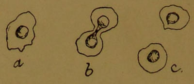
Diagram 15.—Cell Division.
Diagram 15.—Cell Division.
In some cells, probably in all, the centrosome presides over division. Cells, however, do not always divide in the same way. Some simply lengthen, the nucleus also lengthening inside, become constricted in the middle like a dumb-bell, and separate. (See Diagram 15.)
In some cells, likely in all, the centrosome is in charge of division. However, cells don't always divide the same way. Some just elongate, with the nucleus also stretching inside, becoming pinched in the middle like a dumbbell, and then split apart. (See Diagram 15.)

Diagram 16.—Cell Division.
Diagram 16.—Cell Division.
Others manage differently. In them the nucleus simply bursts, and turns its essential elements, a number—always a constant number—of coarse threads, adrift. Meanwhile, two centrosomes have moved to opposite ends of the cell, and there anchored themselves by fibrils; other fibrils springing from them become attached to the nuclear threads, and when all is ready pull them apart, equally divided, to their respective ends, where they re-form into two fresh nuclei. (See Diagram 16.)
Others handle it differently. In them, the nucleus simply breaks apart, scattering its essential elements, a consistent number of coarse threads, into the open. Meanwhile, two centrosomes have positioned themselves at opposite ends of the cell, anchoring themselves with fibrils; other fibrils emerging from them attach to the nuclear threads. Once everything is set, they pull the threads apart, evenly divided, to their respective ends, where they reassemble into two new nuclei. (See Diagram 16.)
Unicellular animals, which are constant in shape and[33] swim instead of flowing when they want to get anywhere, have at first sight nothing in common with those which do the latter. From their surfaces spring fringes of free protoplasmic threads, called cilia, from their fancied resemblance to eyelashes, which serve as motor organs, and beat the water like oars. (See Diagram 2.) Waves of movement, as they lash one after another, all in the same direction, seem to pass over the cell, and it is propelled through the water; while others, which are situated in the neighbourhood of the cell’s mouth, stir the water into eddies, and drive food particles into it.
Unicellular animals, which have a consistent shape and swim instead of just drifting when they want to move, initially seem unrelated to those that do the latter. From their surfaces extend fringes of free protoplasmic threads, called cilia, named for their resemblance to eyelashes, which act as motor organs and paddle through the water like oars. Waves of movement, as they strike one after another in the same direction, seem to flow over the cell, pushing it through the water; while others nearby the cell's mouth create currents, bringing food particles toward it.

Diagram 17.—Cilia of an Epithelial Cell.
Diagram 17.—Cilia of an Epithelial Cell.
These cilia are important, as they are adapted for many purposes in large animals. The cells which line the cavity at the back of the nose, the tubes of the lungs, and other parts of the body, have a few cilia on their free surface, and it is in them that the structure of these organs can best be made out. At the foot of each cilium is a minute globule, from which a fine fibril passes into the cell, and the fibrils, collectively forming a leash, are attached to its opposite end. (See Diagram 17.) It seems highly probable that the globule is a centrosome giving rise to two fibrils, one attached as described, the other passing up one side of the cilium, and fast to its apex. The result of this arrangement is that when the fibrils contract the cilium is bent over with a jerk to the side up which the fibril runs, and when they relax it slowly straightens itself. There is, therefore, no fundamental difference between this and the other mode of progression; both are dependent upon the centrosome.
These cilia are significant because they serve various functions in large animals. The cells lining the back of the nose, the lung tubes, and other body parts have a few cilia on their surface, and that's where the structure of these organs is best observed. At the base of each cilium is a tiny globule, from which a fine fibril extends into the cell, and these fibrils, forming a sort of bundle, are attached to the opposite end. (See Diagram 17.) It seems very likely that the globule acts as a centrosome, generating two fibrils: one attached as described, and the other running up one side of the cilium and anchored at its tip. This setup results in the cilium being jerked to the side along which the fibril runs when the fibrils contract, and it gradually straightens when they relax. Thus, there’s no essential difference between this and other forms of movement; both rely on the centrosome.
Finally we have muscle cells. These are only found in a fairly complicated animal, since they are a product of the division of labour principle, and their sole business is movement. There are two varieties of muscle, but the principle is the same in both—a long thin cell, with fibrils traversing its length whose contraction causes the cell to shorten and thicken, thus reducing the distance between its two ends. At present the development of muscle and the way in which it ‘contracts,’ to use the word accepted in this case for describing a redistribution of bulk, are little understood, and there are accordingly many opinions; but I think careful study will eventually show that some modification of the centrosome, with its contractile fibrils, is responsible for the movement.
Finally, we have muscle cells. These are only found in relatively complex animals, as they are a result of the principle of division of labor, and their primary function is movement. There are two types of muscle, but the concept is the same for both—a long, thin cell with fibrils running through its length, and when these fibrils contract, the cell shortens and thickens, reducing the distance between its two ends. Currently, the development of muscle and how it “contracts” (a term commonly used to describe a redistribution of volume) are not well understood, leading to various opinions; however, I believe that careful study will eventually reveal that some modification of the centrosome, along with its contractile fibrils, is responsible for the movement.

Diagram 18.—Muscle.
Diagram 18.—Muscle.
The two varieties are: the smooth, or involuntary, and the striped, or voluntary, muscle. Smooth muscle consists of spindle-shaped cells with one elongated nucleus. (See Diagram 18, Fig. 1.) It only contracts very slowly, and is not under control of the will; but it is very abundant in the body, since it effects practically all the movements of the alimentary canal and bloodvessels. Voluntary or striped muscle, so called from its appearance under the low power of a microscope, consists of long fibres, each containing many nuclei. (See Diagram 18, Fig. 2.) Its protoplasm is rich in hæmoglobin, and in it, under powerful microscopes, can be made out two kinds of fibrils: Rutherford’s fibrils, the complicated structure of which gives muscle its striped appearance; and Marshall’s fibrils, which are much finer and more difficult to see. The muscle of the heart, though not under control[35] of the will, is striped; but it differs from ordinary striped muscle in being made up of small branched cells with only one nucleus.
The two types are: smooth (involuntary) and striped (voluntary) muscle. Smooth muscle is made up of spindle-shaped cells that have one elongated nucleus. (See Diagram 18, Fig. 1.) It contracts very slowly and is not controlled by the will, but it is quite abundant in the body as it is responsible for nearly all the movements of the digestive tract and blood vessels. Voluntary or striped muscle, named for its appearance under low magnification, consists of long fibers, each containing multiple nuclei. (See Diagram 18, Fig. 2.) Its protoplasm is rich in hemoglobin, and under powerful microscopes, you can see two types of fibrils: Rutherford’s fibrils, which create the striped pattern, and Marshall’s fibrils, which are much finer and harder to distinguish. The heart muscle, while not consciously controlled, is striped as well; however, it differs from regular striped muscle because it is made up of small branched cells that each have only one nucleus.
The way in which the three elements of striped muscle contribute to a contraction is practically unknown, and the subject of much dispute. In fact, one could hardly wish for a better soil for theories, and some which grow in it are very wonderful indeed. We have reason for supposing that there are two contractile substances—one which gives a sharp twitch, the other a slow, hard pull; and on the whole there seems good reason to believe that Rutherford’s fibrils give the sudden movements, while Marshall’s give the more forcible ones; and that the ordinary protoplasm of the cell is restricted to the duty of nourishing the fibrils.
The way the three elements of striped muscle contribute to a contraction is pretty much unknown and is a topic of much debate. In fact, it’s hard to imagine a better breeding ground for theories, and some of them are quite remarkable. We have reason to think that there are two contractile substances—one that provides quick twitches and another that delivers slow, strong pulls. Overall, there seems to be good reason to believe that Rutherford’s fibrils are responsible for the sudden movements, while Marshall’s fibrils create the more powerful ones; and that the regular protoplasm of the cell mainly focuses on nourishing the fibrils.

Diagram 19.—Striped Muscle Fibre, more highly magnified than in Diagram 18.
Diagram 19.—Striped muscle fiber, magnified more than in __A_TAG_PLACEHOLDER_0__.
The muscle cells are modified from among those of the bud forming the middle layers of the embryo. (See Diagram 5.) Other cells of this bud form connective tissue, by, so to speak, spinning long fibres of the substance called collagen, which turns to gelatin when boiled. (See Diagram 20.) This connective tissue permeates the whole body, affording a firm foundation for the many layers of cells which form the skin and the single layer[36] of digestive cells; supporting the other organs throughout, and keeping the different parts of the body in their places, in doing which, however, it is assisted by other fibres which are not collagenous, but elastic. It also forms tracts which become lymph and blood vessels.
The muscle cells are transformed from those in the bud that creates the middle layers of the embryo. (See Diagram 5.) Other cells in this bud create connective tissue by essentially spinning long fibers of a substance called collagen, which turns into gelatin when boiled. (See Diagram 20.) This connective tissue spreads throughout the entire body, providing a solid foundation for the various layers of cells that make up the skin and the single layer[36] of digestive cells; it supports the other organs and keeps different parts of the body in their proper places, aided by other fibers that are not collagenous but elastic. It also creates pathways that develop into lymph and blood vessels.
In parts of the animal which require special support it forms solid rods, the collagen combining with calcium salts to form a clear, hard substance—cartilage. At one period in the development of an animal or animals we find the only solid support is cartilage, but cartilage is not sufficiently rigid for a very large beast, especially on land, so is only used for outlying parts, the main framework being bone.
In areas of the animal that need extra support, it creates solid rods, with collagen combining with calcium salts to make a clear, hard substance—cartilage. At one stage in the development of an animal or animals, cartilage is the only solid support available, but it's not strong enough for a very large creature, particularly on land, so it's only used for peripheral parts, while the main structure is made of bone.

Diagram 20.—A Connective-tissue Cell giving rise to Long Collagenous Fibres.
Diagram 20.—A connective tissue cell producing long collagen fibers.
Bone is formed very much as if Nature were rectifying a mistake. When a rod of cartilage is unequal to its work it is eaten hollow, and fresh connective-tissue cells immigrate and fill up the cavity, eventually laying down a fine network of cells in its place, the meshes of which are filled with inorganic calcium salts, chiefly phosphate of lime. Nature then benefits by experience, and the last bones to be formed are not preceded by any makeshift cartilage, but built up straight away in ordinary connective tissue.
Bone is formed as if Nature is fixing a mistake. When a piece of cartilage can't handle its job, it gets hollowed out, and new connective tissue cells move in to fill the space, eventually creating a fine network of cells there. These meshes are filled with inorganic calcium salts, mainly calcium phosphate. Nature then learns from experience, and the last bones that form aren’t preceded by any temporary cartilage but are built directly from regular connective tissue.
This brings us back again to muscle, for the object of nearly all the voluntary muscle is to cause movement among the bones. For this purpose the muscle cells or fibres are arranged parallel to one another, and bound up together by connective tissue, the whole bundle being[37] known as ‘a muscle.’ The two ends of a muscle are attached to two bones by connective tissue, which sometimes forms a short cord, or tendon. Then, when the muscle contracts, the two places of its attachment are pulled towards one another, and something has to move. But before saying more about the way in which the bones are jointed and muscles attached—in fact, what movements are possible in the human body—it would be as well here to describe the chief properties of muscle and the way in which they are studied.
This brings us back to muscle, since the main job of almost all voluntary muscle is to create movement among the bones. For this purpose, muscle cells or fibers are arranged parallel to one another and held together by connective tissue, with the entire bundle being[37] referred to as ‘a muscle.’ The two ends of a muscle are attached to two bones by connective tissue, which sometimes forms a short cord, or tendon. When the muscle contracts, the two points of attachment are pulled toward each other, and something has to move. But before we dive deeper into how the bones are connected and how the muscles are attached—in other words, what movements are possible in the human body—it would be good to describe the main properties of muscle and how they are studied.
II.

Diagram 21.—Apparatus for recording a Muscular Contraction.
Diagram 21.—Equipment for Recording a Muscle Contraction.
The way in which voluntary muscle is studied is very simple. A frog is killed by thrusting a probe into the brain and down the spinal cord, and a muscle is then dissected out and attached to a piece of apparatus (see Diagram 21) in such a way that on its contracting it raises a lever, and draws a line on a moving surface. The rate at which the surface is moving is ascertained, so that the nature of the curve, which is a graphic record of the contraction, can be analyzed. (See Diagram 22.) For instance, when an electric shock is used to[38] make the muscle contract, we find that a slight shock causes a small contraction, as shown by a low curve, while a stronger one, up to a certain point, causes an increase.
The way we study voluntary muscle is pretty straightforward. A frog is put down by inserting a probe into its brain and down the spinal cord, and then a muscle is cut out and attached to a piece of equipment (see Diagram 21) in a way that allows it to raise a lever when it contracts, drawing a line on a moving surface. We check the speed of the surface so we can analyze the curve, which graphically represents the contraction. (See Diagram 22.) For example, when we use an electric shock to make the muscle contract, a small shock results in a slight contraction, indicated by a low curve, while a stronger shock, up to a point, leads to a larger contraction.

Diagram 22.—Graphic Record of a Response to a Single Stimulus applied at A.
Diagram 22.—Graphic Record of a Response to a Single Stimulus applied at A.
Lower line = tuning-fork records of ⅟₁₀₀″.
Lower line = tuning-fork records of 1/100″.
But having described how muscle is studied, it is only necessary to state a few facts concerning it; to discuss muscle, fully describing the experiments by which its more obscure properties have been elucidated, and the devices by which causes of error have been eliminated, would fill volumes.
But after explaining how muscle is studied, it's only necessary to mention a few facts about it; discussing muscle in detail, fully describing the experiments that have clarified its more obscure properties and the methods used to eliminate sources of error, would take many volumes.

Diagram 23.—Contractions with Two Stimuli at Different Intervals of Time.
Diagram 23.—Contractions with Two Stimuli at Different Time Intervals.
Muscle is thrown into a state of contraction by an impulse reaching it from a nerve, but it contracts quite as readily if excited directly by a mechanical or electrical shock. A second shock causes a second contraction, or, if the muscle is still in a state of contraction owing to the first, causes it to contract still more. (See Diagram 23.) If a number of stimuli are applied to a muscle in such rapid succession that the effect of the preceding one has not passed off by the time the next arrives, it will contract as far as possible, and remain contracted—a state known as tetanus. (See Diagram 24.) A muscle is therefore kept in a state of contraction by a continuous nervous effort, not arranged and then left contracted.
Muscle goes into a state of contraction when an impulse from a nerve reaches it, but it also contracts just as easily if it's stimulated directly by a mechanical or electrical shock. A second shock leads to a second contraction, or if the muscle is still contracted from the first shock, it will contract even more. (See Diagram 23.) If several stimuli are applied to a muscle in such quick succession that the effect of the previous one hasn't faded by the time the next one hits, it will contract as much as possible and stay contracted, a condition known as tetanus. (See Diagram 24.) Therefore, a muscle remains in a contracted state due to continuous nervous stimulation, rather than just being arranged and left contracted.

Diagram 24.—Tetanus.
Diagram 24.—Tetanus.

Diagram 25.—Fatigue Curves.
Diagram 25.—Fatigue Curves.
Fast drum: a, point of stimulation. Every tenth contraction recorded.
Fast drum: a point of stimulation. Every tenth contraction recorded.

Diagram 26.—Effect of Fatigue on Muscular Contraction.
Diagram 26.—Impact of Fatigue on Muscle Contraction.
Slow drum. Every contraction recorded.
Slow drum. Every contraction logged.
Various conditions alter the character of a muscular[40] response. With repeated stimuli at short intervals a muscle fatigues, and each contraction becomes smaller in extent and longer in duration. (See Diagrams 25 and 26.) If the muscle has to lift a load it has a certain check on its contraction, and its relaxation time is shortened. Temperature also affects muscular contraction, moderate increase causing a sharper, and moderate cooling a slower, rise and fall of the lever on stimulation. (See Diagram 27.) Lastly, we have drugs which exert an influence, but the only one of these which it is necessary to mention here is veratria, which makes the slowly contracting fibrils continue their activity after the quick ones have subsided. (See Diagram 28.)
Various factors change how muscles respond[40]. When stimulated repeatedly in quick succession, a muscle tires, and each contraction becomes less powerful and longer in duration. (See Diagrams 25 and 26.) If the muscle needs to lift a weight, it experiences some restriction on its contraction, and the time it takes to relax is reduced. Temperature also influences muscle contraction; a slight increase causes sharper contractions, while moderate cooling results in slower rises and falls of the lever when stimulated. (See Diagram 27.) Lastly, there are drugs that affect muscle function, but the only one worth mentioning here is veratria, which keeps the slowly contracting fibers active even after the quicker ones have stopped. (See Diagram 28.)

Diagram 27.—Effect of Temperature.
Diagram 27.—Temperature Effects.

Diagram 28.—Veratria Curve.
Diagram 28.—Veratria Curve.
Finally, there are the electrical changes in muscle. These, again, may be passed over briefly, since they are not easily understood or described. To put the facts in a nutshell, the part of a muscle which is in activity is negative to all other parts. Thus, if a muscle be dissected out and cut across, the activity at the seat of the injury, while it lasts, causes a current to pass through a galvanometer from uninjured parts to the wounded. (See Diagram 29.) Again, if a muscle be dissected out without injury, connected at two points with a[41] galvanometer, and then stimulated at one end, as the wave of contraction passes along it, first one, then the other, contact becomes negative. (See Diagram 30.) S, Stimulating electrodes; N, contraction which marks the wave of excitation passing along the muscle; G, galvanometer which shows that the seat of activity (N) is negative to the rest of the muscle.
Finally, there are the electrical changes in muscle. These can also be summarized quickly, as they are not easy to understand or describe. In short, the part of a muscle that is active is negatively charged compared to all other parts. So, if a muscle is removed and cut, the activity at the injury site creates a current that flows through a galvanometer from the uninjured areas to the injured one. (See Diagram 29.) Additionally, if a muscle is removed without injury, connected at two points to a [41] galvanometer, and then stimulated at one end, as the contraction wave travels along it, first one contact and then the other becomes negative. (See Diagram 30.) S, Stimulating electrodes; N, contraction which shows the wave of excitation moving along the muscle; G, galvanometer which indicates that the active area (N) is negative compared to the rest of the muscle.

Diagram 29.—Injury Current: Cross-section of Muscle Negative to Rest.
Diagram 29.—Injury Current: Cross-section of Muscle Negative to Rest.

Diagram 30.—Action Current.
Diagram 30.—Action Current.
In passing, it may be mentioned that, as the heart is a muscle slung obliquely across the body, and waves of contraction are continually passing down its long axis, the whole body is affected by continual electrical changes.[42] By very delicate instruments it can be demonstrated that with each beat the two hands alternately become electrically positive and negative to each other.
It's worth noting that since the heart is a muscle positioned diagonally across the body, and waves of contraction constantly move along its length, the entire body is influenced by ongoing electrical changes.[42] Using very sensitive instruments, it's possible to show that with each heartbeat, the two hands alternately become electrically positive and negative with respect to each other.
Whilst dealing with the electrical phenomena of muscle, it may be as well to state that nerve fibres, which are studied with very much the same apparatus, show the same electrical changes, the point of injury or of the greatest activity being negative to all the rest. Single cells are less easily investigated, but in glands it is possible to show that the same rule holds.
While examining the electrical phenomena of muscles, it's worth noting that nerve fibers, which can be studied using very similar equipment, exhibit the same electrical changes, with the injury site or the area of greatest activity being negative compared to the rest. Single cells are more challenging to study, but in glands, it is possible to demonstrate that the same principle applies.
Undoubtedly the most curious fact about the generation of electricity by protoplasm is that, by a modification of muscle and nerve, which causes them to lose their ordinary properties, they are converted into a special organ for giving electric shocks. Armed with powerful batteries of this description, an otherwise rather helpless class of fish are enabled to defend themselves from their enemies, and deal unexpected death to their more agile prey.
Undoubtedly, the most interesting fact about how protoplasm generates electricity is that, through a change in muscle and nerve, they lose their usual functions and turn into a special organ for delivering electric shocks. With these powerful batteries, a generally defenseless group of fish can protect themselves from predators and deliver surprising blows to their quicker prey.
Having now run over a few of the physical properties of protoplasm, we may pass on to a brief investigation of the movements we find in the body of man.
Having now covered some of the physical properties of protoplasm, we can move on to a brief examination of the movements we observe in the human body.
III.
In describing the movements of the body, we shall have to treat them as several and distinct, as indeed they are; but the fact should not be lost sight of that they cannot really be isolated: one idea embraces the whole. Two kinds of movement may, however, be distinguished in the vital functions: movement of the actual cells, such as muscles; and movement of non-protoplasmic elements acted upon by the cells—e.g., lymph.
In describing body movements, we need to view them as separate and distinct, which they truly are; however, we must remember that they can’t really be isolated: one concept encompasses everything. That said, we can differentiate between two types of movement in vital functions: movement of the actual cells, like muscles, and movement of non-protoplasmic elements that are influenced by the cells—e.g., lymph.
There is a parallel to this in the chemical side of life, where we find some phenomena peculiar to the living elements, and others, like digestion, going on in the living body, but outside the cells.
There’s a similar situation in the chemical aspects of life, where we observe some phenomena unique to living elements, while others, like digestion, occur in the living body but outside the cells.
Taking the movements in the natural order—that is, proceeding from the simpler to the more complex—the first to be considered is undoubtedly that of the leucocytes, or general scavengers of the tissues. The body[43] consists, so far as we have defined its anatomy, of three layers of cells, and its shape is that of a tube with hollow walls. (See Diagram 6.) Within the cavity of the body are various organs, such as the muscles, which are formed from the middle layer; and its space is largely reduced by glands, lungs, and other ramifications of the inner layer which forms the alimentary canal.
Taking the movements in the natural order—that is, moving from the simpler to the more complex—the first to consider is definitely that of the white blood cells, or general cleanup crew of the tissues. The body[43] consists, as we’ve defined its anatomy, of three layers of cells, and its shape is like a tube with hollow walls. (See Diagram 6.) Within the body’s cavity are various organs, such as the muscles, which are made from the middle layer; and this space is mostly filled by glands, lungs, and other branches of the inner layer that forms the digestive tract.
These organs hang more or less freely in the body cavity, slung to its walls by enveloping sheets of connective tissue, the whole being bathed in lymph. Now, in such an arrangement the products of wear and tear must accumulate. Cells here and there die for various reasons, and pieces of cells become detached even in adult animals. The interior of a bone is always being eaten away to decrease its weight, or in order that it may be replaced by fresh bone of a closer texture, and in young animals and embryos there are many structures which, useful for a time, have eventually to be removed; as an instance, we may quote the tadpole’s tail. In fact, if the tissues were left to themselves, the body would soon be choked with débris, and to avoid this it is supplied with an army of scavengers, the leucocytes.
These organs hang relatively freely in the body cavity, attached to the walls by layers of connective tissue, all surrounded by lymph. In this setup, the products of wear and tear can build up. Cells die for various reasons here and there, and bits of cells can become detached even in adult animals. The inside of a bone is constantly being broken down to reduce its weight or to be replaced by new bone that is denser. In young animals and embryos, there are many structures that are useful for a time but eventually need to be removed; for example, the tail of a tadpole. In fact, if the tissues were left alone, the body would quickly fill up with debris, and to prevent this, it is equipped with an army of scavengers, the white blood cells.
The leucocytes are detached cells which owe their origin to the middle layer. In size they are, of course, very small, quite invisible to the naked eye. In appearance they resemble unicellular organisms of the amœba type, which we have had occasion to mention several times already (Essay II., Section II.; Essay III., Section I., Diagram 1). They are of several different varieties, some being larger and more active than others; but they all wander about in the lymph and blood like independent animals, creeping in and out between the cells of the organs, and devouring any foreign matter they come across. They sometimes multiply, like independent animals, by division, especially in the presence of inflammation, or when they have much work to do, and a rapid increase in their numbers is needed; and they have been induced to live, and feed, and multiply, outside the body (in which case they must be considered to have become independent organisms), thanks to the careful attentions of the experimenter.
The white blood cells are detached cells that come from the middle layer. They're very small and not visible to the naked eye. In appearance, they look like single-celled organisms of the amoeba type, which we've mentioned several times already (Essay II., Section II.; Essay III., Section I., Diagram 1). There are several different types, with some being larger and more active than others; but they all move around in the lymph and blood like independent creatures, crawling in and out between the cells of the organs and eating any foreign material they encounter. They can multiply, like independent creatures, by dividing, especially during inflammation or when there's a lot of work to do, requiring a rapid increase in their numbers; and they can be made to live, feed, and multiply outside the body (in which case they should be considered to have become independent organisms), thanks to the careful attention of the researcher.
Apart from their duties of devouring the inside layers of bones and clearing away dead tissue, they are supposed by some to assist in the absorption of food by creeping between the cells lining the alimentary canal, and, after throwing out arms to engulf particles of food, returning with their spoils into the body. Perhaps, however, the most interesting, or at any rate most romantic, of their many and important functions are what may be called their emergency duties. Frequently people, especially those who live in smoky towns, draw into their lungs particles of dust and soot, which if left adhering to the walls of the air cavities would cause dangerous irritation. As if by magic a leucocyte will discover the presence of such a nuisance, and, crawling between the cells forming the wall of the lung, in which, by the way, it is outside the body proper, will engulf it and carry it away with him. This exploit, however, pales beside the warfare which goes on in the body between leucocytes and invading bacteria. A bacterium thrives in the blood or lymph, since it finds itself in a warm alkaline fluid containing complex organic substances, by breaking down which it can easily obtain energy. Unfortunately, the products of such a process are frequently virulent poisons, the effect of which upon neighbouring cells produces the distressing symptoms which we associate with disease. No sooner, however, has the bacterium begun to generate poisons, than leucocytes, influenced by chemical attraction (Essay I.), swarm upon it. First come leucocytes of a small kind, full of zymogen granules, which crowd round the bacterium till they have covered it. After a time they creep away, leaving it dead. They are now in an exhausted condition, and no longer contain granules, having doubtless discharged them as a destructive ferment upon their enemy. Then a leucocyte of another kind moves to the attack, or, rather, to clear up the remains, for he is a large, non-granular, active fellow, and eats up the dead bacterium by the simple process of engulfing him whole. (See Diagram 31.)
Apart from their job of breaking down the inner layers of bones and cleaning up dead tissue, some believe they also help absorb food by moving between the cells of the digestive tract and, after extending their arms to capture food particles, returning with their finds into the body. However, perhaps the most fascinating, or at least the most dramatic, of their many important roles are what could be called their emergency duties. Often, people, especially those living in smoky cities, inhale dust and soot particles that, if they stick to the walls of the air sacs, could cause serious irritation. Like something out of a story, a white blood cell spots this problem and crawls between the cells making up the lung wall, where it is technically outside the main body, and engulfs the particles to take them away. This feat, however, pales in comparison to the battle that occurs in the body between white blood cells and invading bacteria. A bacterium thrives in the blood or lymph because it finds itself in a warm, alkaline fluid containing complex organic substances, which it can easily break down for energy. Sadly, the byproducts of this process are often harmful toxins that distress nearby cells and lead to the symptoms we associate with illness. No sooner does the bacterium start producing toxins than white blood cells, drawn in by chemical signals (Essay I.), swarm around it. First come small white blood cells, filled with zymogen granules, which crowd around the bacterium until it's completely covered. After a while, they pull away, leaving the bacterium dead. Now they are spent and no longer have granules, having likely released them as a destructive substance against their foe. Then a different kind of white blood cell steps in to clean up, being a large, non-granular, active cell that simply engulfs the dead bacterium whole. (See Diagram 31.)
A natural question arising out of the study of leucocytes is, What becomes of them? Particles of soot and similar refuse can hardly be considered nutritious,[45] or even digestible, food, and one is rather drawn to the conclusion that the leucocyte performs its functions for the good of the body at large, not of itself, and that when its work is done it must die. Many leucocytes, probably, loaded with unconsidered and undesirable trifles, cast themselves into the alimentary canal, and are got rid of with the useless portions of the food; but they do not always have the luck or energy to get to a natural outlet. An unpleasantly familiar phenomenon is the boil. Here we have some irritating substance under the skin setting up inflammation, and leucocytes swarm up to remove the cause of the trouble. Before, however, this is done, many have perished in the fray, and they have collected in numbers to the formation of what is commonly known as pus, or matter. Their dispersal into the body is now neither easy nor desirable, and the surgeon usually lets them escape from the surface by a touch of the lancet.
A common question that comes up when studying white blood cells is: What happens to them? Particles of soot and similar waste can hardly be seen as nutritious or digestible food, so it leads to the idea that white blood cells do their job for the benefit of the body as a whole, not for themselves, and that once they’ve completed their role, they must die. Many white blood cells, likely burdened with unwanted debris, end up in the digestive system and are expelled along with the useless parts of food; however, they don’t always have the luck or strength to find a natural way out. A well-known issue is the boil. This occurs when an irritating substance is under the skin, causing inflammation, and white blood cells rush in to eliminate the source of the problem. However, many of them perish in the process, accumulating to create what is commonly known as pus. Once dispersed throughout the body, they can’t be easily or desirably handled, so the surgeon typically allows them to exit at the surface with a quick cut from a lancet.

Diagram 31.
Diagram 31.
A, Eosinophile leucocyte; B, bacterium; C, leucocytes killing bacterium with their enzyme; D, leucocytes leaving bacterium dead; E, hyaline leucocyte devouring dead bacterium.
A, Eosinophil white blood cell; B, bacterium; C, white blood cells destroying the bacterium with their enzymes; D, white blood cells departing from the dead bacterium; E, clear white blood cell consuming the dead bacterium.
Such, then, is very briefly the story of the leucocyte, neglecting such problems as the differences between those found in the blood, called white corpuscles to distinguish them from the red corpuscles, with which they have no sort of connection; those found in the lymph, called lymphocytes to distinguish them from[46] those found in the blood; those caught in the act of devouring bone, called osteoclasts; and those found with bacteria inside them, therefore known as phagocytes; and without speculating on how long an individual lives, and whether the different varieties differ in origin or are merely at progressive stages of development. The study of leucocytes is one of the most fascinating in physiology, but we have many other things calling for our attention, and we have said enough about the part they play in the life of the body to justify our passing on to consider another essential movement.
This is a brief overview of the story of the white blood cell, ignoring issues like the differences between those found in the blood, called white corpuscles to differentiate them from red corpuscles, which they have no connection with; those found in lymph, known as lymphocytes to distinguish them from[46] those in the blood; those involved in breaking down bone, called osteoclasts; and those that contain bacteria, known as phagocytes. It also won't go into how long an individual lives or whether the different types originate differently or are simply at various stages of development. The study of white blood cells is one of the most interesting areas of physiology, but we have many other topics that need our focus, and we've covered enough about their role in the body to move on to consider another important process.
IV.
Next in natural order for consideration come the movements of the alimentary canal.
Next in natural order for consideration are the movements of the digestive tract.
So far we have considered this structure as a chemical laboratory, a tube consisting of a single layer of cells which secrete ferments into the lumen, where digestion takes place, and then absorb the products, and we have not yet accounted for the food travelling along the tube, without which its functions, as described in the earlier part of the book, could not be performed. That the passage of the food is not due to gravitation is obvious from the many directions of the tube’s coils—not to quote the old instance of a horse drinking, in which case the liquid first travels upwards. One must therefore conclude in favour of some muscular method of propulsion.
So far, we've looked at this structure as a chemical lab, a tube made of a single layer of cells that release enzymes into the lumen, where digestion occurs, and then absorb the products. We haven't yet addressed the food moving through the tube, which is essential for its functions, as discussed earlier in the book. It's clear that the movement of food isn’t due to gravity, considering the various directions of the tube’s coils—not to mention the classic example of a horse drinking, where the liquid first moves upward. So, we must conclude that some kind of muscular method is responsible for pushing it along.
We have so far described the alimentary canal as a single layer of cells, but it must be obvious that these soft secreting portions of the tube are not capable of vigorous movement. The canal proper is surrounded by a tough sheath of connective tissue which prevents its being overdistended or ruptured, and, by means of a layer—or, rather, two layers—of non-striped muscle which it contains, produces the movements which result in the passage of its contents along the tube. These two layers lie well to the outside of the connective-tissue sheath. The fibres of the inner layer are arranged circularly, so as to form rings round the tube; those of the outer have a longitudinal direction, running, therefore,[47] parallel with its long axis. When the former contract, the diameter of the tube is reduced, while contraction of the latter has the effect of enlarging it. (See Diagram 32.)
We’ve described the digestive tract as a single layer of cells, but it’s clear that these soft, secretory sections of the tube can’t move vigorously. The main canal is surrounded by a tough layer of connective tissue that prevents it from stretching too much or bursting, and it has a layer—or actually, two layers—of smooth muscle that create the movements needed to push its contents through the tube. These two layers sit well outside the connective tissue layer. The fibers in the inner layer are arranged in a circular pattern, forming rings around the tube, while the outer layer runs along the length of the tube. When the inner layer contracts, the tube's diameter decreases, and when the outer layer contracts, the diameter increases.[47]
The movements of the intestine are what is known as peristaltic. Contraction of the muscle fibres is not simultaneous in all parts, but passes in waves along it. Just in front of the food the longitudinal fibres contract, and thus offer less resistance, while just behind the circular fibres reduce the size of the tube, and so get up a pressure. The result of a number of successive waves of contraction passing down the alimentary canal is that the food is propelled along it.
The movements of the intestine are called peristaltic. The muscle fibers don't contract at the same time in all areas, but rather in waves. Right in front of the food, the longitudinal fibers contract, making it easier for the food to move, while just behind, the circular fibers tighten the tube, creating pressure. As a result, a series of wave-like contractions moving down the digestive tract pushes the food along.

Diagram 32.—To illustrate the Passage of Food along the Intestine.
Diagram 32.—Illustrating How Food Passes Through the Intestine.
The arrangement of the muscle varies in places to suit special needs. Where the tube suddenly enlarges to form the stomach, and where the stomach suddenly narrows to the intestine, there are two strong rings of muscle, whose constricting influence converts the enlargement into a closed chamber during gastric digestion; while the coats which actually clothe it here run obliquely, and their activity causes the contents to be slowly churned about inside.
The way the muscle is set up changes in certain areas to meet specific needs. Where the tube widens to create the stomach, and where the stomach narrows into the intestine, there are two strong muscle rings. These rings tighten and turn the enlargement into a closed space during gastric digestion; meanwhile, the layers covering it run at an angle, and their movement slowly stirs the contents inside.
Thus it will be seen that it is not only the voluntary muscles which give the alimentary system its opportunities; without these unobtrusive non-striped cells we should toil for our bread and swallow it in vain.
Thus it will be seen that it is not only the voluntary muscles that give the digestive system its opportunities; without these subtle non-striped cells we would work hard for our food and swallow it in vain.
V.
Our next step, after having surveyed the principle of movement by which the chemical necessities of the body[48] are exposed to its absorbing surface, must be to see how the fluid which transports them is made to pass along the tubes containing it. We have already had occasion to describe how these blood and lymph vessels ramify through all the organs, when we were dealing with the chemical influence of the blood and lymph.
Our next step, after looking at the main way the body’s chemical needs are brought to its absorbing surface, is to examine how the fluid that carries these necessities moves through the tubes that contain it. We’ve already discussed how these blood and lymph vessels spread out through all the organs when we talked about the chemical effects of blood and lymph.
The tubes through which the lymph is brought back to the blood-stream have thin walls, and no muscle of their own. They are subjected, however, to a constantly varying pressure by the movements of the limbs and trunk, and as, owing to valves inside them, the lymph can only escape in one direction, there is a constant flow towards the junction with the bloodvessels.
The tubes that carry lymph back to the bloodstream have thin walls and no muscles of their own. However, they are affected by the constantly changing pressure from the movements of the limbs and trunk. Because of valves inside them, the lymph can only flow in one direction, creating a steady movement towards the junction with the blood vessels.
The bloodvessels are quite different. A far more certain and expeditious current is necessary—hence the steady circulation through a system of closed tubes.
The blood vessels are quite different. A much more certain and quicker flow is needed—hence the consistent circulation through a system of closed tubes.
In order to understand this passage of the blood, it is necessary to keep in mind the great principle with which hydrostatics supplies us, viz., that a liquid always flows from a region of high to a region of lower pressure. The problem of the vascular system is, therefore: How can the pressure within a ring of tube be so arranged as to maintain a regular flow always in the same direction?
To understand this part about blood flow, it's important to remember the key principle from hydrostatics: a liquid always moves from an area of high pressure to an area of low pressure. So, the challenge for the vascular system is: How can we manage the pressure within a tube so that the flow consistently goes in the same direction?
Let us begin with the structure of the system. The tube through which the blood first passes on leaving the heart is composed of four distinct and essential elements: A lining of endothelial cells, which we need not discuss at length; a main substance of tough white fibrous connective tissue; elastic fibres and muscle fibres, the two last arranged in the substance of the connective tissue. All these parts are present in the main arteries which leave the heart, but in the fine meshwork of capillaries to which the arteries give rise by repeated branchings there is nothing left of the outer coats, only the lining of endothelial cells separating the blood from the organ traversed. In the veins which these capillaries unite to form, the connective-tissue sheath reappears, and also some muscle; but the elastic coat is quite absent. The heart is really a double coil of the tube (see Diagram 12), in which the muscular coat is predominant, and is divided into four chambers by the valves, which insure[49] the blood flowing in the right direction when it contracts. (See Diagram 33.)
Let’s start with how the system is structured. The tube that blood first travels through when it leaves the heart has four key and important components: a lining of endothelial cells, which we won’t go into in detail; a core made of tough white fibrous connective tissue; elastic fibers; and muscle fibers, with the last two integrated within the connective tissue. All these components are found in the main arteries that leave the heart, but in the fine network of capillaries that branch off from the arteries, the outer layers are gone, leaving only the lining of endothelial cells that separates the blood from the tissue it passes through. In the veins formed by the merging of these capillaries, the connective tissue layer comes back, along with some muscle; however, the elastic layer is completely missing. The heart itself is actually a double coil of the tube (see Diagram 12), where the muscular layer is the most prominent, and it's divided into four chambers by valves, which ensure[49] that the blood flows in the correct direction when it contracts. (See Diagram 33.)

Diagram 33.—Scheme of Circulation
Diagram 33.—Circulation Scheme
The way in which these structures work is as follows: Two of the chambers of the heart (the auricles) receive blood from the veins, and when full suddenly contract, driving their contents into the other two chambers (the ventricles). The blood does not run back into the veins, although the pressure in them is very low and there are no valves to prevent it, because there is still less pressure in the ventricles, and also because the veins enter the auricles obliquely, and the tendency of the increasing pressure is to close their orifices. Having discharged the blood into the ventricles, the auricles relax, and the pressure within being a minus quantity, they are speedily filled with blood from the veins, blood not being able to return after entering the ventricles, as valves close automatically to prevent it.
Here's how these structures work: Two of the heart's chambers (the atria) receive blood from the veins, and when they are full, they suddenly contract, pushing their contents into the other two chambers (the ventricles). The blood doesn't flow back into the veins, even though the pressure in them is very low and there are no valves to stop it, because the pressure is even lower in the ventricles. Additionally, the veins enter the atria at an angle, and the increasing pressure tends to close their openings. After sending blood into the ventricles, the atria relax, and since the pressure inside is negative, they quickly fill with blood from the veins. Blood can't flow back after entering the ventricles, as valves close automatically to block that.
Stimulated by the blood distending them, the ventricles[50] then contract simultaneously like the auricles, only with much greater force: for the right ventricle has to drive the blood all through the vessels pervading the lungs back to the left auricle; whilst the left ventricle, which is proportionately stronger than the right, has to send its contents to the furthest extremities of the body. They then relax, in order that conditions of their internal pressure may favour another inflow from the auricles, return of blood from the arteries being, as in the preceding case, prevented by valves.
Inspired by the blood filling them up, the ventricles[50] then contract at the same time as the atria, but with much greater force: the right ventricle has to push blood through the vessels in the lungs back to the left atrium, while the left ventricle, which is relatively stronger than the right, must send its blood to the farthest parts of the body. They then relax to create conditions of internal pressure that allow for another inflow from the atria, with the return of blood from the arteries being blocked by valves, just as before.
The pressure in the arteries during life is always fairly high; indeed, the ventricles have to get up a considerable force before the valves leading from them will open. The result of this is not only that the blood is driven along them with a rush, but also that they are slightly distended at each beat; and so, owing to the elasticity of their walls, the blood continues to flow forwards even between the beats of the heart. The rest of the journey is quite simple; the pressure in the capillaries is lower than in the arteries, and the pressure in the veins lower than in the capillaries, and lower in the veins, too, as they approach the heart, till, where they join the auricle, it is actually minus, and the blood has no other course open to it but to return to the auricle. It looks as though accidents might happen in the veins owing to there being so low a pressure there to direct the current, but this is prevented by the presence of valves at intervals, to stop any return.
The pressure in the arteries during life is always pretty high; in fact, the ventricles need to generate a significant force before the valves leading from them will open. As a result, blood is pushed through them quickly, and they are slightly expanded with each heartbeat; because of the elasticity of their walls, the blood keeps flowing even between heartbeats. The rest of the journey is straightforward; the pressure in the capillaries is lower than in the arteries, and the pressure in the veins is lower than in the capillaries, and it continues to decrease as they get closer to the heart, until, when they connect to the auricle, it actually becomes negative, leaving the blood with no option but to return to the auricle. It might seem like accidents could happen in the veins due to such low pressure directing the flow, but this is avoided by valves placed at intervals to prevent any backward flow.
The rate at which the blood travels is another point which has an important bearing on the nutrition. It does its work—i.e., gives out nutriment and picks up refuse—whilst flowing through the capillaries; so here one finds that it moves slowly. On the other hand, the sooner it reaches them the better, so it races fast through the arteries. Finally, its return to the heart need not be delayed, so it is quickened up again through the veins. The principle by which this variation in the rate of flow is obtained is simple and inevitable. If a tube through which liquid is flowing is not the same size all the way along, the liquid will be found to flow faster in the narrow parts than in the wider ones. Now, in branching, the[51] arteries do not keep becoming smaller in regular proportion, and the result is that the capillaries have collectively a diameter five hundred times larger than the aorta; hence the blood flows through them only one-five-hundredth of the pace at which it leaves the heart. But in uniting again to form the veins their cross-section is reduced once more, so that that of the large veins near the heart is only two and a half times larger than that of the aorta, and hence a flow only two and a half times slower results.
The speed at which blood moves is another aspect that significantly affects nutrition. It does its job—i.e., delivers nutrients and removes waste—while flowing through the capillaries, where it moves slowly. On the flip side, the faster it reaches them, the better, so it speeds through the arteries. Lastly, there's no need to delay its return to the heart, so it quickly moves through the veins again. The principle behind this change in flow rate is straightforward and unavoidable. If a tube through which liquid flows isn’t the same size throughout, the liquid will move faster in the narrower sections than in the wider ones. In the branching arteries, they don’t decrease in size uniformly, resulting in the capillaries having a total diameter five hundred times larger than the aorta. Therefore, blood flows through them at only one-five-hundredth of the speed it leaves the heart. However, when they come together to form the veins, their cross-section decreases again, so that the large veins near the heart are only two and a half times larger than the aorta, leading to a flow that is only two and a half times slower.
The pace of the blood-stream must depend, obviously, on the pressure of the blood in the arteries. This pressure is altered either by changing the rapidity of the heart-beat or the diameter of the arteries, which are capable of considerable variation owing to their muscular coat. The regulation of the blood-pressure is managed by the nervous system, so does not belong here, and we may leave it after mentioning one or two facts. High pressure is due to a large quantity of blood being in the arteries, and this may be due either to the rapidity with which it is injected by the heart or to the reduced capacity of the bloodvessels themselves. High pressure, due to the latter cause, throws a great strain upon the heart, owing to the hard work it has in pumping blood into the arteries; with a low pressure the heart beats feebly, having less resistance to overcome.
The speed of the bloodstream clearly depends on the pressure of the blood in the arteries. This pressure changes either by altering how fast the heart beats or by changing the diameter of the arteries, which can vary quite a bit because of their muscular walls. The nervous system manages blood pressure regulation, so we won’t get into that here, but it's worth mentioning a couple of facts. High pressure results from a large amount of blood in the arteries, which can be caused by either how quickly the heart pumps it or by the narrowed capacity of the blood vessels themselves. High pressure from the latter situation puts a lot of strain on the heart because it has to work hard to pump blood into the arteries; with low pressure, the heart beats weakly, facing less resistance.
Blood-pressure can be raised by stimulating the muscular coat and reducing the capacity of the bloodvessels, and lowered by causing the heart to beat more slowly or by removing blood from the body. This latter operation was a favourite way with doctors of the old school; but as our knowledge of physiology, and with it our control over the vital functions, increases, such crude and heroic remedies are able to be replaced by others which are less dangerous.
Blood pressure can increase by stimulating the muscle layer and decreasing the size of the blood vessels, and it can decrease by making the heart beat more slowly or by removing blood from the body. The latter method was a common practice among doctors of the old school; however, as our understanding of physiology and our ability to manage vital functions improve, such extreme and risky treatments can be substituted with safer alternatives.
VI.
Comparisons are rightly regarded as objectionable, so it would hardly be safe to say that the group of movements whose primary object is filling the lungs, and which we must study next, is the most important in the body, especially when we have just been speaking of the circulation, which, however, would be of but little use if the blood could not be oxidized; but we can at least say that its importance cannot be overrated, so far-reaching are its effects.
Comparisons are often viewed as inappropriate, so it wouldn't be accurate to claim that the group of movements focused on filling the lungs, which we will study next, is the most important in the body, especially after discussing circulation, which would be less effective if the blood couldn't be oxygenated. However, we can certainly say that its significance is immense, considering its wide-ranging effects.
The lungs are, as we have described them above, a pair of delicate membranous sacs connected by a tube, the trachea, with the alimentary canal, from which they originally budded out. They are subdivided, though how we need not describe in detail, into a vast number of small compartments, so as to give the maximum surface in the space accorded them, and the whole somewhat resembles a cluster of grapes, the stalks being the branches of the trachea. The membranous parts are pervaded by an elastic network, enveloping the compartments in such a way that it would reduce them permanently to the resemblance of a bunch of raisins rather than grapes, were it not that they are enclosed in an airtight box—the thorax—from the walls of which they cannot shrink without causing a vacuum. Owing, however, to the latter arrangement and the trachea being open to the external world, they are always more or less distended with air.
The lungs are, as we described earlier, a pair of delicate membranous sacs linked by a tube, the trachea, to the digestive system, from which they originally developed. They are divided, though we won't go into the details, into many small compartments to maximize surface area in the space they occupy, and the whole structure somewhat resembles a cluster of grapes, with the trachea branches acting as the stalks. The membranous parts are filled with an elastic network, surrounding the compartments in such a way that they would permanently look more like a bunch of raisins instead of grapes, if it weren't for the airtight enclosure—the thorax—that prevents them from collapsing and creating a vacuum. However, because of this setup and the trachea being open to the outside, they are always somewhat inflated with air.
The thorax, which they thus must always exactly fill, is a conical-shaped box, its walls being the ribs, and its floor a sheet of muscle known as the diaphragm. It contains, besides the lungs, only the heart and large bloodvessels. The problem, therefore, of drawing air into the lungs and (after the gaseous interchange described in Essay II., Section IV., has taken place) of expelling it again, becomes solely a matter of increasing and decreasing the capacity of the thorax. (See Diagram 34.) This can be done in two ways: the diameter through the ribs can be increased, or the diaphragm can be pulled[53] down, increasing its depth. Actually, both these methods come into play together. Diagram 35 will probably give a better idea of how this is done than could easily be conveyed by a verbal description. An attempt is here made to show the action of the ribs and the diaphragm—first, of each separately, then of the two combined. The elasticity of the lungs themselves is sufficient to drive out the tidal air if the diaphragm and the muscles of the ribs are relaxed, though in hard breathing a muscular movement may depress the ribs and a contraction of the abdominal muscles force up the diaphragm.
The thorax, which always needs to be completely filled, is a cone-shaped box, with the ribs forming its walls and a muscle sheet called the diaphragm serving as its floor. In addition to the lungs, it houses the heart and major blood vessels. So, the challenge of getting air into the lungs and then expelling it (after the gas exchange described in Essay II., Section IV. has occurred) is all about changing the size of the thorax. (See Diagram 34.) This can be achieved in two ways: increasing the diameter between the ribs or pulling the diaphragm down, which deepens the thorax. In reality, both methods work together. Diagram 35 will likely illustrate how this is done better than a verbal explanation could. This is an attempt to demonstrate how the ribs and diaphragm work—first separately and then together. The lungs' elasticity is enough to push out the air when both the diaphragm and rib muscles are relaxed, although during heavy breathing, muscular movement might pull down the ribs, and the abdominal muscles can push the diaphragm up.

Diagram 34.—Model (adapted from Rutherford) for showing how the Lungs are filled with Air by altering the Size of the Thorax.
Diagram 34.—Model (adapted from Rutherford) illustrating how the lungs expand with air by altering the size of the thorax.
But though the primary object of raising the ribs and depressing the diaphragm may be to fill the lungs, its secondary influence upon the trunk as a whole is hardly less important. The effect upon the circulation is profound. The compartments of the lungs are enveloped in innumerable capillary bloodvessels, and, as these lie around and between them in the cavity of the thorax, they must, when breath is drawn in, be subjected to a negative pressure before the lung itself, and be the first to experience a positive pressure when the air is expelled. Here, again, a diagram is the best explanation. (See Diagram 36.)
But even though the main goal of lifting the ribs and lowering the diaphragm is to fill the lungs, its impact on the entire torso is also very significant. The effect on circulation is intense. The sections of the lungs are surrounded by countless tiny blood vessels, and as these vessels are positioned around and between the lungs in the thoracic cavity, they must endure a drop in pressure when air is drawn in and be the first to feel a rise in pressure when the air is pushed out. Again, a diagram best explains this. (See Diagram 36.)
The pulmonary vessels, moreover, are not the only[54] ones influenced. The reader who attentively examined Diagram 13 must have been struck by the peculiarities of the circulation through the spleen, intestine and liver, and the obstacles which this repeated breaking up into fine vessels must offer to the flow of blood, as described in Section V. of this essay.
The pulmonary vessels aren't the only ones affected. Anyone who carefully read Diagram 13 must have noticed the unique features of circulation through the spleen, intestines, and liver, along with the challenges that this repeated division into smaller vessels creates for blood flow, as discussed in Section V. of this essay.

Diagram 35.—Showing how the Capacity of the Thorax is increased by raising the Ribs and depressing the Diaphragm.
Diagram 35.—Demonstrating how the capacity of the thorax is expanded by raising the ribs and lowering the diaphragm.

Diagram 36.—Model for showing Effect of Movements of the Thorax on the Pulmonary Circulation.
Diagram 36.—Model showing how chest movements affect blood flow in the lungs.
The liver forms the crux of the situation. (See Diagram 37.)[55] A vein carrying blood from the intestine and spleen is broken up into fine capillaries to pass through that organ, and the pressure in this vein is extremely low. How is a sufficiently rapid flow of blood to be maintained? The answer to this riddle is best given by Diagram 38, which shows how, by the contraction of the diaphragm at each breath, the large veins entering the heart are subjected to a negative pressure which draws blood out of the liver, while, simultaneously, that organ is squeezed and the blood it contains forced out. Obviously this natural pump influences not only the flow of blood, but also that of the lymph, and what was said about the hepatic vessels also holds good for the thoracic duct, up which the lymph, rich with fat absorbed from the intestine, passes to be emptied into the large veins near the heart. So, though vigour in the action of the diaphragm is more favourable to health than necessary to life, deep breathing is an essential factor in the well-being of the body.
The liver is at the center of the issue. (See Diagram 37.)[55] A vein that carries blood from the intestine and spleen splits into small capillaries as it passes through that organ, and the pressure in this vein is very low. How is a steady flow of blood maintained? The answer to this puzzle is best illustrated by Diagram 38, which explains how, with each breath, the contraction of the diaphragm creates negative pressure in the large veins entering the heart, pulling blood out of the liver, while simultaneously squeezing the organ and pushing the blood it contains out. Clearly, this natural pump affects not only blood flow but also lymph flow, and what applies to the hepatic vessels is also true for the thoracic duct, through which lymph, rich in fat absorbed from the intestine, travels to be emptied into the large veins near the heart. Therefore, while vigorous diaphragm action is more beneficial for health than necessary for survival, deep breathing is a crucial element for the body's well-being.

Diagram 37.—A Diagrammatic View of the Circulation through the Organs upon which the Diaphragm presses when it descends.
Diagram 37.—A diagram showing the blood flow through the organs that the diaphragm compresses as it moves downwards.

Diagram 38.—Illustrating the Influence of the Diaphragm upon the Circulation through the Viscera.
Diagram 38.—Illustrating How the Diaphragm Influences Blood Flow through the Organs.
VII.
All the movements as yet described are absolutely necessary to the continuation of life; they are, moreover, independent of the efforts of the will. But there remains yet another kind of movement without which the body, left to itself, would die. This is the movement of the limbs—organs by which the body is able to move from one place to another, to capture its food and convey it, viâ the mouth, to its stomach—in a word, to satisfy its chemical and physical needs.
All the movements described so far are essential for sustaining life; they also happen without any effort from our will. However, there is another type of movement that's crucial for survival; without it, the body would eventually die on its own. This is the movement of the limbs—parts that allow the body to move from one place to another, to gather food and bring it, through the mouth, to the stomach—in short, to meet its chemical and physical needs.
To understand how the limbs work requires a knowledge of their anatomy, for which we have not time or space here; but the principle throughout is that of a system of levers, the bones, worked by the voluntary muscles. Here, as before, a diagram will probably be found to convey more than could ever be expressed in words. (See Diagram 39.)
To understand how the limbs work, you need to know their anatomy, which we don’t have time or space to cover here; but the main idea is that they operate like a system of levers, with the bones being moved by the voluntary muscles. Again, a diagram will likely communicate more than could ever be explained in words. (See Diagram 39.)
The diagram represents very roughly, but it is hoped very plainly, the main principle of the elbow-joint; but for an exact knowledge of the mechanism of the joint, and the comparative strain upon, and therefore strength of, each muscle, the reader must consult some work on anatomy. He will there find, if he goes on to read the description of the hand, what a wonderful precision,[57] complexity, and amount of movement can be obtained by variations of this simple device.
The diagram gives a rough but clear overview of how the elbow joint works. However, to truly understand the mechanics of the joint and the different stresses and strengths of each muscle, the reader should refer to an anatomy book. There, they will discover, as they read the description of the hand, the incredible precision, complexity, and range of motion that can be achieved through variations of this simple structure.[57]

Diagram 39.—Diagram of the Arm.
Diagram 39.—Arm Diagram.
1, Lifting; 2, pressing.
1, Lifting; 2, pressing.
Here, however, we must leave the study of the manner and object of the bodily movements, and proceed to investigate the far more intricate question of how they originate and are controlled.
Here, however, we must move away from the study of how and why the body moves, and instead look into the much more complex issue of where these movements come from and how they are controlled.
I.
Now comes the final problem. Protoplasm forms a structure always changing, always making good its waste by chemical action upon raw material, always capturing raw material or in search of it, always, when it exists in large quantities, and the labour is therefore divided between many cells, economically apportioning the work and the spoils. How is it that all the actions, chemical as well as physical, of a vast number of cells composing a large body are, no matter how complicated, always harmonious, and always with purpose directed to the advantage of the whole animal?
Now we reach the final issue. Protoplasm creates a structure that is always changing, constantly replenishing its waste through chemical reactions with raw materials, always capturing raw materials or searching for them, and when they are abundant, dividing the work and resources efficiently among many cells. How is it that all the actions, both chemical and physical, of countless cells making up a large organism, no matter how complex, are always in harmony and aimed at benefiting the entire animal?
In the first essay in this book we discussed the phenomenon of life, and described briefly the chemical and physical peculiarities of protoplasm. These in the two succeeding essays we have gone into more fully; but there is one characteristic of that interesting substance which yet remains for us to examine in specialized cells, viz., its extreme readiness to respond to changes in its environment.
In the first essay in this book, we talked about the phenomenon of life and briefly described the chemical and physical traits of protoplasm. In the two following essays, we explored these in more detail; however, there is one characteristic of this fascinating substance that we still need to look at in specialized cells: its remarkable ability to react to changes in its environment.
In Essay I. we saw that chemical agents, light, heat, electricity, etc.—had a definite effect upon protoplasm, and that, though they might influence different kinds in different ways, the effect was nevertheless invariable; in a word, the response of protoplasm to circumstances is automatic. But the most remarkable thing about this is that the response is not confined to the protoplasm[59] actually affected, but is transmitted to that nearest to the part stimulated, and again passed on to that beyond, so that a wave of excitation passes through the whole mass, not stopping till it has reached the extreme confines of the cell. It may even pass beyond these and set up activity in neighbouring cells. The power of conductivity once grasped, it may easily be seen that certain cells, by specializing in this direction and adapting their shape to the needs of the body, might by throwing out long threads to reach distant parts set up an organic system of telegraphy.
In Essay I., we observed that chemical agents, light, heat, electricity, etc., have a specific effect on protoplasm. Even though these influences may impact different types of protoplasm in various ways, the response remains consistent; essentially, protoplasm reacts automatically to its environment. The most striking aspect of this is that the response isn't limited to just the protoplasm being directly affected. It gets transmitted to nearby areas that are stimulated and continues to spread farther out, creating a wave of excitation that moves through the entire mass until it reaches the edge of the cell. It may even extend beyond the cell and trigger activity in neighboring cells. Once we understand conductivity, it's easy to see how certain cells could specialize in this area and adapt their shape to the needs of the body, potentially creating a biological system of long-distance communication by extending long threads to connect distant parts.
The organs developed for the control of the body owe their origin to the outer layer. (See Diagram 5.) This was only to be expected. In the second essay, in which we treated of the chemistry of the body, we, of course, touched upon all three layers from which the body is built up; but the one which chiefly occupied our attention was the innermost layer, which is so admirably arranged as a chemical laboratory. In the third essay we dealt chiefly with the middle layer, which both by its position and its bulk might have been guessed to be the foundation of most of the motor organs. Now that we have come to the organs of perception and transmission of impressions, it is only natural to expect that they should be specialized from the cells already in contact with the external world, and which, since they form the envelope of the animal, must allow all such stimuli as reach the subjacent motor layer to pass through them.
The organs that control the body originated from the outer layer. (See Diagram 5.) This was only to be expected. In the second essay, where we discussed the chemistry of the body, we touched on all three layers that make up the body; however, our main focus was the innermost layer, which is beautifully designed as a chemical laboratory. In the third essay, we focused primarily on the middle layer, which, due to its position and size, could have been assumed to be the base of most motor organs. Now that we’ve reached the organs responsible for perception and conveying impressions, it makes sense to expect that they evolved from the cells already in contact with the outside world. Since these cells make up the outer layer of the animal, they must allow all stimuli that reach the underlying motor layer to pass through them.
Hitherto we have not dealt at great length with the development of the organs whose functions we have been describing, either from the point of view of the embryologist or the evolutionist. Nor have we spent much time upon their gross anatomy. With the nervous system we must proceed rather differently; for to understand how its higher functions can be performed they must be traced from their origin step by step, while their complexity is largely vested in the structure of special organs.
So far, we haven't talked in detail about the development of the organs we've described, either from the perspective of embryology or evolution. We also haven't focused much on their overall anatomy. However, we need to approach the nervous system differently; to understand how its higher functions work, we must trace their development step by step, as their complexity is largely due to the structure of specific organs.
The way in which the nervous system was evolved is shown in Diagram 5. Originally, no doubt, the cells of the outer layer, when the latter was in its simplest[60] form—that is to say, only one cell thick, not several, as it is in our skin—would, when influenced in any way directly call forth the activity of the motor cells lying beneath them. (See Diagram 40, Fig. 1.) In Fig. 2, however, we see one cell of the outer layer becoming specialized. It has thrown out a process above the surface of the skin the more readily to catch impressions, and has sent another down into the body the better to distribute them. Diagram 41, Fig. 1 shows the nerve cell at a further stage. The principle is the same, but the cell is removed to a safer place. In Fig. 2 it is not exposed to the outside world at all, but by receiving its impulses second-hand from several cells the same work is done with greater economy and uniformity. Some of the special sense organs are still developed in this way.
The evolution of the nervous system is illustrated in Diagram 5. Initially, it’s likely that the cells of the outer layer, when it was at its most basic[60]—meaning only one cell thick, rather than multiple layers as in our skin—would directly trigger the activity of the motor cells underneath when influenced in any way. (See Diagram 40, Fig. 1.) In Fig. 2, however, we see that one cell of the outer layer has become specialized. It has developed a process extending above the surface of the skin to better receive sensations and has sent another connection down into the body to distribute these sensations more effectively. Diagram 41, Fig. 1 shows the nerve cell at a more advanced stage. The principle remains the same, but the cell has relocated to a safer position. In Fig. 2, it is completely protected from the outside environment, yet it still receives impulses indirectly from several cells, allowing it to perform the same functions more efficiently and consistently. Some of the special sense organs continue to develop in this manner.

Diagram 40.—Showing Origin of a Nerve Cell.
Diagram 40.—Showing the Origin of a Nerve Cell.

Diagram 41.—Showing the Development of a Nerve Cell.
Diagram 41.—Showing the Growth of a Nerve Cell.
Once the nerve cell is developed and safely shifted into the interior of the body, it is clothed with a protecting feltwork of connective tissue, and the nerve fibres are also surrounded by connective-tissue cells which secrete around them the fatty substance which makes nerves look white.
Once the nerve cell has developed and is securely placed inside the body, it's covered with a protective mesh of connective tissue, and the nerve fibers are also enveloped by connective tissue cells that secrete the fatty substance that gives nerves their white appearance.
Such is the nerve cell or intermediary between the world and the muscles; but thence to harmonious movement in a body with complex organs capable of varied actions is a long step. To obtain precision and uniformity throughout the body, all the impressions received must be collected and balanced, and stimuli, the correct[61] outcome of this balancing, must be transmitted to the muscles, glands, etc., whose activity circumstances require. The way in which cells of the outer layer become enclosed to form a central nervous system is shown in Diagram 5; but its development will be better seen in the figures of Diagram 42.
The nerve cell acts as a connector between the world and the muscles; however, getting to smooth movement in a body with complex organs that can perform various actions is a significant leap. To achieve precision and consistency throughout the body, all the incoming signals must be gathered and balanced, and the correct[61] responses from this balancing must be sent to the muscles, glands, etc., that the situation demands. The process by which the cells of the outer layer come together to create a central nervous system is illustrated in Diagram 5; but its development will be clearer in the figures of Diagram 42.

Diagram 42.—To illustrate the Development of the Nervous System.
Diagram 42.—To illustrate the Development of the Nervous System.

Diagram 43.—Cross-section of the Spinal Cord, showing how it gives off Nerves.
Diagram 43.—Cross-section of the Spinal Cord, illustrating how it branches into nerves.

Diagram 44.
Diagram 44.
This diagram shows how certain cells of the outer layer are budded off and transferred to a safe place within the body. In this position the cells are further developed, throwing out one long fibre, which goes to some distant[62] organ of the body, and short fibres, which, though they do not join those of other cells and become continuous, closely interlace and put them into communication. They are also separated from one another by connective tissue, which supports them, holding them suspended with only their fibres approaching one another (Diagram 43). Diagram 44 shows how the bone which replaces the supporting rod (see Diagram 6) throws an arch round the feltwork of connective tissue in which the nerve cells are suspended, giving them still further protection.
This diagram illustrates how certain cells from the outer layer break off and are moved to a secure location within the body. In this position, the cells continue to develop, producing a long fiber that extends to a distant[62] organ of the body, along with shorter fibers that, while not connecting with those of other cells to become continuous, interlace closely and create communication between them. They are also separated by connective tissue, which supports them, keeping them suspended with only their fibers coming close to one another (Diagram 43). Diagram 44 shows how the bone that replaces the supporting rod (see Diagram 6) arches around the network of connective tissue where the nerve cells are suspended, providing them with even more protection.
It will be noticed in the figures of Diagram 42, which is fuller than Diagram 5, that there are three of these buds—one central and two lateral. The central one becomes a tube running the whole length of the animal, while the lateral buds form solid clusters or ganglia, arranged in pairs at intervals beside it (see Diagram 45). Fibres from these ganglia go to the skin, and bring to the nerve cells information from the outside world, which they duly pass on to the cells of the central column. The cells of the central column, when set in motion by the ganglion cells, send out impulses to the muscles, whose contraction is necessary to perform the movement which circumstances indicate. A movement brought about in this way is called reflex.
It can be observed in the figures of Diagram 42, which is more detailed than Diagram 5, that there are three buds—one in the center and two on the sides. The central bud develops into a tube that runs the entire length of the animal, while the lateral buds form solid clusters or ganglia, arranged in pairs at intervals beside it (see Diagram 45). Fibers from these ganglia connect to the skin and transmit information from the outside world to the nerve cells, which then relay it to the cells of the central column. The cells in the central column, when activated by the ganglion cells, send out signals to the muscles, whose contraction is necessary for the movement determined by the situation. A movement triggered in this manner is called a reflex.

Diagram 45.—Central Nerve Tube and Ganglia.
Diagram 45.—Central Nerve Tube and Ganglia.
The reflex movements are, however, not quite the simplest. For instance, the food is moved along the alimentary canal by the contraction of two sets of muscle fibres—an outer longitudinal coat and an inner circular one. Between these two coats are some nerve cells, which are thrown into activity by the presence of food and the iron compounds of the bile secreted by the liver in the tube. These sympathetic cells do not send their[63] impulses to any centre for examination, but at once stimulate the muscle fibres between which they lie, thereby producing the peristaltic movements we have already described. Yet it should be remembered that, though these cells act independently of the central nervous system, they are under its control, and can, if need be, have their action modified for the benefit of the body as a whole.
The reflex movements aren't exactly the simplest. For example, food is pushed through the digestive tract by the contraction of two sets of muscle fibers—an outer longitudinal layer and an inner circular one. In between these layers are some nerve cells that become active when food is present and when bile compounds from the liver are in the tube. These sympathetic cells don't send their[63]signals to a center for processing; instead, they immediately stimulate the muscle fibers around them, which creates the peristaltic movements we've already talked about. However, it's important to remember that even though these cells operate independently from the central nervous system, they are still under its control and can have their actions adjusted when necessary for the overall benefit of the body.
For convenience’ sake, we had better here specify the chief kinds of nervous action. First there is what we may call the immediate nerve action, such as that we have just been describing; secondly there is reflex action, the centres for which are in the spinal cord and the base of the brain; and thirdly there is voluntary movement, which arises out of the interaction of centres in the hemisphere of the brain, where the most complex machinery of all is kept.
For the sake of convenience, let's identify the main types of nervous activity. First, there's what we can call immediate nerve action, like what we've just described; second, there's reflex action, which is controlled by centers in the spinal cord and the base of the brain; and third, there's voluntary movement, which comes from the interaction of centers in the brain's hemisphere, where the most complex systems are located.
II.
Of the first kind we need say no more. The instance of peristaltic movement illustrates it sufficiently; so we can at once begin a more careful examination of reflex action.
Of the first kind, there's nothing more to say. The example of peristaltic movement illustrates it well enough; so we can immediately start a closer look at reflex action.
The simplest instance of reflex action may be taken from the schoolroom. If a boy suddenly sticks a pin into an unsuspecting schoolfellow, the latter invariably starts, and frequently lets fall an exclamation also. In this case the presence of an injurious agency is reported to the nearest motor centre, which is in the spinal cord, and this automatically convulses the body, jerking the limb out of danger.
The simplest example of reflex action can be found in a classroom. If a boy suddenly pokes a pin into an unsuspecting classmate, the latter usually jumps and often lets out a yelp as well. In this case, the presence of something harmful is communicated to the nearest motor center, which is in the spinal cord, and this automatically causes the body to react, pulling the limb away from danger.
This is reflex movement; the nerve fibre, which conveys an intimation of the injurious influence, is a prolongation, or really two prolongations, of a spinal ganglion cell. (See Diagram 46.) The near end of this fibre, which enters the cord, has several branches. Some run a little way up the cord, and some a little way down, so as to communicate with several motor cells; but one branch runs right up the cord, and sends the message on to the brain. Our outraged schoolboy starts a fraction[64] of a second before he is conscious of the pain of being pricked, and this first response is involuntary and unvarying; the sensation, however, is reported to his brain, and the workings of that wonderful organ are less easy to[65] predict. It leads to his taking stock of the aggressor, on the strength of which he decides whether it is safe to attempt a reprisal, and, if so, in what form it will be most effective and least likely to attract the master’s attention. This knotty point settled, the motor cells of the brain send down messages to the motor cells of different parts of the spinal cord, and these in turn set the necessary muscles in motion for delivering a surreptitious kick or aiming a splash of ink, as the case may be. This is voluntary movement.
This is a reflex movement; the nerve fiber that carries the signal about the harmful effect is an extension, or actually two extensions, of a spinal ganglion cell. (See Diagram 46.) The end of this fiber that connects to the spinal cord has several branches. Some go a little way up the cord, and some go a little way down, allowing communication with several motor cells; but one branch goes all the way up the cord and sends the message to the brain. Our outraged schoolboy reacts a fraction of a second before he feels the pain of being pricked, and this initial response is automatic and consistent; however, the sensation is sent to his brain, and the operations of that incredible organ are harder to predict. It leads him to assess the aggressor, which affects his decision about whether it's safe to retaliate and, if so, how to do it in a way that is most effective and least likely to draw the teacher's attention. Once this complicated issue is resolved, the brain’s motor cells send signals to the motor cells in different parts of the spinal cord, which in turn activate the muscles needed to deliver a sneaky kick or splash some ink, depending on the situation. This is voluntary movement.

Diagram 46.—Evolution of a Spinal Ganglion Cell.
Diagram 46.—Development of a Spinal Ganglion Cell.

Diagram 47.—Scheme of the Central Nervous System.
Diagram 47.—Overview of the Central Nervous System.
→ shows the path taken by an impulse in reflex action.
→ shows the route followed by an impulse in reflex action.
↣ shows the path for a voluntary action.
↣ shows the way for a voluntary action.
The difference between reflex and voluntary movement is, as may be seen from the above instances, very much a matter of degree; but we had better leave a comparison between them, and any discussion as to the extent to which the manifestations of consciousness are automatic, until we have finished describing reflex movement, and set forth the little we know about voluntary movement.
The difference between reflex and voluntary movement is, as shown in the examples above, largely a matter of degree. However, it's better to hold off on comparing them and discussing how much consciousness is automatic until we finish describing reflex movement and share what we know about voluntary movement.
Time and space forbid a complete list of reflex movements. The following are, however, a few typical examples of how the body is automatically made to perform such acts as are necessary, and of how such as do not require deliberation are brought about without taxing the intellect.
Time and space don't allow for a complete list of reflex movements. However, here are a few typical examples of how the body automatically performs actions that are necessary, and how actions that don't require thought happen effortlessly.
A reflex action which is unpleasantly familiar is the cough, also the somewhat similar phenomenon of the sneeze. In this case, a foreign body which obstructs the windpipe, or causes irritation to the membrane lining the nose, is, on being reported at the spinal cord, incontinently blown out by an explosive blast of air from the lungs.
A reflex action that we all know too well is coughing, along with the similar action of sneezing. In this situation, a foreign object that blocks the windpipe or irritates the membrane lining the nose is quickly expelled by a powerful burst of air from the lungs when it’s detected by the spinal cord.
An organ which is very important, and at the same time very sensitive—viz., the eye—has many protective reflexes. The external surface of the eye is covered by a very delicate membrane, which must be kept moist and scrupulously clean. Whenever this membrane gets in the least dry, or any dust falls on it, the eyelids are closed for a moment, thereby bathing it with the secretion of the tear glands. Few people are aware, I think, that they blink their eyes on an average twice every minute. The eyes are also closed quite involuntarily by a reflex when any danger threatens them—for instance, a sudden dazzling light, a strong wind, or a blow aimed[66] at the face; and if any foreign substance—say a fly—does get into one of them, the secretion of the tear glands is enormously increased to wash it out.
An organ that is very important and also very sensitive—specifically, the eye—has many protective reflexes. The outer surface of the eye is covered by a delicate membrane that needs to stay moist and clean. Whenever this membrane becomes even slightly dry, or if any dust lands on it, the eyelids automatically close for a moment, which helps to wash it with tears from the tear glands. I think few people realize that they blink on average twice every minute. The eyes also close involuntarily in response to potential danger—for example, a sudden bright light, strong wind, or a blow aimed at the face; and if a foreign object, like a fly, gets into one of them, the tear glands produce a lot more tears to flush it out.
The size of the pupil, again, is quite involuntarily, i.e., reflexly, altered in proportion to the strength of the light.
The size of the pupil is involuntarily, i.e., reflexively, changed based on the amount of light.
Reflex actions are, however, by no means only protective. The act of swallowing is reflex. So is the secretion of the digestive glands when the lining membranes of the stomach are stimulated by the presence of food. The very act of standing depends on the reflex principle, the tendency of the body to collapse and fall being unconsciously perceived and corrected by the spinal cord. Walking is also a reflex action. It may be objected that we think about walking, and do so with intention; but it is of common experience that we can walk along ‘thinking of something else,’ and the way in which an intellectual though absent-minded man will run into people, charge lamp-posts, trip over steps, and tread upon dogs, is sufficient to absolve the organ of thought and intention from any share in the performance.
Reflex actions aren't just protective. The act of swallowing is a reflex. So is the secretion of digestive glands when the lining of the stomach gets stimulated by food. Even standing relies on reflexes; the body’s tendency to collapse is unconsciously detected and corrected by the spinal cord. Walking is also a reflex action. Some might argue that we think about walking and do it intentionally, but it's common to walk while 'thinking about something else.' The way an absent-minded person might bump into people, crash into lampposts, trip over steps, and step on dogs shows that thought and intention don't play a role in the action.
The blood-pressure is also automatically regulated, both the diameter of the bloodvessels and the frequency of the heart-beat being under reflex control; and we may, as a final instance of reflex action, describe one of Nature’s most perfect and merciful contrivances—fainting. Suppose a man receives a severe wound—say, has his hand struck off by a sword—the shock to his system causes an immediate dilatation of the large bloodvessels of the abdomen; this results in a great fall of blood-pressure, and the heart, finding that it has much less resistance to overcome, slackens its beats so that soon the flow of blood is very slow indeed. Hence, it has time to clot over the wound, and the man does not bleed to death. Incidentally, the feeble current of blood is insufficient to keep the most delicate organ of the body, the brain, in its normal state of activity, and the man is relieved from his pain by unconsciousness, which passes off when the heart again quickens its beat. It is perhaps needless to remark that fainting fits are not always and only caused by flesh wounds; they may be due to weakness or other causes.
The blood pressure is also automatically regulated, with both the size of the blood vessels and the heart rate being controlled reflexively. As a final example of reflex action, we can look at one of Nature’s most remarkable and compassionate mechanisms—fainting. Imagine a person receives a severe injury—like having his hand cut off by a sword—the shock to his system causes the large blood vessels in the abdomen to widen immediately. This leads to a significant drop in blood pressure, and since the heart has much less resistance to manage, it slows down its beats so that soon the blood flow is very slow. This gives it time to clot over the wound, preventing the person from bleeding to death. Additionally, the weak blood flow isn’t enough to keep the body’s most sensitive organ, the brain, functioning normally, so the person experiences relief from pain through unconsciousness, which fades away when the heart speeds up again. It's worth mentioning that fainting spells aren’t only caused by physical injuries; they can also happen due to weakness or other factors.
Now, if we consider the instances quoted above, we are able to deduce a few general principles from them. In the first place, it may be noticed that reflex action compels us to perform the movements necessary to our existence whether we like it or no. It is not for us to decide whether we will breathe or not. We must. The strongest-willed man who ever lived, no matter how much a philosopher, could not commit suicide by holding his breath, as Cato boasted he could. Directly he lost consciousness, supposing he managed to hold out till then, the tainted blood bathing the respiratory centre would awake it to activity, and he would start breathing afresh. Again, it is noticeable that many of these actions could not possibly be performed by a voluntary effort. We can, to a certain extent, regulate the depth and frequency of our breathing, and we can blink our eyes voluntarily; but an average man would be quite at a loss what to do if asked to make the pupil of his eye dilate and contract, the glands of his stomach secrete, or his heart alter its rhythm.
Now, if we look at the examples mentioned earlier, we can draw a few general principles from them. First of all, it’s clear that reflex actions force us to perform the movements necessary for our survival, whether we want to or not. We don’t get to choose whether we breathe or not. We have to. Even the strongest-willed person who’s ever lived, no matter how philosophical, couldn't commit suicide by holding their breath, despite what Cato claimed. As soon as they lost consciousness, assuming they could hold out that long, the polluted blood affecting the respiratory center would kick it back into gear, and they would start breathing again. Additionally, it’s important to note that many of these actions cannot be performed through conscious effort. We can somewhat control the depth and frequency of our breathing, and we can blink our eyes voluntarily; but an average person would be completely unsure of how to make the pupil of their eye dilate and contract, cause their stomach glands to secrete, or change the rhythm of their heart.
It is a familiar fact that some reflex actions can be altered by an effort of the will; in other words, an impulse from a brain cell will prevent a nerve cell in the spinal cord from discharging. But it is an equally familiar fact that with continuous stimulation the impulses accumulate and ultimately overcome this resistance. Most people have at some time or other striven to resist the inclination to cough, consequent upon a tickling sensation in the throat, and know that there comes a time when they can restrain themselves no longer. This is because the accumulated stimuli from the throat, having reached a greater strength than the prohibitive impulse from the brain, succeed in compelling the cells in the cord to discharge.
It's well-known that some reflex actions can be changed by willpower; in other words, a signal from a brain cell can stop a nerve cell in the spinal cord from reacting. However, it's also familiar that with continuous stimulation, the signals build up and eventually overpower this resistance. Most people have at some point tried to hold back a cough caused by a tickling feeling in the throat and realize that there comes a point when they can no longer hold it in. This happens because the built-up signals from the throat become stronger than the impulse from the brain, successfully forcing the cells in the spinal cord to react.
Lastly, reflexes can be learnt. When a young child first endeavours to stand upright, the sensation of falling is doubtless conveyed to the brain, and thought taken of how the erect position can be maintained. But it is not until after many experiments and failures that the brain-cells can send messages to the right cells in the cord, and these set the necessary muscles in motion. Experience[68] teaches what must be done, and constant practice eventually enables the spinal cord to act for itself without referring for orders to the brain. It is on the same principle that we learn to ride the bicycle. At first we have to devote our whole attention to keeping our balance, but in a short time we find we are doing it with our mind free to contemplate the scenery.
Lastly, reflexes can be learned. When a young child first tries to stand up, the feeling of falling is definitely sent to the brain, prompting thoughts about how to stay upright. However, it isn't until after many tries and mistakes that the brain cells can communicate with the right cells in the spinal cord, which then activate the necessary muscles. Experience[68] teaches us what needs to be done, and with consistent practice, the spinal cord eventually operates on its own without needing instructions from the brain. It's the same principle as learning to ride a bike. At first, we have to focus entirely on keeping our balance, but soon we find ourselves doing it with our minds free to take in the scenery.
What can be done by reflex action can only be appreciated by observing an animal from which the brain has been removed. A frog which has been treated in this way—the operation, it should be said, if performed under an anæsthetic can cause no pain, either at the moment or afterwards—will live for weeks—in fact, almost indefinitely—if proper precautions be taken. But it is an automaton pure and simple. Unless touched it sits absolutely still. If touched it hops once or twice straight ahead regardless of obstacles. If placed in water it swims, equally regardless of obstacles. If turned on its back it immediately resumes its normal position. If small chips of wood are placed on its back it kicks them off. If the table on which it is sitting be tilted it will crawl up the incline until it reaches a level. But it will starve in the midst of plenty, having lost all power of thought, memory and perception. If diligently fed by hand a frog, a fish, or a bird will live for a long time without any brain, since their repertoire of movements is small and mostly reflex, and their occasions for deliberated action comparatively few. But the higher we get in the scale of life the more the brain takes over the duties of the cord, the less automatic become the greater number of the actions, and hence the more open does the animal’s conduct lie to moral criticism.
What can be achieved through reflex action can only be understood by observing an animal that has had its brain removed. A frog that has undergone this procedure—the process can be done under anesthesia and causes no pain at the time or afterward—can live for weeks, or even almost indefinitely, if the right care is taken. However, it becomes a mere automaton. Unless it is touched, it remains completely still. When touched, it hops once or twice straight ahead, ignoring any obstacles. If placed in water, it swims without regard for barriers. If flipped onto its back, it instantly rights itself. If small pieces of wood are put on its back, it will kick them off. If the surface it is on is tilted, it will crawl up the incline until it reaches a level spot. But it will starve even when surrounded by food, having lost all ability to think, remember, and perceive. If carefully fed by hand, a frog, fish, or bird can survive for a long time without a brain, as their range of movements is limited and mostly reflexive, with few opportunities for deliberate action. However, as we move higher up the animal kingdom, the brain increasingly takes over the functions of the spinal cord, making many actions less automatic, and thus the behavior of the animal is more susceptible to moral judgment.
III.
We have now seen how protoplasm exists in a large body, sharing the work of living amongst specialized cells, and how it responds as a whole to the influences exerted upon it by its surroundings. The next thing to consider is how it is situated with regard to matter which does not form part of its own body; how protected from,[69] and how put into communication with, the rest of the universe.
We have now observed how protoplasm exists in a large body, working together among specialized cells, and how it collectively responds to the influences from its environment. The next thing to consider is how it relates to matter that isn't part of its own body; how it is protected from,[69] and how it communicates with the rest of the universe.
With regard to the former, we have seen that in the single cells, constituting unicellular organisms, there is always a bounding membrane of denser texture than the rest of the protoplasm. As the cell develops its capabilities, we have a shell or case of non-living matter secreted around it, with apertures for communication with the outside world, and increasingly effective protection is provided as protoplasm, whether in the single cell or the body, leaves the water, and has to face the inclemencies of terrestrial life.
Regarding the first point, we've observed that in the single cells that make up unicellular organisms, there is always a membrane that is denser than the surrounding protoplasm. As the cell grows and develops its functions, a shell or casing of non-living material is secreted around it, with openings for communication with the outside world, and more effective protection is provided as protoplasm, whether in a single cell or in a multicellular body, exits the water and has to deal with the harshness of life on land.

Diagram 48.—Showing the Formation of the Skin.
Diagram 48.—Showing How the Skin Develops.

Diagram 49.—Structure of Skin.
Diagram 49.—Skin Structure.
In the schematic embryo (Diagram 6) and other diagrams contained in this volume, the skin has so far been represented as consisting of a single layer of living cells; but we must now admit that the skin of man is quite different. Such a covering would be no protection from heat, cold, or irritating chemicals, while, in order to prevent its drying up, it would have to be kept moist with slime, and we should look very like frogs. In order that an adequate defence may be provided for the body, this layer of cells divides tangentially, forming two layers. The inner of these two then divides tangentially again, and a second layer is interposed between the innermost and that first formed. The skin now consists of three[70] layers, and so the process is repeated until it is several layers thick. (See Diagram 48.) It is the innermost and best-nourished layer which keeps dividing; the other layers, as they get pushed outwards, are only reached by a little lymph which filters between the cells, and are eventually starved even of that. As they get pushed away from the dividing layer, however, they set to work to surround themselves with a horny wall, which thickens and thickens, until eventually there is hardly any cell left. (See Diagram 49.) Finally the cells die and the horny envelopes form a dead cuticle, protecting the living layers beneath, and are ultimately sloughed off when their successors are ready to replace them.
In the schematic embryo (Diagram 6) and other diagrams in this book, the skin has so far been shown as a single layer of living cells; however, we must now acknowledge that human skin is quite different. Such a covering wouldn't protect against heat, cold, or irritating chemicals, and to prevent it from drying out, it would need to be kept moist with slime, making us look a lot like frogs. To provide proper defense for the body, this layer of cells divides tangentially, creating two layers. The inner layer then divides tangentially again, with a second layer forming between the innermost layer and the initially formed one. The skin now consists of three[70] layers, and this process is repeated until it becomes several layers thick. (See Diagram 48.) It is the innermost and best-nourished layer that keeps dividing; the other layers, as they get pushed outward, receive only a little lymph that filters between the cells and eventually become deprived even of that. As they are pushed away from the dividing layer, however, they begin to surround themselves with a hard wall that thickens until eventually, there’s hardly any cell left. (See Diagram 49.) Eventually, the cells die, and the hardened outer layers form a dead cuticle that protects the living layers beneath, ultimately being shed when their replacements are ready to take over.

Diagram 50.—Showing the Development of Hair.
Diagram 50.—Illustrating the Growth of Hair.
Not even a horny layer of dead cells is, however, always sufficient protection, and the growing layer has sometimes to supplement it by hair or feathers. How hair is developed is shown in the accompanying diagram (50). The growing layer sends a strand straight downwards into the connective tissue, which forms the basement of the skin. The cells in the middle of this strand, which behaves like ordinary skin, are the least[71] well nourished, and accordingly die and leave a tube. This tube, if no further development took place, might become a sweat gland; but if it is to give rise to a hair it becomes cup-shaped at the base, enclosing a small loop of bloodvessel. The cells just above the capillary, being better nourished than the rest, grow more rapidly than their neighbours, and the result is that a column of cells which we know as a hair pushes its way up through the tube. (See Diagram 50.)
Not even a thin layer of dead cells is always enough protection, and the growing layer sometimes needs to add hair or feathers. The development of hair is illustrated in the diagram (50). The growing layer sends a strand straight down into the connective tissue, which makes up the base of the skin. The cells in the middle of this strand, which act like regular skin, are the least well-nourished, so they die and leave a tube behind. This tube, if it doesn't develop further, could become a sweat gland; but if it's going to form a hair, it becomes cup-shaped at the base, encasing a small loop of blood vessel. The cells just above the capillary, being better nourished than the others, grow faster than their neighbors, resulting in a column of cells that we recognize as a hair pushing its way up through the tube. (See Diagram 50.)
This outer layer comes everywhere between the main bulk of the body and the outer world. Hair and sweat glands do not by any means represent its only modifications. Teeth are formed from it in somewhat the same manner as hair, while we have already seen that it gives rise to the whole nervous system.
This outer layer exists all around the main part of the body and the outside world. Hair and sweat glands are just some of its adaptations. Teeth are created from it in a similar way to hair, and we've already observed that it develops into the entire nervous system.
The next thing which we have to consider is how knowledge of the external world reaches the central nervous system. Sensations of touch, temperature, and pain are fairly easy to understand, since the nerves which convey such impressions have numerous endings in the skin. End organs of nerves in the joints and muscles doubtless enable the animal to perceive and estimate strain and resistance in moving or lifting things. But the power of perceiving the chemical peculiarities of things; light, involving the formation of visual images, which we call seeing; sound; and position and equilibrium, it is not possible for the whole surface of the body to possess. The principle of division of labour is extended to the task of perception as well as to that of motion; and cells, with their property of responding to light, vibration, chemical stimulation, etc., are grouped together to form special organs, connected with the central nervous system by special nerves.
The next thing we need to consider is how knowledge of the outside world reaches the central nervous system. Sensations like touch, temperature, and pain are pretty straightforward, since the nerves that convey these impressions have many endings in the skin. Nerve endings in the joints and muscles likely help the animal perceive and assess strain and resistance when moving or lifting things. However, the ability to perceive the chemical properties of substances; light, which leads to visual images we call seeing; sound; and balance and position can't be spread across the entire surface of the body. The principle of division of labor applies not only to motion but also to perception; and cells, which respond to light, vibration, chemical changes, etc., are grouped together to form specialized organs, connected to the central nervous system by specific nerves.
Perhaps the most important factor which can influence protoplasm is the chemical nature of its surroundings; and in the first essay, on the general nature of protoplasm, we touched upon the way in which it is drawn towards some substances, and repelled by others.
Perhaps the most important factor that can influence protoplasm is the chemical nature of its surroundings; and in the first essay, on the general nature of protoplasm, we discussed how it is attracted to some substances and repelled by others.
In the body there are two sets of cells deputed to act for the rest in this particular. One set is situated in the membrane lining the nose, over which the air we breathe[72] passes; and these cells examine our gaseous surroundings, and warn us, by what we term ‘smell,’ whether the atmosphere is fit for us or we had better seek a purer. The other set is for the examination of liquids. Against these we are protected by our skin, and, as we do not absorb anything through it, it is devoid of the power of examining the things it touches. But with our food it is different; we must have the power of testing that. Accordingly, there are Customs officers in our mouth in the form of little groups of cells, which report upon the liquids and solids moistened by saliva, and enable the animal to reject pernicious imports. Thus, the stimulation of a small portion of the protoplasm composing a body is transmitted over the whole, and is able to awake in it the necessary response.
In the body, there are two sets of cells designated to act on behalf of the others in this specific function. One set is located in the membrane lining the nose, through which the air we breathe[72] passes. These cells analyze our gaseous environment and alert us, through what we call 'smell,' whether the air is safe to breathe or if we should look for cleaner options. The other set is responsible for examining liquids. We are protected from these by our skin, and since we don’t absorb anything through it, it cannot analyze what it touches. However, our food is different; we need to be able to test that. Therefore, there are "Customs officers" in our mouth, formed by small groups of cells that assess the liquids and solids that come into contact with saliva, allowing us to reject harmful substances. In this way, the stimulation of a small part of the protoplasm in the body is transmitted throughout, triggering the necessary reaction.

Diagram 51.
Diagram 51.

Diagram 52.
Diagram 52.

Diagram 53.
Diagram 53.

Diagram 54.
Diagram 54.
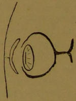
Diagram 55.
Diagram 55.

Diagram 56.
Diagram 56.
So much for the chemical sense organs; they are comparatively simple. But between a single cell, which always makes towards or always hurries out of a ray of light passing through the water in which it swims, and an animal with eyes capable of recognising the colour,[73] shape, size, and distance of objects in space, there really does seem to be a wide gulf. It is not, however, too wide to be bridged.
So much for the chemical sense organs; they're relatively simple. But there's a big difference between a single cell that always moves toward or away from a beam of light in the water where it swims and an animal with eyes that can recognize the color, shape, size, and distance of objects in space, [73]. However, that gap isn’t too wide to cross.
After the single-cell stage has been passed, and we have beasts consisting of an inner layer of cells which is digestive in function, and an outer layer which is protective, motor, and sensory, the power of perceiving light is doubtless vested in the outer layer. When we get beasts consisting of three layers progressing along the straight path of development which leads to man, we find the outer layer becoming too opaque for this purpose, and the torch is handed on to the sensory tube derived from it. (See Diagram 5.) As more and more protection is required, the skin thickens, and the neural tube comes to lie deeper, as in Diagram 51. In order not to lose the light altogether, it has to throw out buds, which concentrate in themselves the peculiar faculty of perceiving it, and at the same time little pits are formed in the skin just over them to help the light to reach them. (See Diagram 52.) In Diagram 53 both the nervous elements and the integumentary are developing their possibilities; and in Diagram 54 a large surface has been prepared for the reception of light, and a lens formed to focus the rays upon it. Diagrams 55 and 56 give the concluding stages in the development of the eye: the formation of the cornea and its protecting eyelids. The two cavities are filled with clear liquids, and the whole eyeball supported by connective tissue.
After the single-cell stage has been passed, and we have organisms made up of an inner layer of cells for digestion and an outer layer that provides protection, movement, and sensory functions, the ability to perceive light is clearly held by the outer layer. As we move to organisms with three layers evolving along the path toward humans, we see the outer layer becoming too opaque for this purpose, and the responsibility shifts to the sensory tube that comes from it. (See Diagram 5.) As more protection is needed, the skin thickens and the neural tube becomes positioned deeper, as in Diagram 51. To avoid completely losing the ability to perceive light, the organism has to develop buds that enhance its capacity to detect it, while small pits form in the skin directly above them to allow light to reach these areas. (See Diagram 52.) In Diagram 53, both the nervous system and the skin are evolving their capabilities; and in Diagram 54, a large area has been prepared to receive light, along with a lens developed to focus the rays onto it. Diagrams 55 and 56 describes the final stages in the development of the eye: the formation of the cornea and its protective eyelids. The two cavities are filled with clear fluids, and the entire eyeball is supported by connective tissue.
So fascinating is everything connected with the eye that the temptation to describe it in detail is great; but in a book of rough outlines, and in consideration of the many important matters yet awaiting their turn, we must confine ourselves to briefly mentioning a few of the more important points concerning it. The light is focussed by the lens upon the nervous curtain at the back, and produces there a picture, as in the photographic camera. Thus we perceive the shape of objects. The different rays of the spectrum affect different elements in this curtain or retina, whereby we get sensations of colour. Finally, the clearness of the picture, its size, the degree of convergence of the two eyes, and the effort of focussing—for[74] the curvature of the surface of the lens can be altered—enable us to estimate the size and distance of an object. And now, though it would take volumes to do justice to the physiology of vision, we must pass on to deal equally briefly with the functions of that no less important organ, the ear.
Everything related to the eye is so fascinating that it's tempting to describe it in detail; however, since this is a book of rough outlines and there are many important topics still to cover, we need to briefly mention a few key points about it. Light is focused by the lens onto the nervous layer at the back, creating an image much like a photographic camera does. This is how we perceive the shape of objects. Different rays of the spectrum interact with various elements in this layer or retina, giving us sensations of color. Additionally, the clarity of the image, its size, the degree of convergence of both eyes, and the effort of focusing—since the curvature of the lens can be adjusted—allow us to estimate the size and distance of an object. Although it would take many volumes to fully cover the physiology of vision, we must now move on to briefly discuss the functions of the equally important organ, the ear.
The essential part of the ear is a membranous bag, formed by the pouching in of the outer layer of cells—as shown in Figs. 1, 2, and 3 of Diagram 57—which comes to lie in a bony chamber beneath the skull, and assumes the somewhat complicated shape depicted in Fig. 4. We have not time, nor is it for our purpose necessary, to trace all the steps in the development of the ear, either external or internal, nor need we spend much time upon its structure, beyond indicating its position. But its position, which is shown in Diagram 58, must be grasped in order to understand how it is influenced by sound.
The main part of the ear is a membranous bag, created by the outer layer of cells folding in on itself—as shown in Figs. 1, 2, and 3 of Diagram 57—which sits in a bony chamber beneath the skull and takes on the somewhat complex shape shown in Fig. 4. We don’t have time, nor is it necessary for our purpose, to go through all the steps in the ear’s development, either external or internal, nor do we need to spend much time on its structure, other than to note its position. However, its position, as shown in Diagram 58, must be understood to grasp how it is affected by sound.

Diagram 57.—Showing Development of the Membranous Labyrinth of the Ear.
Diagram 57.—Illustrating the Development of the Membranous Labyrinth of the Ear.
U, Utricle; C, cochlea; S, saccule; S.C., semicircular canals.
U, Utricle; C, cochlea; S, saccule; S.C., semicircular canals.
It will be seen that the membranous bag, which is fitly termed the labyrinth, is situated in a bony cavity which fits so closely as to be termed the bony labyrinth (C). The membranous labyrinth is filled with a liquid, called endolymph, and the bony labyrinth (C) is also filled with a liquid, called perilymph, in which the membranous bag swims. All this is called the inner ear. The inner ear communicates with a second cavity—the middle ear (B)—by two apertures in the bony wall, which are closed by membranes. The middle ear is full, not of liquid, but of air, and is separated from the external ear, the cavity[75] marked A, which is open to the external world, by another membrane called the tympanum, or drum, of the ear. The middle ear is connected by a tube with the throat, so that the pressure of the air on both sides of the drum may be the same.
It can be seen that the membranous bag, aptly named the labyrinth, is located in a bony cavity that fits so closely it’s called the bony labyrinth (C). The membranous labyrinth is filled with a liquid known as endolymph, while the bony labyrinth (C) contains a liquid called perilymph, in which the membranous bag floats. All of this constitutes the inner ear. The inner ear connects to a second cavity—the middle ear (B)—through two openings in the bony wall, which are covered by membranes. The middle ear is filled with air rather than liquid and is separated from the external ear, the cavity [75] marked A, which is open to the outside world, by another membrane called the tympanum, or eardrum. The middle ear is linked by a tube to the throat to equalize the air pressure on both sides of the eardrum.

Diagram 58.—Showing the Position of the Ear.
Diagram 58.—Illustrating the Location of the Ear.
A., Outer ear; B., middle ear; C., inner ear.
A., outer ear; B., middle ear; C., inner ear.
Now, the object of this arrangement is that the ear may be able to fulfil one of its principal duties, namely, the perception of sound. Sound, as the reader is doubtless aware, is transmitted through the air as waves of condensation and rarefaction, due to the swinging backwards and forwards of its particles; it resembles the passing on of a bump along a line of trucks on the railway when the engine runs up against the end one preparatory to coupling. The magnitude of this oscillation we perceive as the loudness, the frequency as the pitch of a note. Now, when the waves of sound strike against the drum of the ear, they cause it to vibrate backwards and forwards also. Supposing there was no middle ear, and the sound waves beat directly upon the membranous windows of the inner ear, these could not be made to vibrate, as there is liquid behind them, and liquids are incompressible; so, in order that the movements[76] of the drum may be transmitted to the liquids of the inner ear, they are carried across the middle ear by a chain of small bones, by which their extent is curtailed, but their force increased, and brought to bear upon one only of the two openings. The consequence of this is that the membrane closing it is able to vibrate and pass on the vibrations to the liquid within, since when it is pushed in, the membrane covering the other hole is pushed out.
Now, the purpose of this setup is to enable the ear to perform one of its main functions, which is to perceive sound. Sound, as you probably know, travels through the air in waves of compression and rarefaction, caused by the movement of its particles back and forth; it's similar to how a bump travels along a line of train cars when the engine pushes against the last one before coupling. We perceive the strength of this movement as loudness, and the frequency as the pitch of a note. When sound waves hit the eardrum, they make it vibrate back and forth as well. If there were no middle ear, and the sound waves directly struck the membranes of the inner ear, these wouldn't be able to vibrate because there's liquid behind them, and liquids can't be compressed. Therefore, to transmit the vibrations from the eardrum to the liquids in the inner ear, they are carried across the middle ear by a chain of small bones, which reduce their extent but increase their force, directing them at just one of the two openings. As a result, the membrane closing that opening can vibrate and transfer the vibrations to the liquid inside, since when it's pushed in, the membrane covering the other hole is pushed out.

Diagram 59.—The Semicircular Canals.
Diagram 59.—The Semicircular Canals.
Exactly how the different parts of the membranous labyrinth contribute to our perception of sound we do not quite know. It appears as though the difference of pressure in saccule and utricle originally conveyed to the brain a sensation of noise without any idea of quality, while the cochlea was developed later to analyze sounds and give information as to pitch and tone. Whether the rest of the labyrinth has any longer a part to play in the perception of sound, we cannot say with certainty; but it seems pretty certain that the cochlea is the organ for receiving musical impressions. Here, again, though, we are at a loss, for we do not know with certainty how the cochlea acts. In shape it is a long tube, and in the head is coiled spirally—like a snail’s shell to look at. Along its whole length is a ridge of cells with short hairs projecting from their inner surface into the liquid it contains; and to the cells along this ridge a branch of the auditory nerve is distributed. But as to whether one[77] of the cells along this keyboard responds to each of the notes we can distinguish, or whether they are affected as a whole, physiologists are not yet agreed.
We're still not entirely sure how the different parts of the membranous labyrinth help us perceive sound. It seems like the pressure differences in the saccule and utricle initially sent a sensation of noise to the brain without conveying any idea about its quality, while the cochlea developed later to analyze sounds and provide information about pitch and tone. We can't say for certain if the rest of the labyrinth still plays a role in sound perception, but it seems clear that the cochlea is the main organ for receiving musical impressions. However, we're also uncertain about how the cochlea actually works. It's shaped like a long tube and spirals in a way that resembles a snail's shell. Along its entire length, there's a ridge of cells with tiny hairs projecting into the liquid it contains; a branch of the auditory nerve connects to these cells. But whether each of the cells along this ridge responds to individual notes we can distinguish, or if they all react together, is still a point of debate among physiologists.
At least one other important duty the ear performs; it tells us in what position we are, and how our whole head moves or is moved. On the top of the saccule, in Diagram 57, Fig. 4, there are shown three little loops which are called the semicircular canals. They are shown again more clearly by themselves in Diagram 59.
At least one other important job the ear does; it lets us know the position we’re in and how our entire head moves or is moved. On top of the saccule, in Diagram 57, Fig. 4, three small loops are shown, which are called the semicircular canals. They are shown again more clearly by themselves in Diagram 59.
Fig. 1 shows their position with regard to each other. It will be seen that two of them are vertical, with their loops forming a right angle with one another, and that the other is horizontal—in fact, that they lie in the three planes of space. Fig. 2 shows the structure of one of them; it has a swelling at one end (a), and a knob projecting into it where the nerve joins it (b). In Fig. 3 is shown a section through this knob, which gives the key to the use of these structures. A little head of cells projects from the wall of the canal into its lumen, and from these cells hairs bristle out into a dome-like covering of jelly, weighted, to prevent its moving too easily, with small particles of lime. Now, if you take up a round vessel full of liquid—say a bowl of gold-fish—and give it a twist round, you will notice that, though the bowl turns, the water inside does not; the fish remain in their old position. If there were a rod projecting from the side of the bowl, it would, of course, move with it, and if a fish came in its way would strike against it. This is the principle of the semicircular canal. For if we turn our head, the tube of the canal turns, passing over the liquid in it, which of course does not move, though it appears to flow in the opposite direction. The consequence is that the hairs on the side of the knob in the direction in which the head is being moved are pressed upon by the dome of jelly, which, as it floats in the liquid, tends to remain where it is. The nerves, stimulated in this way, inform the animal generally of the movement.
Fig. 1 shows how they are positioned relative to each other. You can see that two of them are vertical, with their loops forming a right angle with each other, while the other one is horizontal—in fact, they are arranged in the three planes of space. Fig. 2 shows the structure of one of them; it has a swelling at one end (a), and a knob extending into it where the nerve connects (b). Fig. 3 displays a section through this knob, which explains how these structures work. A small cluster of cells extends from the wall of the canal into its interior, and from these cells, hairs extend outward into a dome-like layer of jelly, which is weighted down with tiny particles of lime to keep it stable. Now, if you pick up a round container filled with liquid—like a bowl of goldfish—and give it a spin, you'll notice that while the bowl turns, the water inside stays still; the fish remain in their original position. If there were a rod sticking out from the side of the bowl, it would move with it, and if a fish got in the way, it would bump into it. This is the principle behind the semicircular canal. When we turn our head, the canal tube rotates, moving over the liquid inside it, which remains still, though it seems to flow in the opposite direction. As a result, the hairs on the side of the knob toward which the head is turning get pressed by the jelly dome, which, buoyed by the liquid, tends to stay put. The nerves, activated in this way, relay information to the animal about its movement.
These little organs are very important to us, though we have our eyes to correct our ideas of position, and they are still more so to the fish, which dart and turn in[78] the wide expanse of the ocean, and the birds and bats, which wheel about in the air. There are, however, some occasions when we do not feel inclined to bless them; for, inasmuch as they faithfully report every roll and plunge of a ship to a person on board, it is they which are mainly responsible for sea-sickness.
These small organs are really important to us, even though we have our eyes to help us sense our position, and they’re even more crucial for fish that swim and turn in[78] the vast ocean, as well as for birds and bats that fly around in the air. However, there are times when we don’t feel grateful for them because they accurately signal every roll and dip of a ship to someone on board, which is mainly why we experience sea sickness.
And now that we have seen how the body lies with regard to the external world; how it is efficiently protected from its surroundings; how it is placed in communication with them; and have briefly examined the organs by which it makes its chemical and physical investigations, looks out into space, and is kept aware of what is going on therein, we may return to the means whereby it responds as a whole to the stimuli thus reported—the central nervous system—and try to learn how the right response is brought about.
And now that we’ve seen how the body interacts with the outside world, how it’s effectively protected from its environment, how it communicates with it, and have briefly looked at the organs that allow it to conduct chemical and physical investigations, observe the space around it, and stay aware of what’s happening there, we can go back to how it responds as a whole to the stimuli it receives—the central nervous system—and try to understand how the right response is generated.
IV.
There is but one thing more to describe in the mechanism of the body—the connecting link between the last two sections. In the last we saw how the body receives stimuli from the external world; in the one before, that when these stimuli reach the central nervous canal it in turn stimulates the organs to perform such movements as circumstances require. What, therefore, remains to be described is the working of that canal by which these necessary movements are ordered and controlled.
There’s just one more thing to explain about the body’s mechanism—the connection between the last two sections. In the previous section, we looked at how the body receives signals from the outside world; in the one before that, we saw that when these signals reach the central nervous system, it prompts the organs to perform movements as needed. What’s left to describe is how that system directs and manages these necessary movements.
Now, in speaking of reflex action a few pages back, we said that the nerves which bring in stimuli from the periphery distribute them about the neural canal to those cells whose activity, by sending out fresh stimuli to the muscles, produces the requisite movements. These motor cells, however, are not scattered about the spinal cord anyhow. They are collected into clusters, or nuclei, as they are sometimes called, and each cluster has special duties—i.e., a special organ to control. Thus, we say that there are in the central nervous system centres—a nervous centre to control the leg; another to work[79] the diaphragm; another for the muscles of the ribs; more for the arm, hand, etc. And these centres are in communication with one another, so that they may not pull different ways.
Now, when we talked about reflex actions a few pages ago, we mentioned that the nerves bringing in stimuli from the outside send them through the neural canal to specific cells. These cells trigger fresh stimuli to the muscles, creating the necessary movements. However, these motor cells aren't just scattered throughout the spinal cord randomly. They are grouped into clusters, or nuclei, as they're sometimes called, and each cluster has specific functions—like controlling a particular organ. For example, there’s a nervous center in the central nervous system for controlling the leg; another one for operating the diaphragm; another for the muscles of the ribs; and more for the arm, hand, etc. These centers communicate with each other to ensure they work together rather than at cross purposes.
In the first example of reflex action given in Section II. of this essay, the sensation of a pin-prick was first conveyed to the centres controlling the limb injured, by whose activity it was drawn away from the danger. But the nerve which gave the warning which produced this elementary movement distributed the impression that something was wrong to the higher centres, so that the whole body was involved in protecting, doctoring, and avenging the outraged member; from which it would appear that the lower centres are under control of higher ones. And this is the case. If we may be allowed the metaphor, there are captains of tens, who are under the direction of captains of fifties, and the captains of fifties receive their orders from captains of hundreds. The nerve canal, the manner of whose formation as a simple tube is shown in Diagrams 5 and 42, has therefore different functions in different parts, and this to such an extent that considerable differentiation in bulk and structure is produced.
In the first example of reflex action provided in Section II. of this essay, the feeling of a pinprick was initially sent to the centers that control the injured limb, which then moved it away from the danger. However, the nerve that gave the alert for this basic movement also sent the message that something was wrong to the higher centers, engaging the entire body in protecting, healing, and responding to the injured part. This suggests that the lower centers are controlled by the higher ones. And that is indeed the case. If we can use a metaphor, there are captains of tens who report to captains of fifties, and the captains of fifties take their orders from captains of hundreds. The nerve canal, whose formation as a simple tube is illustrated in Diagrams 5 and 42, thus has different functions in various parts, to the extent that significant differences in size and structure occur.
The neural canal may be roughly divided into two parts—a comparatively simple tube, running the greater part of the animal’s length, containing many centres from which nerves run to the organs they control; and a complicated bulbous enlargement at one end, with thickened walls, in which are the centres controlling those in the cord, and thereby managing not so much organs as the whole animal. The former is called the spinal cord, the latter the brain.
The neural canal can be generally divided into two parts—a relatively simple tube that runs along most of the animal's length, containing many centers from which nerves extend to the organs they control; and a complex bulbous enlargement at one end, with thicker walls, housing the centers that control those in the cord, thus managing not just the organs but the entire animal. The first part is called the spinal cord, while the second is the brain.
This division, accustomed as we all are to take it for granted, offers plenty of food for reflection. Why should an animal have such a brain placed in its head? Why, indeed, should it have a head, regarding that member as a group composed of eyes, nose, mouth, ears and brain? The mouth gives us the key to the riddle; the mouth is the essential organ, and all the rest are its accessories.
This division, which we all tend to take for granted, gives us a lot to think about. Why does an animal have such a brain in its head? Why, in fact, should it have a head at all, considering that part is made up of eyes, nose, mouth, ears, and brain? The mouth holds the answer to the puzzle; the mouth is the main organ, and everything else is just extra.
In the first essay we saw that the basis of life was chemical, and in the second that the materials necessary[80] for the chemical action, or food, must, in the higher animals, be taken into the digestive tube through the opening which we call the mouth. Therefore, as it is highly important that only the most beneficial substances shall be received into it, and that all which are actively injurious shall be excluded, it is plain that the organs of chemical perception must be placed in its neighbourhood—the organs of smell to enable the mouth to find its food, and the organs of taste to aid in selecting it. As, moreover, our humble ancestors, the fishes, move literally mouth foremost, it is not surprising to find the organs of space perception, the eyes, also situated in its neighbourhood, especially when one considers that their food is often of a lively character, and requires precision of movement to secure it. The inevitable consequence of thus grouping the more important organs of perception under the fore-end of the neural canal is that it grows and develops more highly here than elsewhere along its length, and soon is in a position to dictate to the rest of the body. Another reason why it must develop is that it must contain centres for turning its impressions to practical account, not only by producing complicated movements in the jaws, eyes and gills, but also by ruling the centres in the cord, and instructing the body to carry the mouth whither it needs to go.
In the first essay, we learned that the foundation of life is chemical, and in the next section, we discussed that the materials needed for chemical action, or food, must be taken into the digestive system through the opening we refer to as the mouth. Therefore, it's crucial that only the most beneficial substances enter it, while actively harmful ones are kept out. It's clear that the organs of chemical perception need to be located nearby—the sense of smell helps the mouth find food, and the sense of taste assists in selecting it. Additionally, since our simple ancestors, the fishes, literally move with their mouths first, it's not surprising that the organs for perceiving space, the eyes, are also located nearby, especially considering that their food often moves and requires precise movements to catch. The inevitable result of placing the more important sensory organs at the front end of the neural canal is that this area grows and develops more than other parts, quickly becoming capable of directing the rest of the body. Another reason for this development is that it must contain centers to effectively use its impressions, not only by controlling complex movements of the jaws, eyes, and gills, but also by managing the centers in the spinal cord to instruct the body on where to move the mouth.

Diagram 60.—Showing Primary Division of Nervous Tube.
Diagram 60.—Displaying the Main Division of the Nervous Tube.
In the preceding diagram (60) the origin of the brain is shown as a dilatation of the end of the neural canal into a bulb with thickened walls, which has already become constricted in places, so that it is subdivided into three. The next diagram (61) is intended to give, in no matter[81] how crude and schematic a way, some idea of the lines on which the development continues. We do not show all, or even half, the structures which go to make up the brain. To do so would be out of place in a book like this. Further, we shall endeavour as far as possible to speak of the brain in general terms, avoiding the five-syllable bastard Græco-Latin names with which the early anatomists have endowed almost every square inch of its substance, and confine ourselves to summing up its functions as briefly as can be done with justice.
In the previous diagram (60), the brain's origin appears as an expansion of the end of the neural canal into a bulb with thickened walls, which has already narrowed in some areas, dividing it into three parts. The next diagram (61) aims to provide, no matter how simplistic and schematic, a general idea of the way development progresses. We won't be showing all, or even half, of the structures that make up the brain. That would be inappropriate for a book like this. Additionally, we'll try to discuss the brain in general terms as much as possible, avoiding the complicated five-syllable Greco-Latin terms that early anatomists used for almost every inch of its structure, and focus on summarizing its functions as briefly as possible while still being fair.

Diagram 61.—Giving a Rough Idea of how the Brain is developed.
Diagram 61.—Understanding Brain Development Basics.
In pursuance of this method, attention must be drawn to the fact that only the foremost of the three original bulbs (marked A in the diagram) and the hindermost (C) continue to grow. The middle one (B) remains comparatively simple. From the foremost lobe buds grow out to form the eyes in the manner which we have already described, and other buds push forwards to meet the nerves from the nose. The latter have, even in the early stages shown in Diagram 61, reached an extraordinary size; and when we come to trace them further, we shall find that they become very complex, and acquire remarkable and unexpected powers, considering their humble origin. Strange changes also take place in the hindermost bulb. It splits along the top, so that the cavity it contains is open like a saucer, though bridged over by a three-lobed body called the cerebellum.
In following this method, it's important to note that only the front of the three original bulbs (marked A in the diagram) and the back one (C) continue to grow. The middle one (B) stays relatively simple. From the front lobe, buds grow out to form the eyes as we've already described, and other buds push forward to connect with the nerves from the nose. Even in the early stages shown in Diagram 61, these nerves have reached an incredible size; and as we trace them further, we'll see that they become quite complex and gain remarkable and unexpected abilities, given their humble beginnings. Strange changes also happen in the back bulb. It splits at the top, creating an open space like a saucer, though it's covered by a three-lobed structure called the cerebellum.
Following the spinal cord up into the brain, we are conscious of no sudden line of demarcation separating the one from the other, only of an increasing size and complexity. The lower parts of the brain send out and receive nerves much as the cord does; three pairs go to the muscles which turn the eyes; other pairs bring in sensations from the face and throat; others control the muscles of the face, tongue and throat. But the brain differs from the cord in being directly connected by nerves, not only with adjacent parts, but also with the distant and more important organs in the interior of the body—heart, lungs, etc.; in containing groups of cells which have stimuli sent on to them from all over the body viâ the cord; and in possessing centres which control those lower down in the nervous system. It therefore not only receives and balances stimuli from all over the body, but, by governing the centres which preside over the bodily movements, is able to wield and direct the body as a whole.
Following the spinal cord into the brain, we notice there’s no clear line separating the two, just an increase in size and complexity. The lower parts of the brain send and receive nerves similarly to the spinal cord; three pairs control the muscles that move the eyes, other pairs bring in sensations from the face and throat, and more control the muscles of the face, tongue, and throat. However, the brain is different from the spinal cord because it connects directly by nerves, not just with nearby areas but also with distant and important organs inside the body—like the heart and lungs; it contains groups of cells that receive signals from all over the body through the spinal cord; and it has centers that control those lower parts of the nervous system. Therefore, it not only receives and processes signals from all over the body but, by managing the centers that oversee bodily movements, can direct and control the body as a whole.
The hinder divisions of the brain, which we shall consider first, have no connection with consciousness or volition. They only produce reflex movements, which, however, owing to the wealth of material they have to work upon, are wonderfully complex and far-reaching.
The back parts of the brain, which we'll discuss first, don't connect to awareness or will. They only create reflex movements that, due to the rich materials they have to operate with, are incredibly complex and extensive.
Let us take a few examples. In the hindermost division of the brain (C in the diagrams) there is the centre which presides over the oxygen supply, the importance of which we saw in the essay on vital chemistry. This centre perceives when the lungs have been filled with a gas, and causes them to be emptied; it perceives when they are empty, and again does not allow them to remain too long in that state, before ordering an inspiration; it notes the quality of the air which is passing through the nose, and it notes the quality of the blood which bathes its own cells. The condition of the blood, indeed, is closely watched. An excessive quantity of carbonic acid gas, poverty of oxygen, even temperature, all produce through it an effect upon the rhythm of the breathing.
Let’s look at a few examples. In the back part of the brain (C in the diagrams), there’s the center that controls the oxygen supply, which we discussed in the essay on vital chemistry. This center detects when the lungs are filled with gas and triggers them to empty; it senses when they’re empty and quickly initiates a breath. It assesses the quality of the air passing through the nose and also the quality of the blood that nourishes its cells. The state of the blood is carefully monitored. High levels of carbon dioxide, low oxygen levels, and even temperature all affect the rhythm of breathing.
Close by the respiratory centre is the centre which controls the circulation. But enough has been said in[83] the section on reflex action, wherein the process of fainting was described, to give an idea of the part it plays in the body; so it need not detain us here.
Close to the respiratory center is the center that controls circulation. But we've already discussed this in[83] the section on reflex action, where we talked about the process of fainting, which illustrates its role in the body; so we won't linger on it here.
We cannot, however, pass over its neighbour, the centre of temperature, so briefly. Its methods not only afford one of the most striking and interesting examples of harmonious regulations by reflex action, but the subject of temperature itself is so important that we must describe in some detail how that of the body is kept level.
We can't overlook its neighboring center of temperature so quickly. Its methods provide one of the most noteworthy and fascinating examples of harmonious regulation through reflex action, and the topic of temperature itself is so crucial that we need to explain in more detail how the body's temperature is maintained at a constant level.
As we said when discussing protoplasm generally, life—that is, the change always going on in the protoplasmic substance—is influenced by temperature: the single cell becomes less active at a low temperature, and dies at a high one; so obviously there is a temperature at which its functions are most easily carried on. Inside the body the cells are all kept at the temperature best for them by the circulation of the blood; but the absolute temperature of the whole body depends upon the heat which is generated within it by chemical action, and the heat which it loses to, or receives from, its surroundings. Under normal conditions this temperature in man is 98·4° F., when the production of heat from its own metabolism is balanced by the loss of heat by radiation. If, however, the atmosphere be very hot, less heat is developed in the body, the general metabolism being slower; and more heat is lost, since by reflex action the skin is bathed in sweat and cooled by its evaporation, and the small bloodvessels under the skin are dilated, so that more blood being brought to the surface, its chance of being cooled by radiation is thereby increased. If, on the other hand, the atmosphere is cool, the loss at the surface is minimized by constriction of the cutaneous bloodvessels, and a checking of the perspiration and consequent evaporation; while internally more heat is generated by increased metabolism. The cells which are mainly responsible for the production of heat are those of the muscles; and when much heat is required they increase in activity, not only in their general tone, but even by a visible movement, which we describe as shivering. So, within reasonable limits, whatever the temperature of its surroundings may be, that of the body remains the same,[84] and though we may raise or lower our temperature by lying in a hot or cold bath, reflex adjustment of the sweat glands, bloodvessels and muscles brings it quickly back to normal when we emerge.
As we mentioned when talking about protoplasm in general, life—that is, the ongoing changes in protoplasmic substance—is affected by temperature: a single cell becomes less active in cold temperatures and dies in high ones; so clearly, there is a temperature at which its functions are most effectively performed. Inside the body, cells are kept at their optimal temperature by the blood circulation; however, the overall temperature of the body depends on the heat generated internally by chemical processes and the heat lost to or gained from the environment. Under normal conditions, the temperature in humans is 98.4° F., when heat production from metabolism balances heat loss through radiation. If the environment is very hot, less heat is produced in the body, as overall metabolism slows down; more heat is lost because the skin sweats and cools through evaporation, and the small blood vessels under the skin widen, allowing more blood to surface and increasing its cooling by radiation. Conversely, if the environment is cool, heat loss at the surface is minimized by narrowing the skin's blood vessels and reducing sweating and evaporation; while internally, more heat is generated through increased metabolism. The cells mainly responsible for producing heat are muscle cells; and when more heat is needed, they become more active not only in general tone but also through visible movement, which we call shivering. So, within reasonable limits, regardless of the surrounding temperature, the body's temperature remains stable,[84] and even though we can raise or lower our temperature by lying in a hot or cold bath, the reflex adjustments of sweat glands, blood vessels, and muscles quickly restore it to normal when we get out.
With a passing mention of the cerebellum, the three-lobed organ shown in Diagram 61, and seen again in a more advanced stage in Diagram 63, we may dismiss the two hinder divisions of the brain.
With a brief mention of the cerebellum, the three-lobed organ shown in Diagram 61, and appearing again in a more advanced stage in Diagram 63, we can overlook the two back parts of the brain.
The cerebellum lies on the upward path of fibres from the cord to the higher centres in the fore-brain. It is a somewhat complicated organ, and its functions are not yet fully known. The older physiologists took a very extreme view of its importance, assigning to it, among other romantic duties, that of providing a habitation for the soul. This opinion on the strength of later research we can hardly endorse. The cerebellum really seems mainly concerned in co-ordinating the action of the muscles, especially in maintaining equilibrium in standing and walking.
The cerebellum is located along the pathway of fibers that connect the spinal cord to the higher centers in the forebrain. It's a fairly complex organ, and its functions are not entirely understood yet. Older physiologists had an exaggerated view of its significance, claiming it had various grand responsibilities, including being a home for the soul. Based on more recent research, we can hardly support that idea. The cerebellum appears to primarily play a role in coordinating muscle actions, particularly in helping maintain balance while standing and walking.
Our knowledge of the whole brain is very far from complete. We should like to know the peculiar function of each little group of cells that can be made out under the microscope, and the paths of all the fibres connecting the different parts of the nervous system. As it is, we have to wait with the best patience we can while they are being investigated, and hope. In few departments, however, have the labours of the physiologist proved more fruitful and interesting than in the study of the fore-brain (A in the diagrams).
Our understanding of the entire brain is still quite incomplete. We want to know the specific function of each small group of cells visible under the microscope, as well as the connections of all the fibers linking different parts of the nervous system. For now, we have to be patient while these areas are being explored, and remain hopeful. However, in few fields have the efforts of physiologists been more rewarding and fascinating than in the study of the forebrain (A in the diagrams).
In the simpler form, as shown in Diagram 61, A, and Diagram 62, Fig. 1, A, the fore-brain is remarkable in that it throws out buds for the two most important sense organs—those of sight and smell. So important are these senses, especially in our humble ancestors, as we have already pointed out, that it is not surprising to find the impressions of the other senses brought on up from the hinder parts of the brain to be compared with them. The fore-brain is, in fact, a sort of terminus whither the whole of the afferent or incoming stimuli are brought, and whence, since information is only received in order to be acted upon, the supreme orders to the body issue.
In a simpler form, as shown in Diagram 61, A and Diagram 62, Fig. 1, A, the forebrain is notable because it develops buds for the two most essential senses—sight and smell. These senses are so crucial, especially for our early ancestors, as we've already mentioned, that it's not surprising to find that impressions from other senses are relayed from the back parts of the brain to be compared with them. The forebrain is essentially a kind of hub where all incoming stimuli are directed, and from which, since information is only gathered to be acted upon, the final commands to the body are issued.
In the fore-brain there are centres for specially governing all the motor organs; but by a strange arrangement the main root of the brain is overwhelmed by its own offshoot, the hemisphere, or lobe which gives rise to the olfactory bud. In fact, so great is the importance of the sense of smell to an animal whose one object in life is to find food, that, instead of the hemisphere being subordinate to its parent, it seems to take over most of the latter’s business, receiving a report of the sensations collected by it, and sending out orders upon its own initiative. Yet, unimposing though the history of this division of the brain may be, it ultimately becomes the seat of consciousness, whereby the mental processes are carried on, and whence all voluntary movements spring.
In the forebrain, there are centers that control all the motor functions. However, in a peculiar twist, the main part of the brain is overshadowed by its own extension, the hemisphere or lobe that produces the olfactory bulb. The sense of smell is so crucial for an animal whose primary goal is to find food that instead of the hemisphere being secondary to its parent, it seems to take over most of the parent's functions. It receives reports on the sensations the parent gathers and sends out commands on its own. While the history of this part of the brain may seem unremarkable, it ultimately becomes the center of consciousness, where mental processes happen and from which all voluntary movements originate.

Diagram 62.—Showing how the Cerebral Hemispheres are developed from A, the Foremost Bulb of the Brain.
Diagram 62.—Illustrating how the Cerebral Hemispheres develop from A, the Front Bulb of the Brain.
Of course, in order to do this, the hemispheres have to grow considerably, and thus we find them enveloping the rest of the fore-brain and swamping it in structure as well as in function. Diagram 62 indicates how this is done, while Diagram 63 shows roughly the proportion and position the different parts of the brain ultimately attain. Finally, Diagram 64, which is rather more realistic, but still much simplified, presents a view of the organ in the head.
Of course, to make this happen, the hemispheres need to grow significantly, and as a result, they end up covering the rest of the forebrain and overwhelming it both in structure and function. Diagram 62 shows how this occurs, while Diagram 63 gives an approximate idea of the size and position the various parts of the brain eventually reach. Lastly, Diagram 64, which is a bit more realistic yet still quite simplified, offers a view of the organ in the head.
The size of the cerebral hemispheres, compared with the rest of the brain, is especially remarkable. So, too, is their endeavour to increase their surface still more by throwing it into deep folds. (See Diagram 64.) These[86] two features vary with the position of the animal in the scale of development; in man, who stands highest in intelligence and dexterity, the hemispheres are very large indeed compared with the other organs, and seamed all over with a maze of winding furrows. Another remarkable feature is the extreme degree to which specialization is carried out. Different parts of the body are represented, each by a small area of the cortex, or surface layer, and we know at what spot on the cortex such sensations as sight and hearing are perceived, and from exactly what little patch the impulse to move each limb emanates. In the accompanying diagrams (65 and 66) these areas are mapped out, their locality being fixed by the principal folds which act as landmarks on the surface of the hemisphere.
The size of the brain's hemispheres, in comparison to the rest of the brain, is particularly impressive. Their attempt to expand their surface area even further by developing deep folds is equally noteworthy. (See Diagram 64.) These[86] two characteristics change depending on the animal's place in the evolutionary hierarchy; in humans, who rank highest in intelligence and skill, the hemispheres are indeed very large when compared to other organs, covered in a complex network of winding grooves. Another striking aspect is the high level of specialization present. Different parts of the body are represented by specific small areas of the cortex, or outer layer, and we know exactly where on the cortex sensations like sight and hearing are processed, as well as where the signals to move each limb originate. In the diagrams provided (65 and 66), these areas are outlined, with their locations determined by the main folds that serve as reference points on the surface of the hemisphere.

Diagram 63.—Relation of Different Parts of the Brain.
Diagram 63.—Relationship of Different Parts of the Brain.
There is another important fact which we must not omit to mention in speaking of this localization: each hemisphere presides over the opposite side of the body. Early in development the nerve fibres from the eye cross over to the opposite side of the brain, and the afferent fibres from the lower parts of the body have accordingly to follow suit. Then, as the efferent fibres—i.e., those which set the muscles in motion—have to bring about the movements in response to information received, they must also cross to get back to the side from which it came. So, if a tumour grows inside the head on the right side,[87] it is the left eye which becomes sightless, or the left hand which grows numb and powerless, according to the part of the cortex which is pressed upon.
There’s another important point we need to mention when discussing this localization: each hemisphere controls the opposite side of the body. Early in development, the nerve fibers from the eye cross over to the other side of the brain, and the incoming fibers from the lower parts of the body have to do the same. Then, as the outgoing fibers—i.e., those that trigger muscle movement—must create responses based on the information received, they also need to cross back to the side from which they originated. So, if a tumor grows inside the head on the right side,[87] it’s the left eye that becomes blind, or the left hand that becomes numb and weak, depending on which part of the cortex is being pressed.
Perhaps the most interesting part of the whole body is that little band of the cortex running upwards from behind the temple to the crown of the head, in which (cf. Diagrams 64, 65 and 66) the motor areas of the limbs, and the perception of those sensations which we have grouped together and called ‘touch,’ are situated. The minute structure of this region is roughly shown in section in Diagram 67, as it has been made out with the microscope; but only a few of the nerve cells are shown, the connective tissue of feltwork in which they are suspended, and the bloodvessels by which they are nourished, being left out. All the structures represented are of course very, very small; the large black patches which represent cells would really be invisible, and the whole field of the diagram only a mere speck, to the naked eye.
Perhaps the most interesting part of the entire body is that small strip of the cortex that runs from behind the temple to the top of the head, where the motor areas for the limbs and the perception of what we refer to as 'touch' are located (cf. Diagrams 64, 65 and 66). The fine structure of this area is roughly illustrated in section in Diagram 67, as seen through a microscope; however, only a few of the nerve cells are shown, with the connective tissue framework in which they are suspended and the blood vessels that nourish them omitted. All the structures depicted are, of course, very, very small; the large black spots representing cells would actually be invisible, and the whole area of the diagram would appear as just a tiny speck to the naked eye.

Diagram 64.—Position of the Brain in the Head.
Diagram 64.—Location of the Brain in the Head.
A represents the nerve by which impulses are brought in. It runs straight up to the surface of the cortex, and[88] there its branches end, interlaced with those of a many-branched distributing cell (B). The two cells (C and D) shaped like pyramids, which send up branched processes from their apexes, receive an impulse from the distributing cell, and transmit it along the fibre which runs downwards from the middle of their base. Where the fibre from the smaller one goes to we are not sure—probably to another part of the brain to insure harmonious working—but the large pyramidal cell sends its fibre right away through the lower parts of the brain, passing the cell-stations they contain, on into the spinal cord, till it reaches the centre there, which immediately works some particular limb.
A represents the nerve that carries impulses in. It goes straight up to the surface of the cortex, and[88] there its branches end, intertwining with those of a multi-branched distributing cell (B). The two pyramid-shaped cells (C and D) send branched processes up from their tops, receive an impulse from the distributing cell, and transmit it along the fiber that runs downward from the middle of their base. We're not sure where the fiber from the smaller cell goes—probably to another part of the brain to ensure smooth functioning—but the larger pyramidal cell sends its fiber straight through the lower parts of the brain, passing the cell-stations it contains, and into the spinal cord, until it reaches the center there, which immediately triggers a specific limb.

Diagram 65.—Map of the Cerebral Hemisphere, showing the Areas in which Various Functions are localized.
Diagram 65.—Map of the Cerebral Hemisphere, indicating the areas responsible for various functions.
Supposing we anæsthetized somebody, throwing him into deep unconsciousness, and then opened his skull, laying bare the brain as is done in Diagram 64, only not quite in such a wholesale manner. If we then stimulated the part of the brain we are now considering at different places with electric needles, using a weak induction current, we should see him moving different members according to the different regions touched—now an arm, now a leg, now the whole head. If we were to place the electrodes in the centre for the hand, and then gradually increase the strength of the current, the activity of the hand centre would throw other centres into activity.[89] The arm would move next, raising the hand towards the face. Then the eyes would turn, and the whole head to meet the hand. Lastly the mouth would open. The movements are those of putting something into the mouth—the ruling passion strong in unconsciousness.
Suppose we anesthetized someone, putting him into a deep unconscious state, and then opened his skull, exposing the brain as is done in Diagram 64, but not quite so extensively. If we then stimulated the part of the brain we are currently examining at different spots with electric needles, using a weak induction current, we would see him move different limbs depending on the areas we touched—first an arm, then a leg, and then the whole head. If we placed the electrodes in the center for the hand and gradually increased the current's strength, the activity in the hand's center would activate other centers. The arm would move next, raising the hand toward the face. Then the eyes would turn, and the whole head would follow the hand. Finally, the mouth would open. The movements mimic putting something into the mouth—the strong instinct dominating even in unconsciousness.[89]

Diagram 66.—Map of the Cerebral Hemisphere, showing the Areas in which Various Functions are localized.
Diagram 66.—Map of the Brain Hemisphere, displaying the Areas where Various Functions are located.
Such experiments were, of course, first made upon animals, but they have been fully verified on the human subject. The story of how this was done is not, however, a romance with a martyr or a criminal for the central figure. The corpus vile was not provided by a volunteer, or kidnapped and bound in a dark cellar, but treated as a patient in the airy wards of a hospital. With increasing knowledge of the brain, it was found that epilepsy was cortical in origin. A little piece of the cortex becomes diseased and hyperexcitable. The sufferer suddenly becomes acutely conscious of one of his members—a hand or a foot, say—not because there is anything the matter with it, but because the corresponding area in the brain is morbidly active, and he refers the sensation to the part from which it receives its nerves. The next moment the limb begins to twitch, and the excitement spreading, as in the experiment we described above, to other centres which are not diseased, they, too, become[90] morbidly active, and the whole body is thrown into convulsions. This is a disease which must be checked as soon as possible. The surgeon accordingly lays bare the part of the brain affected, knowing now where to look; finds the exact spot which is diseased by reproducing the first twitchings of a fit by electric stimulation, and removes the source of the trouble.
Such experiments were initially conducted on animals, but they have been thoroughly validated on humans. The account of how this was achieved is not a dramatic tale featuring a martyr or a criminal as the main character. The corpus vile was not sourced from a volunteer, nor was it kidnapped and confined in a dark basement, but rather treated as a patient in the bright wards of a hospital. With a growing understanding of the brain, it was discovered that epilepsy originates in the cortex. A small part of the cortex becomes diseased and overly excitable. The affected person suddenly becomes intensely aware of a part of their body—a hand or a foot, for instance—not because there’s anything wrong with it, but because the corresponding area in the brain is excessively active, causing them to feel sensations in that part due to its nerve connections. Moments later, the limb starts to twitch, and as the excitement spreads, like in the previously described experiment, to other nearby areas that aren't diseased, those regions also become[90] excessively active, leading to convulsions throughout the body. This is a disease that needs to be addressed as quickly as possible. The surgeon, therefore, exposes the impacted area of the brain, now knowing where to focus; identifies the precise spot that is diseased by reproducing the initial twitches of a seizure using electric stimulation, and removes the source of the problem.

Diagram 67.—Showing the System of Nerve Cells in the Cortex.
Diagram 67.—Displaying the System of Nerve Cells in the Cortex.
A, Afferent fibre; B, distributing cell; C, small pyramidal cell; D, large pyramidal cell.
A, Afferent fiber; B, distributing cell; C, small pyramidal cell; D, large pyramidal cell.
Returning to general considerations, an important point is the way in which the different centres are connected by fibres, which put them into relation. The brain may consist of many centres, just as the body consists of many organs, but both body and brain must live as a whole. If the heart and lungs get out of harmony there is trouble, and if the bridge which connects hearing with motion in the brain breaks down, as occasionally happens for a time in an overworked man, he is mentally at a discount. He can hear and understand, but he cannot write or talk sense: he is sane, but quite helpless, and generally very frightened.
Returning to general considerations, an important point is how the different centers are connected by fibers that relate them to one another. The brain may have many centers, just like the body has many organs, but both the body and the brain need to function as a whole. If the heart and lungs fall out of sync, there’s a problem, and if the connection between hearing and movement in the brain fails, as can happen sometimes in an overworked person, they become mentally impaired. They can hear and understand, but they can't write or speak clearly: they are sane but completely helpless, and usually very scared.
These courts of inquiry are called association centres. It used to be believed that they were all in the fore-part of the brain, under the forehead; but this is evidently not the case. Several men in war or by accident have had the frontal lobes of their brain damaged beyond repair; and when they have been discharged from hospital, where, thanks to the advance made by surgery since anæsthetics and antiseptics were discovered, they have been successfully treated, they have gone back to their work seemingly in no way different from men whose brains were whole. In some cases they have even been reported as having become quicker and sharper than before, probably owing to there being fewer association centres, and thought being accelerated by simpler machinery: facts are thenceforward shaken through a larger-meshed sieve.
These inquiry centers are called association centers. It was once thought that they were all located at the front of the brain, beneath the forehead, but that's clearly not true. Several men have suffered irreparable damage to their frontal lobes during wartime or due to accidents, and when they've been released from the hospital—thanks to advancements in surgery since the discovery of anesthetics and antiseptics—they returned to their work seemingly unaffected, just like men with intact brains. In some cases, they've even been noted to be faster and sharper than before, likely because there are fewer association centers and thought processes are sped up by simpler mechanisms: facts are therefore filtered through a coarser sieve.
A few general considerations, and we have done. There is no centre for memory in the brain. The facts which we remember are not stored as in a box, nor can one imagine how they could be, considering that the physical basis of an idea is molecular change. The whole nervous system is probably concerned in memory, a particular change, which has momentarily occurred in its tissues, being more likely to occur again under certain circumstances than a fresh one, and certain tracks becoming well beaten and more permeable than others. Pleasure and pain are other general phenomena: they are not to be localized in the brain like vision or hearing. Pleasure is the consciousness that the whole body is under favourable conditions; and pain, the knowledge that the protoplasm of certain cells of the body is being acted upon by injurious agents, chemical or physical. There seems to be good evidence that separate nerves convey impressions of injury, distinct from those of touch and temperature; but it is the revolt of the whole body against conditions affecting a part which constitutes pain.
A few general points, and we're done. There isn’t a specific center for memory in the brain. The things we remember aren't stored like items in a box, and it’s hard to imagine how they could be, given that the physical basis of an idea involves molecular changes. The entire nervous system is likely involved in memory, with a particular change that has temporarily occurred in its tissues being more likely to happen again under certain conditions than a new one, creating certain pathways that become well-trodden and easier to access than others. Pleasure and pain are also broad phenomena: they can't be pinpointed in the brain like vision or hearing. Pleasure is the awareness that the whole body is in good conditions; pain is the realization that the protoplasm of specific cells in the body is being affected by harmful agents, whether chemical or physical. There seems to be solid evidence that separate nerves transmit signals of injury, different from those related to touch and temperature; however, it's the body's overall reaction to conditions affecting a part that constitutes pain.
Amidst the maze of perplexities which lies between[92] physiology and psychology, there is, however, one fact which stands out clear and bold: the brain can create nothing. We have seen how matter is taken into the body and matter is cast off from the body. We have seen how energy is released in the body from chemical compounds, and made use of by the body. So now, after a moment’s thought, it must be plain that every stimulus which goes to the brain must have its effect there, and that a man’s thoughts and conduct are entirely dependent on what has, at some time or other, come in from the external world. The association centres can evolve wonderful thoughts, but they are structurally derived from the grosser sense organs, and must get all the material they work upon from them.
Amid the complex web of confusion between[92] physiology and psychology, there's one clear fact: the brain can't create anything from scratch. We've observed how the body takes in matter and also expels it. We've noted how energy is released from chemical compounds in the body and utilized by it. So, after a moment's reflection, it's obvious that every stimulus reaching the brain must have an impact, and that a person's thoughts and actions are completely dependent on what has entered from the outside world at some point. The association centers can produce amazing thoughts, but they are fundamentally built from the basic sense organs and rely entirely on the material provided by them.
The nervous system puts the body into relation with the external world as a whole, but for convenience it is subdivided into the afferent system, by which impressions come in, and the efferent system, by which the muscles are set in motion. Of the two halves, the afferent system has a just right to priority, for the efferent system is merely its consequence. Sights, sounds, smells, etc., reach the brain by afferent paths from the external world, and are there moulded into thoughts. Their effects we see in poetry, architecture, sculpture, or laundry work, according to the method of the brain in treating the raw material it receives, and of a quality corresponding to the fineness with which the brain examines them, and can control the motor organs of the body.
The nervous system connects the body to the external world overall, but for ease, it's split into two parts: the afferent system, which brings in impressions, and the efferent system, which activates the muscles. Of these two parts, the afferent system deserves priority since the efferent system is simply a result of it. Sights, sounds, smells, and so on reach the brain through afferent pathways from the outside world and are shaped into thoughts there. We see their effects in poetry, architecture, sculpture, or everyday tasks, depending on how the brain processes the raw information it receives, which relates to the precision with which the brain analyzes them and controls the body’s motor functions.
Whatever goes in at the afferent door, and some people’s sensory apparatus is much more easily affected than others, produces its effect within. Sometimes the energy is expended in thought, sometimes in action; sometimes it trickles away as laughter. But all these phenomena have a material basis: matter producing changes in matter. ‘Those delicate tissues wherein the soul transacts its earthly business,’ as Stevenson so picturesquely describes the brain, stick to their earthly business. There is no astral department opened yet. A man may evolve a great idea from the data he receives, but he must give it a material coefficient if he does not wish it to be lost to his earth-bound brothers. He may[93] write it in a book, or he may sculpture it in marble; but the most convenient means of communicating with his fellows is by sound, which he can command by expelling the air in his lungs over vibrating cords in his throat. These cords are adjusted at the position and tension to give a desired note; and the cavities of the chest, throat and mouth acting as resonators, a noise is produced, which is shaped by the tongue, lips and teeth into words.
Whatever goes in through the afferent door, and some people's sensory systems are much more easily influenced than others, has an effect inside. Sometimes the energy turns into thoughts, sometimes into actions; sometimes it flows out as laughter. But all these events have a physical foundation: matter causing changes in matter. 'Those delicate tissues where the soul handles its earthly tasks,' as Stevenson vividly describes the brain, stick to their earthly duties. There isn't an astral division opened yet. A person may come up with a great idea from the information he receives, but he needs to give it a tangible form if he doesn't want it to be lost to his earthly peers. He might write it in a book or carve it in marble; however, the easiest way to communicate with others is through sound, which he can produce by pushing air from his lungs over vibrating cords in his throat. These cords are adjusted in position and tension to create a specific note; and the cavities of the chest, throat, and mouth act as resonators, producing a noise that is shaped by the tongue, lips, and teeth into words.
By means of language the human body is enabled to co-operate with others of its kind for the development of the resources of the earth, the shaping of society, and the forming of individual character. But here physiology ends and other sciences begin.
Through language, the human body can work together with others to develop the earth's resources, shape society, and form individual character. But this is where physiology stops and other sciences start.
I.
Such, as far as it can be compressed into four short essays, is the nature of protoplasm. We have sketched out its powers; described how it exists only in the form of a cell; and shown how cells, by forming a community and sharing the work, simplify the business of living, and secure great advantages for themselves individually. But now we have something fresh—the body.
Such, as far as it can be summarized in four brief essays, is the nature of protoplasm. We've outlined its capabilities, explained how it only exists in the form of a cell, and demonstrated how cells, by forming a community and collaborating, make living easier and gain significant benefits for themselves individually. But now we have something new—the body.
The body is an organic whole, like the cell. It is composed of cells, but these cells only develop one of their many powers to its utmost that they may justify their existence in the community; they do not acquire fresh properties. So really the body is merely a mass of protoplasm in which—though in a greater quantity—the same changes are going on that we find in a single cell. Yet how utterly different is the body from the cell! what a wide gulf yawns between a man and an amœba!
The body is a living entity, much like a cell. It consists of cells, but these cells only develop one of their many abilities to the fullest so they can prove their value in the community; they don’t gain new properties. So, essentially, the body is just a collection of protoplasm where—although in larger amounts—the same processes happening in a single cell are occurring. Yet, how completely different the body is from the cell! What a huge gap exists between a human and an amoeba!
The body has powers of its own, distinct from those of protoplasm; and the protoplasm may still be alive when the body it helped to compose is dead. A few thousand pounds of protoplasm could not have built Westminster Abbey, but a few hundred men did; for just as out of protoplasm arises the body, so the body gives rise to the mind, a thing as much above it as it is above the cell.
The body has its own powers, separate from those of protoplasm; and protoplasm might still be living even when the body it helped create is dead. A few thousand pounds of protoplasm couldn’t have built Westminster Abbey, but a few hundred men did; because just like the body comes from protoplasm, the mind emerges from the body, which is as much above it as it is above the cell.
A man requires, as we have described before, air and food. The air must contain the proper amount of oxygen, and the food must consist of liquids and solids.
A man needs, as we mentioned earlier, air and food. The air has to have the right amount of oxygen, and the food has to include both liquids and solids.
The important liquid is water. More than half by weight of the whole body is water, and we are always losing it: through the skin, through the lungs, and through the kidneys. People may drink alcohol with water, but not instead of it. Indeed, the more alcohol they take, the more water they require, for if they take their spirits in an undiluted form, they take water out of the body. If you dip a piece of wet cloth into alcohol for a minute, it dries with remarkable rapidity, because the water has been absorbed out of it. In the same way, if you drink neat spirits, they pass into the blood and stimulate the nervous system, ultimately being excreted by the kidneys; but on their way through the tissues they absorb a great deal of water, which must be replaced.
The essential liquid is water. More than half of our body weight is water, and we are constantly losing it: through our skin, lungs, and kidneys. People can drink alcohol with water, but not as a substitute. In fact, the more alcohol they consume, the more water they need, because drinking spirits in their pure form removes water from the body. If you soak a wet cloth in alcohol for a minute, it dries quickly because the water has been drawn out of it. Similarly, when you drink straight spirits, they enter the bloodstream and stimulate the nervous system, ultimately being filtered out by the kidneys; but as they move through the tissues, they absorb a significant amount of water that needs to be replenished.
Milk is often spoken of as the ideal food, and so it may be for very young animals; but it lacks one important constituent—iron. A young animal is born with enough iron in its body to do without any in its food until it can take something better than milk. If it is weaned late it becomes anæmic. An adult animal requires iron and also a certain amount of solid food. Its alimentary canal is provided with a good deal of muscle; and this muscle, to be kept healthy, must have something to work upon—in fact, be exercised.
Milk is often considered the perfect food, and it might be for very young animals; however, it lacks one crucial component—iron. A newborn animal has enough iron in its body to get by without any in its diet until it can consume something more nutritious than milk. If weaning happens too late, it can lead to anemia. An adult animal needs iron and also requires a certain amount of solid food. Its digestive system has a lot of muscle, and to stay healthy, this muscle needs something to work on—in other words, it needs to be exercised.
A man’s food should contain a certain amount of coarse material—cellulose, for instance, which forms the envelope of vegetable cells. And here we see a distinctive feature of the body. Protoplasm can make no use of cellulose, there is no digestive juice which will act upon it; but its presence in the alimentary canal in the form of husk and small seeds stimulates the walls by contact, and produces peristaltic movements.
A man’s diet should include a certain amount of roughage—like cellulose, which makes up the outer layer of plant cells. This highlights a unique aspect of our body. Protoplasm cannot utilize cellulose, as there's no digestive enzyme that can break it down; however, when it enters the digestive tract as husk and small seeds, it stimulates the walls through contact and triggers peristaltic movements.
In Essay II., which dealt with the chemistry of the body, we said that if we were only to live upon meat we[96] should tax our digestive apparatus severely by having to eat more proteid than we required in order to get enough carbon. The converse, however, is equally true: if we fed only upon vegetables, we should again have to over-eat. Plants most of them contain large stores of carbohydrate; potatoes and rice are rich in starch, onions in sugar; but an exclusively vegetarian diet would necessitate our consuming huge quantities, as its proteid-supply is bad. It is defective not only in amount—for sometimes, as in beans, the proportion is fairly large—but in being so indigestible that a good deal of it passes through the body unabsorbed.
In Essay II., which discussed the body's chemistry, we noted that if we only lived on meat, we would really strain our digestive system by having to eat more protein than needed just to get enough carbon. The opposite is also true: if we only ate vegetables, we'd again have to eat too much. Most plants have a lot of carbohydrates; potatoes and rice are full of starch, and onions have sugar; however, a strictly vegetarian diet would require us to consume massive amounts because the protein supply is poor. It's lacking not just in quantity—sometimes, as with beans, there’s a decent amount—but also in digestibility, meaning a lot of it goes through our bodies without being absorbed.
Both science and experience teach that we live most economically upon the carbohydrates of vegetables and the proteid of animals, and that our food is the better for being cooked. In cooking, parasitic animals which are strong enough to survive the ordeal by acid in the stomach are killed, and the food itself is made more accessible, the indigestible cellulose envelopes of the vegetable cells being burst open, and the collagen of the connective tissues converted into gelatin.
Both science and experience show that we get the most nutritional value from the carbohydrates in vegetables and the protein in animals, and that our food is healthier when cooked. Cooking eliminates parasitic animals that can survive stomach acid, and it also makes food easier to digest by breaking down the tough cellulose walls of plant cells and turning the collagen in connective tissues into gelatin.
No less important than the quality of our food is the quantity; and here, again, we get a good illustration of the necessity for regarding the body as a whole. A healthy man’s appetite is his best guide, and if he follows it he cannot go far wrong. People who arrogate to themselves a wisdom superior to that of Nature little know the harm they do when they force food down an unwilling throat. The cells of the alimentary canal digest and absorb what is sent to them, minding their own business, which is not to criticise the appetite.
The quantity of our food is just as important as its quality, and this gives us a clear example of why we should view the body as a whole. A healthy person's appetite is their best guide, and if they listen to it, they won't go wrong. People who think they know better than Nature often don't realize the damage they cause by forcing food down someone who isn't hungry. The cells in the digestive system digest and absorb what they receive, focusing on their task, which isn’t to judge our appetite.
So the digestive and excretory systems embark on hard and profitless labour, and the whole body suffers.
So the digestive and excretory systems take on difficult and unproductive work, and the entire body suffers.
Passing on to another subject, we find that the body eats that it may work, and works that it may eat. This cycle comes naturally enough to animals which have to go and find their food, but men with a sedentary occupation[97] have, in view of the artificial conditions under which they live, to take constitutional exercise.
Switching to another topic, we see that the body eats to fuel its work and works to enable eating. This cycle comes easily for animals that need to hunt for food, but humans with desk jobs, due to the unnatural conditions they live in, need to engage in regular exercise.
There are many reasons why the numerous and bulky muscles with which the body is endowed must be constantly used. In the third essay in this volume we saw that the flow of the lymph and the blood in the veins is largely dependent upon the movements of the limbs. Muscular exercise, therefore, must be taken to prevent the circulating streams from growing sluggish. Obviously, many evils must arise if they do so. Not only would the muscles be starved by the slowness with which they received their food, but they would also be poisoned by the slowness with which the products of their own metabolism were removed. The blood-stream would become tainted, and the brain, which requires pure blood, would suffer.
There are many reasons why the large and powerful muscles in our body need to be used regularly. In the third essay in this volume, we saw that the flow of lymph and blood in the veins greatly relies on the movement of our limbs. So, we need to engage in physical exercise to keep these circulating fluids from becoming sluggish. Clearly, many problems can arise if that happens. Not only would the muscles struggle to get the nutrients they need due to slow delivery, but they would also be harmed by the slow removal of waste products from their metabolism. The blood would become contaminated, and the brain, which needs clean blood, would be adversely affected.
In other ways, however, than by their action on the vessels themselves do the muscles help the circulation. Exercise has secondary effects upon the circulatory and respiratory centres in the base of the brain, and makes the heart beat more strongly and the diaphragm contract more forcibly. The influence of the latter upon the circulation we have already described; but its vigorous action is required not only to aid the circulation through the liver and viscera, but to inflate the lungs to their fullest extent. In the breathing of a man who takes no exercise only a small part of the air which the lungs hold is pumped out at each breath, the greater part remaining stagnant in chambers which are practically unused. Thus, not only is the revenue of oxygen diminished, but there are numerous little crannies in the body filled with still, warm air which are ideal nurseries for bacteria. The devil of consumption does not allow such dwellings to long remain swept and garnished.
In other ways, however, besides their action on the vessels themselves, the muscles help with circulation. Exercise has secondary effects on the circulatory and respiratory centers in the base of the brain, causing the heart to beat stronger and the diaphragm to contract more forcefully. We have already described how the diaphragm influences circulation; however, its vigorous action is necessary not only to support circulation through the liver and internal organs but also to fully inflate the lungs. In the breathing of someone who doesn't exercise, only a small amount of the air held in the lungs is expelled with each breath, while most of it remains stagnant in areas that are hardly used. As a result, not only is the intake of oxygen reduced, but there are many little pockets in the body filled with still, warm air, creating ideal conditions for bacteria. The danger of consumption does not allow such spaces to stay clean and tidy for long.
Without exercise the sweat glands of the skin will not act, and its pores get closed up. The muscular coats of the alimentary canal, too, reflect in their state of health the condition of the voluntary muscles; laziness is followed by constipation, for if the voluntary muscles rust, so do the involuntary.
Without exercise, the sweat glands in the skin won't function, and the pores get blocked. The muscular layers of the digestive tract also show how healthy the voluntary muscles are; laziness leads to constipation because if the voluntary muscles weaken, the involuntary ones do too.
The muscles, moreover, must be well developed and[98] kept in a healthy condition for their own sake. They form a large part of the whole bulk, and no healthy man can have unhealthy muscles. Ignorant people sneeringly say that they have no ambition to lift weights or bend pokers; but they should remember that they are dependent upon their muscles for their bodily warmth, and also to save their internal organs from being oppressed by their own weight.
The muscles also need to be well-developed and maintained in good health for their own benefit. They make up a significant part of our overall physique, and no healthy person can have unhealthy muscles. Some ignorant people mock the idea of lifting weights or bending metal, but they should keep in mind that their muscles are crucial for maintaining body warmth and preventing their internal organs from being weighed down by their own mass.
The bones of the skeleton do not rest one upon another; they are jointed, and kept slung in position by springing bands of muscle attached to their levers. If these muscles are not properly developed, they tire under the strain of holding the frame up, and a disastrous rearrangement of the organs is made to save labour. The chest is drawn in, the hips and knees thrust forward, and the man stands with cramped chest, compressed viscera, and his diaphragm under dire constraint. The result of the redistribution of weight is that his bones tend to rest upon one another like a column of bricks, and his whole weight is upon his heels. Such an individual cannot walk; he stumps along, jarring his whole body at every step.
The bones of the skeleton don’t just sit on top of each other; they’re linked together and held in place by elastic bands of muscle attached to their levers. If these muscles aren’t well developed, they get exhausted from the effort of keeping the body upright, leading to a harmful rearrangement of the organs to reduce the workload. The chest gets pulled in, the hips and knees push forward, and the person ends up with a tight chest, squished internal organs, and a diaphragm under serious pressure. As a result of this shift in weight, their bones tend to stack on top of each other like a column of bricks, with their entire weight resting on their heels. Someone like this can’t walk properly; they shuffle along, jolting their whole body with every step.
A pleasant contrast is the athlete. The athlete is a man who endeavours to develop the latent powers of his body to the utmost; and in the achievement of this desirable object the physiologist takes great interest. Physiology has revolutionized our ideas of training, as well as many other things, during the last half-century. We now recognise two kinds of training—the preparation which a healthy man makes for an occasion on which unusual exertions will be expected of him, and the slower and permanent strengthening of the whole body, now usually called physical culture. The former is a comparatively short process now that athletes no longer think it de rigueur to live in bestial intemperance when they have no contest in immediate prospect. A little extra exercise, to stimulate excretion and clear away any waste products that may have accumulated in the tissues; a little extra proteid in the diet, since there is at first a slight inclination to growth; a good deal of extra carbohydrate, since the muscles want extra fuel;[99] rest—and the man is ready. Many athletes live continually in training, and are ready at any moment to ‘fight for their lives.’
A nice contrast is the athlete. The athlete is someone who works to develop their body's hidden potential to the fullest; and achieving this goal is of great interest to physiologists. Over the last fifty years, physiology has changed our understanding of training, along with many other things. We now recognize two types of training: the preparation a healthy person undergoes for an event where they will be expected to exert themselves unusually, and the slower, ongoing strengthening of the entire body, now commonly referred to as physical culture. The former is a relatively quick process, especially since athletes no longer believe it's essential to live in excessive indulgence when they don’t have an upcoming competition. A bit of extra exercise to boost elimination and clear any accumulated waste products; a little extra protein in the diet, since there is initially a slight tendency for growth; a good amount of extra carbohydrates, since the muscles need extra fuel; rest—and the person is ready. Many athletes train continuously and are prepared at any moment to ‘fight for their lives.’[99]
The second kind of training is for those who are weakly or those who wish to excel. Its object is not only to improve the health, but to increase the absolute strength and size of the body, and its effects are permanent. It necessitates careful diet and constant exercise for a long period, and proves equally beneficial to both sexes. This book has been written in vain if the reader has not grasped by now that the activity of the muscles implies the activity of all the organs in the body. Accordingly, the whole muscular system is by appropriate methods given frequent exercise: gentle at first; never exhausting; but constantly increasing as the strength grows. The result is a general development of the organs throughout the body, which will in time work a complete metamorphosis in the individual’s physique.
The second type of training is for those who are weak or those who want to excel. Its goal is not just to improve health, but also to boost overall strength and size of the body, and its effects are long-lasting. It requires a careful diet and regular exercise over an extended period, and it benefits both men and women equally. This book has served no purpose if the reader hasn’t realized by now that muscle activity involves the activity of all the organs in the body. Therefore, the entire muscular system is exercised appropriately: starting off gently; never to the point of exhaustion; but gradually increasing as strength improves. The outcome is a comprehensive development of the organs throughout the body, which will eventually lead to a complete transformation in an individual's physique.
Yet by strength alone no athlete can excel; success depends upon skill. He must have the strength to work with, but he must have the knowledge to apply it without wasting energy, and the ability to do this with precision. He must practise well the sport he intends to adopt—in other words, train his central nervous system.
Yet strength alone won't make an athlete stand out; success relies on skill. They need the strength to work with, but also the knowledge to use it efficiently without wasting energy, and the ability to do this accurately. They must practice the sport they choose—essentially, train their central nervous system.
Here, again, we must hark back to the main idea—the unity of the body. We have already dealt in this essay with the bodily needs in the way of food and exercise, and we must now consider the needs of its nervous components. The chief of these is education; but education of the nervous system means, of course, education of the whole body. There are still people who cling to the old fallacy that the mind can be developed at the expense of the body, but a visit to a hospital or a lunatic asylum will afford many opportunities of seeing how Nature avenges ill-treatment of ‘those delicate tissues wherein the soul transacts its earthly business.’ Physical culture must come before mental: Mens sana in corpore sano—hackneyed, but true.
Here, once again, we need to return to the main idea—the unity of the body. In this essay, we have already discussed the physical needs for food and exercise, and now we must look at the needs of its nervous components. The most important of these is education; but educating the nervous system means, of course, educating the whole body. There are still some people who hold onto the outdated belief that the mind can be developed at the expense of the body, but a visit to a hospital or a mental health facility will provide many chances to see how Nature responds to the mistreatment of "those delicate tissues wherein the soul transacts its earthly business." Physical culture must come before mental: Mens sana in corpore sano—overused, but true.
I do not, of course, say that a man must develop equally both his body and mind; only, that the former must be functionally competent. The absurdity of supposing that[100] the brain can benefit by forming part of an unhealthy body is, surely, obvious to all. Determined invalids may produce splendid work, as Darwin did, in spite of ill-health, but not because of it, and men of great mental energy will sometimes wear themselves out prematurely by their restlessness; but starvation and maltreatment of the body will not create intellect, however morbidly it may stimulate the imagination.
I’m not saying that a person has to equally develop both their body and mind; just that the body needs to be functionally capable. It seems obvious that the idea of the brain thriving while being part of an unhealthy body is ridiculous. Determined individuals with health issues can produce amazing work, like Darwin did, despite their poor health, but not because of it. Some highly energetic people often wear themselves out too quickly because of their restlessness. However, neglecting and mistreating the body won’t lead to intelligence, no matter how much it might spark the imagination.
Granting the tenement of a healthy body, the education of the central nervous system must proceed along four distinct lines. A child must learn useful reflex actions, such as walking; have its association centres trained, that it may reason quickly and correctly; be endued, if it is not to live upon a desert island, with a sense of moral responsibility and ethical principles; and have its head stored with useful facts, from the meaning of words and the A B C to the value of the coinage.
To ensure a healthy body, the education of the central nervous system must follow four clear paths. A child should learn practical reflexes, like walking; develop its associative centers to think quickly and accurately; be equipped, if it's not to live in isolation, with a sense of moral responsibility and ethical values; and fill its mind with useful knowledge, from understanding words and the alphabet to recognizing the value of money.
Few people seem to realize how much a child has to learn before it gets to the A B C. It begins life with very little beyond a capacity for learning, and even its sense organs tell it little until it has had practice in using them. If baby is so unfortunate as to get a scratch from a pin, he wriggles, and makes the whole house aware of it; but he does not seem to have a clear idea at all as to where he is hurt. He has to learn the way about his own body. He passes his hand over his face, and learns that he has features with a definite position and magnitude; he then waves his arms in the air, and learns that there is such a thing as empty space; finally, he knocks his knuckles against the edge of his cradle, and learns that there are other things in existence besides himself. Of course, his eyes help him considerably to form his ideas of things, but his eyes tell him nothing until he has learnt how far to believe them by correcting their impressions by touch. He learns the properties of matter by experiment, not intuition.
Few people realize how much a child has to learn before getting to the ABCs. It starts life with very little except the ability to learn, and even its senses reveal little until it practices using them. If a baby is unfortunate enough to get a scratch from a pin, it cries and makes everyone in the house aware of it; but it doesn’t really know where it’s hurt. It has to discover its own body. It runs its hand over its face, figuring out that it has features with specific positions and sizes; then it waves its arms in the air, learning that there’s such a thing as empty space; finally, it bumps its knuckles against the edge of the crib and realizes that there are other things around besides itself. Of course, its eyes help a lot in forming its ideas about things, but they don’t tell it anything until it learns how to trust its sight by checking what it sees with touch. It learns the properties of objects through experimentation, not instinct.
Very interesting experiments have been made upon people who have been born blind, and to whom sight has been given late in life by an operation. They generally take some time to appreciate their good fortune. Things, they say, are all pressed up against their eyes, and they[101] are afraid to move. Objects with which they have carefully been made familiar before the operation—wooden spheres, cubes, cones and prisms—they have been absolutely unable to recognise by sight until they have handled them. They have mistaken sparrows for tea-cups, and it is sometimes only after weeks that they have suddenly discovered that pictures are something more than a mixture of irregular splashes of colour on a flat surface.
Very interesting experiments have been conducted on people who were born blind and then had sight restored later in life through surgery. They typically take some time to realize how lucky they are. They describe everything as being crammed up against their eyes, and they feel hesitant to move. Objects they had become familiar with before the operation—like wooden spheres, cubes, cones, and prisms—they can't recognize by sight until they've touched them. They’ve confused sparrows for teacups, and sometimes it takes weeks for them to realize that pictures are more than just random splashes of color on a flat surface.
Babies have to learn to interpret what they see in much the same way, and take longer about it. The child who cries for the moon is probably not so unreasonable as people think. He focuses his eyes upon the little, bright, sharply-defined disc, and it appears to him, if not actually within arm’s length, at any rate near enough to be caught with a butterfly-net. It is only after he has seen it sink behind a large tree on the distant horizon that he gathers a vague idea of its real size and remoteness.
Babies need to learn how to make sense of what they see, and it takes them longer to do so. The child who cries for the moon may not be as unreasonable as people assume. He notices the small, bright, clearly defined disc, and it seems to him, if not quite within arm’s reach, at least close enough to catch with a butterfly net. It’s only after he watches it disappear behind a big tree on the far-off horizon that he begins to form a vague understanding of its actual size and distance.
The term ‘physical culture,’ as usually applied, is supposed only to mean the development of the muscles and viscera; really it only begins there. After the organs of nutrition have been got into a healthy state the motor organs are developed. Finally the nervous system should be trained. Mere muscle will not make even a good runner. He must practise carefully till he can take his full stride, and do so without wasting energy by any needless movements. Then he must make the action which his brain has decided is the most effective for his build the property of his spinal cord, that in a race he may use his strength economically, with his thoughts free to deal with the tactics of his opponents and the peculiarities of the course, or he will not make his supreme effort at the most advantageous moment. This is only one instance. Many men having a normal body develop organs of perception, not motion: the musician and the wine-taster, as well as the juggler and the athlete, are products of physical culture. Even the philosopher must foster the physical basis of intellect.
The term ‘physical culture’ typically refers to the development of muscles and internal organs, but it begins there. Once the digestive system is in good shape, the muscles are developed next. Finally, the nervous system should be trained. Just having muscles won’t make someone a good runner. They need to practice until they can achieve their full stride without wasting energy on unnecessary movements. Then they must turn the action their brain identifies as the most effective for their body type into a reflex, so during a race, they can use their strength efficiently, while keeping their mind clear to strategize against their opponents and navigate the course's unique features; otherwise, they won’t be able to reach their peak performance at the right moment. This is just one example. Many people with a normal physique develop perception skills rather than motion skills: musicians and wine tasters, as well as jugglers and athletes, are all products of physical culture. Even philosophers need to cultivate the physical foundation of their intellect.
The education of the nervous system goes on all through life; and just as oft-repeated actions become automatic, so are habits of thought formed which are almost as regular; in fact, we might almost call them[102] cerebral reflexes. Without constant exercise, men lose their flexibility of mind as well as of body.
The education of the nervous system continues throughout life; just as repeated actions become automatic, so do thought habits develop that are nearly as consistent. In fact, we could almost refer to them as[102]brain reflexes. Without regular practice, people lose their mental flexibility just like their physical flexibility.
But we have already passed the boundaries of our subject, and it is time for us to pull up, lest we trench further upon another science; for the study of the mind is the province, not of the physiologist, but of the psychologist.
But we've already gone beyond the limits of our topic, and it's time for us to stop, so we don't intrude into another field; because the study of the mind belongs to the psychologist, not the physiologist.
II.
Notwithstanding the strange powers of protoplasm, and notwithstanding that these are accumulated and intensified in the body, as we saw in the last chapter, there are immovable limitations to vital activity.
Despite the unusual abilities of protoplasm, and despite the fact that these are gathered and amplified in the body, as we observed in the last chapter, there are unchangeable limits to vital activity.
This is a fact familiar to all. We can trace diminishing vitality through a series of stages, from slight fatigue right up to death itself. Sleep is perhaps one of the most interesting, though it is little understood. During sleep and the hypnotic trance, we know that the cells of the hemispheres pause in their work and chemically recruit themselves; that there is an interruption of consciousness; and that changes occur in the respiratory and circulatory, and, in fact, in most of the functions. But exactly how these states are induced we do not know. It has been suggested that during sleep less blood passes through the brain; but this is unlikely, and still less probable is it that the nerve cells draw in their processes and shut up like sea-anemones, as another daring theorist supposed. We can only draw parallels between the cells of the central nervous system and any others; all need rest.
This is a well-known fact. We can see a decline in vitality through various stages, from mild fatigue all the way to death. Sleep is particularly intriguing, even though we don't fully understand it. During sleep and hypnotic states, we know that the cells in the brain pause their activities and chemically reset themselves; consciousness is interrupted, and changes happen in breathing, blood circulation, and almost all other functions. However, we don't know exactly how these states are triggered. Some have suggested that less blood flows through the brain during sleep, but that's unlikely. It’s even less likely that nerve cells retract their processes and close up like sea anemones, as one bold theorist proposed. We can only draw comparisons between the cells of the central nervous system and other types; all of them need rest.
The simplest unicellular animals, which we have mentioned so often already, spend their lives in alternate spells of activity and rest. In the third essay we mentioned briefly the weakening of each successive response when a muscle, in which tissue fatigue has chiefly been studied, is stimulated. Before the muscle contracted it contained a form of sugar; when it is tired the sugar is gone, and has been replaced by the products of the chemical action by which the energy was evolved. A[103] period of rest must then follow, for the muscle to be cleansed and replenished. The case of glands, described in Essay II., is somewhat similar. After the gland cell has discharged its ferment, it must spend some time secreting a fresh stock before it is ready to discharge again. In fact, a cell seems to load itself up with supplies, like a locomotive with coal, and, after working till the fuel is nearly exhausted, it has to stop to take in more.
The simplest single-celled organisms, which we've talked about so often already, go through alternating periods of activity and rest. In the third essay, we briefly mentioned how each successive response weakens when a muscle, primarily studied for tissue fatigue, is stimulated. Before the muscle contracts, it contains a form of sugar; when it gets tired, the sugar is gone and replaced by the byproducts of the chemical process that produced energy. A[103] rest period must follow for the muscle to clear out and replenish itself. The situation with glands, described in Essay II., is somewhat similar. After a gland cell has released its enzyme, it needs some time to produce a fresh supply before it can release again. In fact, a cell seems to stock up on supplies like a locomotive loads coal, and after working until the fuel is nearly gone, it has to pause to take in more.
All the cells in the body rest at times; even the cells of the heart, carefully as they are nourished and incessant as their work seems, rest between each beat, and the cells of the nervous system form no exception. The brain no less than the body requires periodic rests to renew its chemical stores, and these rests have to be all the longer, as during the waking hours the brain works harder and less intermittently than any other organ. It is only because the brain is the seat of consciousness and the source of voluntary movements that these phenomena are suspended during sleep.
All the cells in the body need rest at times; even the heart cells, no matter how well they are nourished and how continuous their work seems, take a break between each beat, and the cells of the nervous system are no different. The brain, just like the rest of the body, needs regular breaks to recharge its chemical supplies, and the breaks have to be longer since the brain works harder and less regularly than any other organ while we’re awake. It’s only because the brain is where our consciousness resides and the source of our voluntary movements that these activities pause during sleep.
Death may seem at first sight a very simple affair, the breaking up of protoplasm into simpler non-living compounds; but the death of the body is anything but simple—in fact, it is not always easy to say when the body is dead. Usually, however, it is considered dead when the central nervous system has succumbed, though the muscles may continue to live for several hours.
Death may seem, at first glance, like a straightforward event—the breakdown of protoplasm into simpler, non-living compounds—but the death of the body is anything but simple. In fact, it’s not always easy to determine when the body is actually dead. Generally, though, it’s regarded as dead when the central nervous system has failed, even though the muscles might still function for several hours afterward.
Death may begin in many ways. The loss of some organs will bring death only after a considerable time, while the failure of others disturbs its economy fatally, and causes an almost immediate cessation of the vital functions. Any interference with the normal conditions of the brain, heart, or lungs is very dangerous, and it is injury or disease of one of the three which puts an end to most men’s troubles. If the brain weakens so that it no longer keeps the heart beating or the muscles, which fill and empty the lungs, to their duties, the body, for obvious reasons, can no longer keep up the cycle of changes we call life. On the other hand, if the lungs cannot oxidize the blood, or the heart drive fresh pabulum to the brain, that organ collapses immediately, and, if a stream of[104] pure blood is not quickly restored to it, dies. No return of the circulation can then restore it; the death of the rest of the animal must follow.
Death can start in various ways. Losing some organs might not lead to death for a long time, while the failure of others can disrupt the body's balance fatally and result in an almost immediate stop of vital functions. Any disruption to the normal functioning of the brain, heart, or lungs is very risky, and it's usually an injury or illness of one of these three that ends most people's lives. If the brain weakens to the point where it can no longer keep the heart beating or the muscles that expand and contract the lungs operating, the body can no longer maintain the processes we refer to as life. Conversely, if the lungs can't oxygenate the blood, or if the heart can't supply fresh nutrients to the brain, that organ shuts down right away, and if a stream of[104] pure blood isn't quickly restored, it will die. Once the circulation is lost, it can't be revived; the death of the rest of the body will follow.
Being the most delicate, the cells of the central nervous system usually die first, and we then say that the man is dead. So the body may be, but much of the protoplasm of which it is composed—whole organs, in fact—remains alive; the muscles will respond to electrical stimulation, and in case some people may dispute that this is a sign of life, if pieces of his skin be removed and grafted into another person, they will grow there, produce hair, and become, in fact, a part of the new body. This they could not possibly do if they were dead; we cannot endow inanimate matter with life.
Being the most sensitive, the cells of the central nervous system usually die first, and that's when we say that the person is dead. The body might be lifeless, but much of the protoplasm it consists of—entire organs, in fact—stays alive; the muscles will respond to electrical stimulation. And if some people argue that this isn’t a sign of life, when pieces of skin are taken and grafted onto another person, they will grow there, produce hair, and effectively become part of the new body. They couldn't do this if they were dead; we can't give life to inanimate matter.
As death creeps on over the tissues, the leucocytes die, and in doing so form a ferment which solidifies one of the proteids dissolved in the blood, so that the familiar clotting takes place. By a similar process certain constituents of the muscles are also clotted, the muscles stiffening and passing into what is technically, but also pretty generally, known as rigor mortis. Rigor is said to set in soon after death if the body is kept in a warm place, or if death has been preceded by violent exercise; but death in this instance means only the death of the body. It is at the precise moment that a muscle fibre dies that it passes into rigor. By keeping it cool, so that the processes of life may go on slowly, especially if it be in a healthy condition, its death may be deferred for hours; while, on the other hand, at the end of a severe and protracted battle, exhausted soldiers sometimes die instantaneously on being shot, and are found fixed in the position in which the fatal bullet found them—on their knees, with gun to shoulder, in the act of firing.
As death approaches the tissues, the white blood cells die, creating an enzyme that solidifies one of the proteins dissolved in the blood, leading to the familiar clotting. A similar process causes certain components of the muscles to also clot, which results in the muscles stiffening and entering what’s commonly known as rigor mortis. Rigor is said to begin shortly after death if the body is kept in a warm environment or if death follows intense physical activity; however, in this context, death refers only to the end of bodily function. It's at the exact moment a muscle fiber dies that it goes into rigor. By keeping the body cool, which allows the life processes to continue slowly—especially if the body is in good condition—death can be delayed for hours. On the other hand, after a long and grueling battle, exhausted soldiers sometimes die instantly when shot and are found frozen in the position they were in when the fatal bullet struck—on their knees, with their gun raised, in the act of firing.
But if the manner of death is not to be lightly dealt with, its causes are still more obscure. It seems natural enough that people should be killed by violence or by diseases with an external or septic origin, or even by one particular organ wearing out and involving the whole body in the fate of its part. But why should people die of old age? Why should their vitality ebb till they quietly go out? Life is a mechanical cycle of changes.[105] For a time even after it has stopped growing, the body replaces what it wastes, and keeps itself in a condition of equilibrium. Why, then, without any apparent external cause, does it, after a more or less circumscribed period, enter into a decline? And, finally, could we, by taking the proper precautions, delay or prevent old age and death?
But if the way we die isn’t something to take lightly, the reasons behind it are even less clear. It makes sense that people can be killed by violence or diseases that come from outside or infections, or even by one organ failing and dragging the whole body down with it. But why do people die of old age? Why does their energy fade until they just pass away? Life is a mechanical cycle of changes.[105] For a while, even after it stops growing, the body replaces what it loses and maintains a balanced state. So then, why does it, without any obvious external reason, start to decline after a certain period? And finally, could we, by taking the right steps, delay or prevent aging and death?
In the first place, regarding protoplasm as a chemical structure, why, if kept under favourable conditions, should it ever break down? We have no reason to suppose that it need. It is hard to see how the minute animals, consisting of only one cell, can die of old age, provided that no injurious influence be brought to bear upon them. When an individual has grown to a certain size, it divides in two, and each enters upon life afresh. Why, therefore, should not all the cells of the body continue to renew their youth?
In the first place, if we think of protoplasm as a chemical structure, why should it ever break down if kept under favorable conditions? There's no reason to believe that it has to. It’s hard to understand how tiny animals made up of just one cell can die of old age, as long as they’re not exposed to any harmful influences. When an individual reaches a certain size, it splits in two, and each part starts its life again. So, why shouldn’t all the cells in the body continue to renew their youth?
The reason why the body can only last a certain time, in spite of the many quacks with recipes for immortality—recipes including such items as the avoidance of all trouble, worry, or work—must remain a secret until we know the chemical basis of life. It seems to lie in the cell. If a unicellular organism, as described above, be placed in a vessel of sterilized water and left to live alone under otherwise ideal conditions, it will start dividing and multiplying, as though it meant to reproduce itself indefinitely. After a time, however, the shoal begins to deteriorate; each successive generation is feebler than the last, and eventually all die. If, however, before this happens one of the effete cells be placed in another vessel with a similar individual derived from a different ancestor, the two will fuse and form a single fresh animal with entirely restored vigour, ready to multiply to the same extent as either of its original ancestors. A few individuals of a different stock will in this way revivify the whole brood.
The reason the body can only last a certain amount of time, despite the many charlatans offering recipes for immortality—recipes that suggest avoiding all trouble, worry, or work—must remain a mystery until we understand the chemical basis of life. It seems to be linked to the cell. If a unicellular organism, as mentioned earlier, is placed in a container of sterilized water and is allowed to live alone under otherwise ideal conditions, it will start dividing and multiplying, as if it intends to reproduce itself indefinitely. However, after a while, the group begins to decline; each new generation is weaker than the last, and eventually, they all die. If, though, before this decline happens, one of the weak cells is placed in another container with a similar organism from a different lineage, the two will merge and create a single new organism with completely restored vitality, ready to multiply as much as either of its original progenitors. A few individuals from a different lineage can, in this way, revitalize the entire group.
There is, therefore, evidently something in the cell which wears out after it has divided a certain number of times—something which must be restored by blending with cells of another strain. What this is we do not know, and perhaps never shall. The most we know is[106] that it seems to be something inherent in the nucleus, not the main body of protoplasm of the cell, for some unicellular animals do not fuse, under the circumstances related above, but exchange only pieces of their nuclei, and yet derive the advantages of mutually increased vitality. But if we apply the fact to our conception of the body as a vast colony of cells with a common origin, we find that it has an important bearing upon the duration of its life.
There’s clearly something in the cell that wears out after it divides a certain number of times—something that needs to be replenished by mixing with cells of a different type. We don’t know what this is, and maybe we never will. The most we understand is[106] that it seems to be something inherent in the nucleus, not in the main part of the cell's protoplasm. Some unicellular organisms don’t fuse as described above, but only exchange parts of their nuclei and still gain the benefits of increased vitality. If we consider this fact in terms of our view of the body as a large colony of cells with a shared origin, it significantly impacts how long the body can live.
The single eggcell which gave rise to our schematic embryo in Diagram 3, Fig. 1, was formed by the fusion of two cells shed by two distinct animals. How this one cell grows and multiplies by division is roughly shown in the diagram and those immediately following it; but though the cells do not separate, but hang together and form a body, it is obvious that the colony only amounts to a shoal of unicellular organisms, like that described above as growing weaker with each successive division unless blended with individuals of a different stock. This cannot be done in the body; what would become of our individuality even if such a thing were possible? The body can help to give rise to new bodies, but its own tissues must wear out, and when the colony of cells is exhausted it must die. Careful diet and regular habits, the minimum of wear and tear, may enable the body to run its full term; but they cannot lengthen the lease of life.
The single egg cell that led to our basic embryo in Diagram 3, Fig. 1 was formed by the fusion of two cells from two different animals. The way this one cell grows and divides is roughly illustrated in the diagram and those that follow it; however, even though the cells don’t separate and remain together to form a body, it’s clear that this colony is just a group of unicellular organisms, similar to what was described above, which grow weaker with each division unless mixed with cells from a different source. This mixing cannot happen within the body; what would happen to our individuality even if it were possible? The body can help create new bodies, but its own tissues will deteriorate, and when the colony of cells is depleted, it must die. A careful diet and regular habits, along with minimizing wear and tear, may allow the body to live its full life span, but they can’t extend the life itself.
So far only can the physiologist take us. Physiology may teach us how to develop our powers and economize our strength; it is already beginning to convert medicine from an art into a science; it will, it is to be hoped, shortly work a revolution in our at present barbarous ideas of how to rear and educate children; it may, in short, teach us how to make the very most of life and die easily; but not until, if ever, it understands the physical basis of life, and perhaps not even then, will it be likely to succeed in prolonging a man’s days much beyond the traditional fourscore years.
So far, the physiologist is the only one who can guide us. Physiology can teach us how to enhance our abilities and conserve our energy; it's already starting to shift medicine from an art into a science. Hopefully, it will soon bring about a transformation in our currently outdated views on raising and educating children. In short, it can show us how to get the most out of life and die peacefully; however, not until, if ever, it understands the physical foundation of life—and perhaps not even then—will it likely be able to extend a person's life significantly beyond the traditional eighty years.
While the physiologist is quietly working, making slow but sure progress, his critics, friendly and otherwise, buzz about him like bees. There are some who are in a chronic state of excitement, expecting a revolutionizing discovery from hour to hour; there are others who assure him that he has reached the limit of human powers of comprehension, and can never know much more than he does to-day; and, lastly, there are those who declare that he has done next to nothing, and that his utmost endeavours have failed to effect any real result, and leave all the important secrets of life untouched.
While the physiologist is quietly working, making steady progress, his critics, both supportive and not, swarm around him like bees. Some are in a constant state of excitement, waiting for a groundbreaking discovery any minute; others tell him that he has reached the limits of human understanding and can never know much more than he does today; and finally, there are those who claim that he has accomplished very little, and that his greatest efforts have failed to produce any real results, leaving all the crucial secrets of life uncovered.
No one knows better than the physiologist how mistaken is the oversanguine class first mentioned. In no department of science, certainly not in physiology, is it possible to reach the top of the ladder by a bound; each rung must be mastered in order. In invading the unknown land the scientist must thoroughly explore and effectively occupy as he advances. He must annex as he goes along; the flying columns which try to reach the enemy’s capital by a dash are never heard of again. In physiology the publication of the various steps is sometimes withheld until the objective has been reached, but our knowledge of life is like Solomon’s temple: a David collects the material, and his successor raises the edifice. The world watches it grow. It is not like those bewildering and unstable palaces of the ‘Arabian Nights,’ built by genii in a single night, and often vanishing as mysteriously.
No one understands better than the physiologist how wrong the overly optimistic group mentioned earlier is. In no branch of science, especially not in physiology, can one climb to the top overnight; each step must be learned one at a time. When exploring the unknown, a scientist must thoroughly investigate and effectively claim territory as they go. They must capture as they progress; the quick strikes trying to reach the enemy's stronghold in one go are never heard from again. In physiology, sometimes the publication of various findings is held back until the goal is achieved, but our understanding of life is like Solomon's temple: a David gathers the resources, and his successor builds the structure. The world watches it take shape. It’s not like those confusing and unstable palaces from the ‘Arabian Nights,’ built by genies in a single night, only to disappear just as mysteriously.
In every age there have existed people who declare that men can never know more than they do at the moment. There were plenty of them when the science of physiology was unborn, and there will be plenty more of them a hundred years hence; only then they will refer with tolerant amusement to the crude and elementary ideas of their predecessors at the beginning of the twentieth century.
In every era, there have been people who claim that humans can never know more than what they understand right now. There were many of them when the science of physiology didn’t even exist, and there will be plenty more a hundred years from now; only then, they will look back with a sense of amused tolerance at the basic and simple ideas of those who lived at the start of the twentieth century.
The third class, who take such delight in minimizing the achievements of the physiologist, usually are found, if anyone takes them seriously, to know very little either about the science of physiology itself or the history of its growth. I leave the reader to form his own verdict upon the value of the results obtained from their exceedingly brief and sketchy description in this little volume, with the remark that the science is barely more than three-quarters of a century old, and the most important additions to our knowledge have been made within the last twenty years.
The third group, who love to downplay the accomplishments of physiologists, usually have very little understanding of physiology itself or its development over time if anyone actually takes them seriously. I’ll let the reader decide the value of the results presented in this brief and sketchy overview in this little book, noting that the science is just a bit over seventy-five years old, with the most significant contributions to our understanding made in the last twenty years.
There were, paradoxical as it may sound, great physiologists before then; the work of Harvey, who three centuries ago discovered the circulation of the blood, is above all praise; but how nebulous must have been their ideas may be seen from the following facts: It was only at the beginning of the nineteenth century that the atomic theory of matter was formulated; it was not until twenty years later that the world was startled by a daring chemist who showed that organic compounds obeyed the same natural laws as inorganic; and not until ten years later was the cellular structure of animals, the groundwork in all study of life, recognised.
There were, as strange as it may seem, great physiologists before this time; the work of Harvey, who discovered blood circulation three centuries ago, is highly commendable. However, it’s clear how vague their ideas must have been from the following facts: It was only at the start of the nineteenth century that the atomic theory of matter was developed; it wasn't until twenty years later that a bold chemist shocked the world by demonstrating that organic compounds followed the same natural laws as inorganic ones; and it was only ten years later that the cellular structure of animals, the foundation for all studies of life, was acknowledged.
Even when the science was set upon firm foundations, progress was at first necessarily slow: the organic chemist took some time in examining and classifying the compounds met with in the body—he has not finished yet; and even when the cell theory was grasped, it required much ingenuity and long patience to devise ways of examining organs under the microscope, so that their structure could be made out. The microscope itself was a poor toy fifty years ago, magnifying a diameter ten times where now it magnifies a hundred, and giving[109] only a dim and distorted image. The perfecting of the microscope, and the introduction of anæsthetics and antiseptics, have led to enormous strides being made within the last two decades.
Even when the science was built on solid foundations, progress was initially slow: the organic chemist spent a lot of time examining and classifying the compounds found in the body—he still hasn’t finished; and even after the cell theory was understood, it took a lot of creativity and patience to come up with methods for examining organs under the microscope so that their structure could be seen. The microscope itself was a rudimentary tool fifty years ago, magnifying just ten times, while now it magnifies a hundred times, providing only a blurry and distorted image. The improvements in the microscope, along with the introduction of anesthetics and antiseptics, have resulted in significant advancements over the last two decades.
The result of the advance in chemical knowledge, and the introduction of fresh aids to investigation, led to the discarding of vital force as a working hypothesis. Vital force was the bane of the earlier biologists. They made it accountable for all they could not understand, and with this restatement of their difficulties—a restatement which they called an explanation—refrained from further research. But when it was found that many of these inexplicable phenomena, though refractory, yielded to careful study, and could be explained by chemical and physical laws, the physiologist ceased to say of them, ‘They are problems connected with Life, and therefore explained by Vital Force, which is past man’s understanding,’ and frankly admitted that there were many things which he did not as yet fathom. Recently a vitalistic school has cropped up again, declaring that all that it cannot understand must infallibly be due to some occult agency. It shows remarkable vitality in surviving the shocks of successive discoveries.
The advancements in chemical knowledge and the introduction of new research tools led to the abandonment of vital force as a working hypothesis. Vital force was the downfall of earlier biologists. They blamed it for everything they couldn’t comprehend, and with this rephrasing of their challenges—a rephrasing they labeled an explanation—they stopped further investigation. However, when it became clear that many of these puzzling phenomena, although stubborn, could be understood through careful study and explained by chemical and physical laws, physiologists stopped saying, “These are issues related to Life, and therefore explained by Vital Force, which is beyond man’s understanding,” and openly acknowledged that there were many things they still didn’t fully grasp. Recently, a new vitalistic movement has emerged, claiming that anything it cannot explain must be due to some hidden force. It demonstrates a surprising resilience in surviving the challenges of ongoing discoveries.
Turning once more to the present day, we will conclude with a brief glance round a physiological laboratory, and see by what methods the physiologist is preparing future surprises. The chemical department first claims our attention. The imports and exports of animals are carefully balanced, and the changes produced in the food examined. The animal is enclosed in an airtight chamber, air of known composition being pumped into it, and the air which escapes analyzed. The animal most used for this experiment is man himself, since he will take rest and exercise to order, the latter usually on the treadmill, by which it can be also measured, and can be relied upon not to while away the tedium of his imprisonment by gnawing holes in the walls or upsetting his food.
Turning back to the present day, we’ll wrap up with a quick look around a physiology lab and see how physiologists are preparing for future discoveries. First, let’s focus on the chemistry department. They carefully track the imports and exports of animals and analyze the changes in their diet. The animal is placed in an airtight chamber, where air with a known composition is pumped in, and the escaping air is analyzed. The animal most commonly used for this experiment is humans, as they can follow instructions for rest and exercise, usually on a treadmill. This setup also allows for measurement and ensures they won't pass the time in confinement by damaging the walls or spilling their food.
All the substances used as food, found in or excreted by the body, are being thoroughly studied; but it should be remembered that this is chemistry, not physiology. Physiology is only concerned with protoplasm, and the[110] physiologist who goes deeply into the chemistry of non-living matter has to discipline his mind against forgetting its ceaseless change, and trying to regard it as though it were constant. The actual chemistry of protoplasm will be a very hard nut to crack, and may defy us until we can depict molecules as well actually as we now can symbolically. Some idea of the difficulty may be formed if we consider that it is impossible to imagine a pure sample. From the restless activity which is the condition of its existence, it is always working changes in its surrounding, always mixed with raw material, and always masked by the products of its own metabolism. Even if we withhold the former, it consumes its own substance until the moment of death. It does not even look homogeneous under the microscope.
All the substances used as food, found in or produced by the body, are being thoroughly studied; but it's important to remember that this is chemistry, not physiology. Physiology only deals with protoplasm, and the[110] physiologist who digs deeply into the chemistry of non-living matter has to train their mind to avoid forgetting its constant change and attempting to see it as if it were static. The actual chemistry of protoplasm will be a tough challenge to understand and may elude us until we can represent molecules as accurately as we can symbolically. We can get a sense of the difficulty if we consider that it’s impossible to imagine a pure sample. Due to the constant activity that is essential for its existence, it is always altering its surroundings, always mixed with raw materials, and always obscured by the byproducts of its own metabolism. Even if we set aside the former, it consumes its own substance right up to the moment of death. It doesn’t even appear uniform under the microscope.
Before, however, we can pursue the chemical methods further, it will be necessary to describe the histological. The reader may have already wondered how we managed to find out so much about the cellular structure of the body. It is no easy matter to cut up soft tissue, of the consistency of an unboiled egg, into thin slices which can be examined under the microscope. It is done in the following manner: The bloodvessels of the freshly killed body are injected with a fluid which instantaneously kills and fixes the cells in much the same way as an egg is fixed by being hard-boiled. The natural shape of the cells is thus preserved, and the loss of any of their chemical constituents by putrefaction prevented. The piece of organ is then impregnated with and cast in the middle of a solid block of paraffin wax, which is put into a machine and shaved up into thin slices, about 40,000 to the inch sometimes. One of these shavings is then stuck upon a glass slide, and on the wax being dissolved away with some such substance as benzine, a section of the tissue, about one cell thick, is left on the glass ready for microscopic survey.
Before we can explore the chemical methods further, we need to describe the histological ones. You may already be curious about how we learned so much about the cellular structure of the body. It's not easy to slice soft tissue, which has the consistency of an unboiled egg, into thin sections for examination under a microscope. We do it like this: We inject the blood vessels of a freshly killed body with a fluid that instantly kills and preserves the cells, much like how hard-boiling an egg preserves its shape. This method keeps the natural shape of the cells intact and prevents the loss of any of their chemical components due to decay. The piece of the organ is then soaked in and embedded in the center of a solid block of paraffin wax, which is placed in a machine that slices it into thin sections, sometimes about 40,000 to the inch. One of these slices is then attached to a glass slide, and when the wax is dissolved away using a substance like benzene, a section of the tissue, roughly one cell thick, remains on the glass, ready for microscopic examination.
To do anything like justice to the histological methods would require a volume in itself. When the sections are fixed upon slides, they are treated with a number of reagents to show their chemical and structural peculiarities. One section is stained specially to show the[111] nucleus; another to show the centrosome; another zymogen granules, etc. And, as all these cannot be shown at their best in one cell, the differently treated sections have to be separately drawn or photographed, and the typical structure compiled from several. By careful staining, the chemical composition of the different parts of the cell is being worked out, and the effects of rest, activity, feeding, and other influences, studied.
To really do justice to the histological methods would take a whole book. When the sections are placed on slides, they're treated with various reagents to highlight their chemical and structural features. One section is specially stained to show the[111] nucleus; another illustrates the centrosome; and another focuses on zymogen granules, and so on. Since you can't show all these aspects at their best in a single cell, the differently treated sections need to be separately drawn or photographed, and the typical structure is pieced together from several sources. Through careful staining, researchers are determining the chemical composition of the cell's different parts and examining how factors like rest, activity, and feeding affect them.
Take as an example the effects of a meal. A number of animals of the same litter are fed together out of the same trough. One has been killed before the meal, and the rest are killed at intervals dividing the time which must intervene before their next feeding-time comes round. Series of sections from their organs are prepared, one from each animal being mounted in order upon the same piece of glass, dipped in the same reagents, and examined under the same microscope. From a number of these sections the progressive effects of a meal upon each of the several constituents of the cell are traced out, and some of the chemical processes deduced.
Take, for example, the effects of a meal. Several animals from the same litter are fed together from the same trough. One is killed before the meal, and the others are killed at intervals before it’s time for their next feeding. Samples of their organs are prepared, with one from each animal placed in order on the same slide, treated with the same chemicals, and examined under the same microscope. From these samples, the gradual effects of a meal on each of the cell's components are observed, and some of the chemical processes are inferred.
Turning to the physical side of physiology, it is unnecessary here to say more about the means employed for studying the properties of muscle and nerve than that many of the phenomena occur with such extreme rapidity that they can only be perceived by the photographic plate. In the study of the large organs, the physiologist finds a fascinating employment in devising models in which, so far as possible, all the physical conditions are reproduced, and this not only for the benefit of his pupils, but to help himself in perceiving their meaning. Too much reliance must not be placed on these models, of course, but they have added considerably to our knowledge of the eye and throat.
Turning to the physical aspect of physiology, there's no need to elaborate further on the methods used to study the properties of muscle and nerve, except to say that many of the phenomena happen so quickly that they can only be observed with a photographic plate. In examining large organs, physiologists find it engaging to create models that replicate the physical conditions as closely as possible, not only to benefit their students but also to help themselves grasp their significance. Of course, we shouldn't rely too heavily on these models, but they have greatly enhanced our understanding of the eye and throat.
It requires no great imagination to perceive the difficulties which lie in the way of studying the nervous system. Tracing nerve fibres under the microscope through interminable series of sections is a labour which can neither be hurried nor scamped. It is greatly aided by pathological specimens. An animal which has been through life with only one eye will obviously have central organs of vision showing wide contrasts. Those[112] connected with the blind eye will be undeveloped, because never used, while the corresponding lobes of the brain connected with the other eye will show the effects of doing extra work.
It's not hard to see the challenges involved in studying the nervous system. Following nerve fibers under the microscope through endless series of sections is a task that can’t be rushed or done half-heartedly. This process is greatly supported by pathological specimens. An animal that lived with only one eye will clearly have visual centers in the brain that show significant differences. The areas linked to the blind eye will be underdeveloped because they were never used, while the corresponding lobes of the brain associated with the other eye will show the effects of having to work harder. Those[112]
Many of the problems which meet the physiologist can only be solved by experimenting upon a live animal, and these experiments form by no means the easiest part of his work. The animal must be kept, so far as possible, under physiological conditions—that is to say, free from pain and fright and unpoisoned by drugs. Thanks, however, to an extensive knowledge and skilful use of anæsthetics, the obstacles to this method of investigation have been overcome, and its results have proved very profitable. The absence of pain is a very important factor in an experiment, and even if the physiologist took the wanton delight in inflicting suffering which the imagination of his enemies attributes to him, he would have to restrain its indulgence in his laboratory, or forego the hope of even moderate success. In this country, moreover, the Government will not allow such experiments without its express permission, and the license is very rightly only granted to men whose researches promise an adequate return, and who are likely to conduct them humanely and successfully.
Many of the issues faced by physiologists can only be addressed by conducting experiments on live animals, and these experiments are by no means the simplest aspects of their work. The animals need to be kept, as much as possible, in physiological conditions—that is, free from pain and fear and not affected by drugs. Fortunately, thanks to extensive knowledge and skilled use of anesthetics, the challenges of this investigative method have been overcome, and its results have proven to be very valuable. The absence of pain is a crucial factor in any experiment, and even if a physiologist had the malicious desire to cause suffering, as imagined by his critics, he would have to control that impulse in his lab, or he would risk missing out on even moderate success. Additionally, in this country, the Government does not permit such experiments without explicit approval, and this license is rightly granted only to individuals whose research promises significant benefits and who are likely to conduct their work ethically and effectively.
Physiological research is not a hobby to be lightly taken up. It is not one merry round of exciting tussles with tortured and infuriated cats and dogs; on the contrary, it entails arduous labour and needs infinite patience. The experiments, often tedious in themselves, have to be repeated again and again in as many different ways as possible, until every slight difference in result can be accounted for; and the certainty that both the methods used and the interpretation given will, when published, receive the closest, and not in every case the friendliest, scrutiny by other members of the profession serves as an admirable corrective to jumping at conclusions. It is, however, an occupation of absorbing interest, and the physiologist feels amply repaid if he can think that his labours have added, no matter how little, to that control over Nature which the severe conditions of modern life make every day more pressingly necessary.
Physiological research isn't a hobby to take lightly. It's not just a fun series of exciting struggles with unhappy cats and dogs; instead, it requires hard work and endless patience. The experiments, which can often be boring, need to be repeated over and over in as many different ways as possible, until every small difference in results is explained. The knowledge that both the methods used and the interpretations made will, when published, be closely examined—often not in the friendliest way—by others in the field serves as a strong reminder against jumping to conclusions. However, it's a deeply interesting field, and physiologists feel rewarded if they believe their efforts have contributed, however slightly, to our ability to control nature, a necessity that modern life makes increasingly urgent.
- A.
- Abdominal circulation, 54
- Absorption, 25
- Acid, hydrochloric, 23
- Afferent system of nerves, 92
- Alcohol, 95
- Alimentary canal, length of, 19
- movements of, 46
- origin of, 17
- structure of, 46
- Amœba, 16
- Amœboid movement, 31
- Amount of food, 15, 96
- Animalcula (unicellular micro-organisms), 5, 8, 16, 31, 102, 105
- Aorta, 51
- Arm, 57
- Artery, 48
- Association centres, 91
- Athletes, 98
- Atom, 2
- Auditory mechanism, 74
- Auricles, 49
- B.
- Bacteria, 23, 44
- Beaumont, 25
- Bile, 24, 62
- Blind, experiments with the, 100
- Blood, 26
- circulation of the, 48
- clotting after death, 104
- corpuscles, 29, 45
- course of the, 27, 55
- pressure, 51, 66
- Body, the, 9, 94
- Boils, 45
- Bone, 36, 56, 98
- Brain, 63, 79, 102
- Breathing, 25, 52, 82, 97
- C.
- Canals, semicircular, 77
- Capillaries, 48
- Carbohydrate, 14, 96
- digestion of, 22
- Carbon, 11, 13
- Carbonic acid gas, 13, 25
- Cartilage, 36
- Cavity of the body, 43
- Cell, 4
- a chemical laboratory, 6
- division of, 32, 105
- exhaustion of, 102
- movements of, 5, 31
- nerve, 60, 87
- spinal ganglion, 63
- Centres, nervous, 63, 78, 88, 91
- Centrosome, 31
- Cerebellum, 84
- Cerebral cortex, 86
- hemispheres, 85
- Chemical compounds, 2
- needs of body, 11
- Chemistry of the body, 8
- Chords, vocal, 93
- Cilia, 33
- Circulation, 27, 97
- course of, 28, 55
- mechanism of, 48
- Circulating fluids, 26
- Clotting of blood, 104
- Cochlea, 76
- Cold, 83
- Collagen, 24, 35, 96
- Compounds, chemical, 2
- Conductivity of protoplasm, 59
- Connective tissue, 24, 35, 96
- Conservation of energy, 92
- Contraction of heart, 49
- [114]of muscle, 34
- Cooking, 96
- Cord, spinal, 63, 78
- Corpuscles, red blood, 29
- white blood (leucocytes), 45
- Cortex, 86
- Cough, 65, 67
- D.
- Death, 103
- Diaphragm, 52, 97
- Diet, 11, 95
- Digestion, 11
- by body, 22
- by cell, 16
- Doctor, ix
- Drum of ear, 75
- E.
- Ear, 74
- Education, 99
- Efferent system of nerves, 92
- Egg, white of, 13
- Electric manifestations in tissues, 40
- organs of fish, 42
- stimulation of tissues, 37, 88
- Embryo, development of, 17
- Endolymph, 74
- Enzyme, 15
- Epilepsy, 89
- Equilibrium, 66, 84
- Exercise, 97
- Eye, 73
- education of, 100
- protection of, 65
- F.
- Fainting, 66
- Fat, 14, 24, 27
- Fatigue, 40, 102
- Ferments, 15
- Fibres, connective tissue, 35
- muscle, 34
- nerve, 42, 60
- Fibrils, muscle, 35
- Fish, electric, 42
- Food, 10, 95
- amount of, 15, 96
- vacuole, 16
- Forehead, 91
- Frog experiments on muscle, 37
- reflexes, 68
- G.
- Ganglion, spinal, 62
- Gastric juice, 23
- Gelatin, 24, 35, 96
- Gland cells, 21
- Glands, origin of, 20
- salivary, 22
- sweat, 71
- tear, 65
- H.
- Hæmoglobin, 29
- Hair, 70
- Harvey, 108
- Head, 79
- Hearing, 74
- Heart, 48, 66
- Heat, production of, 13
- regulation of, 83
- Histological methods, 110
- Huxley, 7
- Hydrochloric acid, 23
- I.
- Internal secretion, 29
- Intestine, movements of, 47
- Involuntary movements, 65
- muscle, 34
- Iron, 95
- K.
- Kidneys, 29, 95
- L.
- Labyrinths of ear, 74
- Leucocytes, 42
- Limbs, 56
- Liver, 24
- circulation through, 27, 54
- Living matter, 1
- Localization of function in the brain, 86, 88
- Lungs, 52, 93
- absorption by, 25
- Lymph, 26
- circulation of, 27, 48
- M.
- Marshall’s fibrils, 35
- Meat in diet, 15, 95
- Medicine, ix
- Medulla (hind-brain), 82
- Memory, 91
- Micro-organisms, 5, 8, 16, 31, 102, 105
- Microscope (histological methods), 110
- Milk, 95
- Mixture, 3
- [115]Molecule, 2
- Mouth, 79
- Muscle, 34
- how studied, 37
- of alimentary canal, 46, 95
- Muscles, 97
- of limbs, 56
- stiffening at death of, 104
- N.
- Nerve cells, 60
- centres, 63, 78
- fibres, 42, 60
- Nervous system, 58
- Nitrogen, 12
- Nucleus, 4, 21, 32, 106
- O.
- Olfactory lobe of brain, 85
- sense organs, 72
- Oxygen, 11, 13, 25, 82, 95
- P.
- Pain, 91
- Pancreas, 24
- Paramœcium, 16
- Peptone, 24
- Perilymph, 74
- Peristaltic movements, 24, 63, 95
- Physical culture, 98, 101
- Physiological methods, 37, 88, 109
- Physiology, ix, 107
- Pleasure, 91
- Pressure, blood, 50, 66
- Proteids, 12, 23, 96
- Protoplasm (living matter), 1
- Pulmonary circulation, 53
- R.
- Reflex action, 62, 65
- centres, 82
- Removal of refuse, 27
- Rennet, 21, 24
- Respiration, 25, 52, 82
- Ribs, 52
- Rigor mortis, 104
- Rutherford’s fibrils, 35
- S.
- St. Martin, 25
- Saliva, 22
- Salt, 11, 14, 29
- Scavengers of body, 43
- Sea-sickness, 78
- Secretion, 21
- internal, 29
- reflex control of, 66
- Semicircular canals, 77
- Shivering, 83
- Sight, 73, 100
- Skin, 69
- Sleep, 102
- Smell, 72, 84
- Sneeze, 65
- Sound, 76
- Speech, 93
- Spinal cord, 63, 78
- Spirits, 95
- Spleen, 27, 54
- Standing, 66, 84
- Starch, 14, 96
- Stomach, 23, 47
- Sugar, 14, 96, 102
- Sympathetic nervous system, 62
- T.
- Taste, 72, 84
- Tear glands, 65
- Temperature, 83
- Tendon, 37
- Tetanus, 39
- Thorax, 52
- Thought, 91
- Thyroid gland, 29
- Trachea, 52
- Training, 98
- U.
- Unicellular animals, 5, 8, 16, 31, 102, 105
- V.
- Valves of heart, 48, 50
- Varieties of protoplasm, 6
- Vegetable food, 96
- Veins, 48
- Ventricles, 49
- Veratria, 40
- Vision, 73
- Voluntary movement, 56
- muscle, 34, 56
- W.
- Walking, 66, 84
- Warmth, 83
- Water, 95
Ballière, Tindall and Cox, 8, Henrietta Street, Strand
Ballière, Tindall and Cox, 8 Henrietta Street, Strand
Download ePUB
If you like this ebook, consider a donation!